Complex and Sustained Quantum Beating Patterns in a Classic IVR System: The 3151Level in S1p-Difluorobenzene.
Jonathan Midgley, Julia A. Davies1and Katharine L. Reid
School of Chemistry, University of Nottingham, Nottingham, NG7 2RD, United Kingdom
Abstract
Using picosecond time-resolved photoelectron imaging we have studied the intramolecular vibrational energy redistribution (IVR) dynamics that occur following the excitation of the 3151 level which lies 2068 cm-1 above the S1 origin in p-difluorobenzene. Our technique,
which has superior time resolution to that of earlier studies but retains sufficient energy resolution to identify the behavior of individual vibrational states, enables us to determine six distinct beating periods in photoelectron intensity, only one of which has been observed previously. Analysis shows that the IVR dynamics are restricted among only a handful of vibrational levels, despite the relatively high excitation energy. This is deduced to be a consequence of the high symmetry and rigid structure of p-difluorobenzene.
TOC graphic:
Keywords: Intramolecular, excited state dynamics, photoelectron, ultrafast lasers, time-resolved, vibrational energy
The dynamics that occur following the photoexcitation of vibrational levels in S1
p-difluorobenzene provide a classic example in the literature on intramolecular vibrational energy redistribution (IVR).1-14Among the levels studied, one found at 2068 cm-1above the S1 origin has attracted particular interest.10-13 This level is usually labelled the 3151 level,15
and involves excitation of one quantum in mode 3 (CF stretching) and one quantum in mode 5 (ring breathing). The dynamics following the excitation of the 3151level has been studied in room temperature chemical timing experiments by Parmenter and coworkers, who concluded that it exhibited characteristics of IVR in the regime intermediate between restricted (quantum beating between a few levels) and statistical (exponential decay).7 From their experiments an “IVR lifetime” (which they defined as a dephasing time) of 97 ps was deduced. The authors concluded that it should be possible to observe quantum beating superimposed on an exponential decay if the IVR dynamics were studied in a rotationally cold sample. Later work by Knee and coworkers13 studied a rotationally cold sample using
picosecond time-resolved zero kinetic energy photoelectron (ZEKE) spectroscopy. In this work quantum beats with a period of 105 ps were observed, which decayed slightly over the 800 ps range studied. This behavior is characteristic of two-level restricted IVR on which is superimposed the effects of rotational dephasing.16 The Knee study was conducted using
laser pulses of ~15 ps duration and a bandwidth of ~10 cm-1, and experiments were
constrained by the wavelength limitations of the laser system. This restricted the ion states that could be monitored, and as a consequence out-of-phase beats were not observed and the dark zero order state causing the oscillations could not be assigned. An obvious candidate for this dark state is 3162because the 3151 and 3162 states are known to be in Fermi resonance in S1. However, the energy separation between the eigenstates resulting
from this coupling was determined to be ~6 cm-1by Knight and Kable;17this is not consistent with the observed quantum beating period of ~105 ps which corresponds to a separation of ~0.3 cm-1. In this Letter we describe a re-examination of the observed quantum beating pattern, using 1 ps pulses with a bandwidth of ~13 cm-1, and report significant additional structure providing further insight into the IVR dynamics.
recurrence structure as a function of time (t), including evidence of additional ion vibrational states that are not observed att= 0 growing in att> 0. The latter behavior can be seen more clearly in the photoelectron spectra shown in Fig. 2. In these spectra the energy resolution is improved by the use of a longer wavelength in the ionization step. The photoelectron spectrum observed att= 0 agrees with that observed in previous work,11and assignments of the prominent ion vibrational states observed att= 0 can be made (see Fig. 2). Furthermore, the peaks labelled (i) and (ii), occurring at 1689 and 2132 cm-1in the 50 ps spectrum, have been assigned to the 282and 61282 ion states,18where mode 6 is a CCC in-plane bend and mode 28 is a CH out of in-plane bend.
Fig. 1: Color map of time-dependent photoelectron intensity following the excitation of the 3151level in S
1p-difluorobenzene with a 1 ps pump pulse and ionization with a time delayed
1 ps probe pulse at 255.75 nm.
Fig. 2: Photoelectron spectra following the ionization of S1 p-difluorobenzene in its 3151
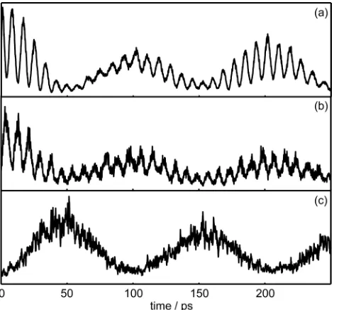
[image:3.595.73.335.538.674.2]As discussed in our earlier work,19 each ion state is sensitive in different ways to the evolution of the prepared vibrational wavepacket as a consequence of the different Franck Condon factors in operation. This is illustrated in Fig. 3, which shows the intensities of the peaks corresponding to three ion states as a function of time. The time profile shown in Fig. 3a corresponds to the 31ion state which tracks the evolution of the S13151bright state. The
time profile shown in Fig. 3b corresponds to the 3161 ion state which predominantly tracks the evolution of the S1 3162 dark state which is not populated at t = 0. The time profile
shown in Fig. 3c corresponds to the 282 ion state (peak (i) in Fig. 2, see above), which predominantly tracks the evolution of an S1dark state other than 3162whose assignment is
[image:4.595.73.312.412.630.2]discussed below. The traces in Figs. 3a-c show an oscillation with a period of 103 ps, in agreement with earlier work,13on which is superimposed a higher frequency oscillation that is clearly resolved in Figs. 3a and 3b. The high frequency components have opposite phases in Figs. 3a and b, whereas the low frequency components have opposite phases in Figs. 3a and 3c. The trace in Fig. 3c has poorer signal-to-noise, but it is clear that the high frequency oscillation has relatively low intensity in this case.
Fig. 3: Time-dependent photoelectron intensity corresponding to: (a) the 31ion peak, (b) the
3161ion peak, and (c) the ion peak labelled (i) in Fig. 2 and assigned to 282.
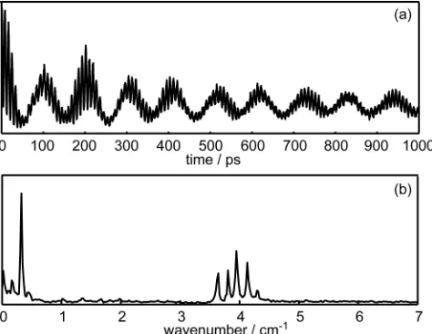
103 ps, the Fourier transform reveals a peak at 0.16 cm-1 that corresponds to a period of 205 ps, as well as four strong peaks between 3.6 and 4.2 cm-1corresponding to periods of 8.5, 8.1, 9.2, and 8.8 ps (in order of decreasing relative contribution), meaning that overall six clear oscillation periods are observed. This is an immediate indication that the prepared wavepacket contains multiple levels, and not just the two known components of the Fermi resonance. Although minor peaks are also observed at 0.44 and 4.3 cm-1in Fig. 4b, they are not observed in Fourier transforms of the time profiles for any of the other ion states. These peaks may indicate very weakly coupled states, or may be artefacts resulting from the Fourier transform procedure. It is clear from the Fourier transform that there is no component close to 6 cm-1which is the previously determined eigenstate energy separation for the 3151/3162Fermi resonance.17 In Fig. 4a, the behavior of the beating pattern over an extended time indicates that the population remains within a small subset of levels, and does not leak out into a bath of states. This is characteristic of a restricted IVR process and is contrary to the expectation for intermediate case IVR. The slight decay in the beat amplitude was also observed by Knee and coworkers13 and is a consequence of rotational
dephasing.16
Fig. 4: (a) The time dependent intensity of the photoelectron peak corresponding to the 31 ion state over an extended time range, and (b) its Fourier transform.
The 282ion state is not expected to have significant Franck Condon overlap with either the 3151 or the 3162 state in S1, and so its time behavior (Fig. 3c) must result from tracking a
[image:5.595.73.310.435.618.2]Knight and Kable,17 which would put the 282 state well outside the ~13 cm-1bandwidth of the pump laser. The level 51282, which is expected to appear at ~2056 cm-1in S1, is however
expected to lie within the bandwidth, is a plausible candidate for coupling to the 3151bright state, and is expected to give rise to population of the 282ion state. The dominance of the 103 ps oscillation in Fig. 3c is consistent with the 5128 state being closest in energy to the prepared 3151bright state, to which it must be strongly coupled. Similar observations have been made in our earlier work.19 Because of the known coupling between 51 and 62 in S1
the presence of 51282 implies the presence of 62282 as well, although the latter state is predicted to have a much smaller Franck Condon overlap with the 282ion state.18
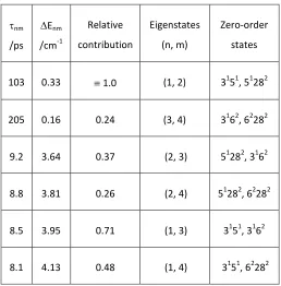
Figure 5 shows an energy level diagram for the four states involved in the IVR process. For such a scheme, the six energy separations between the four eigenstates: E12, E13,E14,
E23, E24, E34, are related to the the observed oscillation periods, nm, bynm h Enm.
The deduced values of nm and Enm together with suggested assignments are listed in
Table 1. This Table also contains the relative contributions of each component which have been determined from a fit to the data shown in Fig. 4a. The eigenstate energy separation resulting from the coupling of the 3151and 3162states is accurately determined in this work to be 3.95 ± 0.3 cm-1. Furthermore, the dark state associated with the 103 ps quantum beats which was unassigned by Knee and coworkers13can now be identified as 51282.
We thus conclude that the 3151 state in S1 p-difluorobenzene is strongly coupled to three
Fig. 5: An energy level diagram for the four states involved in the IVR process. The dotted lines serve to indicate the zero order state (left) that has the largest contribution to each eigenstate (right).
Table 1: Oscillation periods and eigenstate energy separations deduced from the Fourier transform shown in Fig. 4. Likely assignments in terms of the dominant zero-order states are also given.
nm /ps
Enm /cm-1
Relative contribution
Eigenstates (n, m)
Zero-order states
103 0.33 1.0 (1, 2) 3151, 51282
205 0.16 0.24 (3, 4) 3162, 62282
9.2 3.64 0.37 (2, 3) 51282, 3162
8.8 3.81 0.26 (2, 4) 51282, 62282
8.5 3.95 0.71 (1, 3) 3151, 3162
8.1 4.13 0.48 (1, 4) 3151, 62282
Experimental method
[image:7.595.71.329.369.631.2]bandwidth is ~13 cm-1 and the pulse duration is 1 ps. In this work, a pump wavelength of 257.0 nm is used to prepare p-difluorobenzene in the 3151level in S1, whilst the probe beam
is used to ionize the excited molecules using a wavelength in the range 255 to 275 nm. A motorised delay stage (Standa) achieves a variable time delay (-20 to +1000 ps) between the pump and probe laser pulses. The vertically-polarised, co-propagating laser beams are focussed into a photoelectron spectrometer where they are spatially and temporally overlapped with a pulsed molecular beam of p-difluorobenzene seeded in He. Photoelectron images are measured using a velocity map imaging photoelectron spectrometer, which has been described in detail in previous work.22 Time-resolved photoelectron spectra and angular distributions are extracted from the measured images; the latter have been discussed elsewhere.12 In addition, delay stage scans, in which only those photoelectrons striking the central part of the detector are monitored, provide time-dependent photoelectron intensity corresponding to the formation of a selected ion vibrational state; this is achieved by careful choice of the probe wavelength.
Acknowledgements
This work was supported by EPSRC grant EP/E046150.
References
1. Coveleskie, R. A.; Dolson, D. A.; Parmenter, C. S., A Direct View of Intramolecular Vibrational Redistribution in S1Para-Difluorobenzene.J. Chem. Phys.1980,72, 5774-5775.
2. Coveleskie, R. A.; Dolson, D. A.; Parmenter, C. S., Chem. Timing .2. The Picosecond Dynamics of Intramolecular Vibrational Redistribution in S1Para-Difluorobenzene.J. Phys.
Chem.1985,89, 655-665.
3. Coveleskie, R. A.; Parmenter, C. S., Single Vibronic Level Fluorescence Spectra from the S1(1B2u) State Of Para-Difluorobenzene Vapor.J. Molec. Spec.1981,86, 86-114.
4. Davidson, E. R.; Elston, H. J.; Parmenter, C. S., Harmonic Mode Scrambling in Para-Difluorobenzene.Chem. Phys. Lett.1992,197, 123-130.
S1-6. Holtzclaw, K. W.; Parmenter, C. S., A Time-Resolved Fluorescence Search for
Intramolecular Vibrational Redistribution in S1Para-Difluorobenzene Vapor.J. Phys. Chem. 1984,88, 3182-3185.
7. Holtzclaw, K. W.; Parmenter, C. S., Chem. Timing .3. The Picosecond Dynamics of Intramolecular Vibrational Redistribution from 11 Levels in S1Para-Difluorobenzene Vapor.
J. Chem. Phys.1986,84, 1099-1118.
8. Fujii, M.; Ebata, T.; Mikami, N.; Ito, M.; Kable, S. H.; Lawrance, W. D.; Parsons, T. B.; Knight, A. E. W., Mode-Dependent Intramolecular Vibrational Redistribution in the S1State
of Jet-Cooled Para-Difluorobenzene.J. Phys. Chem.1984,88, 2937-2940.
9. Kable, S. H.; Lawrance, W. D.; Knight, A. E. W., Evidence for Mode-Specific
Intramolecular Vibrational Redistribution in S1p-Difluorobenzene.J. Phys. Chem.1982,86,
1244-1247.
10. Nathanson, G. M.; McClelland, G. M., The Role of Rotational Motion in
Intramolecular Energy-Transfer - Measurements of Fluorescence Polarization from p-Difluorobenzene.Chem. Phys. Lett.1985,114, 441-445.
11. Reid, K. L.; Field, T. A.; Towrie, M.; Matousek, P., Photoelectron Angular Distributions as a Probe of Alignment Evolution in a Polyatomic Molecule: Picosecond Time- and Angle-Resolved Photoelectron Spectroscopy of S1Para-Difluorobenzene.J. Chem. Phys.1999,111,
1438-1445.
12. Midgley, J.; Davies, J. A.; Reid, K. L., Comment On "Photoelectron Angular Distributions as a Probe of Alignment in a Polyatomic Molecule: Picosecond Time- and Angle-Resolved Photoelectron Spectroscopy of S1p-Difluorobenzene" J. Chem. Phys. 111,
1438 (1999).J. Chem. Phys.2013,139, 117101.
13. Zhang, X.; Smith, J. M.; Knee, J. L., Picosecond Vibrational Dynamics of Several S1
Bands in Jet-Cooled p-Difluorobenzene.J. Chem. Phys.1994,100, 2429-2436. 14. Baskin, J. S.; Rose, T. S.; Zewail, A. H., Picosecond IVR Dynamics of
para-Difluorobenzene and para-Fluorotoluene in a Molecular-Beam - Comparison with Chem. Timing Data.J. Chem. Phys.1988,88, 1458-1459.
15. Mulliken, R. S., Report on Notation for the Spectra of Polyatomic Molecules.J. Chem. Phys.1955,23, 1997-2011.
17. Knight, A. E. W.; Kable, S. H., The S1-S0(1B2u-1Ag) Transition of para-Difluorobenzene
Cooled in a Supersonic Free Jet Expansion - Excitation And Dispersed Fluorescence-Spectra, Vibrational Assignments, Fermi Resonances, and Forbidden Transitions.J. Chem. Phys.1988,
89, 7139-7160.
18. Midgley, J. PhD, University of Nottingham, 2014.
19. Davies, J. A.; Green, A. M.; Reid, K. L., Deducing Anharmonic Coupling Matrix
Elements from Picosecond Time-Resolved Photoelectron Spectra.Phys. Chem. Chem. Phys.
2010,12, 9872.
20. Keske, J. C.; Pate, B. H., Decoding the Dynamical Information Embedded in Highly Mixed Quantum States.Annu. Rev. Phys. Chem.2000,51, 323-353.