REVIEW
Hypoxia-inducible transcription factors in fish: expression, function
and interconnection with the circadian clock
Bernd Pelster1,2,* and Margit Egg1
ABSTRACT
The hypoxia-inducible transcription factors are key regulators for the physiological response to low oxygen availability. In vertebrates, typically three Hif-α isoforms, Hif-1α, Hif-2α and Hif-3α, are expressed, each of which, together with Hif-1β, may form a functional heterodimer under hypoxic conditions, controlling expression of hundreds of genes. A teleost-specific whole-genome duplication complicates the analysis of isoform-specific functions in fish, but recent studies suggest that the existence of paralogues of a specific isoform opens up the possibility for a subfunctionalization. In contrast to during development inside the uterus, fish eggs are freely accessible and studies analyzing Hif expression in fish embryos during development have revealed that Hif proteins are not only controlling the hypoxic response, but are also crucial for proper development and organ differentiation. Significant advances have been made in our knowledge about tissue-specific functions of Hif proteins, especially with respect to gill or gonadal tissue. The hypoxia signalling pathway is known to be tightly and mutually intertwined with the circadian clock in zebrafish and mammals. Recently, a mechanistic explanation for the hypoxia-induced dampening of the transcriptional clock was detected in zebrafish, including also metabolically induced alterations of cellular redox signalling. In turn, MAP kinase-mediated H2O2 signalling modulates the temporal expression of Hif-1α protein, similar to the redox regulation of the circadian clock itself. Once again, the zebrafish has emerged as an excellent model organism with which to explore these specific functional aspects of basic eukaryotic cell biology.
KEY WORDS: Development, Gill, Gonad, Ion regulation, Metabolic control, Paralogues, Redox, ROS
Introduction
Low oxygen availability occurs during development of many vertebrates, may be the result of high oxygen demand of tissues, is experienced at high altitude, and is associated with pathophysiological conditions such as stroke or cancer (Ramírez-Bergeron and Simon, 2001; Patel and Simon, 2008; Semenza, 2009, 2010; Prabhakar and Semenza, 2015). Aquatic species such as fish, however, are particularly prone to exposure to hypoxic conditions owing to the low oxygen solubility in water, inefficient mixing of water and respiratory activity of aquatic species combined with rhythmic, light-dependent photosynthetic activity (Val and Almeida-Val, 1995; Muusze et al., 1998; Diaz and Rosenberg, 2008; Diaz and Breitburg, 2009; Welker et al., 2013; Jenny et al., 2016). In
aquaculture systems, which are gaining considerable importance for food production, hypoxic conditions have also been identified as a serious problem (Dong et al., 2011). Metabolism of heterotrophic (see Glossary) organisms, however, is largely dependent on the availability of oxygen, and under hypoxic conditions the hypoxia-inducible transcription factors (Hif proteins) are key regulators for the physiological responses to reduced oxygen availability (Semenza, 2010; Prabhakar and Semenza, 2015). In vertebrates, three different Hif-αisoforms (Hif-1α, Hif-2αand Hif-3α) have been detected (Rytkönen et al., 2011; Rytkönen and Storz, 2011).
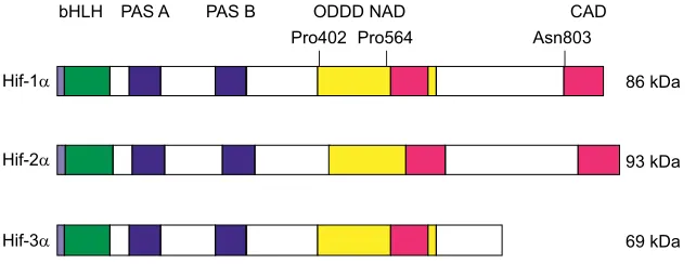
Hif-α proteins are characterized by a basic helix–loop–helix domain, by PAS domains (see Glossary) for dimerization with a constitutively expressed Hif-1β(ARNT; see Glossary) subunit, and by an oxygen-dependent degradation domain (ODDD) (Pugh et al., 1997; Semenza et al., 1997; Schofield and Ratcliffe, 2004); the basic structure of zebrafish (Danio rerio) Hif proteins is shown in Fig. 1. Under normoxic conditions (normoxia; see Glossary), prolyl residues in the ODDD are hydroxylated by prolyl hydroxylases (PHD), allowing for the binding of von Hippel–Lindau protein and subsequent proteasomal degradation in the E3 ubiquitin pathway (Schofield and Ratcliffe, 2004; Ke and Costa, 2006; Kaelin and Ratcliffe, 2008; Prabhakar and Semenza, 2012; Koh and Powis, 2012). Under low oxygen conditions, lack of prolyl hydroxylation results in a stabilization of Hif-αproteins, which then dimerize with Hif-1βand control transcription by binding to hypoxia responsive elements (HREs; see Glossary) in the control region of hundreds of genes, the consensus sequence of the HRE being identified as ACGTG (Wenger et al., 2005; Schödel et al., 2011).
Activation of transcription by Hif-1αand Hif-2αhas been well described, but the role of Hif-3αis less clear. In mammals, Hif-3αis subject to extensive alternative splicing, resulting in a large number of variants (Maynard et al., 2003; Pasanen et al., 2010; Heikkilä et al., 2011). In zebrafish, multiple alternatively spliced transcripts have also been identified, but the full-length version appears to be the one predominantly expressed (Zhang et al., 2014). It was originally believed that these Hif-3α proteins mainly act as inhibitors, and indeed for some of these proteins an inhibitory function was shown (Makino et al., 2007; Lendahl et al., 2009). Owing to a low transactivational activity (Gu et al., 1998), it was suggested that Hif-3α could act as a competitive inhibitor for Hif-1α and Hif-2α by competing for available Hif-1β(Yamashita et al., 2008). Subsequent elegant experiments using zebrafish as a model organism demonstrated that variants of Hif-3αare able to drive transcription by binding to HREs (Zhang et al., 2014). Reporter studies also revealed that all three killifish (Fundulus heteroclitus) Hif proteins act as transcriptional activators, but they differed in their effectiveness (Townley et al., 2017), and Hif-1αhas been shown to control a larger set of genes than Hif-3αin zebrafish (Zhang et al., 2014).
Anin vitroanalysis of the binding behaviour ofF. heteroclitus
Hif proteins to mammalian andF. heteroclitusHREs and to non-canonical HREs suggested that fish Hif proteins may regulate the 1Institute of Zoology, University of Innsbruck, Technikerstr. 25, A-6020 Innsbruck,
Austria.2Center for Molecular Biosciences, University of Innsbruck, A-6020
Innsbruck, Austria.
*Author for correspondence ([email protected])
B.P., 0000-0002-9170-5086
Journal
of
Experimental
expression of genes with HREs that differ from the mammalian consensus sequence (Townley et al., 2017). This is supported by the observation that zebrafish DNA containing ATGTG sequences instead of the canonical ACGTG could be immunoprecipitated with a zebrafish-specific Hif-1αantibody (Greenald et al., 2015). These results suggest that, in fish, which are more likely to face hypoxic conditions than most mammals, which may experience hypoxia at high altitude, additional genes are under the control of Hif proteins. A detailed analysis of these genes might be instructive for a better understanding of the mechanisms of hypoxic adaptation.
Here, we review current knowledge about the mRNA transcription of the differenthifmolecules, and on the expression of Hif proteins. We will start with a discussion of the role of Hif paralogues, because a fish-specific genome duplication resulted in the generation of Hif paralogues, which stimulated research on a possible subfunctionalization. The transparent fish eggs enable an analysis of the role of Hif proteins during development, which is difficult in animals with intrauterine development, and we will focus on the role of Hif proteins in specific tissues. We also discuss the possible function of the different isoforms of Hif proteins with particular reference to ion regulation and metabolism. In addition, we elaborate
advances in understanding the interconnections of the Hif signalling pathway with the circadian clock, which explains daily variations in performance and hypoxia resistance and appears to be crucially involved in overall metabolic control. Recent studies on this topic confirmed that the original observations made in zebrafish also extend to mammals, supporting the more general relevance of studies using fish as model organisms.
HifmRNA versus Hif protein
When talking about Hif protein in mammals, capital letters are generally used in the nomenclature (i.e. HIF), whereas for fish, often Hif is used. The focus of our paper is on fish; therefore, we generally use Hif. When discussing the Hif signalling pathway in fish it is important to discriminate between thehifmRNA molecule and the Hif protein. In mammals, it was soon recognized that Hif signalling is controlled post-translationally. Hypoxia causes a stabilization of Hif-αprotein and the formation of the active Hif-α/Hif-1βdimer, but the transcription of hif genes remains unaltered. In most mammals, mRNA profiling of hif isoforms therefore does not provide any indication for the activation of Hif signalling. In fish, however, the situation is different. Many studies (see below) revealed that in fish, hypoxia often results also in an elevated transcription of hif genes. Thus, in fish, analysis of hif mRNA concentration indeed provides evidence for hypoxia-activated Hif signalling. However, a convincing explanation for the activation of hiftranscription in teleost fish under hypoxic conditions has not yet been presented. In elasmobranchs, repetitive hypoxia or prolonged hypoxia has been shown to result in an increase in hif mRNA concentration, although elevated mRNA levels were not detected after the initial bout of hypoxia. It therefore was speculated that the increase inhiftranscription was required to counteract degradation of Hif protein during prolonged hypoxia (Rytkönen et al., 2012).
Hifparalogues
Analysis of the Hif transcription factor family diversification in metazoans confirmed that Hif-1α is conserved among most metazoans, whereas Hif-2α appeared with the development of chordates/vertebrates. Furthermore, there is strong evidence for a functional divergence between Hif-1α and Hif-2α (Graham and Presnell, 2017). Evolution of vertebrates included two rounds of gene duplications (Holland et al., 1994; Sidow, 1996), and a third round of whole-genome duplications occurred during the evolution of teleosts (Taylor et al., 2001; Meyer and Van de Peer, 2017). Sequence analysis and the analysis of transcriptional expression of hif-α proteins in zebrafish revealed the existence of paralogous Hif-αproteins (hif-1A,
B,hif-2A,B,hif-3A,B) (Rytkönen et al., 2013, 2014), and Hif-αprotein
paralogues appear to be present in all cyprinid fish (see Glossary). In line with this conclusion, paralogues ofhif-1αand hif-2αisoforms have also been identified in six schizothoracine fish (see Glossary) from the Tibetan Plateau, a subgroup of the cyprinids (Guan et al., Glossary
ARNT
Aryl hydrocarbon receptor nuclear translocator protein. CBP/p300
Co-factor activating Hif protein. Cyprinid
A family of freshwater fish. E-box
Enhancer box, containing the nucleotide sequence CGTG. Euteleost
A clade of bony fishes within the class Actinopterygii. FIH-1
Factor inhibiting Hif. Heterotrophic
Organisms that are dependent on the digestion of organic carbon. HRE
Hypoxia responsive element; basic nucleotide sequence ACGTG. Lactacidosis
Accumulation of lactate in tissues due to anaerobic metabolism. Morpholino
Short nucleotide oligomers used to block transcription in zebrafish. Myelencephalon
Most posterior region of the embryonic hindbrain. Normoxia
Oxygen partial pressure near 20 kPa at sea level. PAS
A protein domain first discovered in the proteins period, ARNT and SIM (single minded); allows protein–protein interactions.
Schizothoracine fish
A subgroup within the family of cyprinid fishes.
bHLH PAS A PAS B ODDD NAD CAD
Hif-1α 86 kDa
Hif-2α 93 kDa
Hif-3α 69 kDa
[image:2.612.47.368.619.741.2]Pro402 Pro564 Asn803
Fig. 1. Schematic representation of Hif protein structure according to Lisy and Peet (2008) and Rytkönen et al. (2008).Molecular weight of the zebrafish isoforms is given based on western blot analysis using zebrafish-specific antibodies (Kopp et al., 2011; Köblitz et al., 2015).
Journal
of
Experimental
2014). Many other euteleost (see Glossary) species apparently lost the duplication ofhif-1α,hif-2αand hif-3αisoforms, or retained only truncated versions of an additionalhif-αgene (Rytkönen et al., 2013; Geng et al., 2014; Townley et al., 2017).
In zebrafish, expression profiling of hif-α paralogues revealed that all six paralogues (hif-1A,B,hif-2A,B,hif-3A,B) are transcribed during early developmental stages in a tissue-specific pattern (Rytkönen et al., 2013, 2014). The two paralogues of the Hif-α proteins were differentially transcribed in different tissues, and the paralogues showed clear differences in their transcription pattern in response to hypoxia (Rytkönen et al., 2013, 2014). A possible subfunctionalization of Hif paralogues and eventual specific adaptation to severe hypoxia has also been analyzed in six schizothoracine fish species living in high-altitude freshwater systems of the Tibetan Plateau (Guan et al., 2014). Hif-1αA/B showed a higher responsiveness to hypoxia compared withhif-2αA/ B, but taken together, the data did not provide clear evidence for a subfunctionalization of the paralogues (Rytkönen et al., 2013, 2014; Guan et al., 2014).
In addition, at the protein level, a comparison of amino acid sequence and function of Hif-1αand Hif-2αproteins did not reveal any difference between schizothoracine species living at high altitude on the Tibetan Plateau and zebrafish, a low altitude fish (Guan et al., 2014). Amino acid substitutions in Hif proteins of various fishes revealed that Triplophysa fishes from the Tibetan plateau had a significantly higher rate of synonymous and nonsynonymous substitutions in their Hif-1A and Hif-2B proteins, with evidence for a positive selection pressure on the genes encoding these two proteins (Wang et al., 2015a,b). The authors predicted functional changes in these proteins, but the nature of these changes was not further addressed.
It thus remains to be shown whether preservation of the two paralogues of the Hif-αproteins in cyprinid fish actually resulted in the production of functionally discernable proteins and a subfunctionalization. In mammals, however, sequence variations of the Hif-2αprotein appear to be related to the adaptation to high altitude (Storz et al., 2010; Tissot van Patot and Gassmann, 2011; Bigham and Lee, 2014).
With respect to the presence of hif-1β (arnt) paralogues in zebrafish, expression of genes encoding two ARNT1 proteins has been described (ARNT1a and ARNT1b). The shorter ARNT1a, however, appears to be non-functional inin vitrostudies (Prasch et al., 2006). The duplication ofhif-1βhas apparently been lost in most fishes (Graham and Presnell, 2017).
Expression ofhifisoforms during development
Hif proteins have been detected in early developmental stages of many vertebrates, indicating that this transcription factor is involved in development and organogenesis. Accordingly, in mice, knockout of
hif-1αor ofhif-2αis embryonically lethal between embryonic stages
E9 and E11, but with different phenotypic appearances (Iyer et al., 1998; Kotch et al., 1999; Compernolle et al., 2002; Hu et al., 2003). In yolk-sac fry of Baltic salmon, Salmo salar, mortality has been associated with reduced Hif-1αDNA binding, resulting in reduced vascular endothelial growth factor (VEGF) expression (Vuori et al., 2004). This observation clearly suggests that Hif protein is also involved in fish development. In embryos developing within the uterus, the specific role of Hif transcription factors is difficult to assess, but in free-living fish embryos, which are transparent at least at the onset of development, these studies can readily be performed.
In zebrafish and Wuchang bream (Megalobrama amblycephala), the presence ofhif-1αandhif-2αtranscripts has been shown in the
egg, implicating maternal expression, and the transcripts remained detectable under normoxia throughout development (Rojas et al., 2007; Shen et al., 2010; Kopp et al., 2011). In the developing zebrafish, hif-1α expression is detected in many tissues and is mostly concentrated in the ventricle, certain blood vessels, nervous tissue and notochord, although the expression intensity varies in a stage-dependent manner (Rojas et al., 2007). In some tissues,hif-2α mRNA is also present throughout development, and the expression is mainly found in blood vessels such as intersegmental and brain blood vessels, and in brain tissue, notochord and somites. In some tissues, both isoforms are expressed, but not necessarily at the same time (Rojas et al., 2007). More detailed studies of Rytkönen et al. (2013, 2014) revealed that actually transcripts of two paralogues of all three Hif-α proteins are detectable in zebrafish throughout development, and that previous studies had focused on hif-1B,
hif-2Aandhif-3A.Although the expression ofhif-1A,Bandhif-2A,B
was low in very early stages, at about hatching time, mRNA expression levels ofhif-1Aand ofhif-2Aincreased by approximately 10-fold, whereas hif-1B and hif-2B expression hardly changed (Rytkönen et al., 2013). Similarly, the expression levels of sturgeon
hif-1α and hif-2α increased significantly at about hatching time
(Rytkönen et al., 2013).
The mRNA ofhif-1α,hif-2αandhif-3αhas also been detected in embryos of Chinese sucker (Myxocyprinus asiaticus) and Wuchang bream, and the transcript levels of all three isoforms responded to hypoxia. Normoxic mRNA levels and the changes observed during hypoxia were not consistent between the two species, and although thehif-2αmRNA concentration was very low in normoxic Chinese sucker, it was higher than thehif-1αmRNA concentration in Wuchang bream (Shen et al., 2010; Chen et al., 2012). The Chinese sucker and Wuchang bream have been characterized as hypoxia-sensitive species, whereas the zebrafish, at least transiently and in certain developmental stages, tolerates oxygen deprivation. This suggests that there may be species-specific differences in the role of the different Hif proteins during development, with respect to not only developmental processes, but also the response to hypoxia.
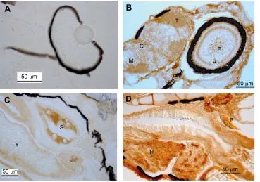
The presence of Hif-α protein has also been assessed during embryogenesis, and in zebrafish embryos, Hif-1αprotein was detected as early as 22 h after fertilization (Robertson et al., 2014). Köblitz et al. (2015) detected expression of all three isoforms (Hif-1α=1B, Hif-2α=Hif-2A, Hif-3α=Hif-3A) 24 h after fertilization. Although the protein concentration of Hif-1αremained almost constant between day 1 and day 9 of normoxic development, the concentration of Hif-2α and of Hif-3α increased during later stages (Köblitz et al., 2015), suggesting that Hif-2α and Hif-3α may play important roles in organogenesis. Immunohistochemical localization of Hif-1αusing the method of Kopp et al. (2010) confirmed the presence of the protein in nervous tissue, including the eye, at 4 days post-fertilization (dpf) (Fig. 2B). Strong staining was also observed in liver and heart tissue, and also in the swimbladder in these early stages raised under normoxic conditions (Fig. 2C,D).
In zebrafish, exposure to hypoxia (5% dissolved oxygen; 0.4 mg l−1) at 18 h after fertilization did not elicit a Hif response, but at 24 h after fertilization hypoxic exposure resulted in stabilization of Hif-1α(Robertson et al., 2014). In another study, however, Hif-1α accumulation was detected in embryos incubated for 12 h after fertilization (Santhakumar et al., 2012). Four hours of hypoxic exposure starting at 2 dpf caused a significant elevation in Hif-1α protein content as compared with the normoxic control, but this change was not observed for Hif-2αand Hif-3α(Köblitz et al., 2015). Similarly, zebrafish larvae raised under hypoxic conditions
Journal
of
Experimental
(PO2=5 kPa) between 3 and 9 dpf showed elevated Hif-1αlevels, but Hif-2αand Hif-3αwere not affected (Köblitz et al., 2015). These data suggest that Hif-2α and Hif-3α play a more important role in developmental processes, whereas Hif-1α is highly responsive to hypoxic conditions. Furthermore, hypoxic stabilization of Hif-1αin embryonic stages of zebrafish occurs in fact much earlier than the first response of cardiac activity to hypoxia in the same species, which was observed at 3 dpf (Jacob et al., 2002). The hypoxic signalling pathway thus appears to be functional long before the coupling between metabolic activity and cardiac activity and haemoglobin-dependent oxygen transport is established (Pelster and Burggren, 1996; Pelster, 1999; Rombough and Drader, 2009).
A recent experimental study, however, revealed that Hif-1αis also required for proper embryonic development in fish. Overexpression
ofhif-1αmRNA in zebrafish resulted in developmental retardation
and in a significant increase in developmental defects (Tan et al., 2017). Knockdown ofhif-1αusing morpholinos (see Glossary) in turn resulted in morphological abnormalities such as small head circumferences, deformed hearts, curved tails and a reduced growth rate (Tan et al., 2017).
Collectively, these results clearly show that Hif-αproteins not only are involved in coordinating the response of fish to reduced oxygen availability, but also control developmental processes and differentiation during embryogenesis and in larval stages under normoxic conditions. To identify the specific function of the three Hif-α proteins during development, however, appears to be challenging, and there appear to be significant species-specific differences. These differences obviously are not simply related to differences in the sensitivity of fish to hypoxia. Complex interactions between different signalling pathways may play a crucial role (see below).
Tissue-specific transcription ofhif-αisoforms and Hif protein concentration in normoxia and hypoxia
In adult fish, Hif-α expression, mostly analyzed at the level of mRNA transcription, has been detected in many tissues; in particular, Hif-1α appears to be almost ubiquitously expressed,
whereas expression of Hif-2α and Hif-3α seems to be more restricted and has received far less attention (Elks et al., 2015). As to be expected from the variable expression of the different Hif-α proteins during development, normoxic presence of these proteins differs between different tissues and is species dependent. In addition, under hypoxic conditions, elevation ofhif-αtranscription and of Hif protein concentration in different tissues varies with PO2and was shown to be temperature and redox state dependent (Nikinmaa et al., 2004; Heise et al., 2006, 2007; Rissanen et al., 2006b). Comparison of the various studies on Hif expression in different fish species is also complicated by the fact that different levels of oxygen availability have been used for the experiments, often depending on the hypoxia tolerance of fish species, ranging from less tolerant species such as salmonids, sea bass or perch, to tolerant species such as epaulette shark, grass carp or oscar, and anoxia-tolerant goldfish and crucian carp. Furthermore, the time of hypoxic exposure varies greatly between different studies, further confusing comparisons.
Nevertheless, the data show that Hif is expressed under hypoxic conditions, and elevated levels ofhifmRNA and Hif protein can be detected as early as 1 h after the onset of hypoxia (Rissanen et al., 2006a). After the initial increase in protein content, prolonged hypoxic exposure typically results in a slow decrease in the expression level of Hif-1α, as has been observed in zebrafish or crucian carp (Sollid et al., 2006; Kopp et al., 2011). In Atlantic croaker,Micropogonias undulatus, however, the expression level of Hif-1α and of Hif-2α was elevated even after 1 to 3 weeks of hypoxia (Rahman and Thomas, 2007, 2011), and this was confirmed in tissues collected from fish caught in the hypoxic waters of the northern Gulf of Mexico (Thomas and Rahman, 2009). The authors therefore discussed the possibility of using the expression levels of hif-1αand hif-2αmRNA or the Hif protein level as a biomarker for the detection of reduced oxygen availability in the environment.
At the tissue level, normoxic presence ofhif-1α,hif-2αand/or hif-3αmRNA has been assessed in a wide range of species, including, for example, zebrafish, goldfish, Atlantic croaker, grass carp, S
L Y
H
L
P T
C
M
E
A
B
C
D
50 μm
50 μm
[image:4.612.48.417.57.316.2]50 μm 50 μm
Fig. 2. Immunohistochemical analysis shows the presence of Hif-1αin various tissues of zebrafish larvae.(A) If the Hif-1α-specific antibody (Kopp et al., 2011) was blocked with expressed Hif-1αprotein (=control), the head region with the eye showed no staining in a 4 dpf larvae, except for unspecific black pigmentation. (B) Staining of the head region with a Hif-1α -specific antibody shows presence of Hif-1α protein in the eye and in parts of the brain of a 4 dpf larva. (C) Hif-1αexpression is also observed in swimbladder and liver of a 5 dpf larva, and (D) heart and liver of a 6 dpf larva. C, cerebellum; dpf, days post-fertilization; E, eye; H, heart; M, myelencephalon (see Glossary); L, liver; P, pancreas; S, swimbladder; T, tectum; Y, yolk. Data are from L. Köblitz, B. Fiechtner and B. Pelster, unpublished results.
Journal
of
Experimental
Chinese sucker and Indian catfish (Clarias batrachus). In most species,hif-αmRNA has been detected, for example, in gill tissue, liver, ovary, brain, spleen and muscle tissue (Law et al., 2006; Rojas et al., 2007; Rahman and Thomas, 2007; Chen et al., 2012; Mohindra et al., 2013). Thus, mRNA of genes encoding all three Hif-αproteins is detected in many fish tissues, but at highly variable concentration levels, and the expression pattern varies with the species. Given the uncertainty of the relationship between transcription and translation, however, the physiological consequences of this phenomenon are not easy to predict.
In fish, hypoxia has repeatedly been shown to affecthif-αmRNA concentrations; for example, after 1 h of hypoxia in Eurasian perch,
Perca fluviatilis, elevated levels ofhif-1αwere detected in brain,
eye, heart and spleen (Rimoldi et al., 2012). In hypoxic sea bass,
hif-1α was elevated in liver, brain, heart muscle and kidney
(Terova et al., 2008), in hypoxic grass carp, increased levels were detected in kidney and eyes (Law et al., 2006), and in Wuchang bream, liver, gill and testis showed elevated levels ofhif-1αmRNA (Shen et al., 2010). In gill tissue of channel catfish (Ictalurus punctatus) after 5 h of hypoxia, mRNA concentration of all expressed Hif-α proteins was elevated (Geng et al., 2014). Most studies in fact agree that in fish, hypoxic exposure results in transcriptional changes causing elevated levels ofhif-αmRNA, but the functional significance of this phenomenon is not yet clear.
β-Adrenergic stimulation in rainbow trout red blood cells has been shown to enhance transcription of hif-1α after 4 h of exposure to hypoxia (Götting and Nikinmaa, 2017). Owing to the presence of the Root effect in fish haemoglobins (Pelster and Randall, 1998; Pelster, 2001), β-adrenergic control of Na+/H+ exchange assures an alkaline red blood cell pH in order to stabilize haemoglobin-based oxygen transport in stress situations to counteract lactacidosis (see Glossary) (see Wells, 2009, for review). Therefore, it is questionable whether the β-adrenergic influence onhif-1αexpression represents a universal phenomenon established in different cell types. Results from a study on the hypoxia-resistant epaulette shark suggest that the stimulation of hif-αtranscription may be necessary to compensate for a breakdown of the Hif protein, because under long-term hypoxia or repeated hypoxia, Hif-controlled transcription of PHD genes and concomitant activation of PHD activity would result in reduced levels of Hif protein (Rytkönen et al., 2012).
Control of Hif activity by factor inhibiting Hif
Transcriptional activity of Hif-α may be controlled by a factor inhibiting Hif (FIH-1; Hif1AN; see Glossary) (Lando et al., 2002), which was also found in fish. FIH-1 is a hydroxylase, which adds a hydroxyl group to the asparagine residue located between the ODDD and CAD of the Hif-α protein. Hydroxylation of this asparagine prevents interaction of the Hif protein with its co-activator CBP/p300 (see Glossary), and thereby significantly reduces the transactivation activity of Hif (Lando et al., 2002; Zhang et al., 2010). In Nile tilapia, hypoxia caused a significant upregulation of hif-1αn expression, suggesting that FIH-1 is involved in the control of Hif-α activity under hypoxic conditions (Li et al., 2017). In channel catfish after 5 h of hypoxia, the mRNA concentration of fih-1 was significantly elevated (Geng et al., 2014). If the increased transcript concentration results in an elevated FIH-1protein concentration and activity, it could be speculated that there is some sort of elaborate fine-tuning in transcriptional activity of Hif proteins during prolonged hypoxia, including increased transcription of hif genes to counteract Hif degradation by elevated PHD activity (see above), but also elevated FIH activity to avoid over-stimulation of Hif-controlled pathways.
Hif expression in gonads
Hypoxia is well known to affect growth of fish (Wang et al., 2009; Roberts et al., 2011). For several species, a significant impairment of reproduction under hypoxic conditions has also been shown, caused by the impairment of gonad development and reduced sperm and egg quality (Wu, 2009; Thomas and Rahman, 2010, 2012; Thomas et al., 2015). Expression profiling using microarrays in zebrafish exposed to either 4 or 14 days of severe hypoxia (1 mg l−1 oxygen) revealed a large number of differentially transcribed genes in testes and in ovaries (Martinovic et al., 2009). Hypoxia has a profound influence on the steroidogenesis pathway and may affect the sex ratio of fish (Shang et al., 2006; Cheung et al., 2014; Robertson et al., 2014). Even genotypic female Japanese medaka, Oryzias latipes, can turn into males if raised under hypoxic conditions (Cheung et al., 2014).
Although many studies have linked hypoxic exposure to impaired fish reproduction and gonadal development, only a few studies have attempted to elucidate the role of Hif transcription factors for this phenomenon. The presence of high levels ofhif-1α,hif-2αand hif-3αmRNA in gonads has been reported for several fish species (Law et al., 2006; Rojas et al., 2007; Rahman and Thomas, 2007; Chen et al., 2012; Zhang et al., 2012; Rytkönen et al., 2014). Overexpression and morpholino knockdown experiments in zebrafish have revealed that Hif-1α controls the expression of various enzymes involved in steroidogenesis (Tan et al., 2017), and thus in the production of estrogen and testosterone (Fig. 3). It has previously been reported that aromatase, an enzyme catalyzing the conversion of testosterone to estradiol, may be controlled by aryl hydrocarbon (=ARNT, Hif-1β) (Kazeto et al., 2001). Accordingly, Hif proteins are clearly involved in the control of estrogen and testosterone production, and hypoxia-induced stabilization of Hif protein therefore most likely contributes to the adverse effects of hypoxia on fish reproduction.
In addition to these enzymes involved in sex hormone synthesis, CYP11b2 (a member of the cytochrome P450 family; aldosterone synthase) was also affected by Hif-1α (Tan et al., 2017), and CYP11b2 and 3β-HSD are both involved in mineralocorticoid and glucocorticoid production of the adrenals. It is therefore conceivable that Hif-1αmight be also be involved in the control of embryonic metabolism and ion regulation.
The role of Hif proteins in ionic regulation
The presence of hif-1αand hif-2αmRNA at reasonably high or sometimes even very high levels in gill tissues has been reported for many fish species (Rojas et al., 2007; Geng et al., 2014; Rytkönen et al., 2014; Townley et al., 2017), fostering the hypothesis that Hif proteins serve a particular function in gills. In anoxia-tolerant crucian carp, Hif-1αappears to be involved in gill remodelling, i.e. in the expansion of gill surface area under hypoxic conditions (Nilsson, 2007). Fish gills are multifunctional organs responsible not only for gas exchange, but also for ion regulation and acid–base regulation (Laurent and Perry, 1991; Evans et al., 2005). Accordingly, in gills, Hif proteins could be involved in the control of ion transport processes, given that hypoxia has repeatedly been shown to inhibit ion homeostasis in fish.
In the Amazonian oscar and in rainbow trout, active uptake of Na+is inhibited under conditions of low oxygen (Wood et al., 2007; Iftikar et al., 2010), and in scaleless carp exposed to hypoxic water, plasma Na+levels dropped (Matey et al., 2008). It may therefore be speculated that Hif proteins are involved in the compromised Na+ and Cl− regulation under hypoxic conditions. However, for members of the slc9 group, i.e. the Na+/H+ exchanger, which
Journal
of
Experimental
plays a crucial role in gill ion regulation, a connection to the Hif transcription factors has not yet been identified. Downregulation of V-ATPase expression, which is involved in Na+ uptake in freshwater fish, under hypoxic conditions has been reported for the zebrafish (van der Meer et al., 2005), but a connection between V-ATPase and Hif has not yet been reported.
Under hypoxic conditions, disturbance of ionic regulation apparently is accompanied by a reduction in gill Na+/K+-ATPase activity (Wood et al., 2007; Matey et al., 2008). Therefore, the disturbance of ionic homeostasis may be the result of a reduced gill Na+/K+-ATPase activity caused by reduced ATP availability under hypoxic conditions, owing to metabolic depression (St-Pierre et al., 2000). A series of glycolytic enzymes is under the control of Hif proteins. Therefore, an indirect connection between Hif proteins and ion homeostasis may result from the influence of Hif proteins on metabolism and ATP production under hypoxic conditions. Analysis of the transcriptome of zebrafish under hypoxic conditions revealed a reduced expression of Na+/K+-ATPase subunits and of Ca2+-ATPase (Ton et al., 2003), which is consistent with the reported decrease in Na+/K+-ATPase activity, if we assume that changes in the transcriptome are indicative of changes in translational activity. In addition, several subunits of F1/
F0-ATPase were reduced in their transcription levels, suggesting a coordinated reduction in energy metabolism under hypoxic conditions in zebrafish (Ton et al., 2003).
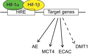
Nevertheless, direct involvement of Hif proteins in ion regulation may also be deduced from transcriptional studies (Fig. 4). A comparison of seven normoxic and hypoxic zebrafish transcriptomes from larval tissue, adult hearts and gill tissue revealed that expression of several ion transport proteins was elevated by hypoxia, namely slc10a4, a sodium co-transporter, slc16a1 and slc16a3, monocarboxylate transporters (mct1 and mct4), and slc4a1a, a bicarbonate transporter, and for slc4a1a and slc16a3 the presence of an HRE in the promoter region was confirmed (Kim et al., 2017). Assuming that transcriptional changes are somehow indicative of translational adjustments, global transcriptional changes recorded under severe hypoxia in zebrafish suggested a switch to a more anaerobic metabolism (Ton et al., 2003), and the enhanced transcription of the respective monocarboxylate transporters suggests that lactate formed under these conditions may be released into the blood stream via monocarboxylate carriers and may be used by other tissues, such as heart, as a fuel.
In addition to Na+and Cl−, freshwater fish also take up Ca2+from the environmental water. Hypoxia impairs Ca2+ uptake in developing zebrafish embryos, and Ca2+ uptake has been shown to remain low even after reoxygenation (Kwong et al., 2016). The decrease in Ca2+uptake was shown to be caused by a decrease in the expression level of an epithelial Ca2+channel (ECAC). Morpholino experiments and analysis of Hif-1αB-deficient zebrafish embryos confirmed that ECAC expression was under the control of Hif-1αB (Fig. 4), which was not the case for expression of the plasma membrane Ca2+-ATPase (PMCA) and the sodium-dependent Ca2+ transporter (NXC1b) (Kwong et al., 2016).
Hif proteins are well known to induce erythropoiesis and haemoglobin formation under hypoxic conditions (Haase, 2010, 2013), and in mammals, Hif-2αhas been shown to control expression of the divalent metal-ion transporter1 (DMT1) and duodenal cytochrome b561 (DCYTB), which is bound to the duodenal membrane and reduces ferric iron (Fe3+) to ferrous iron (Fe2+) (Mastrogiannaki et al., 2013). Iron transport and iron homeostasis are linked to Hif proteins, and Hif-2αmay be of particular importance (Shah and Xie, 2014). An increased expression of genes involved in iron metabolism under hypoxia has also been confirmed for fish (Long et al., 2015), and it is very likely that the control mechanism via Hif proteins might be highly conserved in evolution.
*
*
*
*
*
Relative mRNA
abundance 0
1 2 3 4 5 6 7 8
9
A
*
*
*
0 0.5 1.0 1.5 2.0 2.5
star cyp11a 3b-hsd
star cyp11a 3b-hsd
B
Control
zfhif-MO Control
24 hpf hif-1a+
[image:6.612.64.283.54.360.2]48 hpf hif-1a+
Fig. 3. Artificial increase and artificial knockdown ofhif-1αmRNA transcription revealed that Hif controls expression of enzymes involved in the generation of the sex hormones estradiol and testosterone. (A) Injectinghif-1αmRNA into zebrafish one- to two-cell stage embryos and measuring the mRNA abundance of steroidogenic genes at 24 and 48 h post-fertilisation revealed significant downregulation ofstar, whereascyp11aand 3β-hsdwere upregulated. (B) Using hif-1αmorpholinos (zfHif-MO) to knock downhif-1αexpression resulted in the opposite response:starwas upregulated, whereascyp11aand3β-hsdwere downregulated. *P<0.05. Modified from Tan et al. (2017). Accordingly, stabilization of Hif protein under hypoxic conditions affects sex hormone production and may thus contribute to the negative influence of hypoxia on fish reproduction.
Hif-1α Hif-1β
HRE Target genes
ECAC DMT1 MCT4
AE
Fig. 4. Schematic diagram demonstrating the importance of Hif protein for the transcription of ion transport proteins in fish.The Hif-α/Hif-1βdimer formed under hypoxic conditions binds to a hypoxia response element (HRE; nucleotide consensus sequence ACGTG) and activates transcription of target genes. In fish, an anion exchanger (AE), a monocarboxylate carrier (MCT4) and an epithelial Ca2+channel (ECAC) have been shown to be under the control of Hif protein; divalent metal-ion transporter1 (DMT1) has been identified in mammals, and most likely is also under the control of Hif proteins in
fish.
Journal
of
Experimental
[image:6.612.362.513.552.645.2]Interconnection of Hif signalling and the circadian clock The circadian clock is a conserved pathway among all light-exposed organisms that enables them to maintain an endogenous rhythm of roughly 24 h in order to anticipate temporally varying demands by coordinating biochemistry, physiology as well as behaviour. These endogenous rhythms are synchronized to the environmental light: dark rhythm on a daily basis and were shown to be intimately intertwined with the hypoxic signalling pathway in vertebrates. Thus, the interaction between Hif signalling and the circadian clock was first detected in mouse brain (Chilov et al., 2001). Molecular foundations for this interaction were later described in zebrafish larvae and cells (Egg et al., 2013, 2014; Pelster and Egg, 2015), and this was recently confirmed and specified in more detail for mammals (Peek et al., 2017; Adamovich et al., 2017; Wu et al., 2017). The conserved nature of the mutual relationship between these two essential pathways clearly underlines their critical role in cellular functioning. Hence, on the one hand, decreased oxygen tensions lead to dampened amplitudes of canonical clock gene oscillations, whereas on the other hand, Hif-1α expression itself is tightly controlled by the circadian clock, i.e. protein expression of Hif-1α follows a clear circadian rhythm under normoxic conditions in zebrafish cells and in mammals (Egg et al., 2013; Adamovich et al., 2017; Sandbichler et al., 2018). The protein oscillations of rodent Hif-1αthereby were shown to be based on the daily oscillations in blood and tissue oxygenation. Adamovich et al. (2017) also addressed the specific role of Hif-1αfor circadian time-keeping usingsihif-1αfor knockdown of the protein. They showed that oxygen was able to synchronize circadian rhythms of mouse cell lines and that under rhythmically oscillating O2levels, Hif-1αturned out to be essential for the observed oxygen-induced reset of the circadian clock. Experiments using dexamethasone for an oxygen-independent reset of the circadian clock in the knockdown animals, however, suggested a more complex relationship: some of the core clock genes remained completely unaltered irrespective of the Hif-1α levels generated, whereas expression levels of Rorα, Per1, Per2 and Cry2 were severely lowered insihif-1αknockdowns, although their rhythmicity remained unaffected (Adamovich et al., 2017). These results demonstrate that Hif-1αis definitely responsible for the adaptation of the circadian clock to low oxygen tensions, yet the question regarding whether Hif-1αrepresents an integral component of the clock circuitry itself is not fully answered.
In line with these findings, Sandbichler et al. (2018) showed that the importance of Hif-1αfor the integration of oxygen availability into circadian time-keeping starts far earlier than with the previously reported direct transcriptional transactivation of circadian core clock genes by binding of Hif-1αto E-boxes (see Glossary) of the clock genesper1andper2as well ascry1(Egg et al., 2013, 2014; Pelster and Egg, 2015; Wu et al., 2017; Peek et al., 2017). Hif-1αis known to control metabolism by regulating the expression of many glycolytic enzymes such as phosphofructokinase, pyruvate kinase, lactate dehydrogenase and hexokinase, and glucose transporters such as GLUT1 and GLUT3, thereby orchestrating glycolytic activity in adaptation to the availability of oxygen (Porporato et al., 2011). In a recent study, it was demonstrated that Hif-1α targets the glycolytic enzyme glyceraldehyde-3-phosphate dehydrogenase (GAPDH) even under normoxic conditions, and that knockdown of Hif-1α consequently leads to a reduction of glycolytic metabolism in human synovial fibroblasts (Del Rey et al., 2017). Short-term hypoxic incubation for 2 h of zebrafish cells, where no significantly increased occupation of circadian E-boxes by Hif-1αwas found (Egg et al., 2013), provoked a time-of-day-dependent change in metabolic activity, orchestrated to a
large extent by Hif-1α, which led to a temporal redistribution of available reducing equivalents such as NADH and NADPH. As a consequence, short-term hypoxia applied during the dark phase resulted in a highly oxidized nocturnal redox state, causing a reduced binding affinity of the Clock/BMAL1 complex to E-boxes situated in the gene control region of their target genes (Rutter et al., 2001). Consequently, acute hypoxia-induced metabolic alterations, controlled to a large extent by Hif-1α, might actually form the basis for the hypoxia-induced attenuation of the transcriptional clock by altering the cellular redox state (Sandbichler et al., 2018). The role of glucose metabolism for the attenuation of the transcriptional clock was also confirmed in mammalian systems (Putker et al., 2018). Using different mammalian cell lines, Putker and colleagues showed that redox homeostasis and glucose flux through the pentose phosphate pathway (PPP) are necessary to maintain phase and amplitude of the transcriptional clock (Putker et al., 2018). Inhibition of glycolysis using 2-deoxyglucose (2DG), however, had no major effect and the circadian period remained more or less unaffected by all metabolic perturbations. Hence, Hif-1α, which appeared to exert a regulatory role for glycolysis even under aerobic conditions (Rey et al., 2016), co-regulates the amplitude and phase of the circadian clock in mammalian systems just like in zebrafish cells by altering the glycolytic flux.
Remarkably, application of acute hypoxia did not affect the circadian oscillations of cytosolic H2O2 in zebrafish cells (Sandbichler et al., 2018). Light has been shown to stimulate H2O2 production in cultured cells of various vertebrates, including humans, via photoreduction of flavin-containing acyl-coenzyme A oxidase in peroxisomes (Hockberger et al., 1999), and in zebrafish, H2O2 couples the light signal to the circadian clock, mediated via the mitogen-activated protein (MAP) kinase pathway (Hirayama et al., 2007). It is also known that addition of H2O2to cells or the inhibition of catalase, which indirectly leads to increased intracellular levels of H2O2, result in phase shifts of canonical clock gene oscillations in zebrafish and mammals (Hirayama et al., 2007; Stangherlin and Reddy, 2013; Putker and O’Neill, 2016; Sandbichler et al., 2018). Constant oscillations of H2O2 thus could potentially have a time-keeping function, in particular when considering that the highest levels of cytosolic H2O2 paradoxically coincided with highly oxidized intracellular states, as indicated by the highest ratios of NAD+/NADH, NADP+/NADPH and levels of oxidized peroxiredoxins (Prxox) (Sandbichler et al., 2018).
The circadian rhythms of cytosolic H2O2reported by Sandbichler et al. (2018) basically fit into the picture of clock-controlled redox signalling, which is known to apply also to the mitochondrial release of H2O2into the cytosol of cells (Kil et al., 2015; Rhee and Kil, 2016). Circadian rhythms of redox markers and effectors have been reported previously for free radical acceptors such as reduced peroxiredoxins (Prxred), NADH and NADPH levels or from antioxidant enzymes such as catalase, superoxide dismutase and glutathione peroxidase (Edgar et al., 2012; Wilking et al., 2013; Putker and O’Neill, 2016; Sandbichler et al., 2018). However, a predominant role of cellular redox signalling as an intrinsic part of the circadian system, as, for example, a potential time-keeping function of the cytosolic H2O2oscillation would be, is still under debate and far less accepted (Putker and O’Neill, 2016).
It has been postulated that H2O2 may act as a superimposed co-signal of lesser specificity by integrating cellular processes via recruiting, timing and tuning of signalling pathways (Nathan, 2003; D’Autraux and Toledano, 2007). H2O2not only mediates between light radiation and the circadian clock via the MAP kinase pathway (Hirayama et al., 2007; Uchida et al., 2012), but is also substantially
Journal
of
Experimental
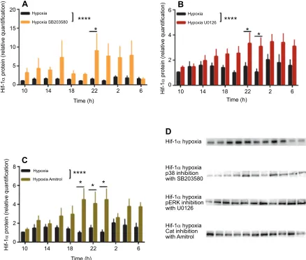
involved in mitochondrial respiration, the hypoxic response and stabilization of Hif-1αprotein (Chang et al., 2008; Niecknig et al., 2012). Accordingly, MAP kinase p38 signalling was shown to be essential for activation of Hif-1α through phosphorylation under hypoxia but not under normoxia, and activation of both p38 as well as Hif-1αappeared to depend on the generation of mitochondrial reactive oxygen species (Kulisz et al., 2002; Emerling et al., 2005; Hamanaka and Chandel, 2009). Only a few studies have investigated the role of p38 for stabilization of Hif-1αprotein, and although inhibition of p38 with the inhibitor SB203580 was supposed to result in decreased Hif-1α protein stability under ischemic conditions (Kietzmann et al., 2016), no effects were found for the expression of Hif-1α protein using the same inhibitor in various breast cancer cell lines under hypoxic conditions (Blancher et al., 2001). In zebrafish fibroblast cells, inhibition of the MAP kinase p38 using the inhibitor SB203580 resulted in significantly increased Hif-1αprotein levels after 2 h of acute hypoxia (Fig. 5A). The inhibitor-induced increase in Hif-1αprotein thereby appeared
to clearly depend on the circadian time at which the hypoxic incubation was administrated and followed the same circadian pattern that was previously reported for Hif-1α(Egg et al., 2013). In contrast, inhibition of the phosphorylated extracellular signal-regulated kinase ( pERK) with the inhibitor U0126 prior to the administration of acute hypoxia led to increased Hif-1α protein levels predominantly during the night-time, so that the circadian pattern of Hif-1αunder hypoxic conditions was significantly altered (Fig. 5B). Inhibition of pERK using the same inhibitor had earlier been reported to abolish the light- and H2O2-induced expression of the clock genescry1aandper2in zebrafish (Hirayama et al., 2007), and might therefore explain the modified circadian expression of Hif-1α protein upon pERK inhibition, considering the known circadian regulation of the transcription factor.
Inhibition of the antioxidant enzyme catalase with Amitrol also resulted in loss of the known circadian rhythm of the transcription factor after acute hypoxia (Fig. 5C), enhancing in particular Hif-1α protein levels during the first half of the night phase. Catalase
Hypoxia
Hypoxia
Hypoxia Amitrol
20
A
B
C
D
6
4
Hif-1
α
protein (relative quantification)
Hif-1
α
protein (relative quantification)
Hif-1
α
protein (relative quantification)
2
0 15
10
5
10
8
6
4
2
0
14 18 22
Time (h) Time (h)
Hif-1α hypoxia
Hif-1α hypoxia p38 inhibition with SB203580
Hif-1α hypoxia pERK inhibition with U0126
Hif-1α hypoxia Cat inhibition with Amitrol
2 6
10 14 18 22
Time (h)
2 6
10 14 18 22 2 6
0
****
****
*
* *
*
Hypoxia SB203580
Hypoxia
****
*
*
[image:8.612.86.524.270.643.2]Hypoxia U0126
Fig. 5. Redox signalling involving the MAP kinase pathway selectively modulates the temporal expression of Hif-1αprotein.(A–C) Hif-1αprotein levels (measured using a custom-prepared antibody; Kopp et al., 2011) after 2 h of hypoxia at each time point over a whole circadian cycle under a light:dark regimen of 14 h light:10 h dark. Black bars indicate control hypoxic levels of Hif-1αprotein, (A) orange bars indicate Hif-1αprotein levels after inhibition of the MAP kinase p38 using the inhibitor SB203585 (Sigma-Aldrich; 10 µmol l−1final concentration), (B) red bars indicate Hif-1αprotein levels after inhibition of the MAP kinase pERK using the inhibitor U0126 (Sigma-Aldrich; 40 µmol l−1final concentration) and (C) olive green bars indicate Hif-1αprotein levels after inhibition of the antioxidant enzyme catalase with Amitrol (3-amino-1,2,4-triazole, 3-AT; 40 µmol l−1final concentration) (mean±s.e.m.;n=4–8). Asterisks mark significant differences calculated by two-way ANOVA (*P<0.05; ****P<0.0001) (M.E., J. Nagy and A. M. Sandbichler, unpublished results). (D) Representative western blots. Similar to the role of H2O2as a second messenger in the regulation of the circadian clock, H2O2-driven MAP kinase signalling mediates the temporal expression of
Hif-1αand by this also the hypoxic response as a function of circadian time.
Journal
of
Experimental
protects cells from the damaging effects of excess H2O2 by degrading H2O2to O2and H2O (Noctor and Foyer, 1998) andcat expression was shown to be strictly light dependent in zebrafish cells, oscillating in antiphase tocry1aandper2(Hirayama et al., 2007). Catalase thereby worked as a negative regulator of the light input pathway into the circadian clock by inhibiting the mRNA expression ofcry1aand per2via the reduction of cellular H2O2, thus regulating the light-induced clock gene transcription. Similar to the circadian system, hypoxia-induced protein stabilization of Hif-1αdepends on H2O2signalling and is mediated specifically by the MAP kinase pathway and the activity of antioxidant enzymes such as catalase (Fig. 5), which also appear to affect Hif-1α protein expression as negative regulators. Hence, according to the impact of H2O2signalling for the circadian system, H2O2seems to influence in particular the temporal expression of Hif-1αprotein, and might also form the basis for the direct light responsiveness of Hif-1αitself (Bozek et al., 2009; Ben-Moshe et al., 2014). Previously, it was
shown that H2O2 acts as a second messenger in the signal transduction of light in mammalian and zebrafish cells (Hockberger et al., 1999; Uchida et al., 2012). Only recently, a study in yeast showed that H2O2 production via a conserved peroxisomal oxidase in combination with Prx was sufficient for the direct sensing of light even in the absence of specific photoreceptors such as cryptochromes or opsins (Bodvard et al., 2017). This implies that light might actually affect cellular physiology in general and circadian clocks in particular, mediated via cellular H2O2signalling, and that the ability for direct light sensing might be common to all cells, as proposed by the authors (Bodvard et al., 2017).
Perspectives
We have argued here that there is abundant evidence that the mutual interaction between the circadian clock and hypoxic signalling is clearly a multiple and redundant one, spanning from transcriptional transactivation (Egg et al., 2013; Peek et al., 2017; Wu et al., 2017)
21% O2 Glycolytic activity
NADP+/NADPH
After 2 h of 1% O2 Glycolytic activity
Prxox
NAD+/NADH
NADP+/NADPH
NAD+/NADH
Ox Red Red Ox
Prxox
Hif-1 Hif-1
Hif-1 Hif-1
Hif-1
Clock Cry Per
Bmal Clock
Cry Per
Bmal Cytostolic H2O2
Prxox Prx
[image:9.612.121.494.263.616.2]ox
Fig. 6. Model for the interaction between the cellular clock and the hypoxic signalling pathway.Left: under normoxia (21% O2), Hif-1αprotein expression follows a circadian rhythm with the highest protein abundances during the night. Glycolytic activity peaks during the day, controlled to a large extent by Hif-1α, which regulates the expression of many glycolytic enzymes such as phosphofructokinase, pyruvate kinase, lactate dehydrogenase, hexokinase and glucose transporters. Markers and effectors of the cellular redox system such as NADP+/NADPH ratio and oxidation cycles of peroxiredoxins (Prxox) oscillate in a circadian manner, whereas the NAD+/NADH ratio does not follow a 24 h rhythm under normoxia. Oxidation maxima and minima of their oscillations occur during different circadian times. Right: after application of acute hypoxia (1% O2) for 2 h Hif-1αprotein peaks twice over one circadian cycle, once during the middle of the day and once in the middle of the night (Egg et al., 2013). The highest glycolytic activity occurs during the night, leading to the alignment of
the NAD+/NADH ratio with the oscillation of the NADP+/NADPH ratio and the nocturnal peak in Prxox, resulting in a highly oxidized cellular environment during the night. This early physiological adaptation to hypoxia precedes the transcriptional repression of the circadian clock through direct binding of Hif-1αto E-boxes located in the gene control regions of theperiod1bandperiod2genes (Egg et al., 2013; Pelster and Egg, 2015). Although cytosolic H2O2oscillations (middle) remain in phase under both conditions, the TTFL is already attenuated after 2 h of hypoxia, similar to the known chronic hypoxia-induced effects on the circadian clock (after Sandbichler et al., 2018). Ox, oxidated state; Red, reduced state; Bmal, transcription factor central to the circadian clock, =ARNT like.
Journal
of
Experimental
over rapid metabolic adaptations (Putker et al., 2018; Sandbichler et al., 2018) to regulation via redox signalling (Hirayama et al., 2007; Chang et al., 2008; Niecknig et al., 2012; Stangherlin and Reddy, 2013; Bodvard et al., 2017), and represents a fundamental aspect of basic eukaryotic cell biology. Owing to their biomedical relevance, mammals have always been a main focus of Hif-oriented research. Careful analysis of the available literature reveals, however, that a lot of information about the Hif signalling pathway was actually obtained from studies using fish, and it has repeatedly been shown that signalling pathways are highly conserved (Idda et al., 2012; Wilkinson and van Eeden, 2014; Elks et al., 2015). Hence, studies using zebrafish as a model organism were the first to clearly demonstrate that Hif-3αis able to transactivate genes, although one transactivation domain is lacking (Zhang et al., 2012, 2014). Similarly, although indications for a connection between the circadian clock and the Hif signalling pathway were described long ago (Chilov et al., 2001), the first mechanistic explanation for this connection was obtained using zebrafish as a model system (Egg et al., 2013). The mutual link between both pathways was only recently confirmed to exist as well in mammalian systems (Adamovich et al., 2017; Peek et al., 2017; Wu et al., 2017). Furthermore, the mutual interaction between the circadian clock and hypoxic signalling appeared to also substantially involve primary metabolism and redox signalling in zebrafish, thus preceding the transcriptional repression of core clock genes by Hif-1α, as summarized in Fig. 6. Similarly, the impacts of glycolysis, the pentose phosphate pathway or redox signalling for circadian time-keeping in mammalian cells and organisms were also described recently (Stangherlin and Reddy, 2013; Putker et al., 2018; Del Rey et al., 2017). Hence, once again, zebrafish larvae and zebrafish-derived cell lines have emerged as reliable experimental models that are as relevant to basic research as mammalian model systems, even when fundamentally new aspects and questions of basic eukaryotic cell biology are being tackled.
Competing interests
The authors declare no competing or financial interests.
Funding
Financial support of the Tiroler Wissenschaftsförderung (TWF) to M.E. is gratefully acknowledged (GZL. 404/2017).
References
Adamovich, Y., Ladeuix, B., Golik, M., Koeners, M. P. and Asher, G.(2017).
Rhythmic oxygen levels reset circadian clocks through Hif1a. Cell Metab.
25, 93-101.
Ben-Moshe, Z., Alon, S., Mracek, P., Faigenbloom, L., Tovin, A., Vatine, G. D.,
Eisenberg, E., Foulkes, N. S. and Gothilf, Y. (2014). The light-induced
transcriptome of the zebrafish pineal gland reveals complex regulation of the circadian clockwork by light.Nucl. Acids Res.42, 3750-3767.
Bigham, A. W. and Lee, F. S.(2014). Human high-altitude adaptation: forward
genetics meets the Hif pathway.Genes Dev.28, 2189-2204.
Blancher, C., Moore, J. W., Robertson, N. and Harris, A. L.(2001). Effects of ras
and von Hippel-Lindau (VHL) gene mutations on hypoxia-inducible factor (Hif )-1a, Hif-2a, and vascular endothelial growth factor expression and their regulation by the phosphatidylinositol 3’-Kinase/Akt signaling pathway.Cancer Res.61, 7349. Bodvard, K., Peeters, K., Roger, F., Romanov, N., Igbaria, A., Welkenhuysen,
N., Palais, G., Reiter, W., Toledano, M. B., Käll, M. et al.(2017). Light-sensing
via hydrogen peroxide and a peroxiredoxin.Nat. Commun.8, 14791.
Bozek, K., Relógio, A., Kielbasa, S. M., Heine, M., Dame, C., Kramer, A. Herzel, H.
(2009). Regulation of clock-controlled genes in mammals.PLoS ONE4, e4882.
Chang, S., Jiang, X., Zhao, C., Lee, C. and Ferriero, D. M.(2008). Exogenous low
dose hydrogen peroxide increases hypoxia-inducible factor-1alpha protein expression and induces preconditioning protection against ischemia in primary cortical neurons.Neurosci. Lett.441, 134-138.
Chen, N., Chen, L. P., Zhang, J., Chen, C., Wei, X. L., Gul, Y., Wang, W. M. and
Wang, H. L.(2012). Molecular characterization and expression analysis of three
hypoxia-inducible factor alpha subunits, Hif-1a/2a/3a of the hypoxia-sensitive freshwater species, Chinese sucker.Gene498, 81-90.
Cheung, C. H. Y., Chiu, J. M. Y. and Wu, R. S. S.(2014). Hypoxia turns genotypic
female medaka fish into phenotypic males.Ecotoxicology23, 1260-1269.
Chilov, D., Hofer, T., Bauer, C., Wenger, R. H. and Gassmann, M.(2001). Hypoxia
affects expression of circadian genes PER1 and CLOCK in mouse brain.FASEB J.15, 2613-2622.
Compernolle, V., Brusselmans, K., Acker, T., Hoet, P., Tjwa, M., Beck, H.,
Plaisance, S., Dor, Y., Keshet, E., Lupu, F. et al.(2002). Loss of Hif-2[alpha] and
inhibition of VEGF impair fetal lung maturation, whereas treatment with VEGF prevents fatal respiratory distress in premature mice.Nat. Med.8, 702-710.
D’Autraux, B. and Toledano, M. B. (2007). ROS as signalling molecules:
mechanisms that generate specificity in ROS homeostasis.Nat. Rev. Mol. Cell Biol.8, 813.
Del Rey, M. J., Valı́n, A., Usategui, A., Garcıa-Herrero, C. M., Sá ́nchez-Aragó, M.,
Cuezva, J. M., Galindo, M., Bravo, B., Cañete, J. D., Blanco, F. J. et al.(2017).
Hif-1a knockdown reduces glycolytic metabolism and induces cell death of human synovial fibroblasts under normoxic conditions.Sci. Rep.7, 3644.
Diaz, R. J. and Breitburg,D. L.(2009). The hypoxic environment. InHypoxia(ed.
J. G. Richards, A. P. Farrell and C. J. Brauner), pp. 1-23. Amsterdam: Elsevier.
Diaz, R. J. and Rosenberg, R.(2008). Spreading dead zones and consequences
for marine ecosystems.Science321, 926-929.
Dong, X. Y., Qin, J. G. and Zhang, X. M.(2011). Fish adaptation to oxygen
variations in aquaculture from hypoxia to hyperoxia.J. Fish. Aquacult.2, 23-28. Edgar, R. S., Green, E. W., Zhao, Y., van Ooijen, G., Olmedo, M., Qin, X., Xu, Y.,
Pan, M., Valekunja, U. K., Feeney, K. A. et al.(2012). Peroxiredoxins are
conserved markers of circadian rhythms.Nature485, 459-464.
Egg, M., Köblitz, L., Hirayama, J., Schwerte, T., Folterbauer, C., Kurz, A.,
Fiechtner, B., Möst, M., Salvenmoser, W., Sassone-Corsi, P. et al.(2013).
Linking oxygen to time: the bidirectional interaction between the hypoxic signaling pathway and the circadian clock.Chronobiol. Int.30, 510-529.
Egg, M., Paulitsch, M., Ennemoser, Y., Wüstenhagen, A., Schwerte, T.,
Sandbichler, A. M., Fiechtner, B., Köblitz, L., Prem, C. and Pelster, B.
(2014). Chronodisruption increases cardiovascular risk in zebrafish via reduced clearance of senescent erythrocytes.Chronobiol. Int.31, 680-689.
Elks, P. M., Renshaw, S. A., Meijer, A. H., Walmsley, S. R. and van Eeden, F. J. (2015). Exploring the Hifs, buts and maybes of hypoxia signalling in disease: lessons from zebrafish models.Dis. Models Mech.8, 1349-1360.
Emerling, B. M., Platanias, L. C., Black, E., Nebreda, A. R., Davis, R. J. and
Chandel, N. S.(2005). Mitochondrial reactive oxygen species activation of p38
mitogen-activated protein kinase is required for hypoxia signaling.Mol. Cell. Biol.
25, 4853-4862.
Evans, D. H., Piermarini, P. M. and Choe, K. P.(2005). The multifunctional fish gill:
dominant site of gas exchange, osmoregulation, acid-base regulation, and excretion of nitrogenous waste?Physiol. Rev.85, 97-177.
Geng, X., Feng, J., Liu, S., Wang, Y., Arias, C. and Liu, Z.(2014). Transcriptional
regulation of hypoxia inducible factors alpha (Hif-a¦) and their inhibiting factor (FIH-1) of channel catfish (Ictalurus punctatus) under hypoxia.Comp. Biochem. Physiol. Part B Biochem. Mol. Biol.169, 38-50.
Götting, M. and Nikinmaa, M.(2017). In vitro study on the regulation of cellular
mRNA levels by changes in transcription rate and transcript stability in fish red blood cells.Comp. Biochem. Physiol. Part B Biochem. Mol. Biol.213, 35-44.
Graham, A. M. and Presnell, J. S. (2017). Hypoxia Inducible Factor (Hif )
transcription factor family expansion, diversification, divergence and selection in eukaryotes.PLoS ONE12, e0179545.
Greenald, D., Jeyakani, J., Pelster, B., Sealy, I., Mathavan, S. and van Eeden, F. (2015). Genome-wide mapping of Hif-1alpha binding sites in zebrafish.BMC Genomics16, 923.
Gu, Y. Z., Moran, S. M., Hogenesch, J. B., Wartman, L. and Bradfield, C. A. (1998). Molecular characterization and chromosomal localization of a third a-class hypoxia inducible factor subunit, Hif3a.Gene Expr.7, 205-213.
Guan, L., Chi, W., Xiao, W., Chen, L. and He, S.(2014). Analysis of
hypoxia-inducible factor alpha polyploidization reveals adaptation to Tibetan plateau in the evolution of schizothoracine fish.BMC Evol. Biol.14, 192.
Haase, V. H.(2010). Hypoxic regulation of erythropoiesis and iron metabolism.
Am. J. Physiol. Renal. Physiol.299, F1-F13.
Haase, V. H.(2013). Regulation of erythropoiesis by hypoxia-inducible factors.
Blood Rev.27, 41-53.
Hamanaka, R. B. and Chandel, N. S.(2009). Mitochondrial reactive oxygen
species regulate hypoxic signaling.Curr. Opin. Cell Biol.21, 894-899.
Heikkilä, M., Pasanen, A., Kivirikko, K. I. and Myllyharju, J.(2011). Roles of the
human hypoxia-inducible factor (Hif )-3a variants in the hypoxia response.CMLS Cell. Mol. Life Sci.68, 3885-3901.
Heise, K., Puntarulo, S., Nikinmaa, M., Lucassen, M., Pörtner, H.-O. and Abele,
D.(2006). Oxidative stress and Hif-1 DNA binding during stressful cold exposure and recovery in the North Sea eelpout (Zoarces viviparus).Comp. Biochem. Physiol. Part A Mol. Integr. Physiol.143, 494-503.
Heise, K., Estevez, E. M. S., Puntarulo, E. S., Galleano, M., Nikinmaa, M.,
Pörtner, H.-O. and Abele, D.(2007). Effects of seasonal and latitudinal cold on
oxidative stress parameters and activation of hypoxia inducible factor (Hif-1) in zoarcid fish.J. Comp. Physiol. B177, 765-777.