Development and Assessment of a Single Tube Internally Controlled
Multiplex PCR Assay to Detect Different Pathogenic Bacteria Involved in
Blood Stream Infections
Mohammad Reza Arabestani
1, *, Hossein Fazzeli
2, Bahram Nasr Esfahani
2, Mohammad
Yousef Alikhani
11 Department of Microbiology, Hamadan University of Medical Sciences, Hamadan, IR Iran 2 Department of Microbiology, Isfahan University of Medical Sciences, Isfahan, IR Iran
*Corresponding author: Mohammad Reza Arabestani, Department of Microbiology, Hamadan University of Medical Sciences, Hamadan, IR Iran. Tel:
98-9188662009, Fax: +98-3116688597, E-mail: [email protected] A B S T R A C T
Background: Bloodstream infections are associated with high morbidity and mortality. Delayed etiological diagnosis and inadequate antimicrobial therapy are associated with treatment failures.
Objectives: This study describes the development and assessment of a new multiplex PCR that includes an Internal Control (IC) for the assurance of the whole workflow from the extraction of the DNA until the revelation of the amplicons.
Materials and Methods: A unique sequence was chosen for each pathogen and used for primer design. Primers for amplification of Enterobacteriaceae, Enterococcus spp, Staphylococcus spp, Acinetobacterbaumanii and IC were designed and tested for sensitivity and specificity on the basis of their standard strains.
Results: The multiplex PCR showed a sensitivity ranging from 1 to 100 target copies per reaction or 50 to 100 colony forming unit (CFU) per ml to the whole blood depending on the bacterial species. The specificity of this method was elevated and no false positive amplification was identified for 17 different species other than the target microorganisms. Moreover, the detection of the IC was observed in the concentration as low as 1 copy per reaction. The correct co-amplification of IC for each single bacterial species showed a correct whole workflow procedure starting from the extraction step.
Conclusion: This new assay permits a rapid and accurate detection of some pathogenic microorganisms, that are among the most commonly detected ones in blood stream infections in Iran, with a simple and cost-effective method which includes the use of an internal control to validate the whole procedure thus avoiding false negative results.
Keywords: Internal Control; Single Tube Multiplex PCR; Bacteremia
Copyright © 2013, Alborz University of Medical Sciences.
Article type: Research Article; Received: 05 Feb 2013; Revised: 20 Feb 2013; Accepted: 06 Apr 2013; Epub: 27 May 2013; Ppub: 05 Aug 2013
Implication for health policy/practice/research/medical education:
This new assay permits a rapid and accurate detection of some pathogenic microorganisms, that are among the most commonly detected ones in blood stream infections in Iran, with a simple and cost- effective method which includes the use of an internal control for the validation of the whole procedure thus avoiding false negative results.
Please cite this paper as:
Arabestani MR, Fazzeli H, Nasr Esfahani B, Alikhani MY. Development and assessment of a single tube internally controlled multi-plex PCR assay for the detection of different pathogenic bacteria involved in blood stream infections.Int J Enterpathog. 2013; 2013; 01(01): 22-7. DOI: 10.17795/ijep10601
Copyright © 2013, Alborz University of Medical Sciences.
This is an Open Access article distributed under the terms of the Creative Commons Attribution License (http://creativecommons.org/licenses/by/3.0), which per-mits unrestricted use, distribution, and reproduction in any medium, provided the original work is properly cited.
1. Background
Bacteremias are recognized globally as a major cause of morbidity and mortality in hospitalized patients (1). Bloodstream infections account for 30-40% of all cases of severe sepsis and septic shock (2). Because of its ability to specifically amplify minute quantities of nucleic acid, PCR has been applied with great success in clinical diagnostics of bacterial infections (3-5). In particular, numerous broad range PCR assays targeting the 16SrDNA were developed to detect and identify the presence of cultivable and uncultivable bacteria from different specimens, and in particular in whole blood in a faster time than standard blood culture (6, 7). Rel-atively simple procedures to extract nucleic acid from clinical specimens provide samples with a reasonable purity without requiring hazardous chemicals and ex-tensive manipulation (8). Nevertheless, extracted clini-cal specimens may contain small impurities that inhibit enzyme based nucleic acid amplification processes as it has been reported in previous studies (9-12). A positive IC result, ensures that negative results are truly nega-tive. Inhibitory specimens can be identified by monitor-ing amplification of a second target nucleic acid, which serves as an internal control (IC). Obtaining a positive signal from the IC target demonstrates successful am-plification, thereby validating a negative result for the microbial target. An internal control sequence (internal quality marker) in each individual sample allows truly internally-controlled DNA extraction and PCR reaction, in which experimental conditions for target and control templates are the same. The use of an internal control to exclude false negative results is highly desirable in or-der to keep the whole workflow unor-der strict control to ensure the quality of the diagnostic process.
2. Objectives
A common cellular gene sequence, which is expected to be present in all specimens, can be used as an IC (13). This ap-proach has the advantage of monitoring the integrity of the nucleic acid target, since in improperly collected, stored, or processed specimens, the endogenous target will be absent (or degraded) and failed to yield a positive amplicon. A dis-advantage is that endogenous sequences may not accurate-ly reflect amplification of the primary target due to differ-ences in the primer sequence, size of the amplified product, and the relative amount of the two targets. Based on these considerations, the current study described the properties of the designed IC and explained how to use and interpret the results during routine clinical testing.
3. Materials and Methods
3.1. Design of Primer
The design of primers was carried out using Mega 4, Allel ID 6 software, Oligo 6 and Oligo analyzer to check out alignment, primer design, annealing temperature and multiplex condition respectively. An amplified 684 base-pair fragment in the Drosophila melanogaster chro-mosome was selected as the IC (GenBank accession num-ber NC_004354.3). A 550 base pair (bp), 370bp, 118bp, 247 bp fragment for Enterobacteriaceae (GenBank accession number NC_000913.2), Enterococcus spp (GenBank acces-sion number NC_017960.1), Staphylococcus spp (GenBank accession number NC_002745.2) and Acinetobacter
bau-manii (GenBank accession number NC_011595.1) were
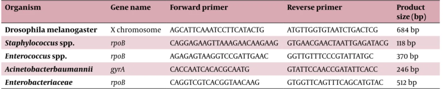
re-spectivelychosen as target sequences for the selected bac-teria. The primer sets and genes target sequences with accession numbers are listed in Table 1.
Table 1. List of Oligonucleotide Primers Used for Conventional Multiplex PCR Amplification.
Organism Gene name Forward primer Reverse primer Product
size (bp) Drosophila melanogaster X chromosome AGCATTCAAATCCTTCATACTG ATGTTGGTGTAATCTGACTCG 684 bp
Staphylococcus spp. rpoB CAGGAGAAGTTAAAGAACAAGAAG GTGAACGAACTAATTGAGATACG 118 bp
Enterococcus spp. rpoB AGAGAGTAAGGTCCGATTGAAC GGTTGTTTCCCGTATTATGC 370 bp
Acinetobacterbaumannii gyrA CACCAATCACACGCAATG GTATTCCAACCGATATTCACC 246 bp
Enterobacteriaceae rpoB CAGGTCGTCACGGTAACAAG GTGGTTCAGTTTCAGCATGTAC 512 bp
3.2. Construction and Preparation of Standard
In-ternal Control
The PTZ75 plasmid was used as the internal control DNA standard. This plasmid was constructed by ligat-ing a PCR amplified fragment accordligat-ing to the instruc-tion of the T/A cloning kit (Fermentas, Lithuania). The cloned fragment comprised a 684bp sequence from the D.melanogaster chromosome: in detail the selected
se-mosome X (Genbank accession number NC_004354.3). A series of 10 fold dilutions of the IC containing plas-mid, ranging from 1 to 108copies/reaction were prepared and stored at - 80º C until used.
3.3. Bacterial Strains and DNA Extraction
Four different bacterial strains were selected as proto-type of Gram positive (Enterococcusfaecalis ATCC 29212,
(Acinetobacterbaumannii ATCC 19606, Escherichia coli ATCC 25922) microorganisms, respectively. These bacteria were grown using the method of standard laboratory condi-tions (14). Each target sequence was cloned by using the T/A cloning kit (Fermentas, Lithuania) as reported below and enumerated by A260 absorbance. In addition, a pan-el of bacterial species, including gram positive and gram negative obtained both from the American Type Culture Collection (ATCC) and from clinical isolates collection (University of Isfahan) were tested.To evaluate the effi-ciencyof different techniques for the extraction of bac-terial DNA, each singlebacbac-terial suspension was treated with 4 diverse commercially available methods (QIAamp DNA mini blood kit (Qiagen, Germany), High Pure PCR Template Preparation Kit (Roche applied bioscience, Germany), Genomic DNA Purification Kit (Fermentas, Lithuania) and DNA extraction kit (Cinagen, Iran).) as fol-lows: 200 µl of bacterial suspension (0.5 McFarland) were subjected to individual extraction protocols as indicated by each manufacturer. 1000 copies number of the DNA sequence, used as IC, were added to each sample before performing the extraction procedure. The final volume of the eluted DNA was adjusted to 100 µl.
3.4. Multiplex PCR
PCR conditions were optimized according to the manu-al of the emermanu-ald Amp MATHS PCR Master Mix (2X premix Bio Inc, Takara, Japan). Details of the procedure are given as follows: 0.2 mM of each primer and 1.25 U of Taq poly-merase; optimal condition for annealing temperature was 60º C. As a template, 5 µl extracted DNA was added thus resulting in a total reaction volume of 25 µl. Ampli-fication started with a cycle of 4 min at 94º C, followed by 35 cycles of denaturation at 94º C for 30 second, an-nealing at 60º C for 30 second and extension at 72º C for 1 min, subsequently final extension at 72º C for 10 min. The amplicons obtained for each set of species were distin-guished by visualizing the PCR products by electrophore-sis in 1% agarose gel and stained with ethidiume bromide (0.5 µg/ml). Stained gels were observed with UV light and images digitalized with (UVI doc HD2 LCD, UVITEC, UK).
Sensitivity was tested with serial dilutions of different plasmids containing the target sequences of the above indicated 4 prototype strains ranging from 1 to 108 cop-ies number/reaction or serial dilutions of DNA extracted from human blood samples in vitro spiked by the 4 pro-totype strains. Specificity was tested with DNA extracted from the 17 bacterial species, including gram positive and gram negative obtained both from the American Type Culture Collection (ATCC) and from clinical isolates col-lection.
4. Results
4.1. Evaluation of Internal Control
The sensitivity of the IC DNA was tested by amplification of serial dilutions of quantified IC sequence containing
plasmid. Specific amplification was obtained from all plasmid samples with concentration ranging from 1 to 108 copies per reactions, showing a high efficiency of am-plification for the IC sequence selected (Figure 1).
1000 bp 700 bp 500 bp
Figure 1. PCR amplification of the Internal Control (IC). The analytical
sensitivity of the PCR was determined with serial dilutions of a plasmid containing the IC sequence ranging from 1to 108 copies. The lower de-tection limit for IC was 1 copy/reaction. The positions of the molecular weight markers (1 kbp) are indicated on the right.
200 180 160 140 120 100
Qiagen Roche Fermentas Cinagen
DNA concentration (ng/µl)
Extraction kites
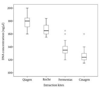
Figure 2. The box plot shows the total DNA concentration (ng/μl)
ob-tained with the 4 different extraction kits. The Qiagen kit yeilded the highest DNA concentration. The boxplot shows that the highest purity with small variations was obtained by the Qiagen kit.
4.2. Qualitative Analysis of Different Extraction
Methods
The quality of extracted bacterial DNA with the four kits was compared for each one of the four different pro-totype bacteria species, by using 5 replicate samples per germ. The mean OD value obtained with, Qiagen, Roche, Ferments and Cinagen kits were 1.93, 1.84, 1.56 and 1.32, respectively. The concentration of bacterial DNA (ng/µl)
obtained with Qiagen, Roche, Fermentas and Cinagen kits are reported in figure 2. There were statistically sig-nificant differences (P < 0.05) among them and the best quality was obtained with QIAamp DNA mini blood kit (Qiagen, Germany).
4.3. Sensitivity and Specificity of the Multiplex PCR
Assay
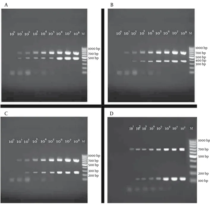
In order to evaluate the sensitivity of the single tube multiplex PCR presented in the current study, the detec-tion limits for all 4 prototype microorganisms were in-vestigated, as reported above. The multiplex PCR assay identified all the 4 prototype bacterial strains success-fully (Figure 3); moreover, no amplification products were detected for the 17 strains investigated for specificity of the method (data not shown). The lower detection limit for Gram positive were 102 copies per reactions or 100 CFU/ml for both E.faecalis (Figure 3 B) and S.aureus (Figure
3 D), whereas the lower detection limit for A.baumannii
(Figure 3 C) and E.coli (Figure 3 A) were 10 copiesor 50 CFU/ml, respectively. The correct amplification of the IC showed that the whole workflow, starting from the ex-traction step, was performed in a reliable way.
5. Discussion
Rapid detection of bacteraemia is one of the most im-portant tools in the clinical diagnosis of blood stream infections. Molecular techniques have been developed to achieve the diagnosis of bacterial infection by detect-ing bacterial DNA in blood faster (5, 15). The purity of the nucleic acid used in PCR is a critical point to achieve the highest sensitivity and to ensure reproducible results over time. Standard commercially available methods and kits are generally suitable for producing clean nucleic acid samples with sufficient concentration (16). The ma-jority of the molecular assays are designed specifically to detect only one single organism, thus providing a high level of sensitivity and specificity (17-20). At the same time, a major limitation of this diagnostic approach is the capability to identify a unique bacterial species. Re-cently, a variety of a broad range PCR or multiplex PCR were applied in order to screen and detect the presence of multiple organismsin clinical samples (4, 21, 22). The major limitations of these methods are their cost to effi-cacy ratio and the fact that most of these techniques are based on time-consuming procedure for the final iden-tification of the microorganism, such as: sequencing, restriction fragment length polymorphism or hybridiza-tion with germ specific probes (23-25).
The current study successfully combined the simulta-neous detection of four different prototype bacteria with primers designed for the identification of A.baumanii
belong, such as Staphylococcus sppandEnterococcus spp., and the most prominent pathogenic members of the family Enterobacteriaceae with a multiplex PCR assay. The current study results showed a detection limit variable between 10 to 100 copies of the target sequencesor 50 to 100 CFU/ml.Similar detection limits were observed for dif-ferent molecular techniques when applied to difdif-ferent samples (26-28).
A previous study conducted by Bonilla et al.showed that the 37,5% of the culture positive samples were negative by PCR (22) and this fact is likely due to inhibition caused by a variety of factors, of which the most prominent is likely the ongoingantibiotic treatment. Moreover, a PCR inhibition was observed in whole blood samples when high level of leukocytosis was present (29). In particular, it was hypoth-esized that the PCR inhibition was probably due to the low performance of the extraction methods utilized or by the competition between the relatively low level of target DNA and the high level of human genomic DNA (29).
Based on these findings, the use of an internal control sequence can be suggested when whole blood samples or patient with special conditions, that are likely to gen-erate PCR inhibition, must be investigated. Several com-pounds in blood have been suggested to possibly inhibit PCR, namely: heme, leukocyte derived host DNA (30), and anticoagulants (31, 32). Inhibition has been shown to affect the sensitivity of LCR-based amplification tests (33), TMA tests (34) and NASBA assays (35). An additional cause of false negative results, other than the presence of inhibiting factors, could be a number of target se-quences below the assay detection limit or the presence of sequence variability (36). To monitor the process in ge-nome amplification based tests, an internal control can be used (37, 38). The result of these studies demonstrates that incorporation of an internal control into PCR-based tests increase sensitivity by enabling the user to identify and retest samples that were scored as negative just for the presence of inhibition phenomena. Furthermore, a positive IC result indicates that the final amplification occurred and thus provides assurance that negative results are truly negative. Testing a dilution series of IC DNA revealed that the assay is able to detect concentra-tions as low as one copy of this sequence and this value is well comparable with the limits detected for the bacte-rial genes: this fact ensures that the IC could be properly detected in all the samples. The panel of the evaluated pathogenic bacteria encompasses several of the most prominent epidemiological causes of invasive infections in selected patient population in the Middle East area (39-42). It is indeed easy to perform the test reported in this manuscript which has an elevated cost effectiveness ratio due to its simplicity. It can consequently be proposed in the routine workflow of microbiology laboratories
locat-1000 bp 700 bp 500 bp
1000 bp 700 bp 500 bp 1000 bp 700 bp 500 bp 400 bp 300 bp
1000 bp 700 bp 500 bp
200 bp 200 bp
100 bp 300 bp
A
B
C
D
Figure 3. Amplification pattern of the 4 prototype bacterial strains. Detection limit of multiplex PCRs are shown in each panel. Each lane contains
se-rial dilutions of plasmid DNA target sequences starting from 1 to 108 copies derived from: (Panel A) E. coli and IC (Panel B) Enterococcus faecalis and IC (Panel C) Acinetobacter baumaniiand IC (Panel D) Staphylococcus aureus and IC. The assays were able to detect as low as10 copies/reaction for E.coli and A.
baumanii while the detection limit was 100 copies/reaction for S.aureus and E.faecalis.
Acknowledgements
The authors wish to thank Prof. Vittorio Sambri and Dr. PaoloGaibani from Regional Reference Centre for Micro-biological Emergencies (CRREM) S. Orsola-Malpighi Uni-versity Hospital, Bologna, Italy for reading and editing the manuscript.
Authors’ Contribution
All authors had same contribution.
Financial Disclosure
There is no conflict of Interest.
Funding Support
This study is supported by Isfahan University of Medical Sciences.
References
Arch Med Res. 2005;36(6):646-59.
2. Bochud Pierre-Yves, Glauser Michel P, Calandra Thierry. Antibiot-ics in sepsis. Intensive Care Medicine. 2001;27(1):S33-S48. 3. DiDomenico N, Link H, Knobel R, Caratsch T, Weschler W, Loewy
ZG, et al. COBAS AMPLICOR: fully automated RNA and DNA am-plification and detection system for routine diagnostic PCR. Clin
Chem. 1996;42(12):1915-23.
4. Harris KA, Hartley JC. Development of broad-range 16S rDNA PCR for use in the routine diagnostic clinical microbiology service. J
Med Microbiol. 2003;52(Pt 8):685-91.
5. Paolucci M, Landini MP, Sambri V. Conventional and molecular techniques for the early diagnosis of bacteraemia. Int J
Antimi-crob Agents. 2010;36 Suppl 2:S6-16.
6. Rampini SK, Bloemberg GV, Keller PM, Buchler AC, Dollenmaier G, Speck RF, et al. Broad-range 16S rRNA gene polymerase chain reaction for diagnosis of culture-negative bacterial infections.
Clin Infect Dis. 2011;53(12):1245-51.
7. Heininger A, Binder M, Ellinger A, Botzenhart K, Unertl K, Dor-ing G. DNase pretreatment of master mix reagents improves the validity of universal 16S rRNA gene PCR results. J Clin Microbiol. 2003;41(4):1763-5.
8. David H. Persing . Diagnostic molecular microbiology: principles
and applications. 1993.
9. Rossen Lone, Nørskov Pernille, Holmstrøm Kim, Rasmussen Ole F. Inhibition of PCR by components of food samples, microbial diagnostic assays and DNA-extraction solutions. International
Journal of Food Microbiology. 1992;17(1):37-45.
10. Panaccio M, Lew A. PCR based diagnosis in the presence of 8% (v/v) blood. Nucleic Acids Res. 1991;19(5):1151.
11. Lantz Pär-Gunnar, Matsson Mikael, Wadström Torkel, Rådström Peter. Removal of PCR inhibitors from human faecal samples through the use of an aqueous two-phase system for sample preparation prior to PCR. Journal of Microbiological Methods. 1997;28(3):159-167.
12. Al-Soud WA, Radstrom P. Purification and characterization of PCR-inhibitory components in blood cells. J Clin Microbiol. 2001;39(2):485-93.
13. Kellogg DE, Sninsky JJ, Kwok S. Quantitation of HIV-1 proviral DNA relative to cellular DNA by the polymerase chain reaction.
Analytical Biochemistry. 1990;189(2):202-208.
14. Murray PR, Baron EJ, Pfaller MA, Tenover FC, Yolken RH. Manual of
clinical microbiology. 1999.
15. Fredricks DN, Relman DA. Improved amplification of microbial DNA from blood cultures by removal of the PCR inhibitor so-dium polyanetholesulfonate. J Clin Microbiol. 1998;36(10):2810-6. 16. Barken KB, Haagensen JA, Tolker-Nielsen T. Advances in nucleic
acid-based diagnostics of bacterial infections. Clin Chim Acta. 2007;384(1-2):1-11.
17. Palomares Concepción, Torres María J, Torres Antonio, Aznar Javi-er, Palomares José C. Rapid detection and identification of Staph-ylococcus aureus from blood culture specimens using real-time fluorescence PCR. Diagn Microbiol Infect Dis. 2003;45(3):183-189. 18. Thomas LC, Gidding HF, Ginn AN, Olma T, Iredell J. Development
of a real-time Staphylococcus aureus and MRSA (SAM-) PCR for routine blood culture. J Microbiol Methods. 2007;68(2):296-302. 19. Elfaki MG, Uz-Zaman T, Al-Hokail AA, Nakeeb SM. Detection of
Brucella DNA in sera from patients with brucellosis by poly-merase chain reaction. Diagn Microbiol Infect Dis. 2005;53(1):1-7. 20. Long F, Zhu XN, Zhang ZM, Shi XM. Development of a
quantita-tive polymerase chain reaction method using a live bacterium as internal control for the detection of Listeria monocytogenes.
Diagn Microbiol Infect Dis. 2008;62(4):374-81.
21. Xirogianni A, Tzanakaki G, Karagianni E, Markoulatos P, Kourea-Kremastinou J. Development of a single-tube polymerase chain reaction assay for the simultaneous detection of Haemophilus influenzae, Pseudomonas aeruginosa, Staphylococcus aureus, and Streptococcus spp. directly in clinical samples. Diagn
Micro-biol Infect Dis. 2009;63(2):121-6.
22. Bonilla H, Kepley R, Pawlak J, Belian B, Raynor A, Saravolatz LD. Rapid diagnosis of septic arthritis using 16S rDNA PCR: a com-parison of 3 methods. Diagn Microbiol Infect Dis. 2011;69(4):390-5. 23. Xu J, Moore JE, Millar BC, Webb H, Shields MD, Goldsmith CE. Em-ployment of broad range 16S rDNA PCR and sequencing in the detection of aetiological agents of meningitis. New Microbiol. 2005;28(2):135-43.
24. Tsai JC, Teng LJ, Hsueh PR. Direct detection of bacterial pathogens
in brain abscesses by polymerase chain reaction amplification and sequencing of partial 16S ribosomal deoxyribonucleic acid fragments. Neurosurgery. 2004;55(5):1154-62.
25. Lu JJ, Perng CL, Lee SY, Wan CC. Use of PCR with universal primers and restriction endonuclease digestions for detection and iden-tification of common bacterial pathogens in cerebrospinal fluid.
J Clin Microbiol. 2000;38(6):2076-80.
26. Rothman RE, Majmudar MD, Kelen GD, Madico G, Gaydos CA, Walker T, et al. Detection of bacteremia in emergency depart-ment patients at risk for infective endocarditis using universal 16S rRNA primers in a decontaminated polymerase chain reac-tion assay. J Infect Dis. 2002;186(11):1677-81.
27. Zucol F, Ammann RA, Berger C, Aebi C, Altwegg M, Niggli FK, et al. Real-time quantitative broad-range PCR assay for detection of the 16S rRNA gene followed by sequencing for species identifica-tion. J Clin Microbiol. 2006;44(8):2750-9.
28. Poppert S, Essig A, Stoehr B, Steingruber A, Wirths B, Juretschko S, et al. Rapid diagnosis of bacterial meningitis by real-time PCR and fluorescence in situ hybridization. J Clin Microbiol. 2005;43(7):3390-7.
29. Jordan Jeanne A, Durso Mary Beth. Real-Time Polymerase Chain Reaction for Detecting Bacterial DNA Directly from Blood of Neo-nates Being Evaluated for Sepsis. The Journal of Molecular
Diagnos-tics. 2005;7(5):575-581.
30. Morata P, Queipo-Ortuno MI, de Dios Colmenero J. Strategy for optimizing DNA amplification in a peripheral blood PCR as-say used for diagnosis of human brucellosis. J Clin Microbiol. 1998;36(9):2443-6.
31. Wang JT, Wang TH, Sheu JC, Lin SM, Lin JT, Chen DS. Effects of anti-coagulants and storage of blood samples on efficacy of the poly-merase chain reaction assay for hepatitis C virus. J Clin Microbiol. 1992;30(3):750-3.
32. Akane A, Matsubara K, Nakamura H, Takahashi S, Kimura K. Identification of the heme compound copurified with deoxy-ribonucleic acid (DNA) from bloodstains, a major inhibitor of polymerase chain reaction (PCR) amplification. J Forensic Sci. 1994;39(2):362-72.
33. Stary A, Tomazic-Allen S, Chouelri B, Burczak J, Steyrer K, Lee H. Comparison of DNA Amplification Methods for the Detection of Chlamydia trachomatis in First-Void Urine From Asymptomatic Military Recruits. Sexually Transmitted Diseases. 1996;23(2):97-102. 34. Manterola JM, Gamboa F, Lonca J, Matas L, Ruiz Manzano J, Rodri-go C, et al. Inhibitory effect of sodium dodecyl sulfate in detec-tion of Mycobacterium tuberculosis by amplificadetec-tion of rRNA. J
Clin Microbiol. 1995;33(12):3338-40.
35. Morre SA, Sillekens P, Jacobs MV, van Aarle P, de Blok S, van Gemen B, et al. RNA amplification by nucleic acid sequence-based ampli-fication with an internal standard enables reliable detection of Chlamydia trachomatis in cervical scrapings and urine samples.
J Clin Microbiol. 1996;34(12):3108-14.
36. Rosenstraus M, Wang Z, Chang SY, DeBonville D, Spadoro JP. An internal control for routine diagnostic PCR: design, properties, and effect on clinical performance. J Clin Microbiol. 1998;36(1):191-7.
37. Wang AM, Doyle MV, Mark DF. Quantitation of mRNA by the poly-merase chain reaction. Proc Natl Acad Sci U S A. 1989;86(24):9717-21.
38. Mulder J, McKinney N, Christopherson C, Sninsky J, Greenfield L, Kwok S. Rapid and simple PCR assay for quantitation of human immunodeficiency virus type 1 RNA in plasma: application to acute retroviral infection. J Clin Microbiol. 1994;32(2):292-300. 39. Erdinc FS, Yetkin MA, Hatipoglu CA, Yucel M, Karakoc AE, Cevik
MA, et al. Erratum to ‘Five-year surveillance of nosocomial in-fections in Ankara Training and Research Hospital [Journal of Hospital Infection 2006;64:391–396]’. Journal of Hospital Infection. 2007;67(2):205-206.
40. Al-Tawfiq JA, Abed MS. Prevalence and antimicrobial resistance of health care associated bloodstream infections at a general hos-pital in Saudi Arabia. Saudi Med. 2009;30:1213-18.
41. Akbar DH, Mushtaq MA, El-Tahawi AT, Bahnasy AA. Staphylococ-cus aureus bacteremia. Saudi Med. 2000;21:171-4.
42. Mamishi S, Pourakbari B, Ashtiani MH, Hashemi FB. Frequency of isolation and antimicrobial susceptibility of bacteria isolated from bloodstream infections at Children's Medical Center, Teh-ran, ITeh-ran, 1996-2000. Int J Antimicrob Agents. 2005;26(5):373-9.