Aliso: A Journal of Systematic and Evolutionary Botany
Volume 14 | Issue 4
Article 5
1995
Using Confocal Microscopy in the Study of Plant
Structure and Development
Mark P. Running
California Institute of Technology
Elliot M. Meyerowitz
California Institute of TechnologyFollow this and additional works at:
http://scholarship.claremont.edu/aliso
Part of the
Botany Commons
Recommended Citation
Running, Mark P. and Meyerowitz, Elliot M. (1995) "Using Confocal Microscopy in the Study of Plant Structure and Development," Aliso: A Journal of Systematic and Evolutionary Botany: Vol. 14: Iss. 4, Article 5.
© 1996, by The Rancho Santa Ana Botanic Garden, Claremont, CA 91711-3157
USING CONFOCAL MICROSCOPY IN THE STUDY OF PLANT STRUCTURE AND DEVELOPMENT
MARK P. RUNNING AND ELLIOT M. MEYEROWITZ
Division of Biology 156-29 California Institute of Technology
Pasadena, CA 91125 Phone: (818) 395-6889, 395-4936
FAX: (818) 449-0756
email: runningm@ starbase 1. cal tech. edu, meyerowitze@ starbasel.caltech.edu
ABSTRACT
The widespread application of confocal laser scanning microscopy has revolutionized biological imaging. We have developed a protocol for using confocal microscopy to examine the development of wild type and mutant Arabidopsis thaliana, overcoming the technical difficulties associated with examining whole-mounted plant tissue. This allowed us to rapidly determine the underlying cellular defects that lead to the morphological changes visible in several mutants, and has led to a greater understanding of the mechanisms involved in the control of floral organ number.
Key words: Arabidopsis, confocal microscopy, flower development, plant development.
INTRODUCTION
Rapid advances in computer technology have rev-olutionized biological studies in recent years. In par-ticular, the development of faster, more powerful, and inexpensive computers have enabled the practical ap-plication and widespread use of sophisticated imaging techniques. One of these techniques is confocal laser scanning microscopy (CLSM), a specialized type of florescence microscopy. In CLSM, a laser is focused on a single point in a sample, and the resulting fluo-rescent light is focused on an aperture in such a way that light emanating from above or below the plane of focus falls outside the aperture and is eliminated. Us-ing computer-driven motors, the microscope can scan and obtain image data from a large series of single points in a two dimensional plane, obtaining an optical section through the sample. A series of optical sections can be combined into a three-dimensional image of the sample.
CLSM has many advantages over histological tech-niques conventionally used in the study of plant anat-omy and development, such as manual sectioning and scanning electron microscopy (SEM). CLSM allows image data to be taken from any and all points in a whole mounted sample, and computers can be used to reconstruct the three dimensional image, perform an-imation and other image processing manipulations to aid in interpretation, and perform virtual sectioning from any angle. Unlike serial sectioning, the integrity of the sample is preserved in CLSM, allowing resam-pling of data using different parameters. Preparing tis-sue for CLSM analysis is also much less labor-inten-sive and much less error-prone than sectioning. SEM
can also be used to examine whole-mounted samples, but in SEM only surface cells are visible, whereas in CLSM internal cell patterns can be discerned. CLSM is also very useful for studying tissues that are more inaccessible to dissection, such as the embryo and ear-ly floral meristem.
We have used CLSM successfully in our studies of wild type and mutant apical and floral meristem de-velopment in a member of the family Brassicaceae,
Arabidopsis thaliana (Clark et al. 1993, 1995; Run-ning et al. 1995; RunRun-ning and Meyerowitz 1996). Ar-abidopsis has emerged as a popular model system for genetic and molecular studies of developmental pro-cesses in plants (Meyerowitz and Sommerville 1994). The plants are very small, so thousands can be grown at once in a single growth chamber. They are also easy to grow, requiring no special care. In addition, they are self fertile, easy to cross, and have a high fecun-dity. They also have an exceptionally short generation time (approximately six weeks) and a very small ge-nome size (as small as the nematode Caenorhabditis elegens and two-thirds the size of the insect Drosoph-ila melanogaster), making genetic characterization and molecular cloning feasible in short time frames.
264
in order to identify the primary defect at the early stages of apical and floral meristem development.
MATERIALS AND METHODS
Arabidopsis Mutant Nomenclature
In Arabidopsis a gene is given a name and a 2 or 3 letter abbreviation. When referring to the wild type gene, the name and its abbreviation are written in ital-ics and capitals; for example, PERIANTHIA (PAN).
When referring to mutant genes, the name is written in italics and lower case; for example, perianthia (pan). Sometimes a number of different genes have similar mutant phenotypes; in this case, the genes are numbered sequentially. For example, there are 3 genes that fall into the CIA VATA (CLV) class, CIA VAT AI (CLVJ), CIAVATA2 (CLV2), and CIAVATA3 (CLV3).
If there are more than one mutant allele of the gene, each allele is given a number which follows the gene name; examples are pan-2, clvl-1, and clv3-2.
Plant Material
For all plants, seeds were sown at least 2 em apart on a 1:1:1 mix of soil:perlite:vermiculite, imbibed at 4 C for 4 days, then placed under 600 foot-candles of constant cool-white fluorescent light at 23 C. Plants were fertilized at regular intervals beginning approxi-mately 7 days after germination. Alleles of clavatal (clvl) and clavata3 (clv3) were obtained as described (Clark et al. 1993, 1995); alleles of perianthia (pan)
and hanaba taranu (han) were isolated in a screen of
Agrobacterium tumefaciens-mediated T-DNA muta-genized Arabidopsis thaliana seeds by Kenneth Feld-mann at the University of Arizona; tsol and wiggum
emerged from screens of seed lines mutagenized by ethyel methanesulphonate at the California Institute of Technology in the lab of E. M. M.
Confocal Microscopy and Other Imaging
A detailed description of tissue preparation for con-focal microscopy is available in Running et al. (1995). Plant tissue is fixed in Formalin:Propionic Acid:Etha-nol (10:5:70), placed under vacuum briefly, then left overnight in fixing solution. The samples are then brought through an ethanol series to 100% ethanol to remove chlorophyll and complete the fixing process. After 4 hours in 100% ethanol the sample is brought through a decreasing ethanol series to water, and
ALISO
stained in 5 ug/ml propidium iodide in a solution of 0.1 M L-arginine buffer at pH 12.4 at 4 C for 1 day or more. The samples are then placed in 0.1 M L-ar-ginine buffer at pH 8 without stain for 4 days at 4 C, changing the solution at least once a day. After an increasing ethanol series to 100%, the sample is placed through a series of ethanol/xylene solutions until the sample is in 100% xylene and completely cleared. The sample is dissected (if necessary) in immersion oil, and mounted in a small drop of immersion oil placed on a cover slip. The cover slip is inverted and placed on a depression slide and secured by nail polish. The samples were viewed with a Zeiss LSM10 microscope using an attenuated argon laser emitting at 514 nm as excitation light. We used a Zeiss Neofluar 40X im-mersion oil lens with a numerical aperture of 1.30. Figures were prepared digitally as described (Sieburth et al. 1995).
RESULTS AND DISCUSSION
Flowers of Arabidopsis, like most other members of the Brassicaceae, initiate four sepals, four petals, six stamens, and two carpels in succession (Smyth et al. 1990; Fig. 1A). The organs emerge in a stereotypical pattern: sepals emerge at 90 degree angles around the floral meristem, with two medial and two lateral sepals present. Petals emerge interior to and in between the sepals, with two medial stamens arising interior to each of the medial sepals, and one lateral stamen aris-ing interior to each of the lateral sepals. Carpels emerge in the center of the floral meristem, with the septum forming along the medial axis.
We are interested in the process by which the flower pattern is established: how certain molecular cues tell cells to grow, divide, and differentiate properly ac-cording to their position in the flower. As geneticists, our strategy is to identify molecules involved in this process by isolating mutants that are defective in the number or pattern of organs. Study of the mutant phe-notype will give us clues into the wild type function of the mutated gene, and in Arabidopsis methods to clone genes identified by mutation are available. Our long term goal is to identify mechanisms by which organ number and pattern is determined.
Several mutants affecting organ number have been isolated, all of which affect floral organ number in various ways. The clavata (clv) class of mutants, com-prising three separate loci, show an increase in organ number in all four whorls of the flower, but has the
c
,
• ···:····
. . . •·.
,..
. ..,
266 ALISO
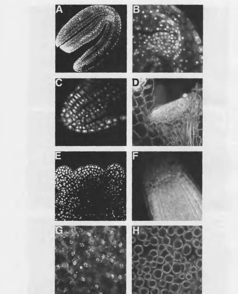
Fig. 2. Examples of wild type Arabidopsis thaliana tissues that can be examined using confocal microscopy.-A. Mature embryo, with
most dramatic effect on carpels, and also show addi-tional whorls of carpels growing interior to the fourth whorl of carpels (Fig. IB; Koornneef et al. I983; Ley-ser and Furner I992; Clark et al. 1993; I995). The
wiggum (wig) mutant also increases organ number, but its most dramatic effect is on the number of sepals and petals (Fig. IC; M.P. R. and E. M. M., unpublished). The hanaba taranu (han) mutant has a different effect: organ number is decreased in all four whorls, but the second and third whorls are most dramatically affected (Fig. ID; Clark et al. I994). The tsol mutant has a near-normal number of first whorl organs, but second, third, and fourth whorl organs fail to initiate (Fig. IE; Z. Liu, M. P. R., and E. M. M., unpublished). Finally,
perianthia (pan) mutant flowers tend to have pentam-erous flowers (5 sepals, petals, stamens), thus increas-ing the number of first and second whorl organs but decreasing the number of third whorl organs compared to wild type (Fig. IF; Running and Meyerowitz I996). Once these mutants were isolated, we were inter-ested in examining their development in detail, to see if we could trace back the cause of the defect, or to perhaps correlate it with changes in the earliest stages of the development of the floral apex. In particular, we were interested in seeing how cell number, cell pat-terns, and meristem size and structure could play a role in determining organ number. We found that examin-ing these mutants with confocal microscopy was the most efficient and effective way of doing this. We de-veloped a protocol that allows nuclear staining of whole mounted plant tissues (Running et al. I995), and have used it successfully in the examination of many different tissues throughout development (Fig. 2). One stage that is particularly amenable to examination with confocal microscopy is the mature embryo (Fig. 2A), where the shoot apical meristem (Fig. 2B) and root meristem (Fig. 2C) particularly easy to image. The structure of the apical meristem can be followed through development, from the vegetative phase, when it is relatively flat (Fig. 2D), to the adult phase (Fig. 2E), where it becomes more rounded in appearance. The size of the root meristem, and the cell pattern and number in the root, does not change throughout the life of the plant (Fig. 2F). The leaf epidermal (Fig. 2G) and leaf mesophyll cells (Fig. 2H) are also visible under confocal microscopy.
The first locus we examined was CLA VAT AI, mu-tations in which lead to plants showing an increase in organ number, particularly in the inner whorls (Fig. lB). By examining the flowers at the first stage of organ initiation, we could readily detect a size change
in the floral meristem (Fig. 3A,B). Specifically, the floral meristem was taller but not wider than wild type at the time the sepals arise. This was due to a greater number of cells, since the size of the cells and the organization of cells into three distinct layers is not affected. The degree of the change in size also corre-lates with the severity of the organ number defect in different alleles: those alleles with the largest increase in organ and whorl number, such as clv 1-4, had the greatest increase in early floral meristem size (Clark et al. 1993). clv3 mutants, which have a phenotype similar to clv I, have a similar increase in meristem size (Clark et al. I995). In examining other tissues in clv
mutants, we found that the apical meristem also has an increased size, though the size and pattern of cells remains intact (Fig. 3C). Increased meristem size is detectable at least as early as the mature embryo stage (Fig. 3D). Root meristems and other areas of active cell division were not detectably affected.
The wig mutant also leads to an increased number of organs in the flower, but its effect is opposite of clv
mutants in that wig primarily affects the outer two whorls, the sepals and petal number showing the great-est deviation from wild type (Fig. IC). We found that
wig also has a readily detectable size change in the early floral meristem (Fig. 3E). In this case, though, the meristem is wider but not taller than wild type, which is what presumably leads to extra organs in the first and second whorl.
The mutant han shows a decrease rather than an increase in organ number (Fig. lD). All four whorls are affected, but the second and third whorls show the strongest effect, even lacking organs sometimes. Our CLSM analysis indicates that at the stage where the second and third whorl organs are initiating, the floral meristem size is much smaller in han mutants com-pared to wild type (Fig. 3F,G). In addition, the apical meristem is smaller, visible as early as in the mature embryo (Fig. 3H,I).
Studies of the clv, wig, and han mutants suggest that one potential mechanism for floral organ number reg-ulation is through regreg-ulation of floral meristem size. Another potential mechanism is identified by the tsol
mutant (Fig. IE). tsol flowers initiate close to the nor-mal number of sepals, but further initiation largely ceases. Instead of petals and reproductive organs, often a mass of undifferentiated tissue appears in the center of the flower. Using CLSM we were able to see that, while the floral meristem size is normal, cell number and pattern are abnormal at the time of sepal initiation (Fig. 3J). Cells are larger and decreased in number,
268
o
.
•
•
•
•
•
•
•
..
,
.
.
.
'.
•••
. ,
.
...
f.
. .
•
..
....
.
~
~.
.
·
----~·
.
.
.
...
..
~
..
...
.
.
,..,~
..
r
·
..
.
·
•
..
-.-
n
..
.
..
..
•
.
.
.
...
.
.
•
•
•
..
••
!-
-
~
·
..
...
'
,,
.
..
8
....
~... .
.:~~~.~
-~~
....
····~~
...
~. t~ ''·~-
....
'·,
••
•.\-k~
-
·.
• ... • • .e • . _. .. •
• • • • • • •t/
r•
-
...
f... _., ;.·~·. il
'··- .
,..
•.:' ,.~
.
.
·
..•.··~
, • . • # •-:.'"r • •
..
~.
" ..
...
·-·~ '. .
.
.
'
,
•.
• ,&..
•o;
.
..
.ALISO
Fig. 3. Confocal microscopy images of wild type and mutant apical and floral meristems.-A. Wild type stage 3 floral meristem. The
and do not fall into obvious cell layers as they do in wild type. At later stages, the sepals continue to grow, but organ morphogenesis breaks down (Fig. 3K). Many nuclei are large and stain more intensely, indi-cating that DNA replication continues but nuclear di-vision and cytokinesis are defective. Thus organized cell division patterns are important to some extent in the initiation and development of floral organs.
Another class of genes is represented by PAN. The most commonly found pan mutant flowers have five sepals, five petals, five stamens, and two carpels, a pattern found in many plant families but not in the Brassicaceae. Unlike the mutants described previously, we were unable to detect a difference in size, cell num-ber, or cell pattern in the floral meristem at the time of sepal initiation (Fig. 3L). Significantly, the organ number increase is more dramatic in pan compared to
wig, in which a size change is readily detectable.
An-other difference is detectable by SEM: the sepal ini-tiation in pan is very regular and predictable, with an adaxial sepal always present, and the rest of the organs equidistant around the ring. In wig and the clv mutants, sepal initiation is much more irregular.
The histological evidence supports the division of the mutants affecting organ number into three classes: one class that affects meristem size and shape (clv, han, wig), one that affects cell division patterns (tso),
and one that acts specifically at the level of organ ini-tiation (pan). Genetic studies also support this classi-fication scheme. Double mutants of genes in different classes have additive phenotypes, and double mutants of genes within the classes have non-additive or syn-ergistic phenotypes. For instance, double mutants be-tween pan and clv or pan and wig have about one additional sepal and petal and one fewer stamen com-pared to clv or wig alone. The wig clv double mutants, though, show dramatically decreased organ number, reduced fertility, and an apex that overproliferates and differentiates into stigmatic tissue.
Our experience has shown that confocal microscopy can be used successfully in the examination of organ number mutants to gain a better understanding of the underlying causes at the cellular level. Conceivably, confocal microscopy could be useful in evolutionary studies in a similar manner. It is possible that changes
in floral organ number among closely related species could be due to one or more of the mechanisms sug-gested by the mutants we have discussed. For instance, a specific size change in the floral meristem will lead to a change in organ number, as in clv or wig mutants. Similarly, a cessation in cell division in specific regions of the floral meristem may lead to missing or-gans in particular locations in the flower, as in tso mu-tants. Confocal microscopy is a rapid, convenient method for looking at cell patterns and meristem struc-ture, and could greatly aid such cross-species studies.
ACKNOWLEDGMENTS
We thank Kenneth Feldmann for the opportunity to screen mutant lines, Detlef Weigel for providing the
wig mutant, and Steven E. Clark, Zhongchi Liu, and
Hajime Sakai for materials and fruitful discussions. We thank Jean-Paul Revel for advice and assistance with the confocal microscope. We thank Jian Hua and Bob-by Williams for comments on the manuscript. This work was supported by NSF grant MCB-9204839 to E. M. M. M. P. R. was a Howard Hughes predoctoral fellow.
LITERATURE CITED
CLARK, S. E., M.P. RUNNING, AND E. M. MEYEROWITZ. 1993.
CLA-VATA1, a regulator of meristem and flower development in Ara-bidopsis. Development 119: 397-418.
- - - , - - - , A N D - - - . 1995. CLAVATA3 is a regulator of shoot and floral meristem development affecting the same pro-cesses as CIA VATA1. Development 121: 2057-2067.
- - - , H. SAKAI, AND E. M. MEYEROWITZ. 199~. Inflorescence development in clavata mutants. In J. L. Bowman [ed.], Arabi-dopsis, An Atlas of Morphology and Development. Springer-Ver-lag, New York, pp. 214-215.
KOORNNEEF, M., J. VAN EDEN, C. J. HANHART, P. STAM, F. J. BRAAKS-MA, AND W. J. FEENSTRA. 1983. Linkage map of Arabidopsis
thal-iana. J. Hered. 74: 265-272.
LEYSER, H. M. 0., AND I. J. FURNER. 1992. Characterization of three shoot apical meristem mutants of Arabidopsis thaliana.
Devel-opment 116: 397-403.
MEYEROWITZ, E. M., AND C. S. SOMERVILLE. 1994. Arabidopsis. Cold Spring Harbor Laboratory Press.
RUNNING, M.P., S. E. CLARK, AND E. M. MEYEROWITZ. 1995. Con-focal Microscopy of the Shoot Apex. In D. W. Galbraith, D. P. Bourque, and H. J. Bohnert [eds.], Methods in Cell Biology, Vol. 49. San Diego: Academic Press, pp. 217-229.
270
---,AND E. M. MEYEROWITZ. 1996. Mutations in the PERIAN-THIA gene of Arabidopsis specifically alter floral organ number and initiation pattern. Development 122: 1261-1269.
SJEBURTH, L. E., M. P. RUNNING, AND E. M. MEYEROWITZ. 1995.
ALISO
Genetic separation of third and fourth whorl functions of Aga-mous. Plant Cell 7: 1249-1258.