ARTIGO ORIGINAL
Revista Científica da Ordem dos Médicos www.actamedicaportuguesa.com
Evaluation of Antiangiogenic Treatment Results
in Choroidal Neovascularization Related to
Pathological Myopia
Avaliação dos Resultados do Tratamento Antiangiogénico na
Neovascularização Coroideia Associada à Miopia Patológica
1. Departamento de Oftalmologia. Hospital de São João/Faculdade de Medicina. Universidade do Porto. Porto. Portugal. Recebido: 22 de Fevereiro de 2013 - Aceite: 22 de Dezembro de 2013 | Copyright © Ordem dos Médicos 2014
Beatriz CARVALHO1, Paulo FREITAS-COSTA1, João PINHEIRO-COSTA1, Manuel FALCÃO1, Ângela CARNEIRO1, Fernando FALCÃO-REIS1
Acta Med Port 2014 Jan-Feb;27(1):49-58
RESUMO
Introdução: A neovascularização coroideia associada à miopia patológica é uma das principais causas de perda de visão central e irreversível em indivíduos jovens. Os objetivos deste estudo são avaliar os resultados a longo prazo do tratamento antiangiogénico, com ranibizumab e/ou bevacizumab, na neovascularização coroideia associada à miopia patológica e caraterizar os fatores preditivos dos resultados funcionais e anatómicos obtidos.
Material e Métodos: Avaliámos os resultados de 84 olhos de 81 doentes com neovascularização coroideia miópica, dos quais 84 (100%) completaram 12 meses de seguimento, 67 (79,8%) 24 meses de seguimento, 54 (64,3%) 36 meses de seguimento, 29 (34,5%) 48 meses de seguimento e 15 (16,7%) 60 meses de seguimento. Procedemos à recolha de dados relativos à melhor acuidade visual corrigida em escala ETDRS, espessura foveal na tomografia de coerência óptica e caraterísticas da angiografia fluoresceínica, inicial e após tratamento.
Resultados: As melhorias na melhor acuidade visual corrigida e na espessura foveal foram significativas para todos os tempos de seguimento (p < 0,05). A média da melhor acuidade visual corrigida inicial era de 43,7 ± 20,1 letras e da espessura foveal inicial de 304,8 ± 127,9µm. As médias da melhor acuidade visual corrigida foram de 55,6 ± 18,5, 52,1 ± 22,3, 52,1 ± 22,6, 50,3 ± 23,8 e 47,8 ± 24,5 para os 12, 24, 36, 48 e 60 meses de tratamento, respetivamente. As médias das espessuras foveais foram de 209,7 ± 86,2, 190,6 ± 76,1, 174,7 ± 60,6, 189,8 ± 96,7 e 159,4 ± 73,3 para os mesmos tempos de seguimento. Apenas a melhor acuidade visual corrigida inicial foi preditiva de melhores resultados na melhor acuidade visual corrigida final (p < 0,001).
Discussão/Conclusão: As injecções intravítreas de anti-VEGF em doentes com neovascularização coroideia miópica cursam com uma melhoria funcional e anatómica significativa e sustentada no tempo. Ensaios clínicos randomizados com follow-up mais extenso são necessários para comprovar a eficácia sustentada destes agentes.
Palavras-chave: Acuidade Visual; Anticorpos Monoclonais Humanizados; Injecções Intravítreas; Miopia Degenerativa; Neovascula-rização Coroideia; Bevacizumab; Ranibizumab.
ABSTRACT
Introduction: Choroidal neovascularization secondary to pathological myopia is one of the leading causes of irreversible central vision loss in younger patients. The purposes of our study is to evaluate the long-term results of antiangiogenic treatment, with ranibizumab and/or bevacizumab, in myopic choroidal neovascularization and define the predictive factors for visual and anatomic outcomes. Material and Methods: In this study were included 84 eyes from 81 patients with myopic choroidal neovascularization. Eighty-four (100%) eyes accomplish 12 months of follow-up, 67 (79.8%) 24 months, 54 (64.3%) 36 months, 29 (34.5%) 48 months, and 15 (16.7%) 60 months. We retrieved data related to best corrected visual acuity measured with ETDRS chart, foveal center thickness on optical coherence tomography and fluorescein angiographic findings, before and after treatment.
Results: The best corrected visual acuity and foveal center thickness improvements were statistically significant for all follow-up times (p < 0.05). Mean baseline best corrected visual acuity was 43.7 ± 20.1 letters and mean baseline foveal center thickness was 304.8 ± 127.9µm. Mean best corrected visual acuity was 55.6 ± 18.5, 52.1 ± 22.3, 52.1 ± 22.6, 50.3 ± 23.8 and 47.8 ± 24.5 for 12, 24, 36, 48 and 60 months of treatment, respectively. Mean foveal center thickness was 209.7 ± 86.2, 190.6 ± 76.1, 174.7 ± 60.6, 189.8 ± 96.7 and 159.4 ± 73.3 for the same follow-up times. Baseline best corrected visual acuity was the only predictive factor for better visual outcome (p < 0.001).
Discussion/Conclusion: Intravitreal anti-VEGF injections in patients with myopic choroidal neovascularization yielded a significant and sustained functional and anatomic improvement. Randomized long-term clinical trials are needed to determine the sustained ef-ficacy of these drugs.
Keywords: Antibodies, Monoclonal, Humanized; Choroidal Neovascularization; Intravitreal Injections; Myopia, Degenerative; Bevaci-zumab; RanibiBevaci-zumab; Visual Acuity.
INTRODUCTION
Pathological myopia (PM) is one the major causes of blindness in developed countries, with a prevalence in the general population of approximately 2%.1,2 It is defined as a
refractive error of at least -6 dioptres, combined with typi-cal degenerative retinal, scleral and choroidal changes.3-5
Approximately 10% of the patients develop choroidal
neo-vascularisation (CNV), the most commonly related compli-cation, which may be responsible for severe and progres-sive loss of vision, mainly in young people and middle-age adults, in the most productive stage of their lives.6-8 Despite
heterogeneity in its natural history, when not treated, CNV has an adverse outcome.9,10
35
35 anos aprom ov
er
as
IC
A PO
RTUG
UESA
ARTIGO ORIGINAL Ocular angiogenesis involves several protein and biochemical mediators, among which vascular endothelial
growth factor (VEGF) plays a major role and its inhibition represents an important therapeutic strategy.3,11 In fact, some
studies have showed that VEGF levels are increased in aqueous humour of patients with active neovascularisation, either in age-related macular degeneration (AMD) or in PM12
Laser photocoagulation, submacular surgery, radiotherapy and macular translocation have all been used as therapeutic options in AMD. Nevertheless, the results obtained are variable and related to high recurrence rates and progressive vision loss, not allowing for a global benefit to be established.13-16 Verteporfin photodynamic therapy,
approved for myopic CNV, has allowed for the achievement of good results in vision recovery, with no collateral damages in the remaining neurosensory retina. Nevertheless, the results lose their significance in the long term, probably due to the higher risk of chorio-retinal atrophy present in these patients, as well as due to photoreceptor and retinal pigment epithelium (RPE) damage.17-20 As a consequence,
new therapies were needed, namely intra-vitreal injection of anti-VEGF agents, that allowed for improvement.4,6,21-25
Anti-angiogenic therapy, using bevacizumab and ranibizumab, is approved for treatment of CNV secondary to AMD, having also shown clear clinical benefits in neovascularisation associated to other inflammatory or vascular diseases.3,9,26 In view of the evidence regarding
efficacy and safety of these agents, it is currently considered as first-line therapy in CNV-related PM.2-4,11,26 Ranibizumab
and bevacizumab are anti-angiogenic monoclonal antibodies used in CNV treatment, with short-term
encouraging results.9,27 Recent studies showed beneficial
and sustained results with bevacizumab therapy in myopic CNV28,29 and there are currently ongoing randomized and
multicenter clinical trials aimed for ranibizumab long-term efficacy assessment.
Our study aimed to assess anti-angiogenic treatment long-term outcome with ranibizumab and/or bevacizumab in CNV-related PM, as well as to describe the predictive factors that contribute to the final functional and anatomical outcome.
MATERIAL AND METHODS
A retrospective analysis of the clinical records of patients with myopic CNV treated with intra-vitreal anti-angiogenic injections at the Ophthalmology Department of the Hospital
de São João between January 2007 and October 2012 was
carried out.
Inclusion criteria included: myopia with a spherical equivalent refractive error > -6.0 dioptres, in phakic eyes or in pseudophakic or aphakic eyes with an axial length ≥ 26.5 mm; subfoveal, juxtafoveal (1 to 199 μm from the center of the central avascular area) or extrafoveal (beyond 200 μm) CNV; active disease in fluorescein angiography (FA); with
or without previous treatment; and a minimum of 12 months follow-up. The presence of active CNV was determined according to FA and OCT (optical coherence tomography) signs.
Exclusion criteria included: CNV secondary to other causes such as AMD, angioid streaks, choroiditis or traumatic; vitreo-retinal surgery during the study period; retinal vasculopathy such as diabetic retinopathy or retinal venous occlusion; loss of patient follow-up; and allergy to
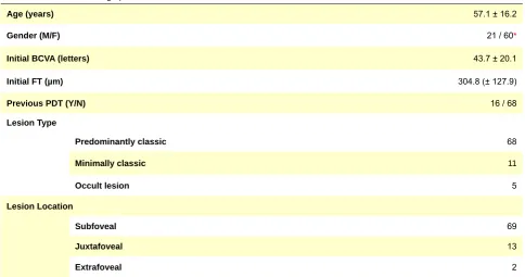
Table 1 - Clinical and Demographic Patients´ characteristics
Age (years) 57.1 ± 16.2
Gender (M/F) 21 / 60*
Initial BCVA (letters) 43.7 ± 20.1
Initial FT (µm) 304.8 (± 127.9)
Previous PDT (Y/N) 16 / 68
Lesion Type
Predominantly classic 68
Minimally classic 11
Occult lesion 5
Lesion Location
Subfoveal 69
Juxtafoveal 13
Extrafoveal 2
FT. Foveal thickness (µm); BCVA. Best corrected visual acuity (ETDRS chart); PDT. Photodynamic therapy
ARTIGO ORIGINAL fluorescein.
We assessed the results regarding 84 eyes in 81 patients, from which 22 were treated with bevacizumab, 29 with ranibizumab and 33 with both drugs. Data regarding stereoscopic fundus evaluation, best corrected visual acuity (BCVA) assessed using a ETDRS chart, foveal thickness (FT) in OCT and fluorescein angiographic signs were collected, initially and upon 3, 6, 12, 24, 36, 48 and 60 months of therapy.
BCVA was measured using the ETDRS chart, according with the refraction protocol used at the Ophthalmology Department of the Hospital de São João from Porto. FT
was measured using OCT Stratus (Zeiss), version 4.0.2 and/or HRA-OCT (Heidelberg Engineering). Through FA, we assessed contrast diffusion and lesion growth and defined an early lesion as a non-fibrotic CNV occupying an area below half of the size of the optic disc. CNV lesions were classified according to angiography as predominantly classic, minimally classic or occult with no classic component (Type 1). All subtypes of angiographic lesions were considered in the study, including those with areas of fibrosis, atrophy or haemorrhage, above half the size of the lesion. Lesions with a fibrotic scar but no active NCV were not included in our study.
Intra-vitreal 1.25 mg bevacizumab (IVB) and/or 0.5 mg ranibizumab (IVR) injections were applied to patients with active CNV in fluorescein angiography or with the presence of intra or sub-retinal fluid in OCT. The patients did not receive any combined therapy with photodynamic therapy (PDT), triamcinolone or any other anti-angiogenic drug. Nevertheless, patients submitted to therapy previous to anti-VEGF injections were not excluded. All patients included in the study were treated following a 1+PRN (pro re nata – as
the circumstance arises) regimen, with IVB and/or IVR, as required after the first injection.
SPSS 20.0 software was used for statistical analysis.
t-Student test was used for paired or independent samples
analysis of continuous variables. Levene test was used to assess for homogeneity of variance. Multivariable linear regression analysis was used for assessing predictive pre-therapy factors for final BCVA; p-values < 0.05 were
considered as statistically significant.
Our study was approved by the Ethics Committee of Porto University, according to the Declaration of Helsinki.
RESULTS
From all studied eyes, 84 (100%) completed 12 months of follow-up, 67 (79.8%) 24 months, 54 (64.3%) 36 months, 29 (34.5%) 48 months and 15 (16.7%) completed 60 months. Twenty-nine (34.5%) were treated with IVR alone, 22 (26.2%) with IVB in isolation and 33 (39.3%) were treated with both drugs (IVR/IVB). The patients´ clinical and demographic characteristics are presented in Table 1. Overall, initial BCVA was on average 43.7 ± 20.1 letters and initial FT was on average 304.8 ± 127.9 µm. Differences on initial BCVA, as well as on initial FT, between the groups with different follow-up times were statistically not significant
(p =0.8).
Mean initial BCVA and mean initial FT were assessed and compared between previously treated or non-treated patients. We found that previously treated-patients presented a lower initial FT (230.4 ± 95.9 µm) when compared with non-treated patients (308.3 ± 1,259 µm) and this was a statistically significant difference (p = 0.04). We
did not find any significant differences regarding initial BCVA (45.9 ± 19.8 and 43.8 ± 20.2, respectively), p = 0.8. We also
evaluated whether lesion location would have any impact on initial BCVA and FT; however, no statistically significant differences were found (p = 0.2 and p = 0.8, respectively).
Upon intra-vitreal therapy, mean BCVA was 55.1 ± 17.9
Figura 1 – Mean BCVA variation vs. initial. n = 84 at 3, 6 and 12 months; n = 67 at 24 months; n = 54 at 36 months; n = 29 at 48 months;
n = 15 at 60 months.
* p value < 0.05
Time of follow-up (months)
Mean BCV
A variation (ETDRS chart)
0 0 5 10 15
12
6 36 60
3 24 48
*
*
*
* *
*
ARTIGO ORIGINAL letters for 3 months of treatment, 54.5 ± 18.9 for 6 months, 55.6 ± 18.5 for 12 months, 52.1 ± 22.3 for 24 months, 52.1
± 22.6 for 36 months, 50.3 ± 23.8 for 48 months and 47.8 ± 24.5 for 60 months. Final BCVA improvement regarding initial BCVA was statistically significant for all follow-up groups (p < 0.05). BCVA variation from initial values is
shown in Fig. 1.
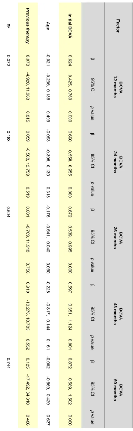
BCVA results were adjusted to pre-treatment variables and we found that the initial BCVA is independently correlated with better results on final BCVA (p < 0.001)
(Table 2).
Mean FT was 227.9 ± 98.9 for 3 months, 224.8 ± 84.9 for 6 months, 209.7 ± 86.2 for 12 months, 190.6 ± 76.1 for 24 months, 174.7 ± 60.6 for 36 months, 189.8 ± 96.7 for 48 months and 159.4 ± 73.3 for 60 months. FT variation from the initial values was statistically significant for all follow-up times (Fig. 2).
Mean number of injections was 5.1 ± 2.6 at 12 months, 2.1 ± 2.6 between 12 and 24 months, 1.9 ± 2.5 between 24 and 36 months, 1.8 ± 2.5 between 36 and 48 months and 1.2 ± 2.2 between 48 and 60 months. The gradual reduction of the number of injections was statistically significant (p <
0.001).
The relationship between the presence of an early lesion and the number of injections was assessed in 81 of the 84 studied eyes. The mean number of injections in the group of patients with an early lesion was lower than in the group of patients with a late lesion (8.1 ± 8.9 and 9.9 ± 10.4, respectively). However, these differences were not statistically significant (p = 0.2). We also assessed whether
the presence of an early lesion was related to a long-term lesion lower growth, defined by a thickness increase in the OCT. From the 28 (34.6%) patients with an early lesion, 19 (67.9%) did not present a lesion growth over the follow-up time and in nine (32.1%) patients the lesion grew. Similarly, from the 33 (40.7%) patients in whom the lesion grew, nine (27.3%) presented an early lesion and 24 (72.7%) presented a late lesion. However, none of these results was statistically significant (p = 0.3).
Overall, BCVA and FT results were also separately assessed for the 29 (34.5%) patients aged 50 or below and for the 55 (65.5%) patients aged above 50 (Table 3). Initial BCVA and FT differences between both groups were statistically not significant (p = 0.3 and p = 0.4, respectively).
BCVA differences for the various follow-up times were statistically not significant between both groups (p = 0.9 at 3
and 6 months; p = 0.7 at 12 months; p = 0.4 at 24 months; p = 0.2 at 36 months; p = 0.3 at 48 months and p = 0.5 at
60 months). As regards FT, we found statistically significant differences at 12, 24 and 60 months, with a lower FT in the over-50 patient’s group (p = 0.03, p = 0.03 and p = 0.01
respectively). Differences were not significant at 3, 6, 36 and 48 months (p = 0.3 at 3 months; p = 0.6 at 6 months; p = 0.1 at 36 months and p = 0.2 at 48 months).
As regards treatment complications, one patient developed an inflammatory reaction (vitritis) with bevacizumab. No serious systemic complications or other
Table 2
-Multivariate regression analysis of pre-treatment factors contributing to BCV
A Factor BCV A 12 months BCV A 24 months BCV A 36 months BCV A 48 months BCV A 60 months β 95% CI p value β 95% CI p value β 95% CI p value β 95% CI p value β 95% CI p value Initial BCV A 0.624
0.425, 0.760
0.000
0.690
0.558, 0.955
0.000
0.672
0.539, 0.995
0.000
0.597
0.351, 1.124
0.001
0.872
0.589, 1.502
0.000
Age
-0.021
-0.236, 0.186
0.409
-0.093
-0.395, 0.130
0.318
-0.176
-0.541, 0.040
0.090
-0.228
-0.817, 0.144
0.161
-0.082
-0.669, 0.429
0.637 Previous therapy 0.073 -4.920, 1 1.963 0.815 0.059 -6.508, 12.759 0.519 0.031 -8.709, 1 1.918 0.756 0.910 -10.276, 18.785 0.552 0.125 -17.492, 34.310 0.486 R 2 0.372 0.483 0.504 0.744 BCV
ARTIGO ORIGINAL
Figura 2 – FT variation vs. initial. n = 84 at 3, 6 and 12 months; n = 67 at 24 months; n = 54 at 36 months; n = 29 at 48 months; n = 15 at
60 months.
* p value < 0.05
Time of follow-up (months)
Mean FT
variation (μm)
0
-150 -100 -50
0 3 6 12 24 36 48 60
* *
*
*
*
*
*
ocular complications occurred during follow-up, namely endophthalmitis, vitreous haemorrhage, retinal detachment, cataracts or glaucoma.
Bevacizumab and/or ranibizumab-treated patient subgroup analysis
Mean BCVA and TF within the different groups, for the various follow-up times are presented in Table 4. BCVA differences found in the ranibizumab group regarding initial values were statistically significant at 3, 6 and 12 months
(p < 0.05). From 24 months of follow-up, differences were
statistically not significant (p = 0.3 at 24 months; p = 0.1 at
36 months; p = 0.1 at 48 months and p = 0.2 at 60 months).
As regards FT, the differences with the initial values were statistically significant at 3, 6, 12, 24 and 36 months (p <
0.05) and were not significant for longer follow-up times (p
= 0.1 at 48 months and p = 1 at 60 months).
Regarding the treatment with bevacizumab, we found that for BCVA differences, comparing final with initial measurements, were statistically significant up to 36 months of follow-up but not at48 and 60 months (p = 0.1 and p = 0.4, respectively). Final FT difference compared with
the initial measurements was statistically significant at 3, 6, 12, 36 and 60 months (p < 0.05). At 24 and 48 months, the
differences between the parameters were not statistically significant (p = 0.2 and p = 0.1, respectively).
In patients treated with both drugs, we found that the difference between final and initial BCVA was statistically significant up to 36 months of follow-up but not at 48 and 60 months (p = 0.9 and p = 0.1, respectively). Final FT was
lower than the initial, with statistically significant differences up to 48 months of follow-up, not significant at 60 months
(p = 0.1).
BCVA values were compared between the groups, for
the same follow-up times and we did not find statistically significant differences (p = 0.7; p = 0.9; p = 0.8; p = 0.8; p = 0.8; p = 0.8; p = 0.7 and p = 0.2 for the initial values,
at 3, 6, 12, 24, 36, 48 and 60 months, respectively). FT at 60 months shows better results in the bevacizumab group
(p = 0.02). We did not find statistically significant differences
for the other follow-up times (p = 0.5; p = 0.6; p = 0.7; p = 0.9; p = 0.2; p = 0.1 at 3, 6, 12, 24, 36 and 48 months,
respectively).
DISCUSSION
When not treated, PM involves a bad prognosis and is related with severe and progressive vision loss.9,10,30
Therefore and taking into account the non-sustained PDT results, the new anti-angiogenic drugs have been increasingly used in the treatment of pathologies with CNV, as in PM.
Ranibizumab and bevacizumab are monoclonal anti-VEGF antibodies used to treat CNV, for their effect reducing cell proliferation, vascular patency and new blood vessel formation.9,12,31 Their efficacy and safety were described
in several retrospective studies and in some prospective clinical trials. Therefore, we have witnessed a word-wide use of these agents in treatment of PM and AMD-related CNV, with significant functional and anatomical improvements.32 Ranibizumab stands for a monoclonal
Fab humanized, recombinant fragment, with a molecular
weight of 49 kDa.33 Specifically designed for intraocular
use, it presents some theoretical advantages when compared with bevacizumab, namely a lower molecular weight, associated with a better and faster penetration into retinal layers, higher affinity for the VEGF-A receptor and lower incidence of systemic effects.12 Bevacizumab
ARTIGO ORIGINAL
designed for intravenous chemotherapy adjuvant treatment of metastatic solid tumours. It is particularly important in myopic CNV treatment, due to promising results described in several case reports and some clinical trials,1,6,8,11,13,34-36
coupled with a significantly lower cost when compared with ranibizumab, allowing for higher accessibility of this therapy to a greater number of patients.10
We found that the anti-angiogenic agents allowed for a significant and sustained BCVA and FT improvement up to 60 months of treatment. Our results are in line with others described for ranibizumab37-41 and for bevacizumab
in shorter follow-up studies.36,42 BCVA variation vs. the initial
value was progressively lower with the various follow-up times, although always higher to the initial BCVA (p < 0.05),
what may be explained by older age, with an AMD-related component and therefore with lower BCVA, as well as by the progression of myopia-related atrophy and by CNV itself. In addition, FT variation was progressively higher, what may be related not only to macular oedema reduction but also to chorio-retinal atrophy in myopic patients.
Previously treated-patients presented lower initial FT when compared with non-treated patients, probably due to macular oedema and CNV reduction (230.4 ± 95.9 µm and
308.3 ± 125.9 µm, respectively; p = 0.04).17,18 However, in
line with the studies by Calvo-Gonzalez et al.,15 Monés JM
et al.39 and Lalloum F et al.,41 we did not find statistically
significant differences in initial visual acuity of previously treated or non-treated patients (45.9 ± 19.8 and 43.8 ± 2.02 letters, respectively; p = 0.7). Lower initial thickness was
not followed by a significantly improved visual acuity, which may be explained by chorio-retinal atrophy progression associated to PDT..17 In addition, we are aware that the
results of this therapy are only demonstrated in the short term, explaining the fact that there are no differences on visual acuity between the groups when anti-angiogenic treatment is started.
It has been described that non-subfoveal CNV may constitute a better outcome predictive factor, as it does not directly affect the central area.4,13,43 In our study, 69 of the 84
eyes (81.1%) presented a subfoveal lesion. However, the location of the lesion did not significantly affect BCVA or FT
(p = 0.2 and p = 0.8, respectively).
Pre-therapy visual acuity had positive effects on long-term visual acuity (p < 0.001). In fact, a better initial BCVA
is related with less damage to the photoreceptors, what may explain the better results obtained.15,25 However, other
Table 3 - Mean BCVA and FT for various follow-up times according to patient’s age (≤ 50 and > 50)
Age ≤ 50
(n = 29)
Initial (n = 29)
3 months (n = 29)
6 months (n = 29)
12 months (n = 29)
24 months (n = 21)
36 months (n = 18)
48 months (n = 9)
60 months (n = 6)
BCVA
(letters) 46.9 ± 20.2 55.4 ± 17.7 55.5 ± 18.2 56.9 ± 18.3 56.7 ± 18.4 58.1 ± 20.7 58.1 ± 18.9 54,6 ± 21,9
FT
(µm) 282.8 ± 94.1 230.9 ± 73.9 225.7 ± 72.3 225.1 ± 70.3 208.2 ± 43.4 190.8 ± 39.9 234.6 ± 130.4 208.2 ± 49.5
Table 3 - Mean BCVA and FT for various follow-up times according to patient’s age (≤ 50 and > 50) (continuation)
Age > 50 (n = 55)
Initial (n = 55)
3 months (n = 55)
6 months (n = 55)
12 months (n = 55)
24 months (n = 46)
36 months (n = 36)
48 months (n = 20)
60 months (n = 9)
BCVA
(letters) 42 ± 19.9 54.6 ± 18.9 53.6 ± 20.3 54.6 ± 19.6 49.5 ± 24.6 48.5 ± 24.3 46.8 ± 25.4 44 ± 26.2
FT
(µm) 316.11 ± 41.8 223.4 ± 107.9 222.4 ± 91,7 200.7 ± 92.5 181.3 ± 87.3 168.5 ± 67.4 156.5 ± 61.2 123.4 ± 54.8
ARTIGO ORIGINAL
Table 4 - Mean BCVA and FT for the various follow-up times according to ranibizumab and/or bevacizumab treatment
Initial
(n = 84) 3 months (n = 84) 6 months (n = 84) 12 months(n = 84)
IVR (n = 29)
IVB (n = 22)
IVR/IVB (n = 33)
IVR (n = 29)
IVB (n=22)
IVR/IVB (n = 33)
IVR (n = 29)
IVB (n = 22)
IVR/IVB (n = 33)
IVR (n = 29)
IVB (n = 22)
IVR/IVB (n = 33)
BCVA (letters) 46.2 ± 22.1 41.6 ± 20.4 42.9 ± 18.2 53.8 ± 22.9 55.9 ± 16.5 55 ± 15.5 53.7 ± 21.8 55.6 ± 20.8 53.8 ± 26.8 52.7 ± 23.2 54.7 ± 17.3 58.1 ± 16.1 FT (µm) 284.9 ± 121.3 307.3 ± 146.1 321.3 ± 124.5 204.7 ± 61.3 258.4 ± 130.7 225.1 ± 96.8 205.6 ± 56.6 236.2 ± 101.6 231.7 ± 94.1 207.5 ± 77.1 207.1 ± 99.7 212.4 ± 86.4
Tabela 4 - Mean BCVA and FT for the various follow-up times according to ranibizumab and/or bevacizumab treatment (continuation)
24 months (n = 67)
36 months (n = 54)
48 months (n = 29)
60 months (n = 14)
IVR (n = 25)
IVB (n = 12)
IVR/IVB (n = 30)
IVR (n = 18)
IVB (n = 12)
IVR/IVB (n = 24)
IVR (n = 4)
IVB (n = 12)
IVR/IVB (n = 13)
IVR (n = 2)
IVB (n = 10)
IVR/IVB (n = 3)
BCVA (letters) 50 ± 24.1 49.2 ± 25.5 54.3 ± 21.4 53.8 ± 24.4 49.2 ± 24.9 51.3 ± 22.9 57.5 ± 25.6 49.7 ± 25.4 48.6 ± 23.4 45 ± 38.2 41.7 ± 23.3 68 ± 14.2 FT (µm) 176.9 ± 64 160 ± 67.1 212 ± 84.5 193.2 ± 54.2 139.8 ± 54.9 179.9 ± 61.7 218.3 ± 50.9 135.4 ± 59.5 210.6 ± 114 213 ± 67.9 115.8 ± 45.3 225.3 ± 6.1
FT: Foveal thickness (µm); BCVA: Best corrected visual acuity (ETDRS chart).
studies show that a lower initial BCVA causes better results upon treatment,13 in relation to a ceiling or a floor effect.
The authors consider that a better initial BCVA will have less chance of improvement (ceiling effect), while patients with a worse initial BCVA will have better chance of improvement (floor effect).
Previous therapy did not significantly affect final BCVA results (p > 0.05). Some studies show that previously
non-treated patients present best results with anti-VEGF therapy.15,24 Previously treated-patients, presumably with
longer evolution and with CNV recurrence, may show higher resistance to anti-angiogenic therapy, explaining worse results.8,15 In addition, PDT predisposes to choroidal
vessel thrombosis and chorio-retinal atrophy.11 Better results were also described in previously-treated patients, possibly due to an early perception of symptoms which may have induced recurrent treatments.8 However, in the
present study, the reduced size of the sample (n = 21) was
not enough to allow for an extrapolation of these results. As it would be expected, taking into account a lower severity of an early lesion, we found that in most of these patients lesions did not grow (67.9%). However, the results were statistically not significant, again probably due to the
reduced size of the sample (p = 0.3).
Although patient’s age is a major prognostic factor, affecting natural history of the disease upon anti-VEGF treatment,10, 36 it was not significantly related with the results
on visual acuity in our study(p > 0.05). Our results may
be due to the fact that most patients are aged above 50 (65.5%), with a mean age of 57.2 (+-16.1). In fact, these results have been confirmed by another study in which the visual outcome in patients aged 50 or below was similar to those aged above 50.44 The patients aged above 50 presented a lower final FT for the various follow-up times, tending to be significant at 12, 24 and 60 months of follow-up
(p = 0.03, p = 0.03 and p = 0.001, respectively). This fact is
ARTIGO ORIGINAL due to previous treatment.
15
Taking into account molecular differences between ranibizumab and bevacizumab, a different clinical efficacy of these two drugs on PM has been suggested.46 However,
we found similar improvements in visual acuity between treatment groups. Our results were in line with those of a prospective, randomized and controlled study by Gharbiya
et al.,9 witha maximum six-month follow-up, that also did not
find any significant BCVA differences between bevacizumab and/or ranibizumab-treated groups. In this study, BCVA at six months was 43.8 ± 9.9 letters for ranibizumab group (n
= 16) and 45.4 ± 9.9 letters for bevacizumab group (n =
16). In our study and for the same follow-up, BCVA was 53.7 ± 21.8 and 55.6 ± 20.8 in ranibizumab (n = 29) and
bevacizumab (n = 22) groups, respectively. However, the
best results in our study are probably related with the best initial BCVA (46.2 ± 22.1 in the ranibizumab group and 41.6 ± 20.4 in the bevacizumab group), when compared to the study by Gharbiya et al. (26.44 ± 12.58 in the ranibizumab
group and 29.50 ± 12.98 in the bevacizumab group). Beyond this, other studies confirmed our results.40,47,48
However, contrary to these studies that did not find any FT differences between the groups, we found that the final FT in the IVB group was better at 60 months of follow-up (115.8 ± 45.3 µm in the IVB group, 213 ± 67.9 µm in the IVR group and 225.3 ± 6.1 in the IVB/IVR group), which may be due to the drug higher molecular weight, consequently affecting the duration of intra-vitreal action. However, from the 15 patients with 60 months of follow-up, 10 were treated only with bevacizumab, which may explain better results in this group. BCVA and FT improvements in the IVR group were significant with shorter follow-up times, a likely consequence of its underlying shorter duration of action when compared with bevacizumab.
In our study, treatment followed a 1+PRN regimen, comprising a bevacizumab and/or ranibizumab injection, as needed, upon the first injection. We found that functional and anatomical benefits were met, alongside with a significant reduction in the yearly number of injections, probably underlying an improvement from the beginning of the treatment. We did not find ocular or systemic serious complications, except in one female patient who developed an inflammatory reaction (vitritis) with bevacizumab. This patient kept a similar visual acuity and was changed to ranibizumab. There are several case reports described in literature regarding inflammatory reactions related with intra-vitreal injections of anti-VEGF agents.49-51 Although
not yet completely clarified, the underlying mechanism is
assumed to be related with an immunological response to drug components. In fact, the risk seems to be higher with bevacizumab, when compared with ranibizumab, probably underlying the additional Fc portion and consequently to the
higher protein content.51,52 Once the patients with PM present
an increased risk of developing ocular complications, a detailed retinal examination is recommended and prophylactic laser therapy in high risk cases before starting intra-vitreal injections may be required.47
We wish to emphasize the following limitations to our study: a retrospective design, the absence of a control group of patients and the small size of our group of patients. We have found difficulties in the comparison between the present study and published research mainly due to different follow-up times, methodologies and treatment criteria. In addition, results are highly dependent on patient´s characteristics, symptom duration, age, CNV area and pre-treatment BCVA. Although we did not measure the area of CNV lesion, we found, in FA and in OCT a reduction in the size of CNV on most treated eyes (59.3%).
CONCLUSION
Our results reflect clinical practice and show that intra-vitreal injection of anti-VEGF agents, following a 1+PRN regimen, result in a significant and sustained improvement in visual acuity in patients with myopic CNV. We should emphasize that benefits obtained with anti-angiogenic treatment were kept for follow-up times of up to 60 months, whose clinical relevance is due to the natural history of disease. Until the present time, only a limited number of studies with exceedingly short follow-up time, had compared both drugs directly, using the same treatment regimen and with no significant differences being found. As such, the ranibizumab cost-benefit ratio is still controversial, when compared with that of bevacizumab.
We consider that multicenter, prospective, randomized clinical trials involving greater samples will be necessary to confirm the results of anti-angiogenic treatment in the long term, as well as to draw firm conclusions on efficacy and safety issues, which are crucial in treatment choices.
CONFLICTS OF INTEREST
The authors declare that there were no conflicts of interest in writing this manuscript.
FINANCIAL SOURCES
There were no external financial sources for writing this manuscript.
REFERENCES
1. Ruiz-Moreno JM, Montero JA, Amat-Peral P. Myopic choroidal neovas-cularization treated by intravitreal bevacizumab: comparison of two dif-ferent initial doses. Graefes Arch Clin Exp Ophthalmol. 2011;249:595–9. 2. Neelam K, Cheung CM, Ohno-Matsui K, Lai TY, Wong TY.
Choroi-dal neovascularization in pathological myopia. Prog Retin Eye Res. 2012;31:495-525.
3. Mitry D, Zambarakji H. Recent trends in the management of
maculopa-thy secondary to pathological myopia. Graefes Arch Clin Exp Ophthal-mol. 2012;50:3–13.
4. Silva R. Myopic maculopaty: a review. Ophtalmologica. 2012;228:197-213.
neovascular-ARTIGO ORIGINAL
ization. Graefes Arch Clin Exp Ophthalmol. 2009;248:1087-90. 6. Parodi MB, Iacono P, Papayannis A, Sheth S, Bandello F. Laser
pho-tocoagulation, photodynamic therapy, and intravitreal bevacizumab for the treatment of juxtafoveal choroidal neovascularization secondary to pathologic myopia. Arch Ophthalmol. 2010;128:437-42.
7. Ng DS, Kwok AK, Chan CW. Anti-vascular endothelial growth fac-tor for myopic choroidal neovascularization. Clin Exp Ophthalmol. 2012;40:e98–110.
8. Voykov B, Gelisken F, Inhoffen W, Voelker M, Bartz-Schmidt KU, Ziems-sen F. Bevacizumab for choroidal neovascularization secondary to pathologic myopia: Is there a decline of the treatment efficacy after 2 years? Graefes Arch Clin Exp Ophthalmol. 2010;248:543–50. 9. Gharbiya M, Giustolisi R, Allievi F, Fantozzi N, Mazzeo L, Scavella V,
et al. Choroidal neovascularization in pathologic myopia: Intravitreal ra-nibizumab versus bevacizumab — a randomized controlled trial. Am J Ophthalmol. 2010;149:458–46.
10. Wang J, Kang Z. Summary of prognostic factors for choroidal neovascu-larization due to pathological myopia treated by intravitreal bevacizumab injection. Graefes Arch Clin Exp Ophtalmol. 2012;250:1717-23. 11. El Matri L, Kort F, Chebil A, Bouraoui R, Merdassi A, Bouladi M.
Intra-vitreal bevacizumab versus photodynamic therapy for myopic choroidal neovascularization in a North-African population. Graefes Arch Clin Exp Ophthalmol. 2011;249:1287–93.
12. Konstantinidis L, Mantel I, Pournaras JA, Zografos L, Ambresin A. In-travitreal ranibizumab (Lucentis®) for the treatment of myopic choroidal neovascularization. Graefes Arch Clin Exp Ophthalmol. 2009;247:311– 8.
13. Bottoni F, Perego E, Airaghi P. Surgical removal of subfoveal choroidal neovascular membranes in high myopia. Graefes Arch Clin Exp Oph-thalmol. 1999;237:573-82.
14. Ruiz-Moreno JM, de la Vega C. Surgical removal of subfoveal cho-roidal neovascularization in highly myopic patients. Br J Ophthalmol. 2011;85:1041-3.
15. Hamelin N, Glacet-Bernard A, Brindeau C. Surgical treatment of sub-foveal neovascularization in myopia: macular translocation vs. surgical removal. Am J Ophthalmol. 2002;133:530-6.
16. Uemura A, Thomas MA. Subretinal surgery for choroidal neovascular-ization in patients with high myopia. Arch Ophtalmol. 2000;118:344-50. 17. Blinder KJ, Blumenkranz MS, Bressler NM, Bressler SB, Donato G,
Lewis H et al. Verteporfin therapy of subfoveal choroidal neovascular-ization in pathologic myopia: 2-year results of a randomized clinical trial - VIP report n.3. Ophthalmology. 2003;110:667-73.
18. Verteporfin in Photodynamic Therapy Study Group. Photodynamic ther-apy of subfoveal choroidal neovascularization in pathologic myopia with verteporfin. 1-year results of a randomized clinical trial - VIP report no.1. Ophthalmology. 2001;108:841-52.
19. Marticorena J, Gomez-Ulla F, Fernandez M, Pazos B, Rodriquez-Cid MJ, Sanchez-Salorio M. Combined photodynamic therapy and intravi-treal triamcinolone acetonide for the treatment of myopic subfoveal cho-roidal neovascularisation. Am J Ophtalmol. 2006;142:335-7.
20. Chan WM, Lai TY, Wong AL, Liu DT, Lam DS. Combined photodynamic therapy and triamcinolone injection for the treatment of choroidal neo-vascularisation secondary to pathological myopia: a pilot study. Br J Ophthalmol. 2007;91:131-3.
21. Baba T, Kubota-Taniai M, Kitahashi M, Okada K, Mitamura Y, Yama-moto S. Two year comparison of photodynamic therapy and intravitreal bevacizumab for treatment of myopic choroidal neovascularization. Br J Ophthalmolol. 2010;94:864-70.
22. Hayashi K, Ohno-Matsui K, Teramukai S. Comparison of visual outcome and regression pattern of myopic choroidal neovascularization after in-travitreal bevacizumab or after photodynamic therapy. Am J Ophthalmol. 2009;148:396-408.
23. Ikuno Y, Nagai Y, Matsuda S, Arisawa A, Sho K, Oshita T. Two-year visual results for older Asian women treated with photodynamic ther-apy or bevacizumab for myopic neovascularization. Am J Ophtalmol. 2010;149:140-6.
24. Yoon JU, Byun YJ, Koh HJ. Intravitreal anti-VEGF versus photodynamic therapy with verteporfin for treatment of myopic choroidal neovascular-ization. Retina. 2010;30:418-24.
25. Nakanishi H, Tsujikawa A, Yodoi Y, Ojima Y, Otani A, Tamura H, et al. Prognostic factors for visual outcomes 2-years after intravitreal bevaci-zumab for myopic choroidal neovascularization. Eye. 2011;25:375–81. 26. Wu PC, Chen YJ. Intravitreal injection of bevacizumab for myopic
cho-roidal neovascularization: 1-year follow-up. Eye. 2009;23:2042–5. 27. Calvo-Gonzalez C, Reche-Frutos J, Donate J, Fernandez-Perez C,
Gar-cia-Feijoo J. Intravitreal ranibizumab for myopic choroidal
neovascular-ization: Factores preditive of visual outcome and need for retreatment. Am J Ophtalmol. 2010;151:529-34.
28. Gharbiya M, Cruciani F, Parisi F, Cuozzo G, Altimari S, Abdolrahimzadeh S. Long-term results of intravitreal bevacizumab for choroidal neovas-cularisation in pathological myopia. Br J Ophthalmol. 2012;96:1068-72. 29. Peiretti E, Vinci M, Fossarello M. Intravitreal bevacizumab as a
treat-ment for choroidal neovascularization secondary to myopia:4-year study results. Can J Ophthalmol. 2012;47:28-33.
30. Yoshida T, Ohno-Matsui K, Yasuzumi K, Kojima A, Shimada N, Futagami S, et al. Myopic choroidal neovascularization: a 10-year follow up. Oph-thalmology. 2003;110:1297-305.
31. Gomi F, Nishida K, Oshima Y, Sakaguchi H, Sawa M, Tsujikawa M, et al. Intravitreal bevacizumab for idiopathic choroidal neovascularization after previous injection with posterior subtenon triamcinolone. Am J Ophthalmol. 2007;143:507-9.
32. Carneiro AM, Mendonça LS, Falcão MS, Fonseca SL, Brandão EM, Falcão-Reis FM. Comparative study of 1+PRN ranibizumab versus bevacizumab in the clinical setting. Clin Ophthalmol. 2012;6:1149-57. 33. Ferrara N, Damico L, Shams N, Lowman H, Kim R. Development of
ranibizumab, an anti-vascular endothelial growth factor antigen binding fragment, as therapy for neovascular age-related macular degeneration. Retina. 2006;26:859–70.
34. Yamamoto I, Rogers AH, Reichel E, Yates PA, Duker JS. Intravitreal bev-acizumab (Avastin) as treatment for subfoveal choroidal neovascularisa-tion secondary to pathological myopia. Br J Ophtalmol. 2007;91:157-60. 35. Chan WM, Lai TY, Liu DT, Lam DS. Intravitreal bevacizumab (avastin)
for myopic choroidal neovascularisation: Six-month results of a prospec-tive pilot study. Ophthalmology. 2007;114:2190-6.
36. Gharbiya M, Allievi F, L Mazzeo, Gabrieli CB. Intravitreal bevacizumab treatment for choroidal neovascularization in pathologic myopia:12-month results. Am J Ophthalmol. 2009;147:84-93.
37. Konstantinidis L, Mantel I, Zografos L, Ambresin A. Intravitreal ranibi-zumab as primary treatment for neovascular membrane associated with idiopathic juxtafoveal retina. Graefes Arch Clin Exp Ophthalmol. 2009;247:1567-9.
38. Silva RM, Ruiz-Moreno JM, Rosa P. Intravitreal ranibizumab for myopic choroidal neovascularization:12-month results. Retina. 2010;30:407-12. 39. Monés JM, Amselem L, Serrano A, Garcia M, Hijano M. Intravitreal
ranibizumab for choroidal neovascularization secondary to pathologic myopia:12-month results. Eye. 2009;23:1275-80.
40. Lai TY, Chan WM, Liu DT, Lam DS. Intravitreal ranibizumab for the pri-mary treatment of choroidal neovascularization secondary to pathologic myopia. Retina. 2009;29:750-6.
41. Lalloum F, Souied EH, Bastuji-Garin S. Intravitreal ranibizumab for choroidal neovascularization complicating pathologic myopia. Retina. 2010;30:399-406.
42. Ikuno Y, Sayanagi K, Soga K. Intravitreal bevacizumab for choroidal neovascularization attributable to pathological myopia:one-year results. Am J Ophthalmol. 2009;147:94-100.
43. Hayashi K, Ohno-Matsui K, Yoshida T. Characteristics of patients with a favorable natural course of myopic choroidal neovascularization. Grae-fes Arch Clin Exp. Ophthalmol. 2005;243:13-9.
44. Arias L, Planas N, Prades S, Caminal JM, Rubio M, Pujol O, et al. Intravitreal bevacizumab (Avastin) for choroidal neovascularisation secondary to pathological myopia:6-month results. Br J Ophthalmol. 2008;92:1035-9.
45. Sayanagi K, Ikuno Y, Soga K, Wakabayashi T, Tano Y. Marginal crack after intravitreal bevacizumab for myopic choroidal neovascularization. Acta Ophthalmol. 2008;85:50-4.
46. Mordenti J, Cuthbertson RA, Ferrara N, Thomsen K, Berleau L, Licko V, et al. Comparisons of the intraocular tissue distribution, pharmaco-kinetics, and safety of 125I-labeled full-length and Fab antibodies in rhesus monkeys following intravitreal administration. Toxicol Pathol. 1999;27:536–44.
47. Chan WM, Lai TY, Liu DT, Lam DS. Intravitreal bevacizumab (Avastin) for myopic choroidal neovascularisation: 1-year results of a prospective pilot study. Br J Ophthalmol. 2009;93:150–4.
48. Gharbiya M, Allievi F, Mazzeo L, Gabrieli CB. Intravitreal bevacizum-ab treatment for choroidal neovascularization in pathologic myopia: 12-month results. Am J Ophthalmol. 2009;147:84 –93.
49. Kelly SP, Barua A. A review of safety incidents in England and Wales for vascular endotelial growth factor inhibitor medications. Eye. 2011;25:710–6.
Ophthal-ARTIGO ORIGINAL
mology. 2011;118:2028–34.
51. Bakri SJ, Larson TA, Edwards AO. Intraocular inflammation following intravitreal injection of bevacizumab. Graefes Arch Clin Exp Ophthalmol. 2008;246:779–81.