191
© 2018 by the Serbian Biological Society
CXC chemokine ligand 12α-mediated increase in insulin secretion and survival of mouse
pancreatic islets in response to oxidative stress through modulation of calcium uptake
Melita Vidaković1,2,*, Ernesto Caballero Garrido2, Mirjana Mihailović1, Jelena Arambašić Jovanović1, Marija
Sinadinović1, Jovana Rajić1, Aleksandra Uskoković1, Svetlana Dinić1, Nevena Grdović1, Miloš Đorđević1, Anja
Tolić1 and Goran Poznanović1
1Department of Molecular Biology, Institute for Biological Research, University of Belgrade, Belgrade, Serbia
2Institute of Bioengineering, Universidad Miguel Hernández de Elche, UNIT of Cell Physiology and Nutrition, Elche, Spain
*Corresponding author: [email protected]
Received: July 11, 2017; Revised: October 23, 2017; Accepted: October 24, 2017; Published online: October 30, 2017
Abstract: We examined whether CXCL12α improves insulin secretion by influencing the Ca2+ oscillation pattern and Ca2+ influx ([Ca2+]
i), thereby enhancing the viability of pancreatic islet cells in oxidative stress. The islets of Langerhans were isolated from male OF1 mice and pretreated with 40 ng/mL of CXCL12α prior to exposure to 7.5 µM hydrogen peroxide, which served to induce oxidative stress. Incubation of islets with CXCL12α induced pancreatic β-cell proliferation and improved the ability of β-cells to withstand oxidative stress. Consecutive treatments of isolated islets with hydrogen peroxide caused a decline in β-cell functioning over time, while the CXCL12α pretreatment of islets exhibited a physiological response to high glucose that was comparable to control islets. The attenuated response of islets to a high D-glucose challenge was observed as a partial to complete abolishment of [Ca2+]
i. Treatments with increasing concentrations of CXCL12α decreased the number of Ca2+ oscillations that lasted longer, thus pointing to an overall increase in [Ca2+]
i, which was followed by increased insulin secretion. In addition, treatment of islets with CXCL12α enhanced the transcription rate for insulin and the CXCR4 gene, pointing to the importance of CXCL12/CXCR4 signaling in the regulation of Ca2+ intake and insulin secretion in pancreatic islet cells. We propose that a potential treatment with CXCL12α could help to remove preexisting glucotoxicity and associated temporary β-cell stunning that might be present at the time of diabetes diagnosis in vivo.
Key words: diabetes; calcium; CXC chemokine ligand 12α; insulin; pancreatic islet cells; voltage-gated calcium channels
How to cite this article: Vidaković M, Garrido EC, Mihailović M, Arambašić-Jovanović J, Sinadinović M, Rajić J, Uskoković A, Dinić S, Grdović N, Đorđević M, Tolić A, Poznanović G. CXC chemokine ligand 12α-mediated increase in insulin secretion and survival of mouse pancreatic islets in response to oxidative stress through modulation of calcium uptake. Arch Biol Sci. 2018;70(1):191-204. IntRoduCtIon
Diabetes is characterized either by a near-absolute absence (type 1 diabetes; T1D) or depletion (type 2 diabetes; T2D) of pancreatic β-cells that is responsi-ble for the condition that is described by insufficient or impaired insulin secretion. Therefore, β-cell mass regulation and improved β-cell viability and function through the stimulation of insulin secretion represent a focal point for prospective diabetes management.
Insulin secretion from the pancreatic β-cells in-duced primarily by glucose is mediated by the known mechanism that involves: raising the ratio of ATP/ ADP, closing the ATP-sensitive K+ (K
ATP)
chan-nels leading to plasma membrane depolarization, voltage-gated calcium (Cav) channels activation and increased Ca2+ influx ([Ca2+]
i) [1]. Finally, increased
[Ca2+]
i triggers the exocytosis of insulin-containing
granules. There is extensive literature that examines the relationship between β-cell electrical activity and intracellular Ca2+ concentration, both in the islets of
Langerhans and in isolated islet cells [2-6]. Increased glucose concentration induces several types of cycli-cal spike activity and Ca2+ oscillations with different
periods in insulin-secreting cells [7]. The Ca2+
oscil-lations are closely linked to multiple key aspects of β-cell functioning in physiological and pathological conditions. There are several Ca2+ oscillation types:
Ca2+ oscillatory behavior of β-cells thereby influencing
[Ca2+]
i and insulin secretion.
Results obtained from β-cell research recently high-lighted the important role of CXC chemokine ligand12 (CXCL12) in enhancing β-cell viability and prolifera-tion. This positioned CXCL12 as a potentially impor-tant molecule for the treatment of diabetes. CXCL12 belongs to the CXC group of chemokines. It was first discovered as a pre-B cell growth-stimulating factor [9,10] that is also important for the proper formation of multiple organ systems. It has a significant role in tissue development, repair and regeneration [11-13]. CXCL12 is a ligand for two transmembrane receptors: CXCR4 and CXCR7 [14,15]. It was shown to stimulate β-cell survival by preventing apoptosis via activation of the prosurvival kinase Akt, which consequently up-regulates the expression of antiapoptotic protein Bcl-2 and phosphorylates the proapoptotic protein Bad [16]. Transgenic mice overexpressing CXCL12 in their β-cells are resistant to β-cell apoptosis and diabetes. Our laboratory revealed a CXCL12-mediated improve-ment of β-cell viability, which is based on CXCL12 an-tinecrotic action through the modulation of PARP-1 activity [17]. Furthermore, CXCL12 is involved in the regulation of insulin secretion [18]. Since diabetes re-sults from an insufficient number of β-cells or a lack of their functionality, the important role of CXCL12 in the process of increased β-cell viability and boosting of insulin secretion in the pancreas is of particular interest for the potential treatment of diabetes.
The present work sought to investigate if and by which mechanism CXCL12 signaling could confer insulinotropic effects under physiological and oxida-tive stress conditions in the intact islets of Langerhans. We hypothesized that the delivery of CXCL12 to intact mouse Langerhans islets would improve viability and increase insulin secretion, influencing the Ca2+
oscil-lation pattern, [Ca2+]
i and overall concentration of the
Ca2+ in the cytosol.
MAteRIAls And Methods
Animals
All protocols followed the regulations approved by the Animal Care Committee of the Universidad Miguel
Hernández according to national and European poli-cies about ethics in animal research. Swiss albino OF1 mice (8-10 weeks old) were used.
Isolation of mouse pancreatic islets
Swiss albino OF1 male mice were killed by cervical dislocation and pancreatic islets were isolated by colla-genase (Sigma, Madrid, Spain) digestion as previously described [19]. Briefly, islet isolation was performed in Hank’s balanced salt solution (HBSS; 115 mM NaCl, 10 mM NaHCO3, 5 mM KCl, 1.2 mM NaH2PO4, 25 mM HEPES, 1.1 mM MgCl2) containing 1% (wt/vol) bovine serum albumin (BSA; fraction V) (Sigma, Ma-drid, Spain) and 5 mM glucose. The pancreas was cannulated by infusion of a 10-mL solution of 1.0 mg/ mL collagenase in supplemented HBSS into the com-mon bile duct. The distended pancreas was excised, transferred to a flask tube and incubated at 37°C with 1.0 mg/mL of collagenase in supplemented HBSS for 20 min. The digestion was stopped by adding cold supplemented HBSS solution. Using a dissection mi-croscope and an external light source, the islets were handpicked (handpicking was repeated three times). The islets were cultured in groups in RPMI 1640 with-out phenol red at 37°C in a humidified atmosphere of 95% O2 and 5% CO2 for 48 h. The medium was supplemented with 10% fetal bovine serum (Thermo Scientific), 2 mM L-glutamine, 200 U/mL penicillin and 0.2 mg/mL streptomycin. To obtain single islet cell suspensions, the isolated islets were collected and washed in PBS before dispersal by mechanical shaking at 37°C for 3 min in 0.05% trypsin, 0.7 mM EDTA. The enzymatic reaction was stopped by the addition of RPMI 1640 culture medium, and after centrifugation at 400 x g for 10 min, the cells were washed in PBS and placed in the appropriate medium, depending on the experimental protocol.
Pancreatic islet cell viability assay
was added to the cell suspension and transferred to a microscope slide. Cells were visualized using Leica DMLB fluorescence microscope.
5-bromo-2ʹ-deoxyuridine (Brdu) incorporation
The isolated islets were dispersed into single cells with trypsin/EDTA as described above. The cells were then centrifuged and resuspended in RPMI 1640 without phenol red (Cambrex, Belgium), which contained 10% fetal bovine serum (Thermo Scientific), 2 mM L-glutamine, 200 U/mL penicillin and 0.2 mg/mL strep-tomycin. Single cell suspensions were plated on glass covers and cultured for 48 h in the presence of 10 µM BrdU (Sigma, Madrid, Spain). The cells were fixed for 2 min with 4% PFA and washed with PBS. The cells were then treated with 2 N HCl for 20 min at 37°C and washed three times with 0.1 M Na2B4O7 (pH 8.5). The cells were permeabilized with 1% Triton X-100 for 5 min. Non-specific interactions were blocked with PBS supplemented with 5% normal goat serum for 1 h. The cells were probed with mouse anti-BrdU (1:20, Dako, M0744) and rabbit anti-insulin (1:200 Abcam, ab181547) antibodies overnight at 4 °C. After 5 con-secutive washing steps, islet cells were incubated with secondary antibodies: goat anti-mouse Alexa Fluor 488 was used at 1:500 and goat anti-Rabbit IgG Alexa Fluor 594 (A-11037) was used at 5 mg/mL for 1 h at room temperature. Nuclei were stained with 1 µM Ethidium Homidimer-1 for 15 min at room tempera-ture. BrdU- or insulin-positive cells were represented as the percentage from a total number of 1000 cells (100%) per coverslip.
Calcium imaging
The isolated pancreatic islets were incubated in the presence of 5 µM fura-2-acetoxymethyl ester (Fura-2 AM; Molecular Probes) for at least 1 h at room temperature in a modified HBSS buffer (pH 7.35), containing 120 mM NaCl, 25 mM NaHCO3, 5 mM KCl, 1.1 mM MgCl2 supplemented with 2.5 mM CaCl2 and increasing concentrations (3, 8 or 11 mM) of D-glucose. Calcium records in the whole islets of Lang-erhans were obtained by imaging intracellular calcium under an inverted epifluorescence microscope (Zeiss, Axiovert 200). Images were acquired every ~3 s with an extended Hamamatsu Digital Camera C4742-95
(Hamamatsu Photonics, Barcelona, Spain), using a dual filter wheel (Sutter Instrument CO, CA, USA) equipped with 340 and 380 nm, 10 nm bandpass filters (Omega optics, Madrid, Spain). Data were acquired using ORCA software from Hamamatsu (Hamamatsu Photonics, Barcelona, Spain). The results were ex-pressed as the ratios of 340 nm/380 nm wavelengths. The results were plotted and analyzed using commer-cially available software (Sigmaplot, Jandel Scientific). Rt-qPCR analysis
for 60 s for 40 cycles. Negative controls lacking the template were used in all RT-qPCR reactions. The expression levels of target genes were related to the averaged expression level of rat β-actin as the house-keeping gene.
Measurement of insulin secretion
For insulin secretion, the 48-h-cultured islets were washed for 2 h with a modified HBSS buffer (pH 7.35) when gassed with 95% O2 and 5% CO2. Groups of 5 islets were then transferred to 400 µl of a buffer solution containing (in mmol/L) 140 NaCl, 4.5 KCl, 2.5 CaCl2, 1 MgCl2, 20 HEPES and the corresponding concentration of glucose (3, 8 or 11 mmol/L), with a final pH of 7.4. Subsequently, 100 μL of the buffer solution with the corresponding glucose concentra-tion and 5% BSA was added, followed by incubaconcentra-tion at room temperature for 3 min and a cool-down period of 15 min on ice. Then the medium was collected, and the insulin content was measured in duplicate samples via radioimmunoassay (RIA) using a Coat-a-Count kit (Siemens, Los Angeles, CA, USA). The protein concentration was measured using the Bradford dye method [20].
statistical analysis
Data are expressed as the mean±SEM. Pairwise com-parisons were made using the Student’s t-test, unless stated otherwise. A probability level of p<0.05 was considered statistically significant.
Results
CXCl12α exerts proliferative and prosurvival effects on pancreatic islet cells
To examine the beneficial effect of CXCL12α on pan-creatic β-cells, the islets of Langerhans were isolated from male OF1 mice and pretreated with 40 ng/mL of CXCL12α prior to exposure to 7.5 µM hydrogen peroxide, which served to induce oxidative stress (Fig. 1). The proliferative potential of the pretreatment with CXCL12α was validated by BrdU incorporation for 48 h, followed by double-staining with anti-BrdU and anti-insulin antibodies (Fig. 1A). Statistically
signifi-cant increases in the number of double-stained BrdU and insulin positive cells after the pretreatment of is-lets with CXCL12α as compared to control cells were observed (inset in Fig. 1A). Overnight incubation of the islets decreased cell viability to about 70%, where-as the treatment with hydrogen peroxide decrewhere-ased cell viability further, which was observed as a 15% higher number of dead cells (Fig. 1B). However, when the islets were incubated overnight with 40 ng/mL of CXCL12α and exposed to oxidative stress, their vi-ability remained at the control level. This result shows that CXCL12α provided for an increased proliferative potential and improved the ability of β-cells to with-stand oxidative stress.
Fig. 1. CXCL12α increases pancreatic beta-cell proliferation in basal conditions and protects islets from hydrogen peroxide-in-duced cell death. A – Proliferation assay of control and islet cells incubated with CXCL12α (40 ng/mL) for 48 h. Cells were double-stained with anti-insulin (INS), displayed in red, and anti-BrdU (BrdU) displayed in green. B – Viability assay of control islet cells or pretreated with recombinant CXCL12α (40 ng/mL) before and after treatment with a 7.5-µM concentration of hydrogen peroxide. C – Alteration of [Ca2+]
CXCl12α preserves islet cell functionality by normalizing their Ca2+ response under oxidative
stress
To assess the functionality of β-cells under oxidative stress, we analyzed the rise in [Ca2+]
i that primes cells
for insulin release. [Ca2+]
i served as a dynamic
param-eter of islet functionality in response to stimulatory glucose concentration. In our experimental setup, we first examined whether a 7.5-µM concentration of hydrogen peroxide influences [Ca2+]
i. Isolated
is-lets were preloaded with Fura-2 AM and subjected to calcium imaging (Fig. 1C). Exposure to hydrogen peroxide completely abolished the influx of Ca2+ so
that the glucose-induced [Ca2+]
i response to 11 mM
of D-glucose was clearly decreased. As shown on Fig. 1C, the initial Ca2+ spike observed after the first
hy-drogen peroxide injection and D-glucose stimulation was absent; however, some oscillations could still be detected. The second hydrogen peroxide treatment of the same islet and exposure to 11 mM of D-glucose did not provoke a rise in [Ca2+]
i, indicating
unrespon-siveness of the islet cells to glucose stimulus.
To check whether CXCL12α increases cell viability by influencing Ca2+ influx, isolated islets were
incubat-ed overnight with 40 ng/mL of CXCL12α, preloadincubat-ed with Fura-2 AM and subjected to calcium imaging. Islet viability was challenged with 25 mM KCl, which depolarizes the cell membrane and impedes Ca2+
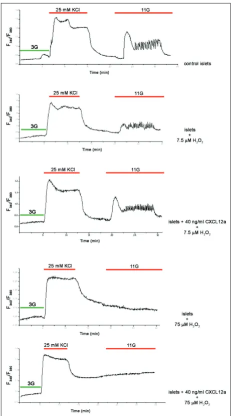
in-flux. The control islets exhibited the usual response to a high glucose challenge, observed as an increase in intracellular Ca2+ concentration (Fig. 2A). In contrast,
islets treated with 7.5 µM hydrogen for 1 h exhibited decreased responsiveness to the high D-glucose treat-ment (Fig. 2B), displayed as an irregular Ca2+ spike
followed by an oscillatory pattern. When the same experimental setup was applied to islets that were in-cubated overnight with 40 ng/mL of CXCL12α prior to exposure to 7.5 µM hydrogen peroxide for 1 h (Fig. 2C), the CXCL12α-pretreated islets exhibited a physi-ological response to high glucose that was comparable to the response elicited in control islets. The protec-tive capacity of CXCL12α was further tested on islets that were exposed to a 10-fold higher concentration (75 µM) of hydrogen peroxide. This concentration of hydrogen peroxide, which induced a high level of oxidative stress, initiated widespread cell death, so that
the islet cells were unable to properly respond to the high D-glucose concentration (Fig. 2D). The pretreat-ment of islet cells with CXCL12α did not preserve cell functionality at the level of oxidative stress induced by 75 µM hydrogen peroxide (Fig. 2E).
CXCl12α modulates Ca2+ oscillations in islet cells
in response to high glucose
The deterioration of pancreatic β-cell function over time in T2D is the consequence of poor glycemic control and the continuous exposure of β-cells to the deleterious effects of reactive oxygen species (ROS). We examined whether CXCL12α could improve the response of pancreatic islet cells in the hyperglycemic state. To that end, [Ca2+]
i was monitored in isolated
islets preloaded with Fura-2 AM (Fig. 3). As can be seen in Fig. 3A, when islets were treated with 3 mM of D-glucose supplemented with 40 ng/mL of CXCL12α, their intracellular Ca2+ concentration did not change,
and islet functionality was preserved, which was confirmed by their response to the high, 11-mM D-glucose concentration. When islets were exposed to a moderate to high, 8-mM D-glucose concentration (Fig. 3B), the initial Ca2+ spike was followed by
con-tinuous oscillations as a result of the repeated entry of Ca2+. A mix of 8 mM D-glucose and 40 ng/mL of
CXCL12α induced changes in the mode of Ca2+ entry
and overall [Ca2+]
i, manifesting as an oscillatory
pat-tern with longer-lasting oscillations with a lower fre-quency. Calculation of the overall Ca2+ uptake revealed
that CXCL12α modulated Ca2+ influx, resulting in
in-creased [Ca2+]
i (inset in Fig. 3B). The same result was
obtained when islet cells were challenged with 11 mM of D-glucose (Fig. 3C). In this setup, the islets were probed with 11 mM D-glucose, as well as in combina-tion with two CXCL12α concentracombina-tions (20 and 40 ng/ mL). Treatments with both CXCL12α concentrations decreased the number of oscillations that also lasted longer, which pointed to an overall increase in [Ca2+]
i
(inset in Fig. 3C), with the 40-ng/mL CXCL12α con-centration showing a more significant effect.
CXCl12α signaling influences the expression of voltage-gated Ca2+ channels in pancreatic islets
The voltage-gated calcium (Cav) channels in β-cells are molecular switches that play a central role in insulin secretion. The observation that CXCL12α is capable of modifying Ca2+ oscillations and increasing [Ca2+]
i led
us to assume that the CXCL12α/receptor axis regulates the behavior of the Cav channels. To examine this
fur-Fig. 3. Direct stimulatory effect of CXCL12α on intracellular calcium concentration. The
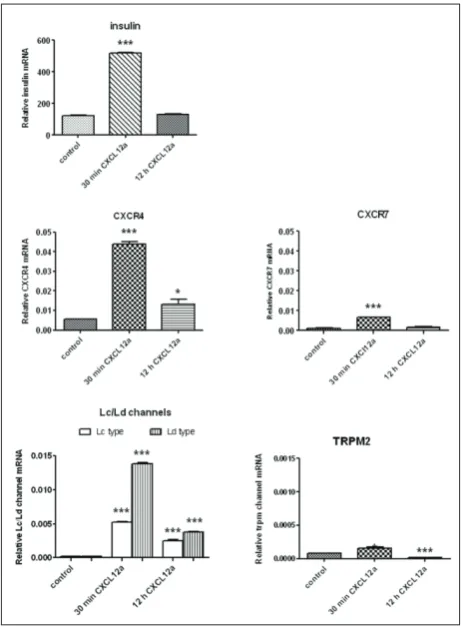
ther, isolated islets were incubated either for 30 min or overnight with 40 ng/mL of CXCL12α, and their mRNA was isolated (Fig. 4). CXCL12 acts via two re-ceptors, CXCR4 and CXCR7. RT-qPCR experiments revealed that the mRNAs encoding for both receptors were significantly increased after the 30-min incuba-tion of islets with CXCL12α (Fig. 4). The overnight incubation with CXCL12α induced a 2-fold increase in CXCR4 mRNA, while the level of CXCR7 mRNA was unchanged. This result revealed that CXCR7 gene transcription was considerably lower in islets as com-pared to CXCR4 gene transcription. Further, RT-qPCR experiments revealed that CXCL12α also induced insu-lin mRNA synthesis in islet cells. The exposure of islets
to CXCL12α for 30 min induced a 4-fold increase in insulin mRNA synthesis, while overnight islet incuba-tion with CXCL12a did not cause a significant increase in insulin gene transcription.
Next, we analyzed the expression of voltage-gated L-type Ca2+ channels that belong to the high-threshold
type of channels and are opened by large depolariza-tions, that contribute to the β-cell Ca2+ current and
insulin secretion. After the 30-min incubation with CXCL12α, the mRNAs for both Lc and Ld subtypes were significantly increased (2.5- and 6-fold, respec-tively). The overnight incubation with CXCL12α induced less pronounced but significant increases in Lc and Ld mRNAs (Fig. 4). We also analyzed the Ca2+-permeable, nonselective cation channel TRPM2,
which is gated by intracellular ADP-ribose and has a key role in hydrogen peroxide-induced Ca2+
tran-sients and cell death [21]. In pancreatic islets under physiological conditions, TRPM2 mRNA was barely detectable. After the 30-min incubation with 40 ng/ mL of CXCL12α, a small, insignificant increase was observed (Fig. 4), and after the overnight incubation with CXCL12α, a significant decrease in TRPM2 gene transcription was measured, pointing to the prosur-vival effect of CXCL12α in islet cells.
CXCl12α stimulated insulin release in the hyperglycemic state
An increase in blood glucose concentrations induces a series of cellular events leading to the depolariza-tion of the β-cell plasma membrane that opens CaV channels to mediate Ca2+ influx and stimulate insulin
secretion. In the next set of experiments we exam-ined whether the increase in [Ca2+]
i, observed after
the incubation of pancreatic islets with CXCL12α, was also linked with insulin secretion. The islets were incubated either for 30 min (Fig. 5A) or overnight (Fig. 5B) with 40 ng/mL of CXCL12α, followed by treatment with 8 mM and 11 mM of D-glucose, and their insulin secretion was measured by RIA. Under physiological conditions, in the presence of 3 mM of D-glucose, CXCL12α did not affect insulin secretion. Stimulatory glucose concentrations induced increased insulin secretion, further enhanced in the presence of CXCL12α (Fig. 5). Incubation with CXCL12α for 30 min induced significant insulin release after
posure to 8 mM and 11 mM of D-glucose (compared to matching controls, 2.18- and 1.53-fold increases, respectively; Fig. 5A). The overnight incubation of islets with CXCL12α induced significant but slightly lower increases in insulin secretion after exposure to 8 mM and 11 mM of D-glucose (compared to matching controls, 1.59- and 1.35-fold increase, respectively) compared to the 30-min incubation with CXCL12α (Fig. 5B). These findings underlined that the presence of CXCL12α in the cell medium increases insulin re-lease in islets by enhancing Ca2+ uptake.
dIsCussIon
Hyperglycemia contributes to the reduction of insulin secretion and to the progression from glucose intoler-ance to T2D [22], causing pancreatic β-cell
dysfunc-tion and decreasing the β-cell mass [23,24]. Therefore, β-cell protection remains the focus of strategies for the prevention and treatment of diabetes [25]. In general, the diabetogenic environment, including insulin re-sistance and low-grade systemic islet inflammation contribute toward the stunning of pancreatic β-cells, i.e. a state of temporary incapacity to properly respond to the presence of increased glucose concentration by secreting insulin [26]. The aim of the present study was to examine whether CXCL12α exhibits a benefi-cial effect on pancreatic β-cells’ ability to release insu-lin and whether it improves the response of pancreatic islets in the hyperglycemic state.
Antidiabetic action of CXCl12α: normalizing Ca2+ uptake in order to preserve the functionality
of islets
In overweight and obese individuals, pancreatic β-cells are exposed to metabolic changes and the consequences thereof, including oxidative stress re-sulting from increased ROS and RNS production. As a ligand of two receptors, CXCR4 and CXCR7, CXCL12 signaling is crucial for β-cell differentiation and pancreatic islet genesis [27]. Recent animal stud-ies revealed the essential role of CXCL12 in duct cell survival, proliferation and migration during pancre-atic regeneration, through its ability to activate Akt, Src and the extracellular signal-regulated protein ki-nase (ERK1/2) [28]. Our group also showed that the role of CXCL12 in diabetes attenuation is based on its ability to activate the antinecrotic/prosurvival pathway after hydrogen peroxide treatment [17]. In this study, we examined oxidative stress as a potential inducer of β-cell dysfunction [29]. Both effects, proliferative burst and increased survival rate of pancreatic β-cells, observed after islet incubation with CXCL12α could be accomplished by the direct influence of CXCL12 on intracellular Ca2+ influx.
It is widely accepted that exogenous ROS induces dynamic changes in Ca2+ concentration in a variety
of cell types [30-35]. This effect could be due to the mobilization of intracellular Ca2+ stores and influx
of extracellular Ca2+. An important feature of
cross-regulation between ROS and Ca2+ is that the ROS
ef-fect on Ca2+ signaling can vary from stimulatory to
repressive, depending on the type of oxidants, their concentrations and the duration of exposure. The
fect of ROS on Ca2+ signaling is also tissue-specific
(reviewed in [36]). We found that CXCL12α preserves mouse islet functionality by normalizing their Ca2+
response under oxidative stress (hydrogen peroxide treatment). In contrast, islet cell treatment with a 10-times higher concentration of hydrogen peroxide (75 µM) induced a constant entry of Ca2+, which
in-dicated cell death as a consequence of exposure to the very high concentration of peroxide treatment. This finding is in accordance with previous studies showing that hydrogen peroxide causes an increase in intracel-lular Ca2+ levels that leads to cell death in a variety of
cell types [37-41]. The mechanism whereby hydrogen peroxide causes an increase in [Ca2+]
i is still under
debate, with several options proposed, including the involvement of voltage-gated Ca2+ channels (Ca
v) [41],
nonspecific changes in membrane calcium perme-ability [42], alteration in Na+-Ca2+ exchange [38], or
changes in Ca2+ release from intracellular stores [43].
Using the insulin-secreting cell line CRI-G1 and very high hydrogen peroxide concentrations (1-10 mM), Herson et al. [44] described the effects of hydrogen peroxide on [Ca2+]
i. They clearly demonstrated that
hydrogen peroxide disrupts calcium homeostasis. In contrast, it has also been reported that hydrogen per-oxide exerts no significant effect on the L-type Ca2+
current in pancreatic β-cells [45].
On the other hand, a chronic exposure of pancre-atic islets to nonphysiological high glucose concentra-tions caused adverse alteraconcentra-tions in β-cell functioning in a phenomenon referred to as glucose toxicity, which may play a secondary pathogenic role in T2D. Several results suggested that one mechanism of glucose toxic-ity in β-cells may be linked to chronic exposure of cells to ROS (or chronic oxidative stress) [46-48]. Both low and high concentrations of glucose have been shown to increase intracellular peroxide levels within the is-lets [49], which indicates that glucose metabolism is essential for the induction of glucose toxicity. This is in agreement with Ihara et al. [50], who reported elevated levels of oxidative stress markers (8-hydroxy-2’-deoxyguanosine and 4-hydroxy-2-nonenal-mod-ified proteins) in the β-cells of Goto-Kakizaki rats. Our experimental setup is suitable for observing β-cell behavior in hyperglycemic conditions, since chronic hyperglycemia causes a progressive decline in β-cell functioning and their ultimately demise [26,51].
Electrical activity and stimulated secretion, coupled with intracellular Ca2+ dynamics that allow
pancre-atic β-cells to secrete insulin, first reported by Dean and Matthews in 1968 [52], are nowadays accepted [53-55]. Cells often respond to changes in stimulus intensity by varying the frequency of Ca2+ waves.
To demonstrate the rescue potential of CXCL12α in preventing β-cell dysfunction and exhaustion due to sustained hyperglycemia, mouse pancreatic islets were exposed to stimulatory glucose concentrations, both moderate to high (8 M) and high (11 M), for an average duration of 45 min [56,57]. In our study, the treatment of β-cells with CXCL12α in hyperglycemic conditions decreased the number and duration of Ca2+
oscillations, with an overall increase in [Ca2+] i. It has
been already shown that CXCL12 stimulates Ca2+ flux
and transients in many cell types [58-60]. Similar to our finding, CXCL12 dose-dependently increased intracellular Ca2+ in adherent IEC-6 cells in the
con-centrations of 20 and 100 ng/mL [61]. Chemokine-induced Ca2+ mobilization was not solely a function
of rat intestinal epithelial cells, since CXCL12 stimu-lated a rapid increase in Ca2+ within 40 s of ligand
stimulation in the intestinal carcinoma cell line CaCo2 cells as well [61]. In human LX2 cells, treatment with CXCL12 led to a nearly two-fold increase in relative Ca2+ influx, whereas in murine JS1 cells CXCL12 did
not promote Ca2+ movement [62]. The latter authors
showed that the CXCL12-influenced hepatic stellate cell contraction is not Ca2+ dependent. In addition,
the Ca2+-influenced CXCL12 secretion has also been
investigated. Schajnovitz et al. [63] reported that Ca2+
is transmitted via gap junctions in contacting cell cul-tures, and that Ca2+ signaling has a role in bone
mar-row stromal cell CXCL12 secretion.
CXCl12α signaling influences the expression of voltage-gated Ca2+ channels in pancreatic
islets and stimulates insulin release in the hyperglycemic state
In a high-glucose environment and/or in the presence of ROS, voltage-gated Ca2+ (Ca
v) channels are
acti-vated, allowing extracellular Ca2+ to enter [64-67]. It is
generally accepted that the subsequent increase in cy-tosolic Ca2+ triggers insulin release, even though there
is some evidence for a [Ca2+]
i-independent second
activity and/or density may result in lower or higher insulin secretion [69-77]. Insulin release must be able to anticipate glucose increase and prevent persistent glucose elevations [26,78]. Insulin secretion analysis in rat insulinoma and primary islet cells revealed that N- and L-type channels are both involved in immediate glucose-induced insulin secretion. However, L-type Cav channel was preferentially coupled to secretion at a later phase. P/Q-type channels were not found to play any role in insulin secretion at any stage [79]. In our experimental setup, the exogenously added CXCL12α influenced [Ca2+]
i and insulin release,
pro-moting less frequent oscillations of [Ca2+]
i with longer
duration, and allowing more Ca2+ to enter the islet.
One explanation for this increase in [Ca2+]
i could be
linked to the ability of exogenously added CXCL12α to enhance gene expression of the Lc (Cav1.2-subunit)/ Ld (Cav1.3-subunit) types of Cav, shown in our ex-periments. The main subtype of the L-type of chan-nels in mouse islet β-cells is the Ld subtype [77], al-though the Lc and Ld subtypes share almost a 70% homology [80]. In agreement with previous reports, herein we showed that CXCL12α increases the tran-scription rate of Cav L-type channels, in particular Ld-type, which contains the α1D subunit and repre-sents the Cav1.3α1 channel subtype. We also observed an increase in N-type channels due to the increased presence of CXCL12. The increased transcriptional rate for the N-type of channels was significant but 7.2-fold less pronounced as compared to the increase in the transcriptional rate for the L-type Cav channels (data not shown). In contrast, Barg et al. [81] found no differences in Ca2+-current density and Cav1.3/
α1D-deficient β-cells, and concluded that Cav1.2 is the principal L-type Ca2+ channel subtype in mouse
β-cells. The functional consequences of the increase in Cav channel transcription rate may be relevant in dis-eases, such as T2D, since β-cells operate with very few Cav channels [55,82]. The observed CXCL12α-related influence could provide β-cells with a mechanism to push Ca2+-coupled regulation of insulin secretion in
response to elevated glucose. Since [Ca2+]
i dynamics are also responsible for
β-cell fate [55], we examined whether the Trpm2 Ca2+
channel is upregulated due to an excess of CXCL12α. Trpm2, a calcium-permeable nonselective cation channel expressed in the plasma membrane is criti-cally involved in ROS-induced processes and is
im-plicated in cell death [83]. In contrast to Cav channels, CXCL12α has no influence on the transcription rate of Trpm2 after 30 min of incubation, in contrast to overnight incubation, which caused a lowering of the transcription of the Trpm2 channel gene. The primary gating mechanism of Trpm2 involves the binding of ADP-ribose polymers (ADPR) [84], but Trpm2 cur-rents can also be activated by reactive oxygen spe-cies (ROS) [85] through ROS-induced ADPR release from mitochondria [86]. Bearing in mind that the role of Trpm2 in pancreatic β-cells involves situations in which β-cells are stressed [87,88], the observed down-regulation of Trpm2 mRNA in our experiments is not surprising. As a small chemokine, CXCL12 exerts its effect via coupling to its receptors, CXCR4 and CXCR7. The 30-min incubation with CXCL12α was followed by an increase in the level of Cxcr4 mRNA. The increased Ca2+ influx in islet cells was
accompa-nied by increased transcription of the Cxcr7 and Ins. Our RT-qPCR results showed significantly increased transcription of both receptor genes after islet incuba-tion with CXCL12α, targeting CXCR4 as the most im-portant receptor responsible for signaling within the islet. This is in accordance with previous reports in-dicating that CXCL12 is involved in the regulation of insulin secretion [89]. β-cell injury induces CXCL12 expression and the secreted CXCL12 causes dediffer-entiation of adjacent -cells into pro-α-cells, which are further transformed into pancreatic β-cells [18, 90].
β-cell Cav channel activity can be modulated by changes in membrane potential and through a vari-ety of signaling pathways, such as protein phospho-rylation, Ca2+-dependent inactivation and through
interaction with G protein (reviewed in [91]). The observation that CXCL12α is capable to modify Ca2+
oscillations and increase [Ca2+]
i as well as influence
the transcription of Lc and Ld Ca2+ channel genes, led
us to assume that the CXCL12α/receptor axis regulates the behavior of the Cav channels. Princen et al. [92] showed that a transient dose-dependent increase in intracellular Ca2+ concentration is an essential
com-ponent of the signal transduction cascade activated upon CXCL12 binding to its receptor CXCR4. The authors showed that in all examined CXCR4+ cell
lines, CXCL12 elicited a transient [Ca2+]
i increase.
to the G-protein-coupled receptor (GPCR) Gαi, and that CXCR4 can couple to other Gα protein subunits such as Gαq, Gαo, and Gαs in order to stimulate Ca2+
flux [93]. However, it has been proven that Ca2+ flux is
associated with the GPCR subunit Gαq but not to Gαi [94]. Quite the opposite, Hsu et al. [95] proposed that the L-type Cav channel in insulin-secreting cells is the major mediator of the somatostatin-induced inhibi-tion of insulin secreinhibi-tion by the activainhibi-tion of G protein-coupled receptors. In the same line, Renstrom et al. [96] reported that stimulation of GPCRs in mouse islet β-cells does not influence the voltage activated Ca2+ influx. Our assumption of the CXCL12/receptor
axis being a mediator in the elevated voltage activated Ca2+ influx in pancreatic β cells is supported by Roe
et al. [97], who reported that decreased L-type chan-nel activity and expression were associated with T2D. Defects in the metabolic regulation of insulin se-cretion contribute to the development of T2D. Rescu-ing stunned β-cells is possibly a key therapeutic target for the treatment and prevention of diabetes progres-sion. It seems reasonable to propose that bringing any degree of hyperglycemia under tight control as early as possible can switch on stunned β-cells, in spite of the fact that glycemic memory and genetic/epigenetic programming may differ in every individual. Identi-fication of the factors that regulate the overall Ca2+
intake, activity and expression of Cav channels and insulin release may provide a better understanding of pancreatic β-cell functioning in normal and diabetic states. CXCL12 displayed the potential to regulate [Ca2+]
i and the transcription of Cav channel genes and
thus may serve as a potential T2D attenuator.
Acknowledgments: This work was supported by the Ministry of
Education, Science and Technological Development of the Repub-lic of Serbia, Grant No. 173020. MV is very grateful to EASD for the research travel fellowship award (Albert Renold fellowship) that enabled this work.
Author contributions: Conception and design of the experiments: MV ECG. Experimental work: MV MDj SD NG MM AU JAJ. Data analysis: MV AT GP JR. Writing of the manuscript: MV GP MS. Conflict of interest disclosure: The authors declare that no com-peting interests exist.
RefeRenCes
1. Muoio DM, Newgard CB. Mechanisms of disease: Molecu-lar and metabolic mechanisms of insulin resistance and beta-cell failure in type 2 diabetes. Nat Rev Mol Cell Biol. 2008;9(3):193-205.
2. Braun M, Ramracheya R, Bengtsson M, Zhang Q, Karanaus-kaite J, Partridge C, Johnson PR, Rorsman P. Voltage-gated ion channels in human pancreatic beta-cells: electrophysi-ological characterization and role in insulin secretion. Dia-betes. 2008;57(6):1618-28.
3. Fridlyand LE, Jacobson DA, Philipson LH. Ion channels and regulation of insulin secretion in human beta-cells: a compu-tational systems analysis. Islets. 2013;5(1):1-15.
4. Fujimoto W, Miki T, Ogura T, Zhang M, Seino Y, Satin LS, Nakaya H, Seino S. Niflumic acid-sensitive ion channels play an important role in the induction of glucose-stimu-lated insulin secretion by cyclic AMP in mice. Diabetologia. 2009;52(5):863-72.
5. Proks P, Ashcroft FM. Modeling K(ATP) channel gating and its regulation. Prog Biophys Mol Biol. 2009;99(1):7-19. 6. Rorsman P, Eliasson L, Kanno T, Zhang Q, Gopel S.
Electro-physiology of pancreatic beta-cells in intact mouse islets of Langerhans. Prog Biophys Mol Biol. 2011;107(2):224-35. 7. Henquin JC, Nenquin M, Ravier MA, Szollosi A.
Shortcom-ings of current models of glucose-induced insulin secretion. Diabetes Obes Metab. 2009;11(Suppl.4):168-79.
8. Fridlyand LE, Jacobson DA, Kuznetsov A, Philipson LH. A model of action potentials and fast Ca2+ dynamics in pan-creatic beta-cells. Biophys J. 2009;96(8):3126-39.
9. Nagasawa T, Kikutani H, Kishimoto T. Molecular cloning and structure of a pre-B-cell growth-stimulating factor. Proc Natl Acad Sci U S A. 1994;91(6):2305-9.
10. Rollins BJ. Chemokines. Blood. 1997;90(3):909-28. 11. Day CE, Guillen C, Willars GB, Wardlaw AJ.
Characteriza-tion of the migraCharacteriza-tion of lung and blood T cells in response CXCL12 in a three-dimensional matrix. Immunology. 2010;130(4):564-71.
12. Rabbany SY, Pastore J, Yamamoto M, Miller T, Rafii S, Aras R, Penn M. Continuous delivery of stromal cell-derived fac-tor-1 from alginate scaffolds accelerates wound healing. Cell Transplant. 2010;19(4):399-408.
13. Sharp CD, Huang M, Glawe J, Patrick DR, Pardue S, Barlow SC, Kevil CG. Stromal cell-derived factor-1/CXCL12 stimu-lates chemorepulsion of NOD/LtJ T-cell adhesion to islet microvascular endothelium. Diabetes. 2008;57(1):102-12. 14. Balabanian K, Lagane B, Infantino S, Chow KY, Harriague
J, Moepps B, Arenzana-Seisdedos F, Thelen M, Bachelerie F. The chemokine SDF-1/CXCL12 binds to and signals through the orphan receptor RDC1 in T lymphocytes. J Biol Chem. 2005;280(42):35760-6.
15. Bleul CC, Farzan M, Choe H, Parolin C, Clark-Lewis I, Sodroski J, Springer TA. The lymphocyte chemoattractant SDF-1 is a ligand for LESTR/fusin and blocks HIV-1 entry. Nature. 1996;382(6594):829-33.
17. Grdovic N, Dinic S, Mihailovic M, Uskokovic A, Jovanovic JA, Poznanovic G, Wagner L, Vidakovic M. CXC chemokine ligand 12 protects pancreatic beta-cells from necrosis through Akt kinase-mediated modulation of poly(ADP-ribose) poly-merase-1 activity. PLoS One. 2014;9(7):e101172.
18. Liu Z, Stanojevic V, Avadhani S, Yano T, Habener JF. Stro-mal cell-derived factor-1 (SDF-1)/chemokine (C-X-C motif) receptor 4 (CXCR4) axis activation induces intra-islet gluca-gon-like peptide-1 (GLP-1) production and enhances beta cell survival. Diabetologia. 2011;54(8):2067-76.
19. Nadal A, Rovira JM, Laribi O, Leon-quinto T, Andreu E, Ripoll C, Soria B. Rapid insulinotropic effect of 17beta-estradiol via a plasma membrane receptor. FASEB J. 1998;12(13):1341-8. 20. Bradford MM. A rapid and sensitive method for the
quantita-tion of microgram quantities of protein utilizing the principle of protein-dye binding. Anal Biochem. 1976;72:248-54. 21. Miller BA. Inhibition of TRPM2 function by PARP
inhibi-tors protects cells from oxidative stress-induced death. Br J Pharmacol. 2004;143(5):515-6.
22. Cnop M, Welsh N, Jonas JC, Jorns A, Lenzen S, Eizirik DL. Mechanisms of pancreatic beta-cell death in type 1 and type 2 diabetes: many differences, few similarities. Diabetes. 2005; 54(Suppl.2):S97-107.
23. Chang-Chen KJ, Mullur R, Bernal-Mizrachi E. Beta-cell fail-ure as a complication of diabetes. Rev Endocr Metab Disord. 2008;9(4):329-43.
24. Robertson RP. Beta-cell deterioration during diabetes: what’s in the gun? Trends Endocrinol Metab. 2009;20(8):388-93. 25. Wajchenberg BL. beta-cell failure in diabetes and preservation
by clinical treatment. Endocr Rev. 2007;28(2):187-218. 26. Ferrannini E. The stunned beta cell: a brief history. Cell
Metab. 2010;11(5):349-52.
27. Kayali AG, Lopez AD, Hao E, Hinton A, Hayek A, King CC. The SDF-1alpha/CXCR4 axis is required for proliferation and maturation of human fetal pancreatic endocrine progenitor cells. PLoS One. 2012;7(6):e38721.
28. Kayali AG, Van Gunst K, Campbell IL, Stotland A, Kritzik M, Liu G, Flodstrom-Tullberg M, Zhang YQ, Sarvetnick N. The stromal cell-derived factor-1alpha/CXCR4 ligand-receptor axis is critical for progenitor survival and migration in the pancreas. J Cell Biol. 2003;163(4):859-69.
29. Sakai K, Matsumoto K, Nishikawa T, Suefuji M, Nakamaru K, Hirashima Y, Kawashima J, Shirotani T, Ichinose K, Brownlee M, Araki E. Mitochondrial reactive oxygen species reduce insulin secretion by pancreatic beta-cells. Biochem Biophys Res Commun. 2003;300(1):216-22.
30. Aley PK, Porter KE, Boyle JP, Kemp PJ, Peers C. Hypoxic modulation of Ca2+ signaling in human venous endothelial cells. Multiple roles for reactive oxygen species. J Biol Chem. 2005;280(14):13349-54.
31. Doan TN, Gentry DL, Taylor AA, Elliott SJ. Hydrogen perox-ide activates agonist-sensitive Ca(2+)-flux pathways in canine venous endothelial cells. Biochem J. 1994;297(Pt 1):209-15. 32. Dreher D, Junod AF. Role of oxygen free radicals in cancer
development. Eur J Cancer. 1996;32A(1):30-8.
33. Gen W, Tani M, Takeshita J, Ebihara Y, Tamaki K. Mecha-nisms of Ca2+ overload induced by extracellular H2O2 in quiescent isolated rat cardiomyocytes. Basic Res Cardiol. 2001;96(6):623-9.
34. Kumasaka S, Shoji H, Okabe E. Novel mechanisms involved in superoxide anion radical-triggered Ca2+ release from cardiac sarcoplasmic reticulum linked to cyclic ADP-ribose stimulation. Antioxid Redox Signal. 1999;1(1):55-69. 35. Suzuki YJ, Forman HJ, Sevanian A. Oxidants as stimulators of
signal transduction. Free Radic Biol Med. 1997;22(1-2):269-85. 36. Yan Y, Wei CL, Zhang WR, Cheng HP, Liu J. Cross-talk
between calcium and reactive oxygen species signaling. Acta Pharmacol Sin. 2006;27(7):821-6.
37. Bielefeldt K, Whiteis CA, Sharma RV, Abboud FM, Conklin JL. Reactive oxygen species and calcium homeostasis in cul-tured human intestinal smooth muscle cells. Am J Physiol. 1997;272(6Pt1):G1439-50.
38. Kaneko M, Matsumoto Y, Hayashi H, Kobayashi A, Yamazaki N. Oxygen free radicals and calcium homeostasis in the heart. Mol Cell Biochem. 1994;139(1):91-100.
39. Klonowski-Stumpe H, Schreiber R, Grolik M, Schulz HU, Haussinger D, Niederau C. Effect of oxidative stress on cel-lular functions and cytosolic free calcium of rat pancreatic acinar cells. Am J Physiol. 1997;272(6Pt1):G1489-98. 40. Leist M, Nicotera P. Calcium and neuronal death. Rev Physiol
Biochem Pharmacol. 1998;132:79-125.
41. Rojanasakul Y, Wang L, Hoffman AH, Shi X, Dalal NS, Banks DE, Ma JK. Mechanisms of hydroxyl free radical-induced cel-lular injury and calcium overloading in alveolar macrophages. Am J Respir Cell Mol Biol. 1993;8(4):377-83.
42. Clague JR, Langer GA. The pathogenesis of free radical-induced calcium leak in cultured rat cardiomyocytes. J Mol Cell Cardiol. 1994;26(1):11-21.
43. Nicotera P, Rossi AD. Nuclear Ca2+: physiological regulation and role in apoptosis. Mol Cell Biochem. 1994;135(1):89-98. 44. Herson PS, Lee K, Pinnock RD, Hughes J, Ashford ML. Hydrogen peroxide induces intracellular calcium overload by activation of a non-selective cation channel in an insulin-secreting cell line. J Biol Chem. 1999;274(2):833-41. 45. Krippeit-Drews P, Britsch S, Lang F, Drews G. Effects of
SH-group reagents on Ca2+ and K+ channel currents of pancreatic B-cells. Biochem Biophys Res Commun. 1994;200(2):860-6. 46. Baumgartner-Parzer SM, Wagner L, Pettermann M, Grillari
J, Gessl A, Waldhausl W. High-glucose--triggered apoptosis in cultured endothelial cells. Diabetes. 1995;44(11):1323-7. 47. Du XL, Sui GZ, Stockklauser-Farber K, Weiss J, Zink S,
Schwippert B, Wu QX, Tschope D, Rosen P. Induction of apoptosis by high proinsulin and glucose in cultured human umbilical vein endothelial cells is mediated by reactive oxygen species. Diabetologia. 1998;41(3):249-56.
48. Tanaka Y, Gleason CE, Tran PO, Harmon JS, Robertson RP. Prevention of glucose toxicity in HIT-T15 cells and Zucker diabetic fatty rats by antioxidants. Proc Natl Acad Sci U S A. 1999;96(19):10857-62.
49. Tanaka Y, Tran PO, Harmon J, Robertson RP. A role for glu-tathione peroxidase in protecting pancreatic beta cells against oxidative stress in a model of glucose toxicity. Proc Natl Acad Sci U S A. 2002;99(19):12363-8.
51. Talchai C, Xuan SH, Lin HV, Sussel L, Accili D. Pancreatic beta Cell Dedifferentiation as a Mechanism of Diabetic beta Cell Failure. Cell. 2012;150(6):1223-34.
52. Dean PM, Matthews EK. Electrical activity in pancreatic islet cells. Nature. 1968;219(5152):389-90.
53. Henquin JC. Triggering and amplifying pathways of regulation of insulin secretion by glucose. Diabetes. 2000;49(11):1751-60. 54. Misler S, Barnett DW, Gillis KD, Pressel DM. Electrophysi-ology of stimulus-secretion coupling in human beta-cells. Diabetes. 1992;41(10):1221-8.
55. Rorsman P, Braun M, Zhang Q. Regulation of calcium in pancreatic alpha- and beta-cells in health and disease. Cell Calcium. 2012;51(3-4):300-8.
56. Hoenig M, MacGregor LC, Matschinsky FM. In vitro exhaus-tion of pancreatic beta-cells. Am J Physiol. 1986;250(5Pt1): E502-11.
57. Martin F, Ribas J, Soria B. Cytosolic Ca2+ gradients in pancre-atic islet-cells stimulated by glucose and carbachol. Biochem Biophys Res Commun. 1997;235(3):465-8.
58. Basu S, Broxmeyer HE. CCR5 ligands modulate CXCL12-induced chemotaxis, adhesion, and Akt phosphoryla-tion of human cord blood CD34+ cells. J Immunol. 2009;183(11):7478-88.
59. Drury LJ, Ziarek JJ, Gravel S, Veldkamp CT, Takekoshi T, Hwang ST, Heveker N, Volkman BF, Dwinell MB. Mono-meric and diMono-meric CXCL12 inhibit metastasis through dis-tinct CXCR4 interactions and signaling pathways. Proc Natl Acad Sci U S A. 2011;108(43):17655-60.
60. Kim CH, Hangoc G, Cooper S, Helgason CD, Yew S, Humphries RK, Krystal G, Broxmeyer HE. Altered respon-siveness to chemokines due to targeted disruption of SHIP. J Clin Invest. 1999;104(12):1751-9.
61. Agle KA, Vongsa RA, Dwinell MB. Calcium mobilization triggered by the chemokine CXCL12 regulates migration in wounded intestinal epithelial monolayers. J Biol Chem. 2010;285(21):16066-75.
62. Saiman Y, Bansal M. Hepatic Stellate Cell-derived CXCL12 promotes T cell adhesion to Sinusoidal Endothelial Cells. Hepatology. 2012;56:258a-259a.
63. Schajnovitz A, Itkin T, D’Uva G, Kalinkovich A, Golan K, Ludin A, Cohen D, Shulman Z, Avigdor A, Nagler A, Kollet O, Seger R, Lapidot T. CXCL12 secretion by bone marrow stromal cells is dependent on cell contact and mediated by connexin-43 and connexin-45 gap junctions. Nat Immunol. 2011;12(5):391-8. 64. Quesada I, Martin F, Soria B. Nutrient modulation of
polar-ized and sustained submembrane Ca2+ microgradients in mouse pancreatic islet cells. J Physiol. 2000; 525(Pt1):159-67. 65. Santos RM, Rosario LM, Nadal A, Garcia-Sancho J, Soria B,
Valdeolmillos M. Widespread synchronous [Ca2+]i oscilla-tions due to bursting electrical activity in single pancreatic islets. Pflugers Arch. 1991;418(4):417-22.
66. Soria B, Tuduri E, Gonzalez A, Hmadcha A, Martin F, Nadal A, Quesada I. Pancreatic islet cells: a model for calcium-dependent peptide release. HFSP J. 2010;4(2):52-60. 67. Valdeolmillos M, Nadal A, Contreras D, Soria B. The
relation-ship between glucose-induced K+ATP channel closure and the rise in [Ca2+]i in single mouse pancreatic beta-cells. J Physiol. 1992;455:173-86.
68. Martin F, Sanchez-Andres JV, Soria B. Slow [Ca2+]i oscilla-tions induced by ketoisocaproate in single mouse pancreatic islets. Diabetes. 1995;44(3):300-5.
69. Ammala C, Ashcroft FM, Rorsman P. Calcium-independent potentiation of insulin release by cyclic AMP in single beta-cells. Nature. 1993;363(6427):356-8.
70. Arkhammar P, Juntti-Berggren L, Larsson O, Welsh M, Nan-berg E, Sjoholm A, Kohler M, Berggren PO. Protein kinase C modulates the insulin secretory process by maintaining a proper function of the beta-cell voltage-activated Ca2+ chan-nels. J Biol Chem. 1994;269(4):2743-9.
71. Huang LP, Bhattacharjee A, Taylor JT, Zhang M, Keyser BM, Marrero L, Li M. Ca2+](i) regulates trafficking of Ca(v)1.3 (alpha(1D) Ca2+ channel) in insulin-secreting cells. Am J Physiol-Cell Ph. 2004;286(2):C213-21.
72. Ji JZ, Yang SN, Huang XH, Li XD, Shen L, Diamant N, Berggren PO, Gaisano HY. Modulation of L-type Ca2+ channels by distinct domains within SNAP-25. Diabetes. 2002;51(5):1425-36.
73. Kang Y, Huang X, Pasyk EA, Ji J, Holz GG, Wheeler MB, Tsu-shima RG, Gaisano HY. Syntaxin-3 and syntaxin-1A inhibit L-type calcium channel activity, insulin biosynthesis and exo-cytosis in beta-cell lines. Diabetologia. 2002;45(2):231-41. 74. Passafaro M, Codignola A, Rogers M, Cooke I, Sher E.
Modu-lation of N-type calcium channels translocation in RINm5F insulinoma cells. Pharmacol Res. 2000;41(3):325-34. 75. Plant TD. Properties and calcium-dependent inactivation
of calcium currents in cultured mouse pancreatic B-cells. J Physiol. 1988;404:731-47.
76. Wiser O, Trus M, Hernandez A, Renstrom E, Barg S, Rors-man P, Atlas D. The voltage-sensitive Lc-type Ca2+ channel is functionally coupled to the exocytotic machinery. P Natl Acad Sci USA. 1999;96(1):248-53.
77. Yang SN, Larsson O, Branstrom R, Bertorello AM, Leibiger B, Leibiger IB, Moede T, Kohler M, Meister B, Berggren PO. Syntaxin 1 interacts with the L(D) subtype of voltage-gated Ca(2+) channels in pancreatic beta cells. Proc Natl Acad Sci U S A. 1999;96(18):10164-9.
78. Henquin JC, Dufrane D, Nenquin M. Nutrient control of insulin secretion in isolated normal human islets. Diabetes. 2006;55(12):3470-7.
79. Taylor JT, Huang L, Keyser BM, Zhuang H, Clarkson CW, Li M. Role of high-voltage-activated calcium channels in glucose-regulated beta-cell calcium homeostasis and insulin release. Am J Physiol Endocrinol Metab. 2005;289(5):E900-8. 80. Dolphin AC. The G.L. Brown Prize Lecture. Voltage-depen-dent calcium channels and their modulation by neurotrans-mitters and G proteins. Exp Physiol. 1995;80(1):1-36. 81. Barg S, Ma X, Eliasson L, Galvanovskis J, Gopel SO,
Ober-muller S, Platzer J, Renstrom E, Trus M, Atlas D, Striessnig J, Rorsman P. Fast exocytosis with few Ca(2+) channels in insulin-secreting mouse pancreatic B cells. Biophys J. 2001;81(6):3308-23.
82. Smith PA, Aschroft FM, Fewtrell CM. Permeation and gating properties of the L-type calcium channel in mouse pancreatic beta cells. J Gen Physiol. 1993;101(5):767-97.
84. Perraud AL, Fleig A, Dunn CA, Bagley LA, Launay P, Schmitz C, Stokes AJ, Zhu Q, Bessman MJ, Penner R, Kinet JP, Scha-renberg AM. ADP-ribose gating of the calcium-permeable LTRPC2 channel revealed by Nudix motif homology. Nature. 2001;411(6837):595-9.
85. Hara Y, Wakamori M, Ishii M, Maeno E, Nishida M, Yoshida T, Yamada H, Shimizu S, Mori E, Kudoh J, Shimizu N, Kurose H, Okada Y, Imoto K, Mori Y. LTRPC2 Ca2+-permeable channel activated by changes in redox status confers suscep-tibility to cell death. Mol Cell. 2002;9(1):163-73.
86. Perraud AL, Takanishi CL, Shen B, Kang S, Smith MK, Schmitz C, Knowles HM, Ferraris D, Li W, Zhang J, Stod-dard BL, Scharenberg AM. Accumulation of free ADP-ribose from mitochondria mediates oxidative stress-induced gating of TRPM2 cation channels. J Biol Chem. 2005;280(7):6138-48. 87. Chen J, Gusdon AM, Thayer TC, Mathews CE. Role of
increased ROS dissipation in prevention of T1D. Ann N Y Acad Sci. 2008;1150:157-66.
88. Lenzen S. Oxidative stress: the vulnerable beta-cell. Biochem Soc Trans. 2008; 36 (Pt 3): 343-7.
89. Duncanson S, Sambanis A. Dual factor delivery of CXCL12 and Exendin-4 for improved survival and function of encap-sulated beta cells under hypoxic conditions. Biotechnol Bio-eng. 2013;110(8): 2292-300.
90. Habener JF, Stanojevic V. alpha-cell role in beta-cell genera-tion and regeneragenera-tion. Islets. 2012;4(3):188-98.
91. Yang SN, Berggren PO. Beta-cell CaV channel regulation in physiology and pathophysiology. Am J Physiol Endocrinol Metab. 2005;288(1):E16-28.
92. Princen K, Hatse S, Vermeire K, De Clercq E, Schols D. Evalu-ation of SDF-1/CXCR4-induced Ca2+ signaling by fluoro-metric imaging plate reader (FLIPR) and flow cytometry. Cytometry A. 2003;51(1):35-45.
93. Rubin JB. Chemokine signaling in cancer: one hump or two? Semin Cancer Biol. 2009;19(2):116-22.
94. Teicher BA, Fricker SP. Cxcl12 (Sdf-1)/Cxcr4 Pathway in Can-cer. Clin Cancer Res. 2010;16(11):2927-31.
95. Hsu WH, Xiang HD, Rajan AS, Kunze DL, Boyd AE, 3rd. Somatostatin inhibits insulin secretion by a G-protein-medi-ated decrease in Ca2+ entry through voltage-dependent Ca2+ channels in the beta cell. J Biol Chem. 1991;266(2):837-43. 96. Renstrom E, Ding WG, Bokvist K, Rorsman P.
Neu-rotransmitter-induced inhibition of exocytosis in insulin-secreting beta cells by activation of calcineurin. Neuron. 1996;17(3):513-22.