© 2020 by the Serbian Biological Society How to cite this article: Drljača J, Vejnović AT, Miljković DM, Popović MJ, Rakić DB, Sekulić SR, Čapo IĐ, Petković BB. Changes in mouse thymus after exposure to tube-restraint stress. Arch Biol Sci. 2020;72(1):5-11.
Changes in mouse thymus after exposure to tube-restraint stress
Jovana N. Drljača1, Ana-Marija T. Vejnović2, Dejan M. Miljković3, Milan J. Popović3, Dušica B. Rakić4,
Slobodan R. Sekulić5,*, Ivan Đ. Čapo3 and Branka B. Petković6
1Faculty of Medicine, University of Novi Sad, Hajduk Veljkova 3, 21000 Novi Sad, Serbia
2Department of Psychiatry and Medical Psychology, Faculty of Medicine, University of Novi Sad, Hajduk Veljkova 3, 21000
Novi Sad, Serbia
3Department of Histology and Embryology, Faculty of Medicine, University of Novi Sad, Hajduk Veljkova 3, 21000 Novi
Sad, Serbia
4Department of General Education Subjects, Faculty of Medicine, University of Novi Sad, Hajduk Veljkova 3, 21000 Novi
Sad, Serbia
5Department of Neurology, Faculty of Medicine, University of Novi Sad, Hajduk Veljkova 3, 21000 Novi Sad, Serbia
6Department of Neurophysiology, Institute for Biological Research “Siniša Stanković” - National Institute of Republic of Serbia,
University of Belgrade, Despota Stefana Blvd. 142, 11060 Belgrade, Serbia
*Corresponding author: [email protected]
Received: July 16, 2019; Revised: August 27, 2019; Accepted: September 10, 2019; Published online: September 13, 2019
Abstract: The thymus is the primary lymphoid organ involved in the regulation of the immune and endocrine systems. It is particularly sensitive to various types of stress, which induce its atrophy. This study deals with the effect of repeated restraint stress on the weight, proliferation and apoptosis of the thymus in mice. During restraint, the animals were placed in 50-mL conical plastic tubes for 2 h every day for either 10 or 20 consecutive days. A significant reduction in thymus weight along with decreased cellularity and pronounced atrophy of the cortical part of the thymus was observed in animals exposed to repeated tube-restraint stress for 10 and 20 consecutive days. The observed changes in the thymus were the same, regardless of the number of days of exposure to stress. These findings provide a more comprehensive view of repeated tube-restraint, with special emphasis on its duration on stress-induced thymus atrophy. The presented findings could serve as a basis for further studies aimed at identifying the mechanisms responsible for the adaptive response of the thymus after repeated exposure to stress.
Keywords: immobilization; physical restraint; thymus; proliferation; apoptosis
5 INTRODUCTION
Adverse factors that affect homeostasis represent stressors; when homeostasis of the immune system is disrupted, the body enters a stressful state. Within the response to the stress condition, the hypothalamic-pituitary-adrenal (HPA) axis is activated, which has a primary suppressive effect on inflammation and the immune response [1]. The main role in the modu-lation of the immunoinflammatory responses is as-sumed by glucocorticoids, steroid hormones produced by the adrenal gland after activation of the HPA axis.
The thymus, as the primary lymphoid organ, has been shown to be sensitive to different types of stress
and is commonly considered to be the “barometer of stress” for the body [2]. Several studies have shown that the stress-induced increase of glucocorticoids has an immunosuppressive effect on the functions of lymphocytes. Glucocorticoids induce thymocyte apoptosis, causing a reduction in the mass and volume of the thymus, followed by hormonal thymectomy [3]. Thymic atrophy could lead to peripheral T cell defi-ciency and can result in reduced thymopoiesis [4].
models in rats and mice, however, chronic immobi-lization stress is considered as one of the most useful models for observing the various physiological and biochemical responses to stress [5]. The mode of im-mobilization varies. It is frequently applied as a rigid fixation of the limbs to a stand, with mice or rats as-suming either supine or prone positions for a period of a couple hours [5-7]. Another type of immobiliza-tion stress was introduced to simulate trapped condi-tions under collapsed buildings after an earthquake, in which animals were restrained in ventilated 50-mL conical plastic tubes [8]. These tubes had a wooden insert for adjusting the length of the tube to further restrict movement in order to avoid adaptation [9]. Compared to other types of immobilization, this re-straint stress model has been widely used in acute and chronic stress studies because it is a readily accessible and reproducible procedure [10]. So far, the thymus and its histological features in mice exposed to this type of chronic restraint stress have been poorly examined.
The aim of this study was to determine the ef-fect of repeated restraint stress using a plastic tube (for 2 h daily for 10 or 20 consecutive days) on the weight, proliferation and apoptosis of the thymus in mice. The findings of this study should contribute to a better insight into the consequences of repeated tube-restraint stress on the thymus, with special emphasis on the duration of exposure. Based on these findings, it should also be possible to predict changes in the immune response considering the important role of the thymus in the development and functioning of the immune system.
MATERIALS AND METHODS Animals
The study was approved by the Ethics Committee of the University of Novi Sad, Faculty of Medicine, Serbia (Approval No. 01-153/6-3), and the experiment was performed and conducted in accordance with the Na-tional Institute of Health Guide for the Care and Use of Laboratory Animals. The experiment was carried out on 28 randomly-selected male albino laboratory mice, strain NMRI. The mice were 8 weeks old, bred and raised in the Animal Facility of Pasteur Institute of Novi Sad, Serbia. The mice, four in a group, were housed in cages made of plexiglass (length 23 cm,
width 17 cm, height 14 cm), under controlled environ-mental conditions, i.e. maintenance of the circadian rhythm (12 h light/dark cycle), room temperature of 20-25°C, air humidity of 55±1.5%, with free access to food and water.
Experimental design
The animals were randomly divided into experimen-tal and control groups. In the experimenexperimen-tal group, the animals were immobilized for 2 h every day either for 10 (experimental subgroup E10) or for 20 days (ex-perimental subgroup E20). The ex(ex-perimental groups consisted of 16 mice, with 8 mice in each experimental subgroup. During restraint, the animals were placed in tubes 3 cm in diameter and 10 cm in length. Along the sidewall of the tube were the holes to provide the animals with an uninterrupted flow of air. Immediately after the period of immobilization, food and water were again provided ad libitum. The control group was com-prised of 12 mice that were not immobilized. The mice were divided into two subgroups, which were killed on the 10th day (control subgroup C10, 6 specimens) and
on the 20th day (control subgroup C20, 6 specimens)
after onset of the experiment. Food and water were provided at the same interval as for the experimental group. Mice in the experimental and control groups were weighed at the beginning (initial body weight) and at the end (final body weight) of the experiment in order to determine the effect of stress on body weight.
Tissue processing and immunohistochemistry
polyclonal secondary anti-rabbit antibody conjugated with horseradish peroxidase (Abcam, ab97051, USA) was used. Visualization was performed using DAB Chromogen (Lab Vision, Thermo Scientific). Mayer’s hematoxylin was used as a counterstain.
Morphometric analysis
Slides of the thymus for every group of animals were analyzed using a Leica DMLB 100T microscope (Lei-ca, Germany) and photographed with a Leica MC 190 camera (Leica, Germany). For morphometric analysis of thymus tissue on H&E-stained slides with sections, two parameters were calculated as follows:
(i) The volume density (VV) of the cortex and medulla in the thymus were calculated in 5 nonad-jacent histological sections(H&E stained), in which 5 microscopic fields were taken for each section (40x magnification). In this way, 25 microscopic fields per thymus of one experimental animal were used. Using the Fiji image software plug (or Grid), the number of crosses lying on the cortex and on the medulla was counted. The volume density was presented as the percentage ratio of the medulla and the cortex.
(ii) The number of cortical thymocytes per area or 10 microscopic fields, were taken on the cortical section of the thymus of each animal at a magnifica-tion of 100x. Using the Fiji plugin Cell Counter, the number of nuclear thymocyte profiles was counted per area of the whole image. The results are presented as the number of cells/mm2.
Statistical analysis
The normality of the data sets was assessed by the Kolmogorov-Smirnov test. To determine the existence of significant differences between groups, the obtained quantitative data on the body weight of animals were analyzed by two-way ANOVA with repeated mea-sures, while the weight of the thymus and the num-ber of cortical thymocytes per area were analyzed by one-way ANOVA. In both cases, Tukey’s HSD test was used for post hoc comparisons. The volume density of the cortex and medulla in the thymus, expressed as the percentage, was analyzed by the Z test. Significant difference was set at p<0.05.
RESULTS
Impact of tube-restraint stress on body and thymus weight
In applying two-way ANOVA with repeated mea-sures, a significant effect of the treatment (F=18.751, df=3, p<0.05), time (F=11.545, df=1, p<0.05) and treatment×time (F=16.807, df=3, p<0.05) on body weight of mice was revealed. At the beginning of the experiment, there was no difference in initial body weight between control and experimental mice. A markedly lower body weight was observed in the ex-perimental animals exposed to repeated tube-restraint stress for 10 and 20 consecutive days, whereas in the controls, an increase in body weight at the end of the experiment was observed in both subgroups (Fig. 1A). This increase in the body weight of control mice was significant when compared to the experimental ani-mals, both on the 10th and 20th day after onset of the
experiment. One more observation was the absence of differences between the body weight of control and experimental animals between days 10 and 20.
Analysis of the total weight of the thymus by one-way ANOVA revealed a significant effect of the treatment (F=17.488, df=3, p<0.05). After post hoc
comparisons, a significant reduction was established in animals exposed to repeated tube-restraint stress for 10 and 20 consecutive days as compared to match-ing controls (Fig. 1B). On the other hand, the thymus weight of control mice as well as stress-exposed ani-mals did not differ between days 10 and 20 after onset of the experiment.
Impact of tube-restraint stress on gross anatomy and histological features of the thymus
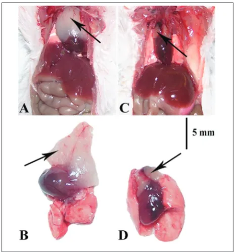
Morphological differences in gross anatomy between control (Fig. 2A and B) and treated animals (Fig. 2C and D) were observed. In control mice, the thymus was composed of two lobes that covered the front 2/3 of the side of the heart, while in the stressed animals the atrophied lobes were situated only in the coronae
cordis area. Using H&E staining, a decrease in the
Impact of tube-restraint stress on thymic medulla and cortex volume densities
A significant difference in the volume density of the cortex (Z=2.140, p<0.05) and medulla (Z=-2.140, p<0.05) in mice exposed to repeated tube-restraint stress for 20 consecutive days was observed by ste-reological analysis of histological sections (Fig. 4). No differences between control and experimental animals after 10 and 20 days of repeated exposure to tube-restraint stress were observed in the medul-lary part of the thymus. This indicated that the noted difference between the animals was the consequence
Fig. 1. Body weight (A) and thymus weight (B) in control and
experimental animals on the 10th day (C10 and E10, respectively) and on the 20th day (C20 and E20, respectively) after onset of the experiment. Results are presented as the means±SD (n=6 speci-mens per control subgroup; n=8 specispeci-mens per experimental subgroup). *p<0.05 indicates significant difference compared to control animals; #p<0.05 indicates significant difference compared to initial mean weight.
Fig. 2. Gross anatomy of the thymus (black arrow) in control
(A, B) and experimental animals (C, D) on the 20th day (C20 and E20, respectively) after onset of the experiment. Scale bar represents 5 mm.
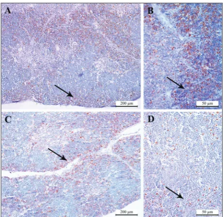
Fig. 3. Histological features of the thymus in control (A, B) and experimental (C, D) animals on the 20th day (C20 and E20, re-spectively) after onset of the experiment; H&E staining. A – C20, 50x; B – C20, 200x; C – E20, 50x; D – E20, 200x. Black and white arrows point to the cortex and medulla of the thymus, respectively. Scale bar represents 400 µm (A, C) and 100 µm (B, D).
of decreased cortical volume density in the stress-exposed animals.
Impact of tube-restraint stress on distribution of PCNA-labelled cells in the thymus
The use of an immunohistochemical marker for cell proliferation revealed certain differences between control and treated mice. In control animals, PCNA nuclear positivity was observed predominantly in sub-cortical thymocytes and to a smaller extent in medulla cells (Fig. 5A and B). In the animals exposed to re-peated tube-restraint stress there was a gradual loss of positivity in subcortical thymocytes, as well as a slight increase in the number of PCNA positive cells in the medulla (Fig. 5C and D).
Impact of tube-restraint stress on the number of cortical thymocytes
The cortical region of the thymus was also character-ized by a reduced number of thymocytes per area after repeated tube-restraint stress (Fig. 6A). Considering the results of morphometric analysis, a significant ef-fect of treatment was revealed by one-way ANOVA (F=3233.697, df=3, p<0.05). Post hoc comparisons indicated a significant difference regarding the num-ber of nuclear profiles of cortical thymocytes in ex-perimental animals compared with matching control animals (Fig. 6B).
DISCUSSION
In the present study, repeated tube-restraint stress (2 h daily for 10 or 20 consecutive days) caused significant weight loss in animals, while non-stressed animals ex-hibited an increase in body weight gain. This result was not related to food deprivation during restraint since the control group was also deprived of food. Weight gain in the control group was probably the result of a normal growth pattern in 8-week-old mice that have not yet reached their final weight. During stress activa-tion, the parvocellular part of the paraventricular nu-cleus delivers corticotropin-releasing hormone (CRH). CRH and its analogs urocortins 1-3 activate CRHR1 and CRHR2 receptors in the sympathetic nervous sys-tem that is responsible for release of catecholamines.
Fig. 5. Distribution of PCNA-labelled cells in the thymus of
con-trol (A, B) and experimental (C, D) animals on the 20th day (C20 and E20, respectively) after onset of the experiment. A – C20, 100x; B – C20, 400x; C – E20, 100x; D – E20, 400x. Black arrows point to the cortex of the thymus. Scale bar represents 200 µm (A, C) and 50 µm (B, D).
Epinephrine and norepinephrine induce hypophagia and weight loss through their effect on liver glycoge-nolysis and gluconeogenesis, as well as lipolysis of the white adipose tissue [11]. Mice in our study exhibited an increase in weight of about 1/7 from the control group, while in another study, weight gain was approxi-mately 1/4 of the control group [12]. Considering the same age of mice at the beginning of the experiment, this discrepancy in weight gain could be caused by the duration of immobilization (4 h daily for 15 consecu-tive days) and the used mouse strain (Swiss ICR mice). Regardless of whether the experiment lasted 10 or 20 days, the same changes in thymus weight were observed. In both experimental subgroups, the thy-muses had approximately one half of the weight com-pared with matching control subgroups. The effects on thymus weight were similar to those reported in previous research using young specimens. Thymus weights were one third [12] or one half [13] of the control group. However, Hu et al. [14] did not note any changes in thymus weight in adult male Sprague-Dawley rats exposed to repeated immobilization stress (2 h daily for 60 days); the absence of differences is an expected finding because involution of the thymus in adults had already occurred before the experiment.
The morphology of the thymus is also important in evaluating stress-related changes. The volume den-sity of the cortex and medulla of the thymus can be used as another parameter that can help in determin-ing atrophic changes of the thymus [15]. Histological examination revealed that thymus cortical thickness is reduced. These results are consistent with the findings that cortical thymocytes are susceptible to stress when compared to medullary thymocytes, which leads to apoptosis in the thymus cortex [15-17]. The medulla remained unchanged, which is consistent with a previ-ous finding [16].
Morphometric analysis of the number of cortical thymocytes per area revealed that a decrease in thy-mus weight in the stress-exposed group was followed by a decrease in the cellularity of the thymus cortex. However, Živković et al. [17] showed that beside the decrease in the number of cortical thymocytes per area after chronic stress induced by swimming, at-rophy of the medulla also occurred [17]. This loss of thymocytes is likely because of the stress-induced,
glucocorticoid-mediated increase in apoptosis, or it was due to reduced migration of bone marrow cells to the thymus and a change in the redistribution of the T-cell subpopulation [18-19].
In this study, we used the immunohistochemical marker PCNA to determine the effects of repeated stress on the proliferative capacity of thymocytes. PCNA is a nuclear protein associated with the cell cycle [20]. The positive PCNA signals in the thymus, particularly in the cortical part, were significantly lower in stressed animals compared to the controls. Decreased expression of PCNA in experimental ani-mals further revealed the pronounced atrophy of the cortical part of the thymus. The effect of stress on the decrease in proliferative ability of thymocytes is a direct indication of the antiproliferative and proapop-totic effect [21-22].
To overcome time-consuming experiments, much effort has been focused on establishing the shortest time of repetitive restraint stress in which the same changes occur when restraint stress lasts longer. Given the slight differences in the morphological and mor-phometric results between the two experimental sub-groups, we propose that repeated tube-restraint stress of 10 days is sufficient to cause intense involution of the thymus. The number of cortical thymocytes per area indicate that a certain degree of adaptive abil-ity of the thymus, possibly its recovery, occurred in the experimental subgroup exposed to tube-restraint stress for 20 consecutive days. Successful adaptation to repeated exposure to the same stimuli is the conse-quence of a marked reduction in HPA axis activation [23-24]. Paskitti et al. [25] showed that thymus atro-phy in rats reaches a plateau after 7 days of exposure to chronic variable stress.
CONCLUSIONS
Funding: This work was supported by the Ministry of Science and Technological Development, Republic of Serbia, Grant No: 175006, and by the Provincial Secretariat for Higher Educa-tion and Scientific Research of Vojvodina, Grant No. 142-451-3630/2017-01/02.
Author contributions: All authors were involved in the planning
and performing the experiment, data analysis and writing of the manuscript.
Conflict of interest disclosure: The authors declare no conflict
of interest.
REFERENCES
1. Bellavance MA, Rivest S. The HPA - Immune axis and the immunomodulatory actions of glucocorticoids in the brain. Front Immunol. 2014;5:136.
2. Yan F, Mo X, Liu J, Ye S, Zeng X, Chen D. Thymic func-tion in the regulafunc-tion of T cells, and molecular mechanisms underlying the modulation of cytokines and stress signaling. Mol Med Rep. 2017;16(5):7175-84.
3. Bjelaković G, Stojanovic I, Jevtovic-Stoimenov T, Pavlović D, Kocić G, Kamenov B, Saranac L, Nikolić J, Bjelaković B, Sokolović D, Basić J. Thymus as a target tissue of glucocorti-coid action: what are the consequences of glucocortiglucocorti-coids thy-mectomy? J Basic Clin Physiol Pharmacol. 2009;20(2):99-125. 4. Marchetti MC, Di Marco B, Cifone G, Migliorati G, Riccardi C. Dexamethasone-induced apoptosis of thymocytes: role of glucocorticoid receptor-associated Src kinase and caspase-8 activation. Blood. 2003;101(2):585-93.
5. Pérez-Mera ML, Guerra-Pestonit B, Rey-Méndez M. Thymic response of C57BL/6 mice to three different stressors. Nova Acta Científica Compostelana (Bioloxía). 1993;4:173-7. 6. Kostic TS, Stojkov NJ, Janjic MM, Maric D, Andric SA. The
adaptive response of adult rat Leydig cells to repeated immo-bilization stress: the role of protein kinase A and steroido-genic acute regulatory protein. Stress. 2008;11(5):370-80. 7. Kolesnikova LI, Kolesnikov SI, Korytov LI, Suslikova MI,
Darenskaya MA, Grebenkina LA, Kolesnikova LR. Oxida-tive stress as a mechanisms of reduced glucose absorption under conditions of immobilization stress. Bull Exp Biol Med. 2017;164(2):132-5.
8. Sheridan J, Feng N, Bonneau R, Allen C, Huneycutt B, Gla-ser R. Restraint stress differentially affects anti-viral cellular and humoral immune responses in mice. J Neuroimmunol. 1991;31(3):245-55.
9. Chmielarz P, Kreiner G, Kusmierczyk J, Kowalska M, Roman A, Tota K, Nalepa I. Depressive-like immobility behavior and genotype × stress interactions in male mice of selected strains. Stress. 2016;19(2):206-13.
10. Kim KS, Han PL. Optimization of chronic stress paradigms using anxiety- and depression-like behavioral parameters. J Neurosci Res. 2006;83(3):497-507.
11. Rabasa C, Dickson S. Impact of stress on metabolism and energy balance. Current Opinion in Behavioral Sciences. 2016;9:71-7.
12. Mehfooz A, Wei Q, Zheng K, Fadlalla MB, Maltasic G, Shi F. Protective roles of Rutin against restraint stress on spermato-genesis in testes of adult mice. Tissue Cell. 2018;50:133-43. 13. Barsoum CS, Raafat MH, Mekawy MA, El Shawarby AM.
The possible protective role of ghrelin on acute stress induced thymic atrophy in mice. Histological and Immu-nohistochemical Study. Cytol Histol Rep. 2019;2:106. 14. Hu Y, Cardounel A, Gursoy E, Anderson P, Kalimi M.
Anti-stress effects of dehydroepiandrosterone: protection of rats against repeated immobilization stress-induced weight loss, glucocorticoid receptor production, and lipid peroxidation. Biochem Pharmacol. 2000;59(7):753-62.
15. Elmore SA. Enhanced histopathology of the thymus. Toxicol Pathol. 2006;34(5):656-65.
16. Ito M, Nishiyama K, Hyodo S, Shigeta S, Ito T. Weight reduc-tion of thymus and deplereduc-tion of lymphocytes of T-dependent areas in peripheral lymphoid tissues of mice infected with Francisella tularensis. Infect Immune. 1985;49(3):812-8. 17. Živković I, Rakin A, Petrović-Đergović D, Miljković B, Mićić
M. The effects of chronic stress on thymus innervation in the adult rat. Acta Histochemica. 2005;106(6):449-58.
18. Bomberger CE, Haar JL. Restraint and sound stress reduce the in vitro migration of prethymic stem cells to thymus supernatant. Thymus. 1992;19(2):111-5.
19. Tarcic N, Levitan G, Ben-Yosef D, Prous D, Ovadia H, Weiss DW. Restraint stress-induced changes in lymphocyte subsets and the expression of adhesion molecules. Neuroimmuno-modulation. 1995;2(5):249-57.
20. Zhu L, Yu T, Qi X, Gao J, Huang K, He X, Luo H, Xu W. Limited link between oxidative stress and ochratoxin A-induced renal injury in an acute toxicity rat model. Tox-ins. 2016;8(12):373.
21. Engler H, Stefanski V. Social stress and T cell maturation in male rats: transient and persistent alterations in thymic function. Psychoneuroendocrinology. 2003;28(8):951-69. 22. Kapitonova Mlu, Kuznetsov SL, Klauchek SV, Mohd Ismail
ZI, Ullah M, Fedorova OV. Accidental thymic involution in the growing body under the effect of different types of stressors. Morfologiia. 2006;130(6):56-61.
23. Missima F, Sforcin JM. Green brazilian propolis action on macrophages and lymphoid organs of chronically stressed mice. Evid Based Complement Alternat Med. 2008;5(1):71-5. 24. Herman JP. Neural control of chronic stress adaptation.
Front Behav Neurosci. 2013;7:61.