Kamalakannan Rajendran et al JMSCR Volume 06 Issue 10 October 2018 Page 464
An analysis of management of solid pseudopapillary neoplasms of Pancreas
in a tertiary centre: 10 years experience
Authors
Kamalakannan Rajendran
1*,
Sugumar Chidambaranathan
2, Jeswanth Sathyanesan
3,
Ravichandran Palaniappan
41Senior Assistant Professor, Instituteof Surgical Gastroenterology & Liver Transplant, Chennai,
Tamil Nadu, India
2,3,4Professors, Institute of Surgical Gastroenterology & Liver Transplant, Govt. Stanley Medical College,
Chennai Tamil Nadu, India Corresponding Author Dr Kamalakannan Rajendran
Senior Assistant Professor, Institute of Surgical Gastroenterology &Liver Transplant, Govt. Stanley Medical College Hospital, Chennai, Tamil Nadu-600001, India
Email: [email protected], Mobile: 9865735009
Abstract
Background & Aim: Solid pseudo papillary neoplasm (SPN) is a rare tumor of the pancreas. The present study was aimed to evaluate the clinical and pathologic characteristics and surgical outcomes of SPNs.
Methods: The clinical data of patients with SPN presented to surgical Gastroenterology Department between 2008 and 2018 were evaluated retrospectively. Clinical and pathological features, radiological findings, surgical intervention, & follow up details were reviewed.
Results: In this study, 18 cases of SPN were identified and prevalence of female was high (16 cases) when compared to the males. The mean age of the cases were 23 years. Predominant symptom was low grade abdominal pain. Among the 18 cases, 12 cases displayed both solid cystic components, 3cases were solid tumors and the remaining 3 cases were cystic tumor. Based on the clinical and radiological findings 12 cases were confirmed for SPN. Further, surgical management was done in all cases and distal pancreatectomy was done in 12cases, whipples surgery in 3 cases , spleen preserving laparoscopic distal pancreatectomy in 1 case, central pancreatectomy in 1case and one case underwent multi organ resection. Six cases with uncertain diagnosis were confirmed by immunohistochemistry. There was one recurrence in patients who underwent multi organ resection and no mortality was observed in our study.
Conclusion: SPN is a rare tumor that develops principally in young women and has a good prognosis. Preoperative diagnosis of SPN is possible in most of the cases based on clinical and radiological features. Surgical resection is the best management. Generally SPNs are associated with long term survival even in advanced stage.
Keywords: Solid pseudo papillary neoplasm, distal pancreatectomy, Central pancreatectomy, Immunohistochemistry (IHC), EUS (Endo ultrasound).
www.jmscr.igmpublication.org Impact Factor (SJIF): 6.379
Kamalakannan Rajendran et al JMSCR Volume 06 Issue 10 October 2018 Page 465 Introduction
Solid Pseudo papillary neoplasm (SPN) of the pancreas is a rare clinical entity with an incidence of 0.13% to 2.7% among the pancreatic tumors.1 First reported by Frantz in 1959, it is an uncommon but distinct pancreatic neoplasm, constitutes only about 5% of cystic pancreatic tumors and about 1 to 2% of exocrine pancreatic neoplasms. SPNs are usually localized pancreatic neoplasm, although 10% to 15% of patients will develop metastases.(2-5) These metastases are often amenable to resection and are associated with long term survival. The aim of this study was to evaluate the clinical and pathological characteristics, diagnosis, treatment and surgical outcomes of SPNs in our institute.
Patients and Methods
Clinical data in our department between May 2008 to May 2018 were retrospectively analyzed. Patient’s clinical and pathological features, radiological findings, surgical intervention, & follow up details were reviewed. Pre operative diagnosis of SPNs was made on basis of clinical and radiological features such as Solid cystic tumor at the tail of pancreas in young females. Pathologic diagnosis of SPN was made based on the presence of following characteristic microscopic features. Solid areas alternating with pseudo papillary formations evidence of cellular degeneration, including cholesterol clefts, aggregates of foamy histiocytes, nuclear grooves and aggregates of hyaline cytoplasmic globule . For some pancreatic tumours in which the diagnosis of SPN was unclear, immune histochemistry (IHC) study was performed. A perioperative surgical complication was defined as occurring within 30 days of operation. A mortality occurring within 30 days of operation was considered a surgical mortality. Complications were classified from Grade I to IV.2 Pancreatic fistula was defined using the recommendations of the International Study Group on Pancreatic Fistula (ISGPF).
Results
In the present study, we have reported 845 cases of pancreatic neoplasms during the period between 2008- 2018 and out of these 18 cases (2.1%) were diagnosed as SPNs. Out of the 18 cases of SPNs 16 cases were female and 2 cases were males. The mean age of cases in the present study was found to be 23 years (Table 1)
Regarding the location of tumor, 12 cases were presented in tail, 3 cases in head , 2 cases at body and one case at the neck of the pancreas (Table 1)
In the present study, the tumor size between 5-10cms were found in 11 cases, followed by less than 5 cms in 5 cases and in 2 cases the tumor size was more than 10cms. Further, the tumor calcification was seen only in 6 cases (Table 1). Regarding the tumor features, solid and cystic type were seen in 12 cases, 3 cases were solid alone and 3 cases were cyst (Table 1).
Table 1: Characteristic features of SPNs of pancreas
Clinical and
Pathological Features
Benign Malignant Total
Sex
Female 14 2 16(89%)
Male 2 2 (11%)
Age
˂ 30 yrs 13 2 15(83%)
˃ 30 yrs 3 0 3(17%)
Location
Head 2 1 3(17%)
Neck 1 1 (5%)
Body 1 1 2(11%)
Tail 12 12(67%)
Size
Less than 5 cm 5 0 5(28%)
5- 10cm 9 2 11(61%)
More than 10 cm 2 0 2(11%)
Calcifications
Present 6 1 7(39%)
Absent 10 1 11(61%)
Tumor Features
Solid & cystic 12 0 12(66%)
Solid 1 2 3(17%)
Kamalakannan Rajendran et al JMSCR Volume 06 Issue 10 October 2018 Page 466 Most of SPNs were symptomatic. Most common
presentation was low grade abdominal pain either with or without abdominal mass. No case presented with mass effect (Table 2).
Table 2 Clinical presentation of SPNs of pancreas
All patients underwent ultra sound examination of abdomen followed by contrast CT abdomen. MRI abdomen was taken for three patients as additional investigations. Preoperative diagnosis of SPN was made in twelve cases based on clinical and radiological features. (Fig.1) CT abdomen shows a well-demarcated heterogeneous mass which was composed of a solid-cystic portion and calcifications.
Fig 1: Computed tomography features of SPN in the tail of pancreas
Surgical management
In the present study, distal pancreatectomy with splenectomy was performed in 12 cases after vaccination against Pneumococcus, Haemophilus influenzae and Meningococcus. Whipple procedure was done in 3 cases. Central pancreatectomy was done in 1case. Laparoscopic spleen preserving distal pancreatectomy was done in1case. One case underwent multi organ resection.Two cases were presented with
malignant features. One at body of the pancreas with involvement of transvers colon, spleen and greater curvature of stomach. Resection of distal pancrease, spleen, segment of transverse colon and sleeve resection of posterior wall of the stomach were performed. Another underwent Whipples procedure. The results were depicted in Table 3.
Table 3: Surgical procedure and its outcome among the SNPs cases
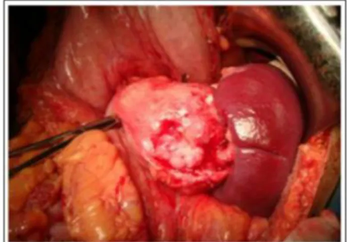
The operative features of SPNs in the present study appeared as an encapsulated beneath a smooth glistening surface and had well-defined margins (Fig 2). The cut surface shows large spongy areas of hemorrhage alternating with both solid and cystic degeneration.
Fig 2: Operative features of SPN in tail of the pancreas
Symptoms No of cases Percentage
Symptomatic 14 77
Incidental 4 23
Abdominal pain 8 38
Mass 4 8
Mass with pain abdomen 4 31
Weight loss 2 15
Nausea/vomiting 2 15
Surgical procedure and
Outcome
Number of patients (n: 18)
Whipples Surgery 3(17%)
Distal Pancreatectomy with splenectomy
12(68%)
Spleen preserving laparoscopic distal pancreatectomy
1(5%)
Central Pancreatectomy 1(5%)
Distal pancreatectomy with multi organ resection
1(5%)
Median blood loss ( in ml ) 200 ml ( 100 – 500 ml ) Median duration of surgery (
in minutes)
150 min ( 120 – 360 min )
Post operative Complications Pancreatic leak-8 (Grade A) Basal pneumonitis – 1,
Kamalakannan Rajendran et al JMSCR Volume 06 Issue 10 October 2018 Page 467 Histology of the SPNs
Histopathologically, the tumor was composed of nests of epithelial cells with a solid pseudopapillary cystic and trabecular pattern, which demonstrated the characteristics of SPN (Fig 3).
Fig 3: Histological features of SPN of pancreas
Immunohistochemistry analysis
In the present study, IHC analysis was done in six patients due to uncertainty in diagnosis. All were positive for vimentin and chromogranin was negative in all patients (Table 4).
Table 4: Immuno histochemistry analysis in the present study
Immunochemistry (6/18) Positive Negative
Vimentin 6 0
chromogranin 0 6
CEA 0 6
CD10 4 2
Beta catenin 5 1
Discussion
SPN is a very rare tumor, with low incidence of 0.13% to 2.7% of all pancreatic tumours.1This retrospective study found only eighteen cases in ten years. Only 2.1% of pancreatic neoplasms were SPNs and this explains the rarity of the tumor. More than two third cases were reported in last five years. This was due to technological advancement in imaging modalities, awareness about SPN and better documentations. SPNs are commonly seen in young female. It predominantly
occurs in adolescent girls with a reported frequency of 86% to 90% (mean age of 25 to 35 years.6-8
In our series, though SPNs were present all over the pancreas, the most common site was tail of pancreas. In literature, the head and tail of the pancreas were the preferential sites of the occurrence of SPNs.6,9 Three patients had SPNs in head of the pancreas. They presented without jaundice though the size was more than 5 cms. The reasons could be less invasive nature and slow growing tumors which mostly push the surrounding structures.
In combination with clinical features, Imaging modalities help to make the clinical diagnosis and differentiate from other pancreatic tumours. Tumour markers like CA199, CEA and CA125 are not elevated. CT scan plays much more important role by providing further information about the size, location, the local invasion and vessel involvement of SPN, ultimately help to provide the final treatment strategy.
MRI is better than CT for distinguishing certain tissue characteristics, such as hemorrhage, cystic degeneration, or the presence of a capsule, particularly as indicated by high signal intensity on T1-weighted imaging and slightly progressive heterogeneous peripheral contrast enhancement, seen after gadolinium administration on dynamic examination .10 Both imaging are complimentary for each other.
Kamalakannan Rajendran et al JMSCR Volume 06 Issue 10 October 2018 Page 468 neoplastic cells by way of the needle tract. They
are only be used for cases of unresectable pancreatic tumours with diagnostic uncertainty to start palliative chemotherapy.
Some studies correlate more than 5 cm in size and male sex are the risk factors for malignancy. 11,12 Most of the SPNs seen in this study were more 5 cm in size and two cases more 10 cm. Average size was 6.4cms. Both malignant SPNs were seen in female with mean size was 7.6 cm. In this series, even benign tumours were larger than malignant. We found that size and sex does not co relate with malignant potential and is not a definitive risk factor for malignant behavior. This kind of presentation was due to slow growing nature, rarely symptomatic and of course, present as a mass after reaching larger size.
Though there was no definitive correlation between nature the of the lesion with risk of malignancy, in our study , Both malignant SPNs were solid in nature. Since the number of SPN cases reported in our series are smaller, This interesting findings should be read carefully. Because the lesion grows slowly and rarely invades adjacent structures, Mass effect caused by obstruction of the duodenum, bile duct or any nearby structure are rare. This enable to do parenchymal sparing surgery. In our series, One laparoscopic spleen preserving distal pancreate-ctomy was done in tail SPN of 3.5 cms in size. It is impossible to predict SPNs with malignant potential without an evidence of distant metastasis, regional lymph node metastasis, or obvious invasion of adjacent organs,. Up to 15% of cases of SPNs have shown aggressive behavior consisting of extension into adjacent blood vessels and organs, local recurrence and distant metastasis.13,14 Some histological features like extensive necrosis ,nuclear atypia, high mitosis, expression of Ki-67 and sarcomatoid areas may be associated with malignant potential.24.
Immunohistochemistry were done in six patients with diagnostic uncertainty. Vimentin was positive in all. According to Kosmahl et al, Positive findings were seen when stained for 1-
vimentin and negative findings when stained for chromogranin.15
Immunohistochemistry study plays an important role, whenever the diagnosis of SPNs are unclear. WHO recommends a panel of beta-catenin (+), CD10 (+), Chromogranin (-) and Vimentin(+) to establish the diagnosis of SPN.
The positive stains for SPN are as follows, CD10 (60%), Vimentin (100%), Beta-Catenin (98%),S OX-11(100%), CD56(96%), Neuron specific enolase (70%)and Snaptophysin (55%)
The negative stains for SPN are as follows, Chromoganin A, CEA, Estrogen receptor and E-Cadherin
On follow up (6 months to 10 years), patients who underwent complete resection did not develop recurrence. Of malignant SPNs, One case which underwent multi organ resection and adjuvant chemotherapy developed multiple liver metastases after eighteen months of follow up. This patient received second line chemotherapy and on follow up. Still, the role of chemotherapy or chemo radiotherapy in the treatment of SPN is unclear 18, 19, 20 and it need to be defined. Another malignant
SPN was still on follow up of 24months without recurrence.
Because the long term survival can be achieved after resection of locally advanced SPN, it would seem prudent to take an aggressive surgical approach aimed at resection of the primary, if it is safely resectable. In many series, it is reported about good survival even after palliative resection.25 Patients with SPN with local recurrence as well as liver and peritoneal metastasis could still have long-term survival, the presence of metastasis in the SPN patients is not a contraindication for surgery in a good risk patient. Surgical debulking favors prolonged survival.16
Kamalakannan Rajendran et al JMSCR Volume 06 Issue 10 October 2018 Page 469 Conclusion
The preoperative diagnosis of SPN is crucial in order to propose the proper management. Preoperative diagnosis of SPN is possible in most of the cases based on clinical and radiological features. Malignant SPNs are diagnosed based on local invasion and metastasis. Since SPNs are slowly growing, less invasive, low grade malignant tumor, organ sparing resection is best option. In advanced cases, aggressive surgical approach is justifiable in good risk patients, since SPNs are associated with long term survival even in advanced stage. It is difficult to identify any prognostic factors to predict survival due to low malignant potential of the tumor and rarity of mortality. The role of chemotherapy and radiotherapy in adjuvant or palliative treatment is yet to be proved.
References
1. Crawford BE 2nd. Solid and papillary epithelial neoplasm of the pancreas, diagnosis by cytology. South Med J 1998; 91: 973-977.
2. Dindo D, Demartines N, Clavien PA. Classification of surgical complications: a new proposal with evaluation in a cohort of 6336 patients and results of a survey. Ann Surg 2004; 240:205–213.
3. Frantz VK. Papillary tumors of the pancreas: Benign or malignant? Tumors of the pancreas. Atlas of tumor Pathology, 1st Series, Fascicles 27 and 28. Frantz VK (ed). Washington. DC, Armed Forces Institute of Pathology, 1959: 32-33
4. Kloppel G, Solcia E, Longnecker DS, Capella C, Sobin LH. World Health Organization, Institutional histological classification of tumors. Histological Typing of Tumors of the Exo- crine Pancreas, 2 nd ed. Springer-Verlag , Berlin, 1996
5. Bassi C, Dervenis C, Butturini G, et al. Postoperative pancreatic fistula: an
international study group (ISGPF) definition. Surgery 2005; 138:8–13.
6. Kosmahl M, Pauser U, Peters K, Sipos B, Luttges J, Kremer B, Kloppel G. Cystic neoplasms of the pancreas and tumor-like lesions with cystic features: a review of 418 cases and a classiffication proposal. Virchows Arch 2004; 445: 168-178
7. Yu CC, Yeh CN, Hwang TL, Jan YY. Clinicopathological study of solid and pseudopapillary tumor of pancreas: Emphasis on magnetic resonance imaging findings. World J Gastroenterol. 2007; 13:1811-1815.
8. Dong DJ, Zhang SZ. Solid-pseudopapillary tumor of the pancreas: CT and MRI features of 3 cases. Hepatobilliary Pancreat Dis Int. 2006;5:300-304.
9. Lee W, Park Y, Choi J, Chi H, Kim B. Solid and papillary neoplasms of the pancreas. Yonsei Medical J. 1996; 37:131-141.
10.Cantisani V, Mortele KJ, Levy A, Glickman JN, Ricci P, Passariello R, Ros PR, Silverman SG. MR imaging features of solid pseudopapillary tumor of the pancreas in adult and pediatric patients. Am J Roentgenol 2003; 181: 395-401 11.Kang CM, Kim KS, Choi JS, et al. Solid
pseudopapillary tumor of the pancreas suggesting malignant potential. Pancreas 2006; 32:276–80. 12.
12.Machado MC, Machado MA, Bacchella T, etal.Solid pseudopapillary neoplasm of the pancreas: distinct patterns of onset, diagnosis, and prognosis for male versus female patients. Surgery 2008; 143:29–34. 13.Lam KY, Lo CY, Fan ST. Pancreatic
Kamalakannan Rajendran et al JMSCR Volume 06 Issue 10 October 2018 Page 470 14.Pasquiou C, Scazec JY, Gentil-Perret A,
et al. Solid pseudopapillary tumors of the pancreas. Pathology reports of 13 cases. GastroenterolClin Biol. 1999; 23:207-214. 15.Kosmahl M, Seada LS, Janig U, Harms D,
Kloppel G. Solid-pseudopapillary tumor of the pancreas: its origin revisited.Virchows Arch 2000; 436: 473-480
16.Rebhandl W, Felberbauer FX, Puig S, Paya K, Hochschorner S, Barlan M, Horcher E. Solid-pseudopapillary tumor of the pancreas (Frantz tumor) in children: Report of four cases and review of the literature. J SurgOncol 2001; 76: 289-296. 17.Gonzalez-Campora R, Rios Martin JJ,
Villar Rodriguez JL, et al. Papillary cystic neoplasm of the pancreas with liver metastasis coexisting with thyroid papillary carcinoma. Arch Pathol Lab Med. 1995;119:268-273.
18.Strauss JF, Hirsch VJ, Rubey CN, and Pollock M. Resection of a solid and papillary epithelial neoplasm of the pancreas following treatment with cis-platinum and 5-fluorouracil: A case report. Med PediatrOncol 1993; 21: 365-367 20 19.Fried P, Cooper J, Balthazar E, Fazzini E, Newall J. A role for radiotherapy in the treatment of solid and papillary neoplasms of the pancreas. Cancer 1985; 56: 2783-2785.
20.Martin RC, Klimstra DS, Brennan MF, Conlon KC. Solid-pseudopap-illary tumor of the pancreas: A surgical enigma? Ann SurgOncol. 2002; 9:35-40. 21.Koito K, Namieno T, Nagakawa T,
Shyonai T, Hirokawa N, Morita K. Solitary cystic tumor of the pancreas: EUS pathologic correlation. GastrointestEndosc 1997; 45: 268-276 22.Brugge WR. Role of endoscopic
ultrasound in the diagnosis of cystic
lesions of the pancreas. Pancreatology 2001; 1: 637-640
23.Das DK, Bhambhani S, Kumar N, Chachra KL, Prakash S, Gupta RK, Tripathi RP. Ultrasound guided percutaneous fine needle aspiration cytology of pancreas: a review of 61 cases.TropGastroenterol 1995; 16: 101-109.
24.Tang LH, Aydin H, Brennan MF, Klimstra DS: Clinically aggressive solid pseudopapillary tumor of the pancreas. A report of two cases with components of undifferentiated carcinoma and a comparative clinicopathologic analysis of 34 conventional cases. Am J SurgPathol 2005, 29:512–519.
25.Ayşe Yagcı, Savas Yakan, Ali Coskun, Nazif Erkan, Mehmet Yıldırım, Evrim Yalcınand Hakan Postacı. Diagnosis and treatment of solid pseudopapillary tumor
of the pancreas: experience of one single
institution from Turkey, World J Surg