Development of Chronic Bronchitis and Emphysema in
b
-Epithelial Na
1
Channel–Overexpressing Mice
Marcus A. Mall1, Jack R. Harkema2, Joanna B. Trojanek1, Diana Treis1, Alessandra Livraghi3, Susanne Schubert1, Zhe Zhou1, Silvia M. Kreda3, Stephen L. Tilley4, Elizabeth J. Hudson3, Wanda K. O’Neal3, and Richard C. Boucher3
1Pediatric Pulmonology and Cystic Fibrosis Center, Department of Pediatrics III, University of Heidelberg, Heidelberg, Germany;2Department of Pathobiology and Diagnostic Investigation, Michigan State University, East Lansing, Michigan; and3Cystic Fibrosis/Pulmonary Research and Treatment Center and4Division of Pulmonary and Critical Care Medicine, Department of Medicine, School of Medicine, The University of North Carolina at Chapel Hill, Chapel Hill, North Carolina
Rationale: Chronic obstructive pulmonary disease is a leading cause of death worldwide, but its pathogenesis is not well understood. Previous studies have shown that airway surface dehydration in b-epithelial Na1 channel (bENaC)–overexpressing mice caused a
chronic lung disease with high neonatal pulmonary mortality and chronic bronchitis in adult survivors.
Objectives: The aim of this study was to identify the initiating lesions and investigate the natural progression of lung disease caused by airway surface dehydration.
Methods: Lung morphology, gene expression, bronchoalveolar la-vage, and lung mechanics were studied at different ages inb ENaC-overexpressing mice.
Measurements and Main Results: Mucus obstruction in b ENaC-overexpressing mice originated in the trachea in the first days of life and was associated with hypoxia, airway epithelial necrosis, and death. In survivingbENaC-overexpressing mice, mucus obstruction extended into the lungs and was accompanied by goblet cell meta-plasia, increased mucin expression, and airway inflammation with transient perinatal increases in tumor necrosis factor-aand macro-phages, IL-13 and eosinophils, and persistent increases in keratinocyte-derived cytokine (KC), neutrophils, and chitinases in the lung.b ENaC-overexpressing mice also developed emphysema with increased lung volumes, distal airspace enlargement, and increased lung compliance. Conclusions: Our studies demonstrate that airway surface dehydration is sufficient to initiate persistent neutrophilic airway inflammation with chronic airways mucus obstruction and to cause transient eosinophilic airway inflammation and emphysema. These results suggest that deficient airway surface hydration may play a critical role in the pathogenesis of chronic obstructive pulmonary diseases of different etiologies and serve as a target for novel therapies.
Keywords:chronic obstructive lung disease; epithelial Na1channels;
airway surface liquid; inflammation; mucus
Cystic fibrosis (CF) lung disease is the most common genetic form of chronic obstructive pulmonary disease (COPD) and is caused by mutations in the cystic fibrosis transmembrane
con-ductance regulator (CFTR) gene (1, 2), which encodes a protein that is a cAMP-dependent Cl2channel and regulates the
epithe-lial Na1channel (ENaC) (3–6). In CF airway epithelia,
CFTR-mediated Cl2 secretion is defective, and ENaC-mediated Na1
absorption is increased (7–9).In vitrostudies of primary human airway cultures suggested that these defects in vectorial ion trans-port resulted in airway surface liquid (ASL) volume depletion and adhesion of dehydrated mucus, which was predicted to im-pair normal ciliary function and efficient mucus clearance in CF airways (10). To further elucidate the role of ASL volume de-pletion in the in vivo pathogenesis of CF, we have previously generated a mouse model with airway-specific overexpression of ENaC (11). In this mouse model, we demonstrated (1) that over-expression of theb-subunit of ENaC (encoded by theScnn1b gene) under control of the Clara cell secretory protein (CCSP) promoter was sufficient to increase airway Na1absorptionin vivo,
(2) that elevated airway Na1 absorption caused ASL volume
depletion and reduced mucus clearance, and (3) that deficient mucus clearance produced spontaneous lung disease sharing key features with CF and other forms of COPD, including substantial pulmonary mortality and airway mucus obstruction, goblet cell metaplasia, chronic neutrophilic inflammation, and impaired clear-ance of bacterial pathogens (11).
Together, the results from thesein vitroandin vivostudies demonstrate that ASL volume depletion is a key mechanism in the pathogenesis of CF lung disease. Furthermore, cigarette smoke has recently been shown to decrease CFTR expression and cAMP-dependent Cl2 secretionin vitroand in nasal
res-piratory epithelia of cigarette smokersin vivo(12). These data indicate that impaired ASL volume regulation may also be im-plicated in the pathogenesis of cigarette smoke–induced chronic bronchitis. The aim of the present study was to define the initial pulmonary lesions and elucidate the sequence of steps in the
AT A GLANCE COMMENTARY
Scientific Knowledge on the Subject
Airway surface dehydration is a key feature of cystic fibrosis and produces chronic bronchitis in mice. The initiating lesions and the natural history of lung disease caused by airway surface dehydration have not been elucidated.
What This Study Adds to the Field
Airway surface dehydration is sufficient to initiate persistent neutrophilic airway inflammation with chronic airways mucus obstruction and to cause transient eosinophilic airway in-flammation and emphysema. These results suggest that de-ficient airway surface hydration may play a critical role in the pathogenesis of chronic obstructive pulmonary diseases of different etiologies and serve as a target for novel therapies.
(Received in original form August 21, 2007; accepted in final form December 13, 2007) Supported by Marie Curie Excellence Grant from the European Commission (MEXT-CT-2004–013666 to M.A.M.), the Deutsche Forschungsgemeinschaft (DFG MA 2,081/3–2 to M.A.M.), the North American Cystic Fibrosis Foundation (MALL04G0 to M.A.M. and R026-CR02 to W.K.O.), and the National Institutes of Health (NIH SCOR P50 HL60280, P01 HL34322, and MTCC P30 DK065988 to R.C.B.).
Correspondence and requests for reprints should be addressed to Marcus A. Mall, M.D., Pediatric Pulmonology and Cystic Fibrosis Center, Department of Pediat-rics III, University of Heidelberg, Im Neuenheimer Feld 153, 69120 Heidelberg, Germany. E-mail: [email protected]
This article has an online supplement, which is accessible from this issue’s table of contents at www.atsjournals.org
Am J Respir Crit Care Med Vol 177. pp 730–742, 2008
in vivo pathogenesis of COPD consequent to airway surface dehydration inbENaC-overexpressing mice. A focus was on the relationships between ASL volume depletion and mucus ob-struction, the presumed cause of mortality in this model. To achieve this goal, we performed quantitative longitudinal stud-ies from fetal to adult ages of the pulmonary phenotype, in-cluding airway mucus obstruction, goblet cell metaplasia, mucin gene expression, airway inflammation, lung volume, alveolar size, and pulmonary function. The results of our studies yielded novel insights into the role of airway surface dehydration in the in vivopathogenesis of CF and possibly other forms of COPD and identified targets for novel therapeutic strategies for the treatment of these diseases. Some of the results of this study have been previously reported in the form of abstracts (13, 14).
METHODS
Experimental Animals
All animal studies were approved by the animal care and use committees of the relevant institutions. The generation ofbENaC-overexpressing mice (line 6608) has been described previously (11). Further details on experimental animals are provided in the online supplement.
Bronchoalveolar Lavage Cell Counts and Cytokine Measurements
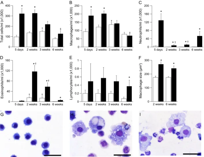
Bronchoalveolar lavage (BAL) was obtained and cell counts were de-termined as previously described (11). Macrophage size was deter-mined as described in the online supplement. Concentrations of tumor necrosis factor (TNF)-a, keratinocyte-derived cytokine (KC), and IL-13 were measured by ELISA (R&D Systems, Minneapolis, MN) or with a Cytometric Bead Array Mouse Inflammation kit (BD Bio-sciences, San Diego, CA).
Morphology
Lungs and tracheae were fixed, paraffin embedded, sectioned, and stained with hematoxylin and eosin (H&E) or Alcian blue periodic acid-Schiff as previously described (11). For transmission electron microscopy (TEM) studies, lungs were fixed and processed, and ultrastructural tissue exam-ination was performed as described in the online supplement.
Airway Morphometry
Tracheae were sectioned longitudinally, and lungs were sectioned trans-versally at the level of the proximal intrapulmonary main axial airway near the hilus and at the distal intrapulmonary axial airway. Airway mucus obstruction was assessed by determining mucus volume density as previously described (15). Airway epithelial glycogen content, epithelial height, and numeric cell densities of goblet cells and degenerative airway epithelial cells were determined as described in the online supplement.
Lung Volume and Mean Linear Intercepts
Lungs were inflated with 4% buffered formalin to 25 cm of fixative pressure, and lung volumes were determined by the volume displace-ment method (16). Lungs were processed for histology, sectioned, and stained with H&E. Mean linear intercepts were determined as previously described (17). Further details are provided in the online supplement.
Immunohistochemistry
Immunohistochemical staining for Ym1 was performed on formalin-fixed, paraffin-embedded lung sections using rabbit polyclonal anti-Ym1 antibody (18) as described in the online supplement.
Hypoxia Detection in the Lung
Tissue hypoxia was assessed by intraperitoneal injection of 3-d-old neonatal mice with pimonidazole hydrochloride (Hypoxyprobe-1; Chemicon, Temecula, CA) and immunostaining of frozen lung sections using fluorescein isothiocyanate–labeled monoclonal antibody (MAb) 1 (Hypoxyprobe-1 Plus Kit; Chemicon) as described in the online sup-plement.
Blood Gas Analyses
Blood gas measurements in neonatal mice (3–5 d) were performed using a blood gas analyzer (Radiometer ABL 500; Diamond Diagnos-tics, Holliston, MA) as described in the online supplement.
Western Blotting
Acidic mammalian chitinase (AMCase) protein expression was assessed in cell-free BAL fluid (BALF) frombENaC-overexpressing mice and wild-type littermates by Western blotting using an anti-AMCase rabbit serum (19) as described in the online supplement.
Real-time Reverse Transcriptase–Polymerase Chain Reaction
Quantitative reverse transcriptase–polymerase chain reaction for Muc5ac, Muc5b, Muc4, Gob5, eotaxin-1, IL-13, IFN-g, Ym1, Ym2, AMCase, andb-actin was performed on an Applied Biosystems 7500 Real Time PCR System using TaqMan universal polymerase chain reaction master mix and inventoried TaqMan gene expression assays (Applied Biosystems, Darmstadt, Germany) as described in the online supple-ment. Relative fold changes in target gene expression betweenb ENaC-overexpressing mice and wild-type littermates were determined by normalization to expression of the reference geneb-actin as previously described (20).
Pulmonary Function Studies
Invasive measurements of lung mechanics were performed in anesthe-tized and paralyzed 8-week-old adult mice using a computer-controlled small animal ventilator (Flexi Vent system; Scireq, Montreal, PQ, Canada) to determine dynamic resistance, dynamic compliance, pressure– volume curves, and static compliance of the lung, as previously de-scribed (21, 22).
Statistics
Data were analyzed with SigmaStat version 3.1 (Systat Software, Erkrath, Germany) and are reported as mean6SEM. Statistical analyses were performed using Student’sttest, Mann-Whitney rank sum test, one-way analysis of variance, Kruskal-Wallis analysis of variance on Ranks, or chi-square test as appropriate, andP,0.05 was accepted to indicate statistical significance.
RESULTS
ASL Volume Depletion–induced Mucus Obstruction, Goblet Cell Metaplasia, and Mucus Hypersecretion
To identify the onset and temporal evolution of goblet cell meta-plasia, mucus hypersecretion, and mucus obstruction caused by ASL volume depletion, we performed longitudinal morphomet-ric studies and studies on airway mucin expression in fetal (Embryonic Day [E] 18.5), newborn (Postnatal Day [PN] 0.5), neonatal (PN 3.5), juvenile (1–3 wk of age), and adult (6 wk of age)bENaC-overexpressing mice and littermate control mice. The onset of mortality in bENaC-overexpressing mice com-menced at 3 d of age with an overall mortality of approximately 50% in the first 2 weeks of life (Figure 1A). Compared with wild-type (Figure 1B), mortality at 3 days was associated with light microscopic evidence of tracheal plugging (Figures 1C and 1D), increased mucus volume density (Figure 1E), and mucus plugging of the larynx (Figure 1F) but not goblet cell metaplasia (Figure 1G). The extent of tracheal and laryngeal mucus ob-struction was significantly more severe in age-matched deceased compared with bENaC-overexpressing mice killed as part of this study (Figure 1F), indicating that death occurred due to mucus obstruction of the glottis and subsequent asphyxia. In contrast, neither intrapulmonary airway mucus obstruction nor goblet cell metaplasia were detected in the lungs from fetal, newborn (data not shown), or neonatal (PN 3.5) b ENaC-overexpressing mice (Figures 2A, 2G, and 2I).
(Figure 2). Mucus obstruction was most prominent in proximal intrapulmonary main axial airways (large-diameter bronchiole; Figures 2B and 2G) but also extended into the more distal conducting airways (small-diameter, preterminal bronchioles; Figures 2C and 2H) and persisted in 3- and 6-week-oldb ENaC-overexpressing mice (Figures 2D, 2E, 2G, and 2H). In contrast to early tracheal mucus plugging, intrapulmonary airway mucus obstruction was accompanied by goblet cell metaplasia in bENaC-overexpressing mice. Wild-type mice exhibited a tran-sient increase in airway goblet cell numbers that peaked at 2 weeks of age, persisted in proximal main axial airways (large-diameter bronchioles), and waned in the more distal small-diameter bronchioles of adult animals (Figures 2I and 2J). In bENaC-overexpressing mice, goblet cell numbers were
signifi-cantly increased in proximal large-diameter bronchioles at the age of 3 and 6 weeks and in distal small-diameter bronchioles at all time points (Figures 2I and 2J). The onset of goblet cell meta-plasia in 2-week-old bENaC-overexpressing mice was paral-leled by epithelial cell hypertrophy with epithelial thickening that persisted in adult animals (Figure 2K).
To explore the relationship between mucus obstruction and goblet cell metaplasia with mucin expression, we determined mRNA transcript levels of the airway mucins Muc5ac, Muc5b, and Muc4 and the goblet cell marker Gob-5 (Clca-3) in lungs from newborn to adultbENaC-overexpressing mice and control mice. In wild-type mice, expression of Muc5ac and Gob-5 was significantly elevated at the age of 1 week, and expression of Muc5b and Muc4 was significantly elevated at the age of 3 weeks when compared with newborn mice (Figures 3A–3D). Although induction of Muc5ac, Muc4, and Gob-5 was transient, with peak levels at 1 and 3 weeks, expression of Muc5b remained elevated in adult wild-type mice (Figures 3A–3D). InbENaC-overexpressing mice, mRNA expression levels of Muc5ac, Muc5b, Muc4, and Gob-5 followed similar time courses and were significantly in-creased compared with control mice from the age of 3 weeks onward (Figures 3A–3D).
Taken together, these data demonstrate that ASL volume de-pletion caused airway mucus obstruction, goblet cell metaplasia, and increased mucin gene expression in an age-dependent fash-ion. In the first week of life, airway Na1hyperabsorption caused
tracheal mucus plugging and high mortality in the absence of gob-let cell metaplasia and elevated mucin gene expression (Figures 1–3). Subsequently, in surviving bENaC-overexpressing mice, mucus obstruction extended into intrapulmonary airways and was accompanied by secondary goblet cell metaplasia and in-creased mucin expression (Figures 2 and 3).
Mucus Obstruction Causes Hypoxia
Because histologic evaluation identified tracheal mucus obstruc-tion in a large fracobstruc-tion ofbENaC-overexpressing mice as early as 3 days (seeFigures 1C–1F) and because the majority of deaths inbENaC-overexpressing mice occurred in the first week of life (seeFigure 1A), we performed blood gas analyses in neonatal mice (PN 3.5–5.5) to determine the effects of tracheal mucus plugging on ventilation. These analyses showed that PO2, oxygen
saturation, and base excess were significantly reduced in blood samples frombENaC-overexpressing mice compared with wild-type littermates (Table 1). These results demonstrate that tracheal mucus plugging was sufficient to cause systemic hypoxia. Re-duced base excess, in parallel with hypoxia, suggested metabolic compensation of respiratory alkalosis, indicating that respiratory insufficiency was a chronic process in bENaC-overexpressing mice.
Airway Na1Hyperabsorption and Epithelial Necrosis
High-resolution light and TEM studies revealed that airway Na1hyperabsorption was accompanied by early airway
epithe-lial degeneration and necrosis in the intralobular airways but not in the trachea (Figure 4). Swollen, highly vacuolated, degen-erative airway epithelial cells were observed in high numbers in neonatal bENaC-overexpressing mice but rarely in wild-type control mice (Figures 4A and 4B). In bENaC-overexpressing mice, degenerative epithelial cells were absent in the fetus (E 18.5; data not shown), present in newborn mice (PN 0.5), peaked at the age of 3 days, and were rarely observed in the airways of olderbENaC-overexpressing animals (Figure 4C). Furthermore, the intraepithelial content of PAS-positive material was reduced in newbornbENaC-overexpressing mice, indicating a reduction in airway epithelial glycogen content compared with wild-type Figure 1. Pulmonary mortality and tracheal mucus obstruction in
b-epithelial Na1 channel (bENaC)–overexpressing mice. (A) Survival curves frombENaC-overexpressing mice (circles; n535) and wild-type littermates (triangles; n536). (B–D) Histology (Alcian blue periodic acid-Schiff) of tracheae from wild-type (B) andbENaC-overexpressing mice (C) killed at the age of 3 days and abENaC-overexpressing mouse that died spontaneously at 4 days (D). Larynx is indicated byarrows.
Scale bars, 1,000mm. Representative for n56–14 mice per group. (E–
G) Summary of tracheal mucus content as determined from volume density measurements (E), frequency of laryngeal mucus plugging (F), and goblet cell counts (G) in tracheae from wild-type (open bars), bENaC-overexpressing mice killed at 3 days (shaded bars), andb ENaC-overexpressing mice that died spontaneously at the ages of 3 to 7 days
(solid bars). n511 – 16 mice per group. *P,0.001 versus wild-type.
control mice (Figure 4D). Ultrastructural examination by TEM identified abnormalities in Clara cells but not in ciliated cells. These abnormalities included depletion of epithelial glycogen stores and vacuolarization of the endoplasmatic reticulum, in-dicative of hydropic degeneration of Clara cells in newborn bENaC-overexpressing but not wild-type mice (Figures 4E and 4F). Compared with wild-type mice (Figure 4G), the majority of degenerative Clara cells of 3-day-old bENaC-overexpressing mice had pyknotic nuclei (Figure 4H), and immunostaining for activated caspase 3 as a marker for apoptosis was negative (data not shown), indicating that these cells underwent necrosis.
To assess a possible role of tissue hypoxia in airway epithelial necrosis, 3-day-old mice were treated with the hypoxia probe pimonidazole hydrochloride, and lung sections were subse-quently evaluated by immunofluorescence using monoclonal antibodies that detect protein adducts of this probe in hypoxic cells. Strong immunoreactive signals were detected in the air-way epithelium ofbENaC-overexpressing mice but not in
wild-type littermates (Figure 5). Immunoreactive signals were not detected in alveolar epithelia or in lungs from mice that were not treated with the hypoxia probe in either genotype. These data demonstrate that airway epithelial cells were hypoxic in neonatal bENaC-overexpressing mice and suggest that Clara cell necrosis resulted from cellular hypoxia.
Taken together, these data demonstrate that Na1
hyper-absorption induced by CCSP promoter–driven overexpression of bENaC resulted in neonatal depletion of glycogen stores, cellular hypoxia, hydropic degeneration, and necrosis of Clara cells. Degenerative and/or necrotic cells were not observed in mouse airways where CCSP promoter-driven overexpression of transgenes did not result in Na1 hyperabsorption (11) (i.e., in aENaC- andgENaC-overexpressing mice; data not shown). In bENaC-overexpressing mice, necrotic Clara cells were sub-sequently cleared from the airways and replaced by Clara cells and goblet cells in juvenile and adult bENaC-overexpressing mice (seeFigures 2B–2E, 4C).
Development of Chronic Airway Inflammation
In histologic lung sections, necrotic lesions in the airways of neonatal bENaC-overexpressing mice were accompanied by acute cellular inflammation of large- and small-diameter bron-chioles (bronchiolitis), consisting of intramural and intraluminal infiltration of neutrophils and airway luminal macrophages engulfing necrotic cellular debris. To study the evolution of airway inflammation, we performed longitudinal BAL studies in 5-day-old to adult mice and measured the absolute numbers and relative distributions of different inflammatory cell types (Fig-ure 6). BAL macrophages were significantly increased at 5 days and 2 weeks and were normalized in numbers but remained morphologically activated (i.e., hypertrophic cells with highly vac-ulolated cytoplasm) in 3- and 6-week-oldbENaC-overexpressing mice compared with control mice (Figures 6B, 6G–6I). Neutro-phils were recruited in parallel with macrophages, peaked at 5 days, and remained significantly elevated at all time points in bENaC-overexpressing mice (Figure 6C). Two- to three-week-old wild-type mice exhibited a transient airway eosinophilia that waned in adult mice, as previously reported (23). Eosinophilia
was significantly increased inbENaC-overexpressing mice. In con-trast to persistent neutrophilic inflammation, eosinophilia waned in adultbENaC-overexpressing animals (Figure 6D). Lymphocyte counts tended to be higher inbENaC-overexpressing mice than in control mice, but a significant difference was only observed in adult animals (Figure 6E).
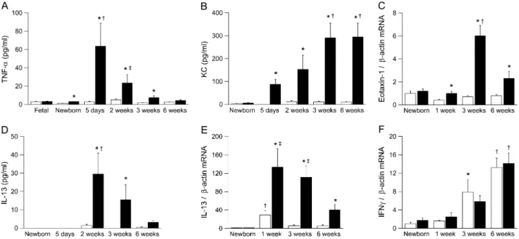
We determined protein and/or mRNA expression of proin-flammatory cytokines, including TNF-a, KC (Cxcl1), eotaxin-1 (Ccl11), IL-13, and IFN-gin BAL or lung homogenates from newborn to adultbENaC-overexpressing mice and control mice (Figure 7). None of the cytokines was elevated in fetal (E 18.5) or newborn (PN 0.5) bENaC-overexpressing mice compared with wild-type littermates. TNF-a was transiently elevated in BAL from bENaC-overexpressing mice, with peak levels in neonatal mice (i.e., at the age when necrotic lesions were most prominent) (Figures 4C and 7A). The neutrophil-attracting che-mokine KC was significantly elevated inbENaC-overexpressing mice from the first week of life, and increased KC levels were sustained into adulthood (Figure 7B). Expression of the eosin-ophil-attracting chemokine eotaxin 1 was increased inb ENaC-overexpressing mice, but the fold increase over wild-type mice was less pronounced than KC expression (Figure 7C). mRNA expression of the Th2 cytokine IL-13 was transiently induced in neonatal wild-type mice and waned in older animals, indicating that wild-type animals were Th2 biased during development (24) (Figure 7E). InbENaC-overexpressing mice, IL-13 mRNA expression followed the same time course but was significantly increased at all time points compared with control mice. Sig-nificantly elevated IL-13 protein levels were observed in 2- to 3-week old mice but not in adultbENaC-overexpressing mice (Figures 7D and 7E). In contrast, mRNA expression of the Th1 cytokine IFN-g increased with age in wild-type and b ENaC-overexpressing mice but did not differ between genotypes at any time point (Figure 7F). Taken together, the transient elevation of IL-13, followed by sustained elevation of IFN-g mRNA ex-pression, indicated a shift from Th2 toward Th1 response during normal development (24).
Airway Inflammation Is Associated with Expression of Chitinases
In airway mucus plugs ofbENaC-overexpressing mice, we fre-quently observed eosinophilic crystalline material (Figure 8A) similar to that identified as the crystallized product of various members of mammalian chitinase family, including Ym1, Ym2, and AMCase (18, 19, 25, 26). Immunohistochemically, Ym1-positive staining was present in the crystals and in the luminal airway mucus ofbENaC-overexpressing mice but not in control mice (Figures 8B and 8C). Positive Ym1 immunostaining was also observed in nonciliated airway epithelial cells (mainly mucous cells) lining proximal large-diameter bronchiolar airways and in macrophages within distal and proximal airway lumens and alveolar airspaces. In contrast, in wild-type littermates, Ym1 protein expression was confined to alveolar macrophages. By TEM, acicular (needle-shaped) crystals were identified in the cytoplasm of macrophages (Figure 8D). Immunohistochemical Figure 3. Time course of airway mucin expression inb-epithelial Na1
channel (bENaC)–overexpressing mice. (A–D) Transcripts levels of Muc5ac, Muc5b, Muc4, and Gob5 in lungs from wild-type (open bars) andbENaC-overexpressing (solid bars) mice at birth, 1 week, 3 weeks, and 6 weeks of age. Data are expressed as fold changes from newborn wild-type mice. n56–14 mice per group. (A) Muc5ac. *P,0.001 versus littermate control mice at same age;†P,0.05 versus newborn, 3-week-old, and 6-week-old wild-type mice;‡P,0.05 versus newborn bENaC-overexpressing mice. (B) Muc5b. *P ,0.01 versus wild-type mice of same age;†P,0.05 versus newborn mice of same genotype. (C) Muc4. *P,0.001 versus wild-type mice of same age;†P,0.05 versus wild-type mice of same age;‡P,0.05 versus newborn and 1-week-old wild-type mice;xP ,0.05 versus 1-week-oldb
ENaC-over-expressing mice. (D) Gob5 expression. *P,0.05 versus wild-type mice of same age;†P,0.05 versus newborn mice of same genotype.
TABLE 1. BLOOD GAS ANALYSIS FROM NEONATAL b-EPITHELIAL NA1 CHANNEL–OVEREXPRESSING MICE AND WILD-TYPE LITTERMATES*
Genotype pH PCO2(mm Hg) PO2(mm Hg) HCO32(mmol/L) BE (mmol/L) Sat O2(%)
Wild-type 7.4260.01 48.761.7 49.962.0 30.860.6 5.660.5 83.761.8
bENaC overexpressing 7.4060.01 48.861.4 43.162.4† 29.260.6 3.860.5† 77.062.4†
Definition of abbreviations: BE5base excess;bENaC5 b-epithelial Na1channel; Sat O
25O2saturation.
* n58–14 mice per group.
reaction toYm1 was minimal or absent in the lungs of newborn mice, strongest in 3-week-old mice, and waned in adultb ENaC-overexpressing mice. Moreover, Western blot analysis revealed increased levels of AMCase in BALF harvested from adult
bENaC-overexpressing mice in comparison to wild-type litter-mates (Figure 8E).
To further elucidate the role of Ym1, Ym2, and AMCase in ASL depletion–induced airway inflammation, we determined their longitudinal mRNA expression profiles in lungs from newborn to adultbENaC-overexpressing mice and control mice (Figures 8F–8H). Ym1 transcript levels increased continuously with age in wild-type mice and were significantly increased from the age of 3 weeks inbENaC-overexpressing animals (Figure 8F). In contrast to sustained expression of Ym1, Ym2 transcript levels were only transiently induced in wild-type littermates, with peak levels at 3 weeks and waning in adult mice (Figure 8G). Similar to Ym1, Ym2 expression was significantly increased in 3- and 6-week-old bENaC-overexpressing mice compared with control mice. AMCase expression was not regulated during development in wild-type mice but was significantly increased in 3-week-old and olderb ENaC-overexpressing mice (Figure 8H). These studies demonstrate that the expression of all three chitinase family members was signifi-cantly increased in airway inflammation induced by ASL volume depletion. However, their distinct temporal regulation in lungs from wild-type and bENaC-overexpressing mice suggests that Ym1, Ym2, and AMCase may have unique functions in pulmonary homeostasis and in the pathogenesis of airway disease.
Na1Hyperabsorption Is Associated with Emphysema
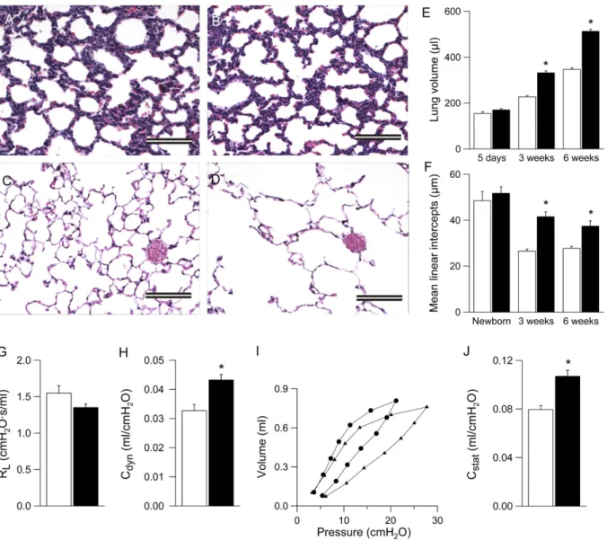
Because distal airspaces were enlarged in lungs from adult bENaC-overexpressing mice (11), we performed quantitative longitudinal studies on lung volumes and mean linear intercepts as a parameter for alveolar diameter in lungs ranging from neo-natal to adultbENaC-overexpressing and control mice. In histol-ogic sections, alveolar size and architecture were normal at birth, but alveoli were substantially enlarged in 3-week-old and adult bENaC-overexpressing mice versus control mice (Figures 9A–9D). Similarly, lung volumes were normal in bENaC-overexpressing neonates (PN 5) but were increased significantly relative to control mice at the age of 3 weeks and remained enlarged in Figure 5. Hypoxia of airway epithelia in neonatal b-epithelial Na1 channel (bENaC)-overexpressing mice. Tissue hypoxia was determined by injection of 3-day-old neonatal mice with the hypoxia probe pimonidazole hydrochloride, and subsequent immunostaining of lung sections with an antibody that detects the probe in hypoxic cells and evaluation by confocal microscopy. Images depict differential interfer-ence contrast microscopy (DIC) (left panels) and fluorescein isothiocya-nate planes (right panels) of lung sections frombENaC-overexpressing (A) and wild-type (B) mice. Specific immunoreactive signals were ob-served in the epithelial cells lining the airways ofbENaC-overexpressing mice, but not in wild-type lungs (right panels). Asterisks indicate the position of the airway lumen.Scale bar, 100mm.
adultbENaC-overexpressing mice compared with control mice (Figure 9E). Mean linear intercepts were normal at birth but failed to become smaller in 3- and 6-week-old b ENaC-over-expressing mice (Figure 9F).
Assessment of pulmonary mechanics by invasive pulmonary function testing in adult mice revealed that the dynamic compli-ance was significantly increased in adultbENaC-overexpressing mice compared with control mice (Figure 9H). Pressure– volume curves were also shifted to the left in adultb ENaC-overexpressing mice, reflecting a significantly increased static compliance compared with wild-type littermates (Figures 9I and 9J). Taken together, these morphologic and functional data demonstrate that ASL depletion causes pulmonary emphysema that evolves in parallel to airway mucus obstruction and in-flammation inbENaC-overexpressing mice.
DISCUSSION
This longitudinal characterization of the evolution of lung disease inbENaC-overexpressing micein vivorevealed sequential
patho-genetic events in airways disease caused by airway Na1
hyper-absorption and ASL dehydration. Our morphometric studies defined severe airway mucus obstruction in the first days of life as the earliest and death-causing lesion inbENaC-overexpressing mice. The initial mucus plugging was restricted to the trachea and occurred in the absence of goblet cell metaplasia and elevated mucin gene expression compared with wild-type mice (seeFigures 1–3,,). These results indicate that Na1hyperabsorption-induced
mucus accumulation was caused by deficient clearance of consti-tutively secreted mucus and demonstrate that ASL depletion alone (i.e., in the absence of mucus hypersecretion) is sufficient to cause severe airway mucus obstruction with airflow limitation resulting in systemic hypoxia and pulmonary mortality.
mice and patients with CF, in whom mucus plugging is not restricted to the trachea but is observed initially in the small airways, may reflect species differences in the anatomic struc-ture of the tracheobronchial tree. In mice, due to the paucity of airway branching, the narrowest cross-sectional surface area of the tracheobronchial tree is found at the level of the trachea, whereas the extensive branching of human airways produces significant restrictions in surface area at the level of the terminal bronchiole (28). Accordingly, the locus of initial mucus plaque formation in infants with CF and neonatalb ENaC-overexpress-ing mice is consistent with the concept that ASL volume depletion–induced mucus obstruction occurs in regions of the tracheobronchial tree with critical reductions of total airway caliber. Alternatively, the observed differences in localization of early mucus plugging between patients with CF andb ENaC-overexpressing mice may be related to regional differences in relative expression of abnormal Na1 transport, goblet cell
numbers, and/or mucin secretory rates.
The histopathologic search for early changes in the intra-pulmonary airways revealed that epithelial cells of b ENaC-overexpressing neonates were depleted of glycogen stores and that a subset of cells subsequently underwent hydropic degener-ation and necrosis in the absence of detectable intrapulmonary mucus obstruction (Figure 4). The following observations suggest that epithelial necrosis is a direct consequence of increased airway Na1absorption. First, glycogen depletion and epithelial
degeneration were confined to CCSP-positive Clara cells (i.e., the cell type in which CCSP-driven overexpression ofbENaC was induced). Second, epithelial degeneration was not observed in airways from mice that overexpressedaENaC orgENaC and did not exhibit increased airway Na1absorption (data not shown).
Third, previous studies in human CF airways demonstrated that increased airway Na1 absorption caused increased epithelial
energy consumption (29). Collectively, these data suggest that reduced oxygen tension due to formation of tracheal mucus plugs in the first days of life (seeFigure 1 and Table 1), coupled with increased O2 demands of bENaC-overexpressing Clara cells,
produced cellular hypoxia (Figure 5) that resulted in necrotic degeneration of a susceptible cell population (likely Clara cells) inbENaC-overexpressing neonates.
We speculate that release of proinflammatory stimuli by ne-crotic epithelial cells played a key role in the recruitment of mac-rophages and in the initiation of airway inflammation in neonatal bENaC-overexpressing mice (Figures 4, 6, and 7). Macrophages are a major source of TNF-a, which acts as a potent proinflam-matory cytokine in the lung. Therefore, Na1hyperabsorption–
induced epithelial necrosis generated a previously unrecognized trigger for an inflammatory response (30). Such a mechanism could contribute to early inflammation observed in a number of infants with CF in the absence of detectable bacterial infection. Although necrosis is not a commonly mentioned feature of CF pathogenesis, ultrastructural studies have detected necrotic debris in the small airways of patients with CF in the absence of bacterial infection (31).
pared with wild-type animals (32–34). In wild-type andb ENaC-overexpressing mice, eosinophil numbers in BALF and IL-13 waned with age, followed by an increase in pulmonary mRNA expression of the Th1 cytokine IFN-g. Consistent with the data of George and colleagues (23), our data suggest that the immune system of juvenile wild-type mice during normal development is temporarily biased/polarized toward a Th2 response (24). To our knowledge, these are the first data demonstrating that ASL depletion, in addition to producing chronic airway neutrophilia, exaggerates Th2-driven airway inflammation in a Th2-biased host. We propose that ASL depletion impaired clearance of inhaled airborne allergens and triggered Th2-driven inflamma-tion in young bENaC-overexpressing mice. Release of IL-13, which induced expression of eotaxin-1, resulted in the recruit-ment of eosinophils into the lung (35).
The chronic lung disease in adult bENaC-overexpressing mice, including airway mucus plugging, goblet cell metaplasia, epithelial hypertrophy, elevated mucin gene expression, and
airway neutrophilia, persisted after necrotic cells were detect-able and Th2-dependent mechanisms waned (Figures 2, 3, 6, and 7). In this context, the present study supports the idea that ASL depletion–induced goblet cell metaplasia and mucus hypersecretion are secondary changes that develop in parallel to, or consequent to, persistent airway inflammation (11, 36, 37). The mechanisms of persistent neutrophilic inflammation and goblet cell metaplasia are not known. We previously hypothe-sized that chronic airway inflammation and mucus hypersecretion may be perpetuated by a failure to clear inhaled environmental particles and irritants triggering the release of proinflammatory chemokines like MIP-2 and KC from macrophages and/or airway epithelia inbENaC-overexpressing mice (11, 38). We predict that future studies in whichbENaC-overexpressing mice are exposed to defined intrapulmonary doses of particulates will help to further elucidate the relative role of air pollution in the patho-genesis of chronic obstructive lung disease caused by ASL depletion and reduced mucus clearance.
Figure 8. Pulmonary expression of chitinases in b-epithelial Na1 channel (bENaC)-overexpressing mice. (A) Eosinophilic crystals in airway epithelium and overlying mucus and intracytoplasmatic material in nonciliated epithelial cells (arrows) in a large-diameter main axial airway from abENaC-overexpressing mouse (hematoxylin and eosin). e5Epithelium; m5mucus,scale bar, 50mm. (B,C) Immunolocalization with polyclonal antibody for Ym1 showing strong Ym1 staining in airway mucus, nonciliated epithelial cells, and alveolar macrophages (arrows) of b ENaC-overexpressing mice (B). a5Alveoli. (C) In wild-type mice, weak Ym1-positive staining was limited to alveolar macrophages (arrow).Scale bars, 25mm. (D) Transmission electron microscopy of a macrophage in the airway lumen from abENaC-overexpressing mouse containing intracellular electron-dense crystals (arrows). Scale bar, 2.5 mm. (E) Western blot for AMCase in bronchoalveolar lavage from wild-type and b ENaC-overexpressing mice.Upper panel: Membrane stained with Ponceau S to control for equivalent loading (z60 kD band likely represents albumin).
Lower panel: AMCase (molecular weightz52 kD) is more abundant in bronchoalveolar lavage fluid frombENaC-overexpressing mice than in
wild-type littermates. Representative for n56–8 mice per group. (F–H) Expression levels of Ym1, Ym2, and AMCase transcripts in lungs from wild-type
(open bars) andbENaC-overexpressing (solid bars) mice at birth, 1 week, 3 weeks, and 6 weeks of age. Data are expressed as fold changes from
Chronic airway inflammation inbENaC-overexpressing mice was also associated with the formation of eosinophilic crystals and increased expression of various members of a recently iden-tified family of mammalian chitinases and chitinase-like proteins, including Ym1, Ym2, and AMCase (18, 19, 25, 26) (Figure 8). Early studies of these proteins in the lung identified elevated expression of Ym1 in mouse models with chronic granulomatous disease, where Ym1 was shown to be a neutrophil granule protein, and it was suggested that crystal formation was due to excess neutrophil turnover at sites of inflammation (25). Sub-sequently, Ym1 was detected in alveolar macrophages from wild-type mice (39). Recent studies supported the involvement of these chitinases in airway inflammation by demonstrating that (1) all three proteins are up-regulated in the context of Th2-driven airway inflammation, (2) AMCase expression is elevated in lungs from patients with asthma, and (3) AMCase polymorphisms are associated with human asthma (19, 26, 40, 41). Because chitin is expressed in the walls of fungi and parasites and chitinases are expressed in alveolar macrophages, neutrophils, and the airway
epithelium, it is possible that chitinases take part in the innate immune defense against these pathogens (42). Conversely, high expression of chitinases during inflammation may have deleteri-ous effects, as indicated by studies showing that inhibition of AMCase activity ameliorates disease severity in murine asthma models (19). Indeed, precipitation and formation of crystals, frequently greater that 100mm in size, in the airways ofb ENaC-overexpressing mice may cause mechanical injury of epithelial cells and phagocytes and thus promote chronic inflammation. Although the focus of recent studies on the role of chitinases in airway disease has been on classic Th2-driven inflammatory models, our studies suggest that alternative stimuli (e.g., dehy-dration) can promote chitinase crystal formation.
Our longitudinal studies demonstrate for the first time that increased airway Na1absorption and ASL depletion cause
em-physema (Figure 9). Although lung volumes, alveolar architec-ture, and alveolar size were normal in bENaC-overexpressing neonates, the subsequent increase in lung volume and relative distal airspace enlargement together with an increase in lung Figure 9. Development of emphysema inb-epithelial Na1channel (bENaC)-overexpressing mice. (A–D) Lung histology (hematoxylin and eosin) from newborn (A,B) and 6-week-old (C,D) wild-type (A,C), andbENaC-overexpressing (B,D) mice.Scale bars, 100mm. (E,F) Lung volume (E) and mean linear intercepts (F) in neonatal to adultbENaC-overexpressing mice (solid bars) and wild-type control mice (open bars). n55–12 mice per group. *P,0.001 versus wild-type mice of same age. (G) Pulmonary resistance (RL), (H) dynamic compliance (Cdyn), (I) pressure–volume curves, and (J) static compliance (Cstat) were measured in adultbENaC-overexpressing mice (solid barsinG,H, andJ;circlesinI) and littermate control mice
compliance show that bENaC-overexpressing mice develop emphysema in the first weeks of life.
We speculate that several mechanisms may contribute to the development of emphysemain vivoinbENaC-overexpressing mice (Figure 9). First, emphysema in adultbENaC-overexpressing mice may reflect failure of alveolar septation during develop-ment. Because overexpression ofbENaC under control of the CCSP promoter is turned on several days before birth (43), increased pulmonary Na1 absorption in the prenatal period
may cause a slight deflation of the lungs with reduced trans-pulmonary pressure, which may reduce a stimulus for postnatal growth of alveolar walls. Second, the observation that lungs seem to be consistently hyperinflated even in the absence of constant pressure fixation indicates that early airway mucus plugging caused persistent air-trapping, which may result in mechanical overdistention of distal airspaces with irreversible disruption of alveolar septi, loss of pulmonary elastance, and alveolar remodeling. Third, it is possible that chronic pulmonary inflammation contributes to the development of emphysema in bENaC-overexpressing mice. Several leukocyte-derived pro-teases, including neutrophil elastase and macrophage elastase, have been shown to cause emphysema in mice (44–46). Previous studies demonstrated that overexpression of several proinflam-matory mediators, including TNF-a and IL-13, in genetically modified mice causes an imbalance in the pulmonary protease/ antiprotease system and emphysema (47, 48) and indicated that proteases/antiproteases may play a key role during lung de-velopment (49). Because TNF-a, IL-13, and neutrophils are increased and macrophages are morphologically activated in the lungs ofbENaC-overexpressing mice, we speculate that dis-ruption of the protease/antiprotease balance in the developing lung may cause ASL depletion–induced emphysema. Although further dissection of the relative roles of these mechanisms and their relationship to airway Na1hyperabsorption is required, it
is noteworthy that emphysema, together with mucus plugging, constitutes an early and invariable feature in the CF lung (27). Recent studies indicated that improvement of ASL hydration by preventive treatment with the specific ENaC blocker ami-loride can reduce mortality, airway inflammation, and airway mucus obstruction inbENaC-overexpressing mice (50). Future studies are required to determine if improved hydration of airway surfaces can prevent emphysema formation inb ENaC-overexpressing mice.
On the basis of the similarities in the pulmonary phenotype between adult bENaC-overexpressing mice and chronic bron-chitis, including airway mucus obstruction, goblet cell metaplasia, chronic inflammation, and emphysema (51), we speculate that ASL depletion may play a critical role in the pathogenesis of reduced mucus clearance observed in the airways of smokers and patients with chronic bronchitis (52, 53). In contrast to CF, no intrinsic ion transport abnormalities have been reported in air-ways from patients with chronic bronchitis. However, cigarette smoke has been shown to decrease CFTR expression and cAMP-dependant Cl2secretion in airway epitheliain vitroandin vivo
(12, 54), providing a mechanism for ASL depletion. Furthermore, cigarette smoke induces hypersecretion of mucin macromole-cules. In the presence of limited ion transport compensation (55), the mucins secreted ‘‘dry’’ onto airway surfaces (56–58) are not properly hydrated, producing ‘‘secondary’’ ASL depletion. The ASL depletion consequent to both mechanisms would be pre-dicted to produce the slow mucus clearance and mucus adhesion characteristic of COPD (52, 53).
The observation that ASL depletion can cause concomitant neutrophilic and Th2-driven airway inflammation is remarkable in the context of the debate over whether COPD and asthma are distinct disease entities, as proposed by the ‘‘British
hypothesis,’’ or whether they are based on a common etiologic background, as put forward by the ‘‘Dutch hypothesis’’ (59). Our results point to the possibility of a single etiologic hy-pothesis for seemingly diverse chronic obstructive airway dis-eases, including CF, COPD, and asthma, in which a defect in mechanical clearance of inhaled particulates, allergens, or pathogens, caused by primary or secondary ASL depletion, plays a critical role in disease pathogenesis. In this context, the interplay between deficient mechanical lung clearance and the host immune response determines whether a stimulus triggers a neutrophil-dominated airway disease, a Th2-driven airway disease, or both.
In summary, the longitudinal evaluation of the spontaneous course of lung disease inbENaC-overexpressing mice provides novel insights into thein vivopathogenesis of chronic obstruc-tive lung disease. First, we show that ASL depletion is sufficient to initiate severe airway mucus obstruction in the absence of goblet cell metaplasia or mucus hypersecretion. Second, we show that airway mucus plugging/hypoxia is associated with epithelial necrosis, constituting a mechanism to initiate airway inflammation in the absence of infection. Third, we demonstrate that ASL depletion causes exaggerated eosinophilic airway inflammation in a Th2-biased host. Finally, we show that in-creased airway Na1 absorption can cause emphysema. Taken
together, these results suggest that deficient airway surface hydration plays a critical role in the pathogenesis and serves as a novel therapeutic target in of chronic obstructive pulmo-nary diseases of different etiologies.
Conflict of Interest Statement:M.A.M is listed on a patent application filed by the University of North Carolina at Chapel Hill, describing thebENaC-overexpressing mouse. ThebENaC-overexpressing mouse has been deposited at JAX for general disposition. J.R.H. does not have a financial relationship with a commercial entity that has an interest in the subject of this manuscript. J.B.T. does not have a financial relationship with a commercial entity that has an interest in the subject of this manuscript. D.T. does not have a financial relationship with a commercial entity that has an interest in the subject of this manuscript. A.L. does not have a financial relationship with a commercial entity that has an interest in the subject of this manuscript. S.S. does not have a financial relationship with a commercial entity that has an interest in the subject of this manuscript. Z.Z. does not have a financial relationship with a commercial entity that has an interest in the subject of this manuscript. S.M.K. does not have a financial relationship with a commer-cial entity that has an interest in the subject of this manuscript. S.L.T. does not have a financial relationship with a commercial entity that has an interest in the subject of this manuscript. E.J.H. does not have a financial relationship with a commercial entity that has an interest in the subject of this manuscript. W.K.O. does not have a financial relationship with a commercial entity that has an interest in the subject of this manuscript. R.C.B. is listed on a patent application filed by the University of North Carolina at Chapel Hill describing theb ENaC-overexpressing mouse.
Acknowledgment: The authors thank Lori Bramble, Brian Brighton, Kim Burns, Ralph Common, Brigitte Ha¨usle-Vior, Stephanie Hirtz, Michelle Perry, Amy Porter, and Jolanthe Schatterny for expert technical assistance; Lisa Brown for editing of the manuscript; and Dr. Andreas Kulozik for general support.
References
1. Kerem B, Rommens JM, Buchanan JA, Markiewicz D, Cox TK, Chakravarti A, Buchwald M, Tsui LC. Identification of the cystic
fibrosis gene: genetic analysis.Science1989;245:1073–1080.
2. Welsh MJ, Ramsey BW, Accurso F, Cutting GR. Cystic fibrosis. In: Scriver CR, Beaudet AL, Sly WS, and Valle D, editors. The metabolic and molecular bases of inherited disease, 8th ed. New York: McGraw-Hill; 2001. pp. 5121–5188.
3. Anderson MP, Gregory RJ, Thompson S, Souza DW, Paul S, Mulligan RC, Smith AE, Welsh MJ. Demonstration that CFTR is a chloride
channel by alteration of its anion selectivity.Science1991;253:202–
205.
4. Canessa CM, Schild L, Buell G, Thorens B, Gautschl I, Horisberger JD,
Rossier BC. Amiloride-sensitive epithelial Na1channel is made of
three homologous subunits.Nature1994;367:463–467.
5. Stutts MJ, Canessa CM, Olsen JC, Hamrick M, Cohn JA, Rossier BC, Boucher RC. CFTR as a cAMP-dependent regulator of sodium
6. Mall M, Hipper A, Greger R, Kunzelmann K. Wild type but notDF508
CFTR inhibits Na1 conductance when coexpressed in Xenopus
oocytes.FEBS Lett1996;381:47–52.
7. Knowles MR, Stutts MJ, Spock A, Fischer N, Gatzy JT, Boucher RC. Abnormal ion permeation through cystic fibrosis respiratory
epithe-lium.Science1983;221:1067–1070.
8. Boucher RC, Stutts MJ, Knowles MR, Cantley L, Gatzy JT. Na1
transport in cystic fibrosis respiratory epithelia: abnormal basal rate
and response to adenylate cyclase activation.J Clin Invest1986;78:
1245–1252.
9. Mall M, Bleich M, Greger R, Schreiber R, Kunzelmann K. The amiloride
inhibitable Na1conductance is reduced by CFTR in normal but not in
cystic fibrosis airways.J Clin Invest1998;102:15–21.
10. Matsui H, Grubb BR, Tarran R, Randell SH, Gatzy JT, Davis CW, Boucher RC. Evidence for periciliary liquid layer depletion, not ab-normal ion composition, in the pathogenesis of cystic fibrosis airways
disease.Cell1998;95:1005–1015.
11. Mall M, Grubb BR, Harkema JR, O’Neal WK, Boucher RC. Increased
airway epithelial Na1absorption produces cystic fibrosis-like lung
disease in mice.Nat Med2004;10:487–493.
12. Cantin AM, Hanrahan JW, Bilodeau G, Ellis L, Dupuis A, Liao J, Zielenski J, Durie P. Cystic fibrosis transmembrane conductance
reg-ulator function is suppressed in cigarette smokers.Am J Respir Crit
Care Med2006;173:1139–1144.
13. Livraghi A, O’Neal WK, Mall M, Boucher RC. Airway inflammation
in Scnn1b transgenic mice [abstract].Pediatr Pulmonol Suppl 2005;
28:261.
14. Mall MA, Harkema JR, Trojanek J, Treis D, Schubert S, Zhou Z, Tilley SL, Livraghi A, O’Neal WK, Boucher RC. Initial pulmonary lesions and spontaneous course of lung disease caused by airway surface
liquid depletion in bENaC overexpressing mice [abstract].Pediatr
Pulmonol Suppl2007;30:279–280.
15. Harkema JR, Plopper CG, Hyde DM, St George JA. Regional differ-ences in quantities of histochemically detectable mucosubstances in nasal, paranasal, and nasopharyngeal epithelium of the bonnet
monkey.J Histochem Cytochem1987;35:279–286.
16. Scherle W. A simple method for volumetry of organs in quantitative
stereology.Mikroskopie1970;26:57–60.
17. Dunnill MS. Quantitative methods in the study of pulmonary pathology.
Thorax1962;17:320–328.
18. Ward JM, Yoon M, Anver MR, Haines DC, Kudo G, Gonzalez FJ, Kimura S. Hyalinosis and Ym1/Ym2 gene expression in the stomach and respiratory tract of 129S4/SvJae and wild-type and CYP1A2-null
B6, 129 mice.Am J Pathol2001;158:323–332.
19. Zhu Z, Zheng T, Homer RJ, Kim YK, Chen NY, Cohn L, Hamid Q, Elias JA. Acidic mammalian chitinase in asthmatic Th2 inflammation
and IL-13 pathway activation.Science2004;304:1678–1682.
20. Pfaffl MW. A new mathematical model for relative quantification in
real-time RT-PCR.Nucleic Acids Res2001;29:E45.
21. Hartney JM, Coggins KG, Tilley SL, Jania LA, Lovgren AK, Audoly LP, Koller BH. Prostaglandin E2 protects lower airways against
bronchoconstriction.Am J Physiol Lung Cell Mol Physiol2006;290:
L105–L113.
22. Lovgren AK, Jania LA, Hartney JM, Parsons KK, Audoly LP, Fitzgerald GA, Tilley SL, Koller BH. COX-2-derived prostacyclin protects
against bleomycin-induced pulmonary fibrosis.Am J Physiol Lung
Cell Mol Physiol2006;291:L144–L156.
23. George CL, White ML, Kulhankova K, Mahajan A, Thorne PS, Snyder JM, Kline JN. Early exposure to a nonhygienic environment alters
pulmonary immunity and allergic responses.Am J Physiol Lung Cell
Mol Physiol2006;291:L512–L522.
24. Adkins B, Leclerc C, Marshall-Clarke S. Neonatal adaptive immunity
comes of age.Nat Rev Immunol2004;4:553–564.
25. Harbord M, Novelli M, Canas B, Power D, Davis C, Godovac-Zimmermann J, Roes J, Segal AW. Ym1 is a neutrophil granule
pro-tein that crystallizes in p47phox-deficient mice.J Biol Chem2002;277:
5468–5475.
26. Webb DC, McKenzie AN, Foster PS. Expression of the Ym2 lectin-binding protein is dependent on interleukin (IL)-4 and IL-13 signal
transduction: identification of a novel allergy-associated protein. J
Biol Chem2001;276:41969–41976.
27. Zuelzer WW, Newton WA. The pathogenesis of fibrocystic disease of the pancreas: a study of 36 cases with special reference to the
pulmonary lesions.Pediatrics1949;4:53–69.
28. Weibel ER. Morphometry of the human lung. Berlin: Springer Verlag; 1963.
29. Stutts MJ, Knowles MR, Gatzy JT, Boucher RC. Oxygen consumption
and ouabain binding sites in cystic fibrosis nasal epithelium.Pediatr
Res1986;20:1316–1320.
30. Chen CJ, Kono H, Golenbock D, Reed G, Akira S, Rock KL. Identification of a key pathway required for the sterile inflammatory
response triggered by dying cells.Nat Med2007;13:851–856.
31. Simel DL, Mastin JP, Pratt PC, Wisseman CL, Shelburne JD, Spock A, Ingram P. Scanning electron microscopic study of the airways in normal children and in patients with cystic fibrosis and other lung
diseases.Pediatr Pathol1984;2:47–64.
32. Wills-Karp M, Luyimbazi J, Xu X, Schofield B, Neben TY, Karp CL, Donaldson DD. Interleukin-13: central mediator of allergic asthma.
Science1998;282:2258–2261.
33. Grunig G, Warnock M, Wakil AE, Venkayya R, Brombacher F, Rennick DM, Sheppard D, Mohrs M, Donaldson DD, Locksley RM,et al.Requirement for IL-13 independently of IL-4 in
experi-mental asthma.Science1998;282:2261–2263.
34. Zhu Z, Homer RJ, Wang Z, Chen Q, Geba GP, Wang J, Zhang Y, Elias JA. Pulmonary expression of interleukin-13 causes inflammation, mucus hypersecretion, subepithelial fibrosis, physiologic
abnormali-ties, and eotaxin production.J Clin Invest1999;103:779–788.
35. Rothenberg ME, Hogan SP. The eosinophil. Annu Rev Immunol
2006;24:147–174.
36. Takeyama K, Agusti C, Ueki I, Lausier J, Cardell LO, Nadel JA. Neutrophil-dependent goblet cell degranulation: role of
membrane-bound elastase and adhesion molecules.Am J Physiol1998;275:L294–
L302.
37. Voynow JA, Young LR, Wang Y, Horger T, Rose MC, Fischer BM. Neutrophil elastase increases MUC5AC mRNA and protein
expres-sion in respiratory epithelial cells.Am J Physiol1999;276:L835–L843.
38. Fujii T, Hayashi S, Hogg JC, Vincent R, Van Eeden SF. Particulate matter induces cytokine expression in human bronchial epithelial
cells.Am J Respir Cell Mol Biol2001;25:265–271.
39. Nio J, Fujimoto W, Konno A, Kon Y, Owhashi M, Iwanaga T. Cellular expression of murine Ym1 and Ym2, chitinase family proteins, as
revealed by in situ hybridization and immunohistochemistry.
Histo-chem Cell Biol2004;121:473–482.
40. Homer RJ, Zhu Z, Cohn L, Lee CG, White WI, Chen S, Elias JA. Differential expression of chitinases identify subsets of murine airway
epithelial cells in allergic inflammation.Am J Physiol Lung Cell Mol
Physiol2006;291:L502–L511.
41. Bierbaum S, Nickel R, Koch A, Lau S, Deichmann KA, Wahn U, Superti-Furga A, Heinzmann A. Polymorphisms and haplotypes of
acid mammalian chitinase are associated with bronchial asthma.Am J
Respir Crit Care Med2005;172:1505–1509.
42. Reese TA, Liang HE, Tager AM, Luster AD, Van Rooijen N, Voehringer D, Locksley RM. Chitin induces accumulation in tissue
of innate immune cells associated with allergy.Nature2007;447:92–96.
43. Hackett BP, Gitlin JD. 59Flanking region of the Clara cell secretory
protein gene specifies a unique temporal and spatial pattern of gene
expression in the developing pulmonary epithelium.Am J Respir Cell
Mol Biol1994;11:123–129.
44. Shapiro SD, Goldstein NM, Houghton AM, Kobayashi DK, Kelley D, Belaaouaj A. Neutrophil elastase contributes to cigarette
smoke-induced emphysema in mice.Am J Pathol2003;163:2329–2335.
45. Hautamaki RD, Kobayashi DK, Senior RM, Shapiro SD. Requirement for macrophage elastase for cigarette smoke-induced emphysema in
mice.Science1997;277:2002–2004.
46. Churg A, Wright JL. Proteases and emphysema.Curr Opin Pulm Med
2005;11:153–159.
47. Fujita M, Shannon JM, Irvin CG, Fagan KA, Cool C, Augustin A, Mason RJ. Overexpression of tumor necrosis factor-alpha produces
an increase in lung volumes and pulmonary hypertension. Am J
Physiol Lung Cell Mol Physiol2001;280:L39–L49.
48. Zheng T, Zhu Z, Wang Z, Homer RJ, Ma B, Riese RJ Jr, Chapman HA Jr, Shapiro SD, Elias JA. Inducible targeting of IL-13 to the adult lung causes matrix metalloproteinase- and cathepsin-dependent
em-physema.J Clin Invest2000;106:1081–1093.
49. Ryu J, Vicencio AG, Yeager ME, Kashgarian M, Haddad GG, Eickelberg O. Differential expression of matrix metalloproteinases and their
inhibitors in human and mouse lung development.Thromb Haemost
2005;94:175–183.
50. Zhou Z, Treis D, Schubert S, Harm M, Schatterny J, Hirtz S, Duerr J, Boucher RC, Mall MA. Preventive but not late ENaC blocker therapy reduces mortality and morbidity of cystic fibrosis-like lung
51. Hogg JC, Chu F, Utokaparch S, Woods R, Elliott WM, Buzatu L,
Cherniack RM, Rogers RM, Sciurba FC, Coxson HO, et al.The
nature of small-airway obstruction in chronic obstructive pulmonary
disease.N Engl J Med2004;350:2645–2653.
52. Goodman RM, Yergin BM, Landa JF, Golivanux MH, Sackner MA. Relationship of smoking history and pulmonary function tests to tra-cheal mucous velocity in nonsmokers, young smokers, ex-smokers, and
patients with chronic bronchitis.Am Rev Respir Dis1978;117:205–214.
53. Wanner A, Salathe M, O’Riordan TG. Mucociliary clearance in the
airways.Am J Respir Crit Care Med1996;154:1868–1902.
54. Welsh MJ. Cigarette smoke inhibition of ion transport in canine tracheal
epithelium.J Clin Invest1983;71:1614–1623.
55. Knowles M, Murray G, Shallal J, Askin F, Ranga V, Gatzy J, Boucher R. Bioelectric properties and ion flow across excised human bronchi.
J Appl Physiol1984;56:868–877.
56. Verdugo P. Mucin exocytosis.Am Rev Respir Dis1991;144:S33–S37.
57. Tarran R, Grubb BR, Gatzy JT, Davis CW, Boucher RC. The relative roles of passive surface forces and active ion transport in the
modulation of airway surface liquid volume and composition.J Gen
Physiol2001;118:223–236.
58. Boucher RC. Relationship of airway epithelial ion transport to chronic
bronchitis.Proc Am Thorac Soc2004;1:66–70.
59. Vestbo J, Prescott E. Update on the ‘‘Dutch hypothesis’’ for chronic