GENERATION OF CLINICALLY RELEVANT MODELS OF BURN-ASSOCIATED COMORBIDITIES FOR ANALYSIS OF IMMUNE DYSFUNCTION AFTER BURN INJURY
Laurel Brianne Kartchner
A dissertation submitted to the faculty at the University of North Carolina at Chapel Hill in partial fulfillment of the requirements for the degree of Doctor in Philosophy in the Department
of Microbiology and Immunology.
Chapel Hill 2018
© 2018
ABSTRACT
Laurel Brianne Kartchner: Generation of clinically relevant models of burn-associated comorbidities for analysis of immune dysfunction after burn injury
(Under the direction of Bruce A. Cairns and Robert Maile)
Burn injury is a significant form of trauma that leads to alterations in the functionality of multiple body systems. One vital system that promotes both healing and protection from invasive pathogens is the immune system. After burn injury the immune system is severely impeded. However, models of burn injury are unable to successfully recapitulate phenotypes seen among burn-injured patient populations. Studies indicate that presence of burn-associated comorbidities can greatly improve the translatability of models and that study of comorbidities is essential for improving treatment of patients.
Additionally, we have worked to establish a model of inhalation injury to examine inhalation-dependent alterations in the immune profile that take place both independently and concomitant with burn injury. We have found that inhalation and burn injury independently contribute to damage in our murine model of inhalation. We examined the pulmonary compartment and found that burn and inhalation independently affect the recruitment of
neutrophils to either the airspace or the lung tissue. We also found that inhibition of nitric oxide production can ameliorate damage that takes place after inhalation injury, representing a
ACKNOWLEDGEMENTS
I would like to thank Drs. Bruce Cairns and Rob Maile for supporting me in many ways. They have encouraged me to continue on the path to complete my Ph.D. at so many steps along the way, even when I didn’t believe I had the strength to continue. Thank you to Dr. Tsu-Shuen Tsao, my first research mentor, the first one to call me “Doctor,” and the man who reminded me that changing your goals is a major aspect of growing. Thanks to Dixie Flannery, Bob Bourret and Natalie Nesbitt for supporting all the graduate students. Thanks to Crystal Neely, April Mendoza, and Cindy Gode who helped me in my early graduate school career and who continue to love and support me with each year. Thank you to the members of the Cairns lab who have helped me in countless experiments and have provided helpful feedback over the years. Thanks to my undergraduates who have made me a better mentor and who have all become
independently successful. Thank you to Julia Dunn, who for years kept me on my toes while also supporting me as we worked side by side, both at and away from the bench. Thank you to my family, for loving, visiting and calling me to make sure that I knew how supported I was at every step along the way, especially to my parents who have done this since the very beginning.
TABLE OF CONTENTS
ABSTRACT ... iii
LIST OF FIGURES ... ix
LIST OF ABBREVIATIONS x
CHAPTER 1: INTRODUCTION ... 1
REFERENCES ... 12
CHAPTER 2: ONE-HIT WONDER: LATE AFTER BURN INJURY, GRANULOCYTES CAN CLEAR ONE BACTERIAL INFECTION BUT CANNOT CONTROL A SUBSEQUENT INFECTION ... 23
OVERVIEW ... 23
INTRODUCTION ... 25
MATERIALS AND METHODS ... 28
RESULTS ... 31
DISCUSSION ... 36
REFERENCES ... 45
CHAPTER 3: DEVELOPMENT AND METHODOLOGY OF A CLINICALLY- RELEVANT MURINE MODEL OF INHALATION INJURY ... 49
SUMMARY ... 49
INTRODUCTION ... 51
PROTOCOL ... 52
DISCUSSION ... 55
REFERENCES ... 57
CHAPTER 4: IMMUNE-MEDIATED MECHANISMS OF PULMONARY DAMAGE AFTER BURN AND INHALATION INJURIES INDEPENDENTLY LEAD TO DAMAGE GENERATED ... 59
OVERVIEW ... 59
METHODS ... 62
RESULTS ... 65
DISCUSSION ... 67
REFERENCES ... 45
CHAPTER 5: DISCUSSION ... 80
CONCLUDING REMARKS ... 80
MODELS GENERATED ... 81
COMMONALITIES BETWEEN MODELS OF BURN INJURY ... 82
CONCLUSION ... 84
LIST OF FIGURES
Figure 1.1: Model of inflammation following burn injury 4
Figure 2.1: Repeated, but not single infection, leads to susceptibility to
bacterial infection in a murine model of burn injury 39
Figure 2.2: Single IT infection leads to increased neutrophil numbers in
the lung BAL late after burn injury 40
Figure 2.3: Double infection leads to increased neutrophil and macrophage
numbers in the lung late after burn injury 41
Figure 2.4: Neutrophils from burn mice are not able to be activated after
secondary infection to increase RONS production 42
Figure 2.5: Neutrophil and macrophage IL-10 and IL-12 are differentially
expressed after single and double infections in burn mice 43
Figure 3.1: Experimental setup for successful generation of inhalation injury
of murine animals 56
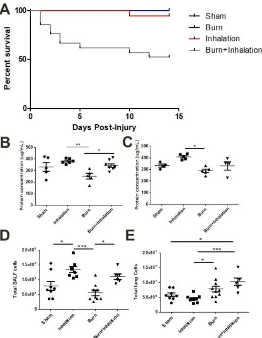
Figure 4.1: Combined injury results in increased mortality over time, while major
markers of pulmonary damage are elevated in an inhalation-dependent manner 72 Figure 4.2: Innate immune cells are recruited to the airspace and to the
pulmonary tissue in an injury-dependent manner 73
Figure 4.3: Multiple cytokines and chemokines are elevated in an inhalation-
dependent manner 96hrs after inhalation injury 74
Figure 4.4: Nitric oxide generation is involved in pathology of inhalation injury 75 Figure 4.5: Complete ablation of nitric oxide machinery results in increased
mortality after inhalation injury 76
LIST OF ABBREVIATIONS
RONS Reactive oxygen and nitrogen species NOS Nitric oxide species
PAMP Pathogen associated molecular pattern DAMP Damage associated molecular pattern
IL Interleukin
SIRS Systemic inflammatory response syndrome
CARS Compensatory anti-inflammatory response syndrome MARS Mixed antagonistic response syndrome
CFU Colony forming units DHR Dihydrorhodamine FBS Fetal bovine serum
LB Luria broth
MFI Mean fluorescence intensity
NO Nitric oxide
PAK Pseudomonas aeruginosa PAK strain TBSA Total body surface area
TLR Toll-like receptor CXCL CX chemokine ligand CCL C chemokine ligand
CHAPTER 1: INTRODUCTION
Each year approximately 486,000 individuals experience a burn injury requiring medical treatment [1]. These patients have an extended hospital stay relative to other forms of trauma, resulting in a treatment that is not only difficult and lengthy, but costly [2]. The average treatment cost per burn patient can exceed $88,000 USD [3]. One leading cause of lengthened hospital stay after burn injury is the presence of a burn-associated comorbidity. Patients who present with comorbidities of burn injury often require additional therapeutic intervention and experience complicated clinical outcomes relative to other patients [4].
When a patient experiences a significant burn injury, the wound tissue releases damage-associated molecular patterns (DAMPS) that can alter many systems throughout the body [5]. One major compartment affected by DAMPS is the immune system. Burn injury is known to lead to significant systemic immune dysregulation [5-8]. DAMPS released into the body lead to an overall systemic inflammatory response syndrome (SIRS), in which the immune system becomes activated in response to damage [9, 10]. Immune dysregulation and activation after burn injury is often typified by the presence of altered levels of cytokines, chemokines, and immune cells [11].
In the case of significant trauma such as burn injury, damaged tissue causes the release of a cytokine storm, which not only activates the SIRS response, but also triggers an overall
CARS responses were thought to be biphasic in nature, with a SIRS phenotype arising early after injury and a CARS response characterizing the late response to injury [13]. However, recent developments in the field indicate that typical injury results in the simultaneous initiation of both the SIRS and CARS response, with a different phenotype overriding the overall response at any given time. The ability to detect simultaneous presence of both pro- and anti-inflammatory mediators of immunity has been designated a mixed antagonist response syndrome (MARS) [14-21]. Although the MARS phenotype more fully encompasses the immune responses that take place after injury, it is possible to detect overriding immune responses characterized as being largely SIRS or CARS at any given timepoint after injury. However, it is important to emphasize the complexity of the immune response during the treatment and recovery of patients due to the presence of both pro-and anti-inflammatory signals.
are successfully recapitulate phenotypes seen among the patient population, and to that end modifications of models currently utilized are necessary.
There are major markers that have been identified as being key and typical of patient responses after injury. Specifically, we have previously demonstrated that aberrant production of IL-10 from several cell types, including macrophages and neutrophils, are likely a major factor contributing to the immunopathology responsible for susceptibility to infection [16, 22, 34, 35], whereby our group has indicated the prognostic potential of IL-10/IL-12 protein ratios within bronchial washes on predicting lung and systemic infections following severe burn trauma (Figure 1.1, [16, 17, 22]).Recent gene expression analysis of peripheral blood mononuclear cells (PBMCs) from burn patients has shown a strong correlation between increased IL-10, increased arginase 1 (ARG1), reduced IL-12 and reduced nitric oxide synthase 2 (NOS2) expression with burn severity and susceptibility to bacterial infection. As burn injury generates numerous inflammatory stimuli including DAMPs that activate innate immune cells [17, 36],
When patients present at the hospital with a burn injury, their treatment is often
immune-mediated disease progression. Animal models permit the study of systems that are inaccessible in human patients and also allow for testing of various potential therapeutic targets to improve overall outcomes of disease and/or injury. Therefore, improved models, such as those that include comorbidities of burn injury are necessary in order to improve understanding of burn-mediated immunopathology.
Burn and Inhalation injury
Approximately 10-20% of all burn patients present to the hospital with a burn-associated inhalation injury [45]. Presence of an inhalation injury can cause a 20% increase in mortality among burn patients, significantly lengthen hospital stay, and result in increased incidence of bacterial infection [46-48]. In fact, inhalation injury is a leading cause of burn-associated mortality [49, 50]. Many models of inhalation injury have been generated, however few improvements in treatment of patients with inhalation injury have been achieved [45].
Additionally, while multiple studies have examined the immune-mediated effects of inhalation injury [51-53], few have examined the combined effects of burn and inhalation injury in spite of the fact that combined injury results in damage more severe than can be attributed to each injury alone [39].
Studies have indicated that presence of severe inhalation injury leads to increased levels of multiple immune-related cytokines in collected bronchoalveolar lavage fluid (BAL),
BAL [54-58]. Presence of neutrophils in BAL after inhalation injury has additionally been associated with overall immune dysfunction and susceptibility to bacterial infection [59-61]. Neutrophil presence has additionally been associated with protein deposition in the airway, and associated pulmonary damage [60, 62, 63].
Neutrophils present in the lungs after inhalation injury release proteases, oxidants, and their downstream products such as nitrate and nitrite, which can lead to significant damage to the lungs [64, 65]. Multiple studies have detected indicators of oxidative stress in the pulmonary compartment after inhalation injury [45, 66, 67], and that inhibition of molecules involved in production of oxidants such as nitric oxide synthase (NOS) can lead to improved outcome after inhalation injury [68, 69]. Indeed, inhibition of inducible NOS (iNOS) has been proposed and tested as a potential therapeutic treatment of inhalation injury in ovine models [70, 71]. It has been reported that multiple studies are currently examining the therapeutic potential of inhibition of NOS, modulators of reactive nitrogen species, and antioxidants in treatment of inhalation injury [72]. These studies are important in order to be able to generate potential improvements in treatment of inhalation injury.
Burn and infection
among burn patients is the presence of a bacterial pneumonia [80], and studies have indicated that repeated infectious episodes significantly increase mortality among patients [81]. In order to decrease infection-associated mortality among burn patients it is essential that further studies be conducted.
Many studies have been conducted using mouse models to examine burn-dependent changes in the immune system and how these changes alter the response to bacterial infection [22, 82-84]. Animal models have been successful at recapitulating immunosuppression and susceptibility to bacterial infection early after burn injury [85-91]. Various labs have sought to identify potential vaccines and treatments that could generate bacterial-specific immune responses to improve outcomes in the face of infection. Multiple groups have applied bacterial byproducts such as flagellin or PilA from Pseudomonas aeruginosa to generate vaccines to decrease bacterial burden and associated mortality after burn injury [92-94]. Additional groups have focused instead on altering host responses in order to promote a response that will result in increased clearance of bacterial presence and improve outcomes. Burn injury leads to
significantly altered metabolism among patients [76, 86], which is known to alter the immune profile [95, 96]. In order to improve the overall immune response some labs have targeted host metabolic responses through insulin therapy to decrease overall systemic inflammation and promote bacterial clearance [97, 98]. Other labs have chosen to more directly target the immune response by administering granulocyte colony-stimulating factor (G-CSF) to improve survival after injury and promote clearance of bacteria [29].
31, 33]. Some labs postulate that this protection is largely due to the recruitment of neutrophils to the pulmonary vasculature after burn injury [27, 28], and have demonstrated that elimination of the neutrophil population using an antibody against Ly6G results in loss of this protective phenotype. Additional studies have indicated that the presence of burn and an additional
comorbidity of burn injury results in the loss of the burn-associated protective phenotype as well [37, 38, 99].
Burn and alcohol
Ingestion of alcohol is known to impair judgement and is often associated with experiencing a trauma [100]. Therefore, patients who present to the burn center have a high probability of being intoxicated. Studies have indicated that intoxicated patients who present to the hospital with a burn injury have significantly increased mortality, frequent intubations, delayed healing, increased infectious complications and lengthy hospital stays relative to non-intoxicated patients [101-107]. Burn injury is known to alter metabolism of patients, an effect that is further exacerbated with alcohol consumption [108].
systemic as well and lead to loss of protection and an inability to fight bacterial infectious agents. Alcohol exposure is known to alter levels of reactive oxygen and nitrogen species, glutathione metabolism, bone formation and wound healing [108], each of which is known to be involved in regulation of immune cell activity [124-127]. It is therefore not surprising that intoxication leads to alteration of the immune compartment through cellular mechanisms involving each of these pathways. Some groups postulate that modulation of these pathways through use of compounds such as glutathione precursor supplementation may improve negative consequences of burn injury during intoxication [108]. The immune compartment is a potentially powerful therapeutic target to promote improved healing and decreased mortality after burn injury while intoxicated [128].
Burn and ageing
Recent studies indicate that burn injury of the aged population is associated with worsened physical conditioning and quality of life after injury, and increased mortality [129-131]. Treatment strategies for older patients who have experienced a burn injury have not led to significant changes in length of hospitalization or mortality [132].
It has long been noted that elderly patients experience altered immune responses relative to young patients due to immunosenescence [133], a condition further complicated by the
presence of a burn injury [134]. Elderly patients commonly have elevated levels of inflammatory cytokines, and upon experiencing a burn injury, aged patients will generate a stronger
pro-inflammatory cytokine response than their younger adult-aged counterparts [134].
injury [135-137]. These differences are related to alterations in immune cell phenotype and function and play an important role in cellular polarization in response to burn injury [138]. Age-dependent alterations in cytokine responses are associated with delays in wound healing, and imply that studies should examine therapies focused on decreasing the expression of these cytokines in order to promote healing [139, 140]. Multiple groups have found that in aged mice, alterations in the cytokine response to burn injury may lead to deficiency in some, but not all immune cell migration to the wound site, potentially altering progression of wound healing and preventing a typical healing response [141, 142], a response that leads to increased susceptibility to bacterial infection in aged mice [143]. Additional studies have indicated an important role of multiple molecular immune markers in promoting a healing response after burn injury, such as hypoxia inducible factor-1 (HIF-1) which mediates migration of immune cells from the bone marrow to the site of injury to promote the healing response [142]. It is possible that immune mediators such as HIF-1 would be viable targets of immunotherapeutic treatment after burn injury. Additional studies indicate that cytokine therapy may potentially be successful in the treatment of elderly burn patients [144], however it is important to note that in an aged population cells do not respond as robustly to cytokines present [143].
Discussion
Patients who require treatment for burn injuries experience substantial immune
injury. In addition to the comorbidities discussed here, additional research is needed to study the combined effects of burn and radiation [41, 145, 146], the effects of sex in responses to burn [147, 148], burn associated with diabetes [85, 149, 150], and the presence of multiple
simultaneous comorbidities [44].
The innate arm of the immune system is altered after burn injury, and comorbidities commonly promote further dysregulation of innate immunity. Multiple studies have indicated that immune cells of myeloid lineage are dysregulated in response to burn injury. Burn injury leads to altered recruitment of neutrophils to the pulmonary compartment, as well as altered hematopoiesis. Neutrophils are a powerful cell type that is responsible for protecting the body from bacterial insult[151]. Studies have shown that neutrophils are both necessary and sufficient to clear multiple bacterial pathogens [152, 153], and dysfunction of the myeloid arm of the immune system is commonly associated with comorbidities of burn injury.
In Chapters 2-4 we discuss unique models that we have generated in our laboratory
to study burn and its associated comorbidities of inhalation injury and infection. We have
REFERENCES
1. Branch, T.A.a.H.C.S. National Hospital Ambulatory Medical Care Survey: 2011 Emergency Department Summary Tables. 2011 [cited 2017 November 7th]; Available from: https://www.cdc.gov/nchs/ahcd/web_tables.htm#2011.
2. Haikonen, K. and P.M. Lillsunde, Burden of Fire Injuries in Finland: Lost Productivity and Benefits. J Public Health Res, 2016. 5(2): p. 705.
3. Hop, M.J., et al., Costs of burn care: a systematic review. Wound Repair Regen, 2014. 22(4): p. 436-50.
4. Thananopavarn, P. and J.J. Hill, 3rd, Rehabilitation of the Complex Burn Patient with Multiple Injuries or Comorbidities. Clin Plast Surg, 2017. 44(4): p. 695-701.
5. Rani, M., et al., Damage-associated molecular patterns (DAMPs) released after burn are associated with inflammation and monocyte activation. Burns, 2017. 43(2): p. 297-303. 6. Schwacha, M.G., et al., Genetic variability in the immune-inflammatory response after
major burn injury. Shock, 2005. 23(2): p. 123-8.
7. Eitas, T.K., et al., Differential regulation of innate immune cytokine production through pharmacological activation of Nuclear Factor-Erythroid-2-Related Factor 2 (NRF2) in burn patient immune cells and monocytes. PLoS One, 2017. 12(9): p. e0184164.
8. Fontaine, M., et al., Innate danger signals in acute injury: From bench to bedside. Anaesth Crit Care Pain Med, 2016. 35(4): p. 283-92.
9. Dahiya, P., Burns as a model of SIRS. Front Biosci (Landmark Ed), 2009. 14: p. 4962-7. 10. Xu, Y.C., C.Q. Luo, and X. Li, Systemic inflammatory response syndrome following burns
is mediated by brain natriuretic peptide/natriuretic peptide A receptor-induced shock factor 1 signaling pathway. Clin Exp Pharmacol Physiol, 2016. 43(10): p. 921-9.
11. Chaudhry, H., et al., Role of cytokines as a double-edged sword in sepsis. In Vivo, 2013. 27(6): p. 669-84.
12. Ward, N.S., B. Casserly, and A. Ayala, The compensatory anti-inflammatory response syndrome (CARS) in critically ill patients. Clin Chest Med, 2008. 29(4): p. 617-25, viii. 13. Binkowska, A.M., G. Michalak, and R. Slotwinski, Current views on the mechanisms of
immune responses to trauma and infection. Cent Eur J Immunol, 2015. 40(2): p. 206-16. 14. Finnerty, C.C., et al., Temporal cytokine profiles in severely burned patients: a comparison
15. Jeschke, M.G., et al., Long-term persistance of the pathophysiologic response to severe burn injury. PLoS One, 2011. 6(7): p. e21245.
16. Jones, S.W., et al., Bronchoscopy-derived correlates of lung injury following inhalational injuries: a prospective observational study. PLoS One, 2013. 8(5): p. e64250.
17. Maile, R., et al., Association between early airway damage-associated molecular patterns and subsequent bacterial infection in patients with inhalational and burn injury. Am J Physiol Lung Cell Mol Physiol, 2015. 308(9): p. L855-60.
18. Mendoza, A.E., et al., Burn injury induces high levels of phosphorylated insulin-like growth factor binding protein-1. Int J Burns Trauma, 2013. 3(4): p. 180-9.
19. Xiao, W., et al., A genomic storm in critically injured humans. J Exp Med, 2011. 208(13): p. 2581-90.
20. Ostanin, A.A., et al., Inflammatory Syndromes (SIRS, MARS, CARS) in Patients with Surgical Infection. Russ J Immunol, 2000. 5(3): p. 289-300.
21. Tamayo, E., et al., Pro- and anti-inflammatory responses are regulated simultaneously from the first moments of septic shock. Eur Cytokine Netw, 2011. 22(2): p. 82-7.
22. Neely, C.J., et al., Flagellin treatment prevents increased susceptibility to systemic bacterial infection after injury by inhibiting anti-inflammatory IL-10+ IL-12- neutrophil polarization. PLoS One, 2014. 9(1): p. e85623.
23. Rae, L., P. Fidler, and N. Gibran, The Physiologic Basis of Burn Shock and the Need for Aggressive Fluid Resuscitation. Crit Care Clin, 2016. 32(4): p. 491-505.
24. O'Dea, K.P., et al., Circulating Microvesicles Are Elevated Acutely following Major Burns Injury and Associated with Clinical Severity. PLoS One, 2016. 11(12): p. e0167801. 25. Liu, Y.W., et al., Activation of Adenosine 2A receptor inhibits neutrophil apoptosis in an
autophagy-dependent manner in mice with systemic inflammatory response syndrome. Sci Rep, 2016. 6: p. 33614.
26. Burmeister, D.M., et al., Impact of Isolated Burns on Major Organs: A Large Animal Model Characterized. Shock, 2016. 46(3 Suppl 1): p. 137-47.
27. Gardner, J.C., et al., G-CSF drives a posttraumatic immune program that protects the host from infection. J Immunol, 2014. 192(5): p. 2405-17.
28. Huber, N.L., et al., Prior thermal injury accelerates endotoxin-induced inflammatory cytokine production and intestinal nuclear factor-kappaB activation in mice. J Burn Care Res, 2012. 33(2): p. 279-85.
30. Alexander, J.W., Effect of thermal injury upon the early resistance to infection. J Surg Res, 1968. 8(3): p. 128-37.
31. Noel, G., et al., A ribonucleotide reductase inhibitor reverses burn-induced inflammatory defects. Shock, 2010. 34(5): p. 535-44.
32. Stieritz, D.D. and I.A. Holder, Experimental studies of the pathogenesis of infections due to Pseudomonas aeruginosa: description of a burned mouse model. J Infect Dis, 1975. 131(6): p. 688-91.
33. Pinto, M. and T. Zehavi-Willner, Thermal injury-induced non-specific resistance to fatal Pseudomonas aeruginosa burn-infection in mice. Jpn J Exp Med, 1989. 59(5): p. 189-96. 34. Dunn, J.L., et al., Direct detection of blood nitric oxide reveals a burn-dependent decrease
of nitric oxide in response to Pseudomonas aeruginosa infection. Burns, 2016.
35. Maile, R., et al., Lymphopenia-induced homeostatic proliferation of CD8+ T cells is a mechanism for effective allogeneic skin graft rejection following burn injury. J Immunol, 2006. 176(11): p. 6717-26.
36. Yao, X., et al., Estrogen-provided cardiac protection following burn trauma is mediated through a reduction in mitochondria-derived DAMPs. Am J Physiol Heart Circ Physiol, 2014. 306(6): p. H882-94.
37. Mendoza, A.E., et al., Radiation combined with thermal injury induces immature myeloid cells. Shock, 2012. 38(5): p. 532-42.
38. Linz, B.M., et al., Innate Immune Cell Recovery Is Positively Regulated by NLRP12 during Emergency Hematopoiesis. J Immunol, 2017. 198(6): p. 2426-2433.
39. Clark, C.J., et al., Role of pulmonary alveolar macrophage activation in acute lung injury after burns and smoke inhalation. Lancet, 1988. 2(8616): p. 872-4.
40. Lee-Chiong, T.L., Jr. and R.A. Matthay, Burns and smoke inhalation. Curr Opin Pulm Med, 1995. 1(2): p. 96-101.
41. DiCarlo, A.L., N. Ramakrishnan, and R.J. Hatchett, Radiation combined injury: overview of NIAID research. Health Phys, 2010. 98(6): p. 863-7.
42. Kok, Y.O., et al., Establishing a treatment protocol for concomitant major burn and trauma patients: a tropical Asian hospital's experience. Burns Trauma, 2017. 5: p. 21.
43. Lundgren, R.S., et al., Influence of comorbidities and age on outcome following burn injury in older adults. J Burn Care Res, 2009. 30(2): p. 307-14.
45. Walker, P.F., et al., Diagnosis and management of inhalation injury: an updated review. Crit Care, 2015. 19: p. 351.
46. Dries, D.J. and F.W. Endorf, Inhalation injury: epidemiology, pathology, treatment strategies. Scand J Trauma Resusc Emerg Med, 2013. 21: p. 31.
47. Knowlin, L., et al., The measured effect magnitude of co-morbidities on burn injury mortality. Burns, 2016. 42(7): p. 1433-1438.
48. Travis, T.E., et al., Factors impacting the likelihood of death in patients with small TBSA burns. J Burn Care Res, 2015. 36(1): p. 203-12.
49. Herndon, D.N., P.B. Thompson, and D.L. Traber, Pulmonary injury in burned patients. Crit Care Clin, 1985. 1(1): p. 79-96.
50. Jones, S.W., et al., Inhalation Injury: Pathophysiology, Diagnosis, and Treatment. Clin Plast Surg, 2017. 44(3): p. 505-511.
51. Davis, C.S., et al., Early pulmonary immune hyporesponsiveness is associated with mortality after burn and smoke inhalation injury. J Burn Care Res, 2012. 33(1): p. 26-35. 52. Davis, C.S., et al., Inhalation injury severity and systemic immune perturbations in burned
adults. Ann Surg, 2013. 257(6): p. 1137-46.
53. Perng, D.W., et al., Inflammatory role of AMP-activated protein kinase signaling in an experimental model of toxic smoke inhalation injury. Crit Care Med, 2013. 41(1): p. 120-32.
54. Albright, J.M., et al., The acute pulmonary inflammatory response to the graded severity of smoke inhalation injury. Crit Care Med, 2012. 40(4): p. 1113-21.
55. Quinn, D.A., et al., Combined smoke inhalation and scald burn in the rat. J Burn Care Rehabil, 2003. 24(4): p. 208-16.
56. Ischiropoulos, H., et al., Role of neutrophils and nitric oxide in lung alveolar injury from smoke inhalation. Am J Respir Crit Care Med, 1994. 150(2): p. 337-41.
57. Zhu, F., et al., A rat model of smoke inhalation injury. Inhal Toxicol, 2012. 24(6): p. 356-64.
58. Ramos, C., et al., Oxidative stress and lung injury induced by short-term exposure to wood smoke in guinea pigs. Toxicol Mech Methods, 2013. 23(9): p. 711-22.
59. Murakami, K., et al., A novel animal model of sepsis after acute lung injury in sheep. Crit Care Med, 2002. 30(9): p. 2083-90.
61. Lange, M., et al., Assessment of vascular permeability in an ovine model of acute lung injury and pneumonia-induced Pseudomonas aeruginosa sepsis. Crit Care Med, 2008. 36(4): p. 1284-9.
62. Grommes, J. and O. Soehnlein, Contribution of neutrophils to acute lung injury. Mol Med, 2011. 17(3-4): p. 293-307.
63. Demling, R.H., Smoke inhalation lung injury: an update. Eplasty, 2008. 8: p. e27.
64. Alpard, S.K., et al., New clinically relevant sheep model of severe respiratory failure secondary to combined smoke inhalation/cutaneous flame burn injury. Crit Care Med, 2000. 28(5): p. 1469-76.
65. Katahira, J., et al., Role of anti-L-selectin antibody in burn and smoke inhalation injury in sheep. Am J Physiol Lung Cell Mol Physiol, 2002. 283(5): p. L1043-50.
66. Park, M.S., et al., Assessment of oxidative stress in lungs from sheep after inhalation of wood smoke. Toxicology, 2004. 195(2-3): p. 97-112.
67. Lee, A.S. and R.B. Mellins, Lung injury from smoke inhalation. Paediatr Respir Rev, 2006. 7(2): p. 123-8.
68. Booke, M., et al., Nitric oxide synthase inhibition versus norepinephrine for the treatment of hyperdynamic sepsis in sheep. Crit Care Med, 1996. 24(5): p. 835-44.
69. Enkhbaatar, P., et al., The inhibition of inducible nitric oxide synthase in ovine sepsis model. Shock, 2006. 25(5): p. 522-7.
70. Soejima, K., et al., Role of nitric oxide in vascular permeability after combined burns and smoke inhalation injury. Am J Respir Crit Care Med, 2001. 163(3 Pt 1): p. 745-52.
71. Enkhbaatar, P., et al., The inducible nitric oxide synthase inhibitor BBS-2 prevents acute lung injury in sheep after burn and smoke inhalation injury. Am J Respir Crit Care Med, 2003. 167(7): p. 1021-6.
72. Enkhbaatar, P., et al., Pathophysiology, research challenges, and clinical management of smoke inhalation injury. Lancet, 2016. 388(10052): p. 1437-1446.
73. Lax, S., et al., Bacterial colonization and succession in a newly opened hospital. Sci Transl Med, 2017. 9(391).
74. Sanon, M.A. and S. Watkins, Nurses' uniforms: How many bacteria do they carry after one shift? J Public Health Epidemiol, 2012. 4(10): p. 311-315.
76. Rogobete, A.F., et al., The influence of metabolic imbalances and oxidative stress on the outcome of critically ill polytrauma patients: a review. Burns Trauma, 2017. 5: p. 8. 77. Stoilova, Y.D., et al., Immunological and microbiological investigations of patients with
burn injuries. Folia Med (Plovdiv), 2007. 49(1-2): p. 49-58.
78. Yan, S., et al., Prediction of multiple infections after severe burn trauma: a prospective cohort study. Ann Surg, 2015. 261(4): p. 781-92.
79. Xiu, F. and M.G. Jeschke, Perturbed mononuclear phagocyte system in severely burned and septic patients. Shock, 2013. 40(2): p. 81-8.
80. Shupp, J.W., et al., Epidemiology of bloodstream infections in burn-injured patients: a review of the national burn repository. J Burn Care Res, 2010. 31(4): p. 521-8.
81. Mueller, E.W., et al., Effect from multiple episodes of inadequate empiric antibiotic therapy for ventilator-associated pneumonia on morbidity and mortality among critically ill trauma patients. J Trauma, 2005. 58(1): p. 94-101.
82. Dunn, J.L., et al., Direct detection of blood nitric oxide reveals a burn-dependent decrease of nitric oxide in response to Pseudomonas aeruginosa infection. Burns, 2016. 42(7): p. 1522-1527.
83. Calum, H., et al., Thermal injury induces impaired function in polymorphonuclear neutrophil granulocytes and reduced control of burn wound infection. Clin Exp Immunol, 2009. 156(1): p. 102-10.
84. Wilkinson, R.A. and J.A. Fishman, Effect of thermal injury with Pseudomonas aeruginosa infection on pulmonary and systemic bacterial clearance. J Trauma, 1999. 47(5): p. 912-7. 85. Abdullahi, A., S. Amini-Nik, and M.G. Jeschke, Animal models in burn research. Cell Mol
Life Sci, 2014. 71(17): p. 3241-55.
86. Williams, F.N., D.N. Herndon, and M.G. Jeschke, The hypermetabolic response to burn injury and interventions to modify this response. Clin Plast Surg, 2009. 36(4): p. 583-96. 87. Cairns, B.A., et al., Toll-like receptor 2 and 4 ligation results in complex altered cytokine
profiles early and late after burn injury. J Trauma, 2008. 64(4): p. 1069-77; discussion 1077-8.
88. Adediran, S.G., et al., Early infection during burn-induced inflammatory response results in increased mortality and p38-mediated neutrophil dysfunction. Am J Physiol Regul Integr Comp Physiol, 2010. 299(3): p. R918-25.
90. Orman, M.A., et al., Long-term dynamic profiling of inflammatory mediators in double-hit burn and sepsis animal models. Cytokine, 2012. 58(2): p. 307-15.
91. Moore, C.B., et al., Downregulation of immune signaling genes in patients with large surface burn injury. J Burn Care Res, 2007. 28(6): p. 879-87.
92. Laghaei, P., et al., Immunogenicity and protective efficacy of Pseudomonas aeruginosa type a and b flagellin vaccines in a burned mouse model. Mol Immunol, 2016. 74: p. 71-81.
93. Faezi, S., et al., Protective efficacy of Pseudomonas aeruginosa type-A flagellin in the murine burn wound model of infection. APMIS, 2014. 122(2): p. 115-27.
94. Korpi, F., et al., Active Immunization with Recombinant PilA protein Protects Against Pseudomonas aeruginosa Infection in a Mouse Burn Wound Model. J Microbiol Biotechnol, 2015.
95. Buck, M.D., et al., Metabolic Instruction of Immunity. Cell, 2017. 169(4): p. 570-586. 96. Ganeshan, K. and A. Chawla, Metabolic regulation of immune responses. Annu Rev
Immunol, 2014. 32: p. 609-34.
97. Przkora, R., et al., Insulin attenuates the cytokine response in a burn wound infection model. Shock, 2007. 27(2): p. 205-8.
98. Deng, H.P. and J.K. Chai, The effects and mechanisms of insulin on systemic inflammatory response and immune cells in severe trauma, burn injury, and sepsis. Int Immunopharmacol, 2009. 9(11): p. 1251-9.
99. Murphy, T.J., et al., Linking the "two-hit" response following injury to enhanced TLR4 reactivity. J Leukoc Biol, 2005. 77(1): p. 16-23.
100. Savola, O., O. Niemela, and M. Hillbom, Alcohol intake and the pattern of trauma in young adults and working aged people admitted after trauma. Alcohol Alcohol, 2005. 40(4): p. 269-73.
101. McGill, V., et al., The impact of substance use on mortality and morbidity from thermal injury. J Trauma, 1995. 38(6): p. 931-4.
102. Choudhry, M.A. and I.H. Chaudry, Alcohol intoxication and post-burn complications. Front Biosci, 2006. 11: p. 998-1005.
104. Grobmyer, S.R., et al., Alcohol, drug intoxication, or both at the time of burn injury as a predictor of complications and mortality in hospitalized patients with burns. J Burn Care Rehabil, 1996. 17(6 Pt 1): p. 532-9.
105. Swenson, J.R., et al., Drug and alcohol abuse in patients with acute burn injuries. Psychosomatics, 1991. 32(3): p. 287-93.
106. Silver, G.M., et al., Adverse clinical outcomes associated with elevated blood alcohol levels at the time of burn injury. J Burn Care Res, 2008. 29(5): p. 784-9.
107. Griffin, R., et al., The association between blood alcohol level and infectious complications among burn patients. J Burn Care Res, 2009. 30(3): p. 395-9.
108. Jung, M.K., et al., Alcohol exposure and mechanisms of tissue injury and repair. Alcohol Clin Exp Res, 2011. 35(3): p. 392-9.
109. Albright, J.M., et al., Implications of formal alcohol screening in burn patients. J Burn Care Res, 2009. 30(1): p. 62-9.
110. Jones, J.D., et al., Alcohol use and burn injury. J Burn Care Rehabil, 1991. 12(2): p. 148-52.
111. Steenkamp, W.C., N.J. Botha, and A.E. Van der Merwe, The prevalence of alcohol dependence in burned adult patients. Burns, 1994. 20(6): p. 522-5.
112. Haum, A., et al., Alcohol and drug abuse in burn injuries. Burns, 1995. 21(3): p. 194-9. 113. Li, X., J.L. Rendon, and M.A. Choudhry, T cell IFN-gamma suppression following alcohol
and burn injury is independent of miRNA155. PLoS One, 2014. 9(8): p. e105314.
114. Li, X., et al., Inflammatory response in multiple organs in a mouse model of acute alcohol intoxication and burn injury. J Burn Care Res, 2011. 32(4): p. 489-97.
115. Hammer, A.M., et al., The First Line of Defense: The Effects of Alcohol on Post-Burn Intestinal Barrier, Immune Cells, and Microbiome. Alcohol Res, 2015. 37(2): p. 209-22. 116. Li, X., et al., Intestine immune homeostasis after alcohol and burn injury. Shock, 2015.
43(6): p. 540-8.
117. Molina, P.E., Alcohol binging exacerbates adipose tissue inflammation following burn injury. Alcohol Clin Exp Res, 2014. 38(1): p. 33-5.
118. Qin, Y., et al., Adipose inflammation and macrophage infiltration after binge ethanol and burn injury. Alcohol Clin Exp Res, 2014. 38(1): p. 204-13.
120. Chen, M.M., et al., An alteration of the gut-liver axis drives pulmonary inflammation after intoxication and burn injury in mice. Am J Physiol Gastrointest Liver Physiol, 2014. 307(7): p. G711-8.
121. Murdoch, E.L., et al., Prolonged chemokine expression and excessive neutrophil infiltration in the lungs of burn-injured mice exposed to ethanol and pulmonary infection. Shock, 2011. 35(4): p. 403-10.
122. Chen, M.M., et al., Alcohol potentiates postburn remote organ damage through shifts in fluid compartments mediated by bradykinin. Shock, 2015. 43(1): p. 80-4.
123. Choudhry, M.A., X. Li, and I.H. Chaudry, A role for corticosterone in impaired intestinal immunity and barrier function in a rodent model of acute alcohol intoxication and burn injury. J Neuroimmune Pharmacol, 2006. 1(4): p. 428-34.
124. Droge, W. and R. Breitkreutz, Glutathione and immune function. Proc Nutr Soc, 2000. 59(4): p. 595-600.
125. Yang, Y., et al., Reactive oxygen species in the immune system. Int Rev Immunol, 2013. 32(3): p. 249-70.
126. Park, J.E. and A. Barbul, Understanding the role of immune regulation in wound healing. Am J Surg, 2004. 187(5A): p. 11S-16S.
127. Pacifici, R., The immune system and bone. Arch Biochem Biophys, 2010. 503(1): p. 41-53.
128. Chen, M.M., et al., Pulmonary inflammation after ethanol exposure and burn injury is attenuated in the absence of IL-6. Alcohol, 2013. 47(3): p. 223-9.
129. Edgar, D.W., et al., The influence of advancing age on quality of life and rate of recovery after treatment for burn. Burns, 2013. 39(6): p. 1067-72.
130. Simsek, M.E., et al., Outcomes of elderly burn patients requiring hospitalization. Aging Male, 2015. 18(2): p. 97-9.
131. Keck, M., et al., Burn treatment in the elderly. Burns, 2009. 35(8): p. 1071-9.
132. Duke, J., et al., Rates of hospitalisations and mortality of older adults admitted with burn injuries in Western Australian from 1983 to 2008. Australas J Ageing, 2012. 31(2): p. 83-9.
133. Fulop, T., et al., Frailty, Inflammation and Immunosenescence. Interdiscip Top Gerontol Geriatr, 2015. 41: p. 26-40.
135. Wang, L., et al., Age-dependent differences of interleukin-6 activity in cardiac function after burn complicated by sepsis. Burns, 2010. 36(2): p. 232-8.
136. Brubaker, A.L., S.R. Carter, and E.J. Kovacs, Experimental Approaches to Tissue Injury and Repair in Advanced Age. Methods Mol Biol, 2015. 1343: p. 35-51.
137. Plackett, T.P., et al., Aging enhances lymphocyte cytokine defects after injury. Faseb j, 2003. 17(6): p. 688-9.
138. Mahbub, S., C.R. Deburghgraeve, and E.J. Kovacs, Advanced age impairs macrophage polarization. J Interferon Cytokine Res, 2012. 32(1): p. 18-26.
139. Simonetti, O., et al., Delayed wound healing in aged skin rat models after thermal injury is associated with an increased MMP-9, K6 and CD44 expression. Burns, 2013. 39(4): p. 776-87.
140. Gomez, C.R., et al., Interleukin-6 contributes to age-related alteration of cytokine production by macrophages. Mediators Inflamm, 2010. 2010: p. 475139.
141. Shallo, H., et al., Monocyte chemoattractant protein-1 (MCP-1) and macrophage infiltration into the skin after burn injury in aged mice. Burns, 2003. 29(7): p. 641-7. 142. Zhang, X., et al., Aging impairs the mobilization and homing of bone marrow-derived
angiogenic cells to burn wounds. J Mol Med (Berl), 2011. 89(10): p. 985-95.
143. Brubaker, A.L., et al., Reduced neutrophil chemotaxis and infiltration contributes to delayed resolution of cutaneous wound infection with advanced age. J Immunol, 2013. 190(4): p. 1746-57.
144. Gomez, C.R., T.P. Plackett, and E.J. Kovacs, Aging and estrogen: modulation of inflammatory responses after injury. Exp Gerontol, 2007. 42(5): p. 451-6.
145. Tajima, G., et al., Immune system phenotyping of radiation and radiation combined injury in outbred mice. Radiat Res, 2013. 179(1): p. 101-12.
146. Palmer, J.L., et al., Development of a combined radiation and burn injury model. J Burn Care Res, 2011. 32(2): p. 317-23.
147. O'Keefe, G.E., J.L. Hunt, and G.F. Purdue, An evaluation of risk factors for mortality after burn trauma and the identification of gender-dependent differences in outcomes. J Am Coll Surg, 2001. 192(2): p. 153-60.
148. McGwin, G., Jr., et al., Gender differences in mortality following burn injury. Shock, 2002. 18(4): p. 311-5.
150. Hamed, S., et al., Topical Erythropoietin Treatment Accelerates the Healing of Cutaneous Burn Wounds in Diabetic Pigs Through an Aquaporin-3-Dependent Mechanism. Diabetes, 2017. 66(8): p. 2254-2265.
151. Rada, B., Interactions between Neutrophils and Pseudomonas aeruginosa in Cystic Fibrosis. Pathogens, 2017. 6(1).
152. Koh, A.Y., et al., Inescapable need for neutrophils as mediators of cellular innate immunity to acute Pseudomonas aeruginosa pneumonia. Infect Immun, 2009. 77(12): p. 5300-10. 153. Lavoie, E.G., T. Wangdi, and B.I. Kazmierczak, Innate immune responses to Pseudomonas
CHAPTER 2: ONE-HIT WONDER: LATE AFTER BURN INJURY, GRANULOCYTES CAN CLEAR ONE BACTERIAL INFECTION BUT CANNOT CONTROL A
SUBSEQUENT INFECTION2
Overview
Objective: Many studies have described the acute immune response to burn injury in both human and animal models, yet few studies are able to recapitulate long term immune suppression and bacterial susceptibility seen in burn patients. We hypothesized that patients suffer immune exhaustion due to repeated microbial challenge, therefore single challenge in animal models will not recapitulate this clinical phenotype. Herein we describe a novel mouse model of repeated infection after burn injury, which reveals immune suppression resulting in decreased pulmonary clearance of bacteria.
Methods: Wildtype mice receiving a full-thickness contact burn were infected with Pseudomonas aeruginosa 14 days and/or 17 days after burn or sham injury. Pulmonary macrophages and neutrophils were enumerated and evaluated for immune activation and
function.
Results: In the double infection model, burn mice were unable to clear bacteria compared to sham injured or singly infected burn mice. After a second infection, neutrophils and
Introduction
Burn injury causes significant lengthy hospital stays among patients. Every year 486,000 patients in the United States seek medical attention for burn injury [1]. Burn injury commonly creates a significant wound that is slow to heal and causes significant immune dysregulation and immunosuppression. Patients that experience this immunosuppression often acquire additional associated complications such as colonization with nosocomial bacteria. Infection of burn patients is also commonly associated with increased morbidity and mortality; patients who have more than one incidence of bacterial infection during the course of their hospital stay can experience a 42% increase in mortality [2].
Bacterial pneumonia is a leading cause of mortality among burn patients [3].One common source of bacterial pneumonia within the burn patient population is Pseudomonas aeruginosa, a gram-negative opportunistic infection [4, 5]. Full clearance of pseudomonal infections requires the activation and integration of the immune system [6, 7]. Studies have demonstrated that neutrophils are the primary innate immune cells responsible for preventing and clearing bacterial infections, and that a neutrophil response is both necessary and sufficient for clearance of P. aeruginosa infections [8].
Immediately after burn injury patients experience a general activation of the immune response, with one model defining this as early systemic inflammatory response syndrome (SIRS) [9-11]. This response is typically associated with cytokine storm, immune cell
human population using animal models. In fact, multiple studies in animal models indicate that after injury, burn mice are more capable of responding to infection than their sham counterparts [20-26]. Recent studies indicate that the SIRS/CARS paradigm may not accurately represent the complex immune response in burn patients because pro- and anti- inflammatory mediators are often detected simultaneously [27-32] and patients experience a mixed antagonist response syndrome (MARS) at all time points. We retain the SIRCS/CARS terminology for this study to define the “early” and “late” phases after injury and the immune bias of the MARS response after burn injury.
Additional studies indicate that SIRS and CARS among burn patients leads to release of immune cytokines and alterations in the immune profile, and that poor outcomes following infection within patient populations can be predicted by production of the cytokines interleukin 10 (IL-10) and interleukin 12 (IL-12) [30, 33-36]. Additionally, murine studies have indicated that these cytokines play an important role in burn-associated responses to bacterial infection [14, 34, 37, 38]. Researchers have demonstrated that treatments resulting in decreased IL-10 production after burn injury lead to increased bacterial clearance and improved outcome [14, 39, 40] and that current therapeutic targets exist capable of altering cytokine production after burn injury [36]. These findings indicate that IL-10 and IL-12 are important markers and potential targets for therapeutic interventions.
It is evident that there is an increased neutrophil presence in the lung vasculature early and late after burn injury [20-22]. Late after injury these neutrophils are part of the overall heightened immune response and can result in improved outcome in burn mice following single infection [20, 21], an effect that is lost with the elimination of the protective neutrophil
obtained from patients late after burn injury is not reflected in the animal model. We have additionally demonstrated that in the presence of burn-associated comorbidities such as irradiation or smoke exposure, infection with bacterial exposure can result in a loss of the protective effect of burn injury [41, 42].
In a nosocomial environment, the skin microbiota of patients commonly changes to match that of their environment and the nurses with whom they commonly interact [43, 44]. Burn wounds represent a disrupted barrier to the environment, and burn patients have a high incidence of infection (39% in our burn unit) due to large burn wounds and necessary surgery, resulting in subsequent immune dysregulation [45-47]. In contrast, mice utilized in
Material and methods
Animals
Female C57BL/6 mice aged eight to twelve weeks old, weighing >18 grams were purchased (Taconic Farms) and used for this study. Mice were shaved dorsally, given a subcutaneous injection of morphine (3mg/kg body weight, West-Ward Pharmaceuticals) and underwent a 20% total body surface area burn as previously described [14, 41, 48, 49]. A full-thickness contact burn was achieved using a 65 gram copper rod (1.9 cm in diameter) that had been heated to 100°C and applied to the dorsum/flank for four applications, each lasting 10 seconds. Animals were then placed in individual cages, given food and water ad libitum and monitored twice daily. All sham animals underwent the same procedure with the exception of the application of the burn injury. All animals were housed in a specific pathogen-free environment and all procedures were approved by the Institutional Animal Care and Use Committee at the University of North Carolina at Chapel Hill in accordance with NIH guidelines for the care and use of Laboratory animals (NIH Publications No. 8023, revised 1978).
Bacterial Inocula preparation and infection
A wildtype strain (PAK) of P. aeruginosa was utilized for all infections as previously described [14]. Bacteria from frozen cultures were grown overnight in Luria Broth (LB). The following morning cultures were diluted 50x and grown for approximately two hours until mid-log phase growth was achieved (OD600=0.6-1.2). Cultures were then centrifuged at 14000 rpm
verified by optical density at 600nm. Bacterial concentration was then confirmed by serial dilution and plating on LB agar plates.
Mice infected intraperitoneally received a 1mL injection of P. aeruginosa at a
concentration of 5x10⁵ CFU/mL. Uninfected mice were given an intraperitoneal injection with 1mL of PBS+1%PP as a control. Mice infected intratracheally were anesthetized with an intraperitoneal injection of Avertin (0.475mg/g body weight: Sigma-Aldrich). Mice were then placed on an intubation platform, and infected by visualization of the vocal cords with a laryngoscope (Model LS-2, Penn Century Inc.), and inserting a MicroSprayer® Aerosolizer (Model 1A-1C and FMJ-250 High-Pressure Syringe Penn Century, Inc.) through the vocal cords, after which a 50uL volume was aerosolized into their lungs using either a bacterial innocula (2x10⁷ CFU/ML in PBS+1%PP) or vehicle (PBS+1%PP).
Enumeration of bacteria
At time of sacrifice, the left lobe of the liver, the lungs, and the spleen were removed and placed in 0.5mL of LB broth on ice. Tissues were homogenized using a BulletBlender (Next Advance) and three 3.2mm stainless steel beads per tube of tissue. Tissue homogenate was serially diluted and plated on LB agar for quantification. Plates were incubated overnight at 37°C.
BAL and Whole Lung tissue collection
and a catheter (22G x 1”, Exel) was placed into the trachea and tied off. A syringe with 1mL 0.6mM EDTA in PBS was connected to the catheter and 0.6 mL of the fluid was flushed into the lungs, the lungs were massaged, and then the fluid was withdrawn into the syringe to obtain a primary wash. This procedure was repeated three times. Two additional washes were performed. Samples were spun down for cellular analysis and the supernatant was collected for assay via Bradford assay and enzyme-linked immune-sorbent assay (ELISA). Lungs were removed from the animals and minced using sterile razor blades. Lungs were then placed in 4mL of PBS supplemented with 10% Fetal Bovine Serum (PBS+FBS), 0.1µg/mouse DNase, 1500 u/mouse collagenase and shaken at 250rpm at 37°C for 1h for digestion of tissue as previously described [14]. Samples were then filtered using a 100µm cell strainer and then pelleted. Pelleted cells then underwent ACK lysis for removal of red blood cells and then samples were washed and
resuspended in PBS+FBS for staining for flow cytometric analysis. Cells collected from BAL and whole lung tissue were counted using a hemocytometer with 0.01% trypan blue viability dye.
Flow cytometric analysis
Cells were incubated with anti-mouse CD16/32 Block (eBiosciences) to block Fc
receptors as previously described [14, 49]. Cells were then stained with antibodies against CD45, CD11c, CD11b, Ly6G, F4/80 and/or NOS2. Cells were then fixed in 1% paraformaldehyde and examined using a Dako CyAn (Beckmann-Coulter) and then data was analyzed using Summit software (Beckman-Coulter). Initial exclusion of CD45- cells was conducted, and then
species (RONS), analysis was performed using dihydrorhodamine-123 (DHR123). Samples were stained using fluorochrome-conjugated antibodies as previously stated. Prior to fixation in
paraformaldehyde, the samples were resuspended in DHR123 (1.875 mg/mL, Invitrogen). The samples were then split into stimulated and unstimulated samples. Samples were stimulated using 98nm Phorbol myristate acetate (PMA) for 30 minutes at 25˚C in the dark. All samples were then fixed in a 1% paraformaldehyde solution, and analyzed using flow cytometry. Reactive Oxygen and Nitrogen Species (RONS) expression was determined for each cell population present, as previously described [53].
Statistical Analysis
All data were visually displayed in GraphPad Prism Version 5.0 for Windows and analyzed by Student’s t-Test or One-Way Analysis of Variance (ANOVA) with a Tukey post-test. Data are represented as mean+/- standard error of the mean (SEM). Statistical significance is indicated as * p < 0.05, ** p < 0.005, *** p < 0.001.
Results
Repeated infection leads to susceptibility to bacterial dissemination in a murine model of burn
injury.
repeated bacterial exposure, we employed an infection strategy in which mice were initially infected (14 days after burn injury) intratracheally (IT) with P. aeruginosa, and then received a high-dose intraperitoneal (IP) infection with P. aeruginosa (17 days after burn injury). After a single IP infection, sham injured mice exhibited significant mortality and high, disseminated bacterial burden in the lungs and distal organs (Figure 2.1A). Administration of an initial IT infection 3 days prior to the IP infection reversed this phenotype and protected the sham mice from death (Figure 2.1A). In contrast, after a single IP infection, burn-injured mice experienced burn “priming” protection against a dose of bacteria that was fatal to sham counterparts, as predicted by earlier studies [20-22, 25, 26, 49] with significantly reduced bacterial dissemination (Figure 2.1B). However, burn mice that had an initial IT infection were unable to clear a
secondary IP infection and demonstrated increased mortality and systemic bacterial burden (Figure 2.1C). This suggests that burn “priming” is transient, and a secondary infection causes burn-mediated immune protection to collapse in comparison with sham mice.
Repeated infection leads to increased neutrophil and macrophage numbers in the lungs late after
burn injury.
tissue to characterize burn-associated responses to a single IT bacterial infection 24 hours after infection; we observed a significant increase in the number of live cells in whole lung in sham and burn mice after IT infection (Figure 2.2A). Using specific flow cytometric quantification of innate immune cells in the lung (representative staining shown in Figure 2B), we found that there were no burn or infection-dependent changes in the total number of macrophages present in the BAL or whole lung tissue (Figure 2.2C and 2.2D). However, infection led to a marked increase in the numbers of neutrophils present in BAL collected from both sham and burn-injured mice (Figure 2.2E). Additionally, we found that infection led to an increased number of neutrophils present in the whole lung tissue collected in mice, a phenotype augmented by burn-injury (Figure 2.2F). These data agree with earlier studies which demonstrate that an increased number of neutrophils are responsible for the burn “priming” after burn [14, 20, 22].
Neutrophils from burn mice are not able to be activated after secondary infection to increase
RONS production.
In addition, we sought to examine immune changes that took place in the actual tissue of the lungs. Enumeration of single cell suspensions generated from whole lung tissue found that there was an infection-dependent increase in the total number of neutrophils present in the tissue (Figure 2.3C). We additionally noted that there was an infection-dependent and injury-dependent change in the total number of macrophages present in the tissue of the whole lung, implying that macrophages could potentially mediate burn-dependent differences in response to infection (Figure 2.3D). We then examined macrophage and neutrophil function to determine their microbicidal potential. We utilized DHR123 to quantify the levels of RONS present in cells collected from whole lung tissue. We found that burn injury alone causes increased neutrophil RONS production as characterized by mean fluorescent intensity (Figure 2.4A&B). Additionally, we found that after burn animals are infected, basal RONS generation decreases, with doubly-infected animals generating significantly decreased RONS relative to undoubly-infected burn
counterparts. We therefore hypothesized that diminishment of RONS production is the source for lost burn-mediated protection. We found nominal changes in macrophage RONS production (Figure 2.4C&D). Loss of RONS activity in neutrophils also correlates with changes in
Neutrophil and macrophage IL-10 and IL-12 are differentially expressed after single and double
infections in burn mice.
We have previously demonstrated that aberrant production of IL-10 and reduced IL-12 cytokines is likely a major factor contributing to the immunopathology responsible for
Discussion
Murine models are frequently utilized to examine molecular and cellular mechanisms of burn-associated immune dysfunction [14, 49, 54-56]. However, clinically relevant animal model of injury should recapitulate phenotypes seen among human patients. Multiple studies have indicated that current murine models of burn injury are insufficient to elicit immunosuppression late after burn injury [20, 57]. In our model burn mice required repeated exposure to bacterial insult in order for burn-mediated immunosuppression to become apparent. This phenotype is similar to previously reported clinical data. Previous studies have indicated that mortality is only significantly increased among the burn population after a secondary bacterial challenge [2]. This clinical result in conjunction with our data implies that the immune compartment is sufficient to protect patients against a single bacterial insult, but that a second infection will result in
immunosuppression.
the compartment would experience immune cell exhaustion. Upon initial examination we found that despite the presence of neutrophils in whole lung tissue, a single intratracheal infection resulted in comparable recruitment of cells to the airspace in both sham and burn-treated animals. We also found that intratracheal infection resulted in recruitment of cells to the whole lung tissue, with slightly elevated numbers of neutrophils present in the whole lung tissue of burn-treated animals. Increased neutrophil numbers in the lungs correlated with improved bacterial clearance in burn mice after a single infection when compared to their sham-injured counterparts. Upon subsequent infection, sham mice “catch up” by recruiting more neutrophils to combat infection; functionally we do not observe any difference between RONS or cytokine production between the first and second infection. In contrast, the neutrophils recruited to combat a second infection late after burn injury produce less RONS and IL-12 than those that were recruited after the first bacterial hit. These results support our hypothesis that despite burn-induced “priming” the immune system is unable to mount a successful immune response to a secondary bacterial exposure.
Upon infection of a host, bacterial agents such as P. aeruginosa will release pathogen associated molecular patterns (PAMPS) that are able to alter the immune response. Multiple studies have indicated that the presence of PAMPS in a host is sufficient to result in altered immune cell signaling, cytokine production and changes in RONS-associated machinery [58, 59]. Studies have additionally indicated that burn injury leads to increased RONS and pro-inflammatory cytokine production in immune cells which can mediate additional tissue injury [60-63]. Some studies have indicated that it is possible to decrease RONS activity shortly after burn injury and decrease damage to lungs while still permitting successful clearance of
type responsible for burn-mediated hyperactivity [20, 22]. In this study we report increased total numbers of macrophages and neutrophils present in the whole lung tissue after burn injury repeated infection. We additionally found that burn injury increased basal RONS and pro- and anti- inflammatory cytokine activity of neutrophils, but that upon repeated infection this was lost. We also found that upon each incidence of infection these cells decreased their baseline
production of RONS. Repeated incidence of infection would cause cells to be repeatedly exposed to PAMPS and result in changes in intracellular signaling leading to changes in RONS
arginase 1 gene (ARG1)/NOS2 gene expression ratios and indicate that they have a powerful predictive ability for susceptibility to infection in burn patients. When we examined neutrophil levels of NOS2 we found that infection of sham-injured mice resulted in decreased NOS2 expression. This phenotype was not found in burn injured-mice after a single exposure to P. aeruginosa, however loss of NOS2 levels took place after repeated infection, potentially indicative of exhaustion arising in burn-injured mice and associated loss of burn-mediated protection.
We and others have previously reported that cells from burn-injured animals exhibit altered signaling of toll-like receptors (TLRs), the receptors responsible for detection of PAMPS and subsequent activation of the immune system [14, 34, 47, 64, 65]. Alteration of TLR function after burn injury have additionally been shown to lead to alterations in cytokine production in a model of burn injury [66]. It is possible that alterations in innate immune cell TLR levels result in alterations in the immune response, production of cytokines, and subsequent response to bacterial infection. Future studies will establish the link between TLR signaling and
FIGURES
Figure 2.1: Repeated, but not single infection, leads to susceptibility to bacterial infection in
a murine model of burn injury. Mice (n=4-6 per group) underwent sham or burn injury and
were infected with either A) intraperitoneal (IP) infection with P. aeruginosa 17 days after injury (ShamP); or intratracheal (IT) inoculum of P. aeruginosa 14 days after injury and a subsequent a
IP infection with P. aeruginosa 17 days after injury (ShamT/P ); B) IP infection with P.
aeruginosa 17 days after injury (ShamP or BurnP); or C) IT with P. aeruginosa 14 days after
harvested from surviving mice for P. aeruginosa quantification by colony forming unit (CFU) analysis. Dashed line represents lower limit of detection for the CFU assay. Data shown are +/-SEM. *p<0.05, **p<0.01, and representative of three repeated experiments. Numbers of initial mice in representative figures; survival plots A-C, ShamP, n=9; ShamT/P, n=9; BurnP, n=10;
Figure 2.2: Single IT infection leads to increased neutrophil numbers in the lung BAL late
after burn injury. Mice (N=4-6 per group) underwent sham or burn injury and were infected
(14 days after burn injury) IT with P. aeruginosa (ShamT or BurnT) or uninfected (Sham or
Burn) and lungs harvested 24 hours later; A) total cells enumerated counted on a
haemocytometer, B) representative flow cytometry of BAL or whole lung tissue used to identify neutrophils and macrophages; cells are shown after gating on live singlets and common
leucocyte CD45+ marker; C-F) number of macrophages or neutrophils from either BAL or whole lung were quantified for each injury and infection group. Data shown are +/-SEM. *p<0.05, **p<0.01, and representative of three repeated experiments. Numbers of mice in representative figures; A, C-F), Sham, n=4; ShamT, n=4; Burn, n=4; BurnT, n=4.
Figure 2.3: Double infection leads to increased neutrophil and macrophage numbers in the
lung late after burn injury. Mice (N=4-6 per group) underwent sham or burn injury and were
given IT inoculation with PBS+1%PP or with P. aeruginosa (ShamT/P or BurnT/P) 14 days after
injury, followed by IP infection IP with P. aeruginosa (ShamP or BurnP) at 17 days after injury,
or left uninfected (Sham or Burn). Lungs were harvested 24 hours later; A-D) number of macrophages or neutrophils from either BAL or whole lung were quantified for each injury and infection group. Data shown are +/-SEM. *p<0.05, **p<0.01, ***p<0.005 and representative of three repeated experiments. Numbers of mice in representative figures; A-D), Sham, n=5;
Figure 2.4: Neutrophils from burn mice are not able to be activated after secondary
infection to increase RONS production. Mice (N=4-6 per group) underwent sham or burn
injury and were infected (17 days after injury) either IP with P. aeruginosa (ShamP or BurnP) or
IT with P. aeruginosa 14 days after injury followed by IP infection at 17 days after injury (ShamT/P or BurnT/P), or left uninfected (Sham or Burn). Lungs were harvested 24 hours later;
in the absence or presence of Phorbol myristate acetate (PMA) stimulation (“+Stim.”), D) NOS2 expression per cell was quantified in neutrophils using flow cytometry. Data shown are +/-SEM. *p<0.05, **p<0.01, and representative of three repeated experiments. Numbers of mice in
representative figures; A-E), Sham, n=4; ShamP, n=5; ShamT/P, n=5; Burn, n=6; BurnP, n=6;
BurnT/P, n=6.
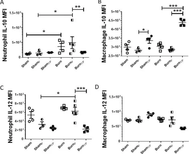
Figure 2.5: Neutrophil and macrophage IL-10 and IL-12 are differentially expressed after
single and double infections in burn mice. Mice (N=4-6 per group) underwent sham or burn
injury and were infected (17 days after injury) either IP with P. aeruginosa (ShamP or BurnP) or
IT with P. aeruginosa 14 days after injury followed by IP infection at 17 days after injury (ShamT/P or BurnT/P), or left uninfected (Sham or Burn). Lungs were harvested 24 hours later;
A-D) intracellular IL-10 and IL-12 expression per cell was measured by flow cytometry (MFI). Data shown are +/-SEM. *p<0.05, **p<0.01, ***p<0.005 and representative of three repeated experiments. Numbers of mice in representative figures; A-D), Sham, n=4; ShamP, n=5; ShamT/P,