Original Research Article
Role of CT enterography in evaluation of small bowel disorders
Ritu N. Misra, Sunil Kr. Bajaj*
INTRODUCTION
The mesenteric small intestine remains the only gastrointestinal tract segment for which diagnostic study is not principally dependent on endoscopic viewing. Since radiologists assume primary responsibility in the diagnostic evaluation of the small bowel, it is essential that methods capable of accurately demonstrating small bowel morphology are appropriately applied. Jacob Gersham Cohem introduced combination of air and simple water suspension of barium sulphate through duodenal tube to produce double contrast effect that was termed enteroclysis barium contrast studies and enteroclysis, in particular, were the mainstay of small bowel imaging in the past.1
Cross sectional imaging of the small bowel (CT and MRI) offer distinct advantages over fluoroscopic barium examination including depiction of the intraluminal pathologies, entire bowel wall and extra intestinal
manifestations coupled with the advantage of multiplanar imaging. Computed Tomographic Enterography (CTE) combines the improved spatial and temporal resolution of MDCT with neutral oral contrast and intravenously administered contrast material, thus permitting excellent assessment of the small bowel wall and lumen.
Luminal distension comparable to enteroclysis can be achieved with oral hyperhydration, thereby obviating nasoenteric intubation and making CT Enterography a useful, well tolerated study.2 The present study aims to
assess the feasibility and usefulness of CT enterography with orally administered iso-osmotic mannitol as neutral oral contrast in demonstrating small bowel diseases.
METHODS
The prospective study was carried out in the Department of Radiodiagnosis in collaboration with Department of Surgery, over a period of two years (2015-2017). Department ofRadiology, VMMC and Safdarjung Hospital, New Delhi, India
Received: 18 December 2018
Accepted: 25 December 2018
*Correspondence:
Dr. Sunil Kumar Bajaj,
E-mail: [email protected]
Copyright: © the author(s), publisher and licensee Medip Academy. This is an open-access article distributed under the terms of the Creative Commons Attribution Non-Commercial License, which permits unrestricted non-commercial use, distribution, and reproduction in any medium, provided the original work is properly cited.
ABSTRACT
Background: Small bowel pathologies are an enigma for clinicians and difficult to assess and evaluate for clinicians. In order to establish the efficacy of MDCT Enterography in diagnostic characterisation of small bowel lesions, the current study was undertaken.
Methods: A prospective observational cross-sectional study was carried out in a tertiary care hospital. 30 patients with clinically suspected small bowel disease underwent CT enterography using iso-osmotic mannitol as neutral enteral contrast. CT enterography diagnoses were compared with clinical, surgical and histopathological results.
Results: CT enterography showed a sensitivity (95.83%), specificity (100%), positive predictive value (100%), negative predictive value (85.71%), accuracy (96.66%) in diagnosis of small bowel diseases.
Conclusions: CTE is a non-invasive well tolerated and reliable imaging modality for the depiction of small-bowel diseases. It provides excellent visualization of luminal, mural and extraintestinal findings.
Keywords: CT Enterography, Enterography, Small Bowel Disorders
Inclusion criteria adult patients with clinically suspected small bowel disease were included in the study. A patient not consenting for imaging study as protocol, contraindication to contrast enhanced CT i.e. allergy to intravenous iodinated contrast media, pregnancy, hemodynamic instability, renal failure, inability for sufficient breath hold, acute high-grade intestinal obstruction/ perforation and acute intestinal infections are excluded.
Included 30 adults presenting with clinically suspected small bowel disease. They comprised of 14 male and 16 female patients of age ranged from 16-70years with a mean age of 31.03years. Most patients belonged to the 21-30years age group.
Procedure
Bowel preparation was done using low residue diet, ample fluids, laxative on the day prior to the examination and nothing by mouth on the day of the examination. Iso-osmotic mannitol at room temperature was used as neutral enteral contrast agent. 20% of 450ml mannitol diluted with enough plain water to make 1500ml. Patient was asked to drink solution every 5minutes to deliver continuous oral infusion over a period of 50minutes. An antispasmodic agent (Inj. buscopan 20mg IV) was given intravenously before CT acquisition. Thereafter, patient was taken up for CT scanning. 100-120ml of non-ionic iodinated contrast media was injected intravenously through a 20-gauge cannula at a rate of 4.5ml/sec.
Philips BrillianceTM 40-slice multidetector CT scanner
was used. Imaging was done in the enteric phase i.e the delay between the start of contrast material administration and the start of helical scanning was 50seconds. Images were obtained from the domes of the diaphragm to the lower margin of the symphysis pubis during a single breath hold. Reformatted images were obtained in the coronal plane. Both axial and multiplanar reformatted images were originally viewed with the standard window level (60HU) and width setting (360HU).
All images were specifically evaluated for the following parameters
• The degree of small-bowel luminal distension, • The presence of focal or diffuse bowel wall
thickening,
• Small-bowel narrowing /stenosis, • Small-bowel masses,
• Mesenteric stranding, • Mesenteric lymph nodes, • Peritoneal/visceral deposit, • Enhancement pattern.
Small-bowel thickening was considered to be present if the thickness of the bowel wall was greater than 3mm in at least two planes. Small-bowel stenosis was considered
to be present if segmental narrowing of the bowel lumen could be seen on at least two planes. A small bowel mass was considered to be present if a flat, sessile, pedunculated or polypoidal structure could be identified on at least two planes and could be differentiated from small bowel folds.
Visceral metastasis was considered to be present if nodules were identified within the peritoneal cavity or hypodense lesions were identified within the liver. Mesenteric stranding was considered to be present if an infiltration of the mesenteric fat was observed. An enlarged mesenteric lymph node was considered to be present when a lymph node was greater than 10mm in short-axis diameter. A lymph node was considered necrotic if there was peripheral enhancement associated with central hypodensity.
Enhancement pattern of bowel wall was characterized as hyperenhancement or mural stratification. Diagnosis was confirmed using clinical/surgical/ histopathological follow up findings as reference standard wherever relevant. Standardized statistical methods were used for the final analysis. The sensitivity, specificity, accuracy, positive and negative predictive value of CT enterography were calculated.
RESULTS
There were 14 male and 16 female patients in the study. Age ranged from 16-70years with a mean age of 31.03years. Most patients belonged to the 21-30years age group. Overall, abdominal pain was most common presentation (76.66%), followed by sub-acute intestinal obstruction (53.33%) and anorexia (53.33%). Optimal distension of the ileal loops was achieved in 28 patients (93.33%) and that for the jejunal loops was achieved in 22 patients (73.33%). The optimal distension of jejunoileal loops is shown in Figure 1.
Figure 1: Optimal distension of the jejuno-ileal loops.
patients (46.66%). Small bowel involvement was seen in all the patients (n=14). Additional large bowel involvement was seen in 4 patients. The involved segments of bowel showed circumferential wall
thickening (n=11), hyperenhancement (n=14), matting of bowel loops (n=1), strictures (n=12). A total of 28 strictures were demonstrated measuring 7-13mm in thickness with a mean thickness of 9.4mm.
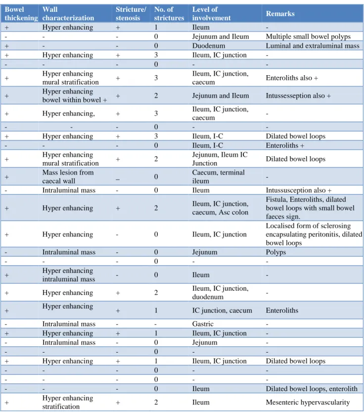
Table 1: MDCT enterography findings: gastro-intestinal tract.
Bowel thickening
Wall
characterization
Stricture/ stenosis
No. of strictures
Level of
involvement Remarks
+ Hyper enhancing + 1 Ileum -
- - - 0 Jejunum and Ileum Multiple small bowel polyps + - - 0 Duodenum Luminal and extraluminal mass + Hyper enhancing + 3 Ileum, IC junction -
- - - 0 - -
+ Hyper enhancing
mural stratification + 3
Ileum, IC junction,
caecum Enteroliths also + + Hyper enhancing
bowel within bowel + + 2 Jejunum and Ileum Intussesseption also + + Hyper enhancing, + 3 Ileum, IC junction,
caecum -
- - - 0 - -
+ Hyper enhancing + 3 Ileum, I-C Dilated bowel loops - - - 0 Ileum, I-C Enteroliths + + Hyper enhancing
mural stratification + 2
Jejunum, Ileum IC
Junction Dilated bowel loops + Mass lesion from
caecal wall _ 0
Caecum, terminal
ileum -
- Intraluminal mass - 0 Ileum Intussusception also +
+ Hyper enhancing + 2 Ileum, IC junction, caecum, Asc colon
Fistula, Enteroliths, dilated bowel loops with small bowel faeces sign.
+ Hyper enhancing - 0 Ileum, IC junction
Localised form of sclerosing encapsulating peritonitis, dilated bowel loops
- Intraluminal mass - 0 Jejunum Polyps
- - - 0 - -
+ Hyper enhancing
intraluminal mass - 0 Ileum - + Hyper enhancing + 2 Ileum, IC junction,
duodenum - + Hyper enhancing
+ 1 IC junction, caecum Enteroliths - Intraluminal mass - - Gastric -
+ Hyper enhancing + 1 Ileum, IC junction - - Intraluminal mass - 0 Jejunum -
- - - 0 - -
+ Hyper enhancing + 1 Ileum, IC junction Dilated bowel loops
- - - 0 - -
- - - 0 - -
- - - 0 Ileum Dilated bowel loops, enterolith + Hyper enhancing
stratification + 2 Ileum Mesenteric hypervascularity
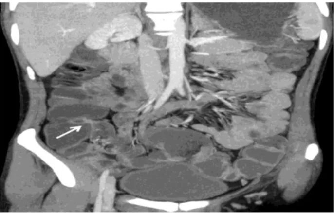
Figure 2 depicts small wall enhancement and thickening in tubercular stricture. Strictures were seen in the Ileo-caecal junction (n=12), ileum (n=13), jejunum (n=2), duodenum (n=1). Consequent small bowel dilatation was
(n=1), pleural effusion (n=1), tree-in bud opacities (n=1). Bilateral psoas abscesses were present in 1 patient. Seven patients with findings of abdominal tuberculosis underwent FNAC from lymph node, out of which 6 patients were confirmed to be tubercular. In one patient FNAC was inconclusive. In rest of the 7 patient’s diagnosis of abdominal tuberculosis was made based on clinical response to anti-tubercular treatment. One patient with ileal perforation underwent diagnostic laparotomy with biopsy, histopathological findings confirmed tuberculosis. The sensitivity and specificity for diagnosing abdominal tuberculosis by CT enterography was 92.85% and 100% respectively.
Figure 2: Focal ileal wall thickening with enhancement and luminal narrowing in a case of
tubercular stricture.
Figure 3: Small bowel carcinoid seen as focal enhancing mass in ileal wall (thin arrow) associated
with adenopathy (thick arrow).
CT enterography enabled the diagnosis of small-bowel masses in 7 patients. Small bowel adenocarcinoma (n=1) presented as intraluminal enhancing mass of soft tissue attenuation in the ileum causing intussusception. Diagnosis was confirmed by surgery and histopathology. Small bowel gastrointestinal stromal tumours were seen in 2 patients arising from the duodenum and jejunum respectively. They presented as intraluminal and extraluminal lobulated enhancing masses. FNAC confirmed the diagnosis of GIST in the duodenum while
surgery and histopathology confirmed jejunal GIST in the other patient. Small bowel carcinoid was diagnosed in one patient and presented as small, intensely enhancing, nodular masses in the terminal ileum (Figure 3).
PET CT was positive for somatostatin receptors. The patient underwent surgery and histopathology was confirmatory for carcinoid. Polyposis was seen in three patients. In one known patient of Peutz Jegher’s syndrome (post-operative status) who had come for evaluation of recurrence, multiple jejunal polyps were seen as moderately enhancing soft tissue masses. A combination of multiple small gastric mucosal polyps, abdominal wall desmoids and frontal osteomas was seen in one patient suspected with Gardner’s syndrome (Figure 4 (A), (B)). Thus, CT enterography accurately depicted small bowel masses in six cases. Crohn’s disease was diagnosed histo-pathologically in one patient. CT enterography depicted skip lesions in terminal ileum, mural thickening, stratification, strictures, fibrofatty proliferation, hyper enhancing mucosa and mesenteric hypervascularity (comb sign) suggestive of Crohn’s disease (Figure 5).
Figure 4 (A) and (B): A combination of multiple small gastric mucosal polyps, abdominal wall desmoids and
frontal osteomas seen in Gardner’s syndrome.
Figure 5: Ileocecal thickening with maintained wall stratification in a case of Crohn’s disease: Surrounding fat proliferation and mesenteric
hypervascularity is also seen.
Figure 6: Ileo-caecal low grade stricture (arrow)
demonstrated by adequate luminal distension.
Negative findings from CT enterography were obtained in 6 patients who presented with recurrent episodes of abdominal pain, diarrhoea, vomiting and anaemia. Clinical course remained uneventful in all. Low grade obstruction (spontaneously resolving obstruction) was seen in 14 patients. CT enterography clearly depicted the site, level, and cause of the obstruction in clinically suspected patients of tuberculosis. Figure 6 shows mild luminal narrowing at the level of ileocecal junction demonstrated by the adequate luminal distension. In the present study sensitivity and specificity in evaluation of low-grade obstruction was 100%. The remaining two cases were of gall stone ileus and ileal perforation respectively. One patient presented with gastrointestinal bleeding in form of melena.
Table 2: Follow up and final diagnosis in patients undergoing CT Enterography.
CT enterography diagnosis Type of follow up
Duration
follow up Final diagnosis Result
Small bowel TB Clinical 6 months Small bowel TB True +ve Small bowel TB with polyposis Clinical/surgical 6 months Small bowel TB with polyposis True +ve Duodenal GIST Surgical 2 months Duodenal GIST True +ve Abdominal TB Clinical 9 months Abdominal TB True +ve Normal study Clinical 3 months Normal True -ve Abdominal TB Clinical 9 months Abdominal TB True +ve Small bowel TB with transient
jejunal intussusception. Clinical 9 months Small bowel TB True +ve Abdominal TB Clinical 9 months Abdominal TB True +ve Normal study Clinical 3 months Normal True -ve Abdominal TB Clinical 9 months Abdominal TB True +ve Abdominal TB Clinical 6 months Abdominal TB True +ve Abdominal TB Clinical/FNAC 9 months Abdominal TB True +ve Caecal malignancy Clinical/FNAC 3 months Adenocarcinoma True +ve Adenocarcinoma Surgical 3 months Adenocarcinoma True +ve Ileal perforation Surgical 2 months Small bowel TB with ileal
perforation. True +ve Abdominal TB Clinical 6 months Abdominal TB True +ve Small bowel polyposis Surgical 2 months Small bowel polyposis with
Peutz Jeghers syndrome. True +ve Abdominal TB with
Chiladiti`s syndrome Clinical 6 months
Abdominal TB with
CT enterography showed caecal wall thickening. FNAC revealed adenocarcinoma. Comparison of CT entero-graphic diagnosis and final diagnosis is summarized in Table 2.
The overall results of this study regarding CT enterography in small bowel diseases were summarized as sensitivity (95.83%), specificity (100%), positive predictive value (100%), negative predictive value (85.71%), accuracy (96.66%).
DISCUSSION
The present MDCT study carried out on 30 patients using oral iso-osmotic mannitol was remarkably well tolerated by all the patients. The taste of mannitol was reported as sweet and was acceptable to all. Optimal distension of the ileal loops was achieved in 93.33% patients and that for the jejunal loops was achieved in 73.33% patients. It has been emphasized in a study by Paulsen SR et al, that there was no difference in the degree of small bowel loop distension between patients who underwent naso-jejunal intubation and those who ingested large volumes of water.2 The current CT enterography study depicted a
varied spectrum of pathologic processes that affected the small bowel in all the 24 patients in whom they were present and ruled out the presence of small bowel disease in 6 patients.
In 7 of these patients, CT enterography depicted small-bowel disease where the combination of clinical and ultrasonography examinations failed. CT, with its ability to provide a comprehensive overview of abdominal structures was the imaging modality of choice for evaluation of such patients.3 Till date, there was paucity
of literature regarding the relevance of CT enterography in evaluation of small bowel tuberculosis. The present study showed 92.85% sensitivity and 100% specificity for diagnosis of abdominal tuberculosis.
Helical CT study done by Leder RA et al, showed that in 90% of cases ileo-caecal involvement was present. The study revealed ileo-caecal involvement in all cases (100%) which are similar to previous study results. The most common radiological finding was the presence of strictures seen in 85.17% of this case of abdominal tuberculosis. A study by Nagi B et al, noted strictures in 62.7% of cases of abdominal tuberculosis.5 CT
Enterography clearly demonstrated the site of bowel involvement, number and length of strictures, bowel dilatation, enterolithiasis, mural enhancement pattern and presence of a fistula in all the cases of abdominal tuberculosis. Extramural findings of lymphadenopathy, ascites and abscesses were clearly visualized.
In a study by Hara AK et al, CT enterography depicted more than twice as many cases of Crohn’s disease than with small bowel follow through examination.6 In another
study of 20 patients by Wold PB et al, CT enterography
correctly depicted more cases of Crohn’s disease (10 of 13) than did SBFT (eight of 13).7 There are not enough
studies that have evaluated the role of CTE for the detection of small bowel tumors. The overall sensitivity and specificity of study by Pilleul F et al, was 84.7% and 96.9%. The sensitivity of CT enterography for depiction of small bowel masses was seen to be 85.71% in the present study which was comparable.8 Anzidei M et al,
reviewed MDCT and MRI protocols for the evaluation of small bowel tumours.9 They have stressed on the fact that
since most carcinoids are small (2cm) at the time of the examination, optimal distension of the bowel lumen must be achieved in order to properly identify the nodules. Coulier B et al, reported analysis of the arterial phase of MDCT study appearing primordial to detect the sometimes very small but intensively enhancing primary tumor.10 In this study, nodular enhancing masses were
present in the ileum. Mesenteric lymph nodes with a desmoplastic reaction in the retroperitoneum was also seen. Relationship of the masses to the adjoining mesenteric vessels was evident which was crucial for surgical planning.
Tumours of the small bowel are rare, accounting for about 3%-6% of all gastrointestinal neoplasm. The clinical symptoms are often nonspecific, and results of routine diagnostic tests are inconclusive. Traditionally, small-bowel follow through with enteroclysis has been used for imaging patients suspected of having small-bowel tumours.11 In this study, 14 patients were suspected
of having low-grade shall-bowel obstruction. CT enterography was able to demonstrate the site, level, and cause of the obstruction in all the patients with a sensitivity and specificity of 100%. CT clearly depicts pathologic processes involving not only the bowel wall but also the mesentery, mesenteric vessels, and peritoneal cavity. Identification of dilated proximal bowel and collapsed distal bowel was diagnostic for bowel obstruction.12
CT criteria for SBO are the presence of dilated small bowel loops (diameter >2.5cm from outer wall to outer wall) proximally to normal-calibre or collapsed loops distally.13 If a transition zone between the dilated
proximal and collapsed distal bowel was detected, the diagnosis was more certain. The reported sensitivity of CT in the detection of small bowel obstruction ranges from 78% to 100% for complete or high-grade obstruction.14 In a high-grade obstruction, there was a
50% difference in calibre between the proximal dilated bowel and the distal collapsed bowel.15 Also, a
high-grade obstruction that has been present for several days leads to complete evacuation of the contents of the bowel segments distal to the obstruction point.16
obstruction.17 The overall sensitivity, specificity, positive
predictive value, negative predictive value and accuracy were 95.83%, 100%, 100%, 85.71% and 96.66% respectively. This compares well with literature.18 The
role of CT enterography continues to evolve within the milieu of traditional examinations and competing techniques. CT enterography has started to replace the small bowel follow-through examination and routine CT at the institution in the investigation of a gamut of small bowel disorders because of its superior performance. Furthermore, because of the good tolerability and high sensitivity of CT enterography and clinician’s preference for this examination, author foresee it becoming the modality of choice in patients with a presumed diagnosis of small bowel disorder.
CONCLUSION
CTE should be routinely indicated for better localization of small bowel lesions, delineation of extent, and mural enhancement pattern for suggesting disease activity. It can replace routine CT for evaluation of small bowel tumors. It can be reliably recommended in cases of low-grade small bowel obstruction.
Funding: No funding sources Conflict of interest: None declared
Ethical approval: The study was approved by the Institutional Ethics Committee
REFERENCES
1. Eisenberg RL, Margulis AR. Brief history of gastrointestinal radiology. Radiographics. 1991;11(1):121-32.
2. Paulsen SR, Huprich JE, Fletcher JG, Booya F, Young BM, Fidler JL, et al. CT enterography as a diagnostic tool in evaluating small bowel disorders: review of clinical experience with over 700 cases. Radiographics. 2006;26(3):641-57.
3. Suri S, Gupta S, Suri R. Computed tomography in abdominal tuberculosis. Brit J Radiol. 1999;72(853):92-8.
4. Leder RA, Low VH. Tuberculosis of the abdomen. Radiol Clin North Am. 1995;33(4):691-705. 5. Nagi B, Kochhar R, Bhasin DK, Singh K.
Colorectal tuberculosis. Euro Radiol. 2003;13(8):1907-12.
6. Hara AK, Leighton JA, Sharma VK, Heigh RI, Fleischer DE. Imaging of small bowel disease: comparison of capsule endoscopy, standard endoscopy, barium examination, and CT. Radiographics. 2005;25(3):697-711.
7. Wold PB, Fletcher JG, Johnson CD, Sandborn WJ. Assessment of small bowel Crohn disease:
non-invasive peroral CT enterography compared with other imaging methods and endoscopy-feasibility study. Radiol. 2003;229(1):275-81.
8. Pilleul F, Penigaud M, Milot L, Saurin JC, Chayvialle JA, Valette PJ. Possible small-bowel neoplasms: contrast-enhanced and water-enhanced multidetector CT enteroclysis. Radiol. 2006;241(3):796-801.
9. Anzidei M, Napoli A, Zini C, Kirchin MA, Catalano C, Passariello R. Malignant tumours of the small intestine: a review of histopathology, multidetector CT and MRI aspects. Brit J Radiol. 2011;84(1004):677-90.
10. Coulier B, Pringot J, Gielen I, Coppens JP, Maldague P, Broze B, et al. Carcinoid tumor of the small intestine: MDCT findings with pathologic correlation. J Belge Radiol. 2007;90(6):507.
11. Minordi LM, Vecchioli A, Mirk P, Filigrana E, Poloni G, Bonomo L. Multidetector CT in small-bowel neoplasms. Radiol Med. 2007;112:1013-25. 12. Maglinte DD, Balthazar EJ, Kelvin FM, Megibow
AJ. The role of radiology in the diagnosis of small-bowel obstruction. AJR. Am J Roentgenol. 1997;168(5):1171-80.
13. Fukuya T, Hawes DR, Lu CC, Chang PJ, Barloon TJ. CT diagnosis of small-bowel obstruction: efficacy in 60 patients. AJR. 1992;158(4):765-9. 14. Gazelle GS, Goldberg MA, Wittenberg J, Halpern
EF, Pinkney L, Mueller PR. Efficacy of CT in distinguishing small-bowel obstruction from other causes of small-bowel dilatation. AJR. 1994;162(1):43-7.
15. Singh J, Kumar R, Kalyanpur A. " Small bowel faeces sign"-act sign in small bowel obstruction. Ind J Radiol Imaging. 2006;16(1):71.
16. Lee JK, eds. Computed body tomography with MRI correlation. Lippincott Williams and Wilkins; 2006:771.
17. Zhang LH, Zhang SZ, Hu HJ, Gao M, Zhang M, Cao Q, et al. Multi-detector CT enterography with iso-osmotic mannitol as oral contrast for detecting small bowel disease. WJG. 2005;11(15):2324. 18. Boudiaf M, Jaff A, Soyer P, Bouhnik Y, Hamzi L,
Rymer R. Small-bowel diseases: prospective evaluation of multi–detector row helical CT enteroclysis in 107 consecutive patients. Radiol. 2004;233(2):338-44.