PROBING SUBUNIT COMMUNICATION VIA A SINGLY LIGATED STATE OF DIMERIC THYMIDYLATE SYNTHASE
Bradley T. Falk
A dissertation submitted to the faculty at the University of North Carolina at Chapel Hill in partial fulfillment of the requirements for the degree of Doctor of Philosophy in the Department of Biochemistry and Biophysics in the School of Medicine.
Chapel Hill 2017
iii
ABSTRACT
Bradley T. Falk: Probing subunit communication via a singly ligated state of thymidylate synthase
(Under the direction of Andrew L. Lee)
Enzymes are highly dynamic molecules that undergo motions on a wide variety of timescales that are linked to all aspects of their function from mediating substrate binding and enabling chemistry to facilitating regulation by effector ligands. NMR studies on a handful of small monomeric enzymes have highlighted the role of dynamics in simple systems, but most enzymes possess added complexity stemming from size, oligomeric structures, multistep and/or multisubstrate mechanisms, and allosteric regulation. In recent years there has been a resurgence of interest in understanding the mechanisms of protein allostery. Canonically, allostery occurs in oligomeric proteins: ligand binding in one subunit alters binding or activity in a symmetry related second subunit. For such systems it is often straightforward to measure cooperative binding thermodynamics, but gaining detailed structural and/or dynamic mechanisms for intersubunit communication has proved to be much more difficult. This work has focused on applying NMR techniques to probe the inner workings of cooperativity and intersubunit
communication in a large dimeric enzyme, Escherichia coli thymidylate synthase. A combined ITC and NMR approach showed that binding of substrate dUMP and cofactor mTHF occurs in both binding sites and dUMP binds with equal affinity. However, the two dUMP binding events are characterized by slightly different ΔCp showing that the two binding sites are not formally
iv
v
vi
ACKNOWLEDGEMENTS
Finishing my Ph.D. would have been impossible without the guidance and support of my wonderful mentors, teachers, family, and friends. I would first like to thank my advisor, Andrew Lee, for the opportunity to work in his lab. My interest in Drew's work was a primary factor in my decision to attend UNC-CH for graduate school, and it has been one of the best decisions in my life. Drew was very accommodating and allowed me to start my rotation early and extend it a couple more weeks. My rotation project was very involved and made me learn fast and work hard, and I really fell in love with the research and the lab. Drew was very helpful in providing me with direction in my research and also allowing me the freedom to explore other paths where the science took me. He fostered my growth as an independent researcher by allowing me to work independently on sometimes a random assortment of experiments, but he was always available when I needed help and was always interested in what I was working on. Drew was good seeing things that I missed and helping me realize my mistakes or oversights without making me feel bad for making mistakes. I have loved my time in the Lee lab and I am grateful and honored to have been given the opportunity to pursue exciting science, take my project in a direction I found interesting, and play with magnets.
vii
them on my own, and always checked my parameter sets when I was unsure. Paul was my second opinion for data interpretation and was always available to discuss ideas or work through problems when science wasn’t behaving. Paul’s knowledge and experience is an incredible asset and I am glad to have had the opportunity to work so closely with him.
My decision to pursue a Ph.D. was greatly influenced by my undergraduate mentor, J. Ellis Bell. Dr. Bell gave me a position in his lab the summer I graduated high school and I worked in his lab every summer and every semester for the next four years. His guidance helped me develop laboratory skills and further develop my scientific reasoning and curiosity. My research in his lab sparked my interest in allostery and large proteins and trying to understand how communication was achieved over long distances. Every time I asked Dr. Bell a question he made me tell him what I thought the answer was, and oftentimes never actually gave me an answer. This taught me how to design experiments, interpret data and in general how to be a scientist.
I am also indebted to my high school chemistry teacher, Ms. J, for starting my journey as a researcher. At the end of the first semester of my sophomore year, and based on my aptitude and interest in chemistry lab, she told me to join the independent research class at my high school. I followed her direction and worked with another teacher at school on a quick project that I could finish in time for the science fair competitions in the spring. This was my first taste of research and I continued doing it for the rest of high school. If Ms. J had not told me to do research I do not know how my education and career tracks may have evolved so I am glad that she helped nurture my curiosity.
viii
experiments, that time we spent most of the meeting pointing out all of the flaws in my
presentation, all impacted my progression and growth over the past several years. Their insights and suggestions helped me work through some difficult problems and I am grateful for their scrutiny in making sure my science was solid and that they made sure I could explain and defend my conclusions.
My work would not have been possible without the technical expertise of the managers of the core facilities at UNC. I have relied on the support of Greg Young and Karl Koshlap,
managers of the biomolecular NMR facilities and Ashutosh Tripaty, manager of the
Macromolecular Interactions Facility. Also, I acknowledge the assistance of Michael Miley, director of the X-ray crystallography core facility for his help in my foray into crystallography.
The administrative aspects of graduate school would have been daunting without the help and organization of Lisa Phillippie, an administrator in the Biochemistry department. She was responsible for enrolling me and my classmates in classes each semester, scheduling lunches with seminar speakers, helping with residency and other forms, and generally taking care of all the administrative aspects for the department. Without her I would not have been nearly as successful in my first couple of years and she made the transition to graduate school easy and allowed us to focus on being students and scientists while she worked her administrative magic.
The emotional rollercoaster that is graduate school can be physically and psychologically exhausting, and so I am thankful to have been surrounded by supportive coworkers, friends and classmates for late night gripe sessions, existential crisis management, and most importantly sharing in each other’s excitement of successful experiments. I am thankful for the late night and long weekend basement NMR hangouts with Bo Zhao and Alex Guseman, the advice and
ix
and Scott Langford - for being an attentive audience at our joint group meetings, and the bus rides and Friday breakfasts with Tejash Patel. Also to my roommate Sterling Swygert, for a fun 2 years of friendship and hopefully more.
I have also had the pleasure of working with four wonderful rotation students; Steve Capuzzi, Kelly Bird, Jeff Bonin, and Drew Cesta. I would like to thank them for the opportunity to teach them and for being able to keep up with me (or trying to) when my ideas were scattered and I was blasting them with information. Working with my students let me step back from my projects and work on something else so I could rest and rethink my own work. I had a lot of fun mentoring my students and I wish them the best of luck in their scientific endeavors.
x
TABLE OF CONTENTS
List of Figures ... xiii
List of Tables ... xvii
List of Abbreviations and Symbols ... xvii
1. Introduction ...1
1.1. Allostery and dynamics...1
1.2. Structure and function of thymidylate synthase ...5
1.3. Allostery and dynamics in bacterial thymidylate synthase ...11
1.4. Human thymidylate synthase and drug resistance ...14
1.5. Synopsis of this work ...18
2. Studies of ligand binding cooperativity...21
2.1. Introduction ...21
2.2. Materials and methods ...22
2.3. dUMP binding cooperativity...30
2.4. Cofactor binding cooperativity ...37
2.5. Conclusions ...41
3. The use of mixed labeled dimers to study subunit communication...43
3.1. Introduction ...43
3.2. Materials and methods ...45
3.3. Generation and characterization of TS mixed dimers ...48
xi
3.5. Chemical shift correction to account for effects of mutation ...52
3.6. Use of the diligand1 complex to validate the mixed dimer strategy ...54
3.7. Imbalanced chemical shift response to dUMP binding ...55
3.8. Ligand state multiplets reveal dUMP1 is an extreme state ...58
3.9. Subunits’ response to diligand binding is equally balanced ...64
3.10. Insights into allostery from ligand state peak multiplets ...66
3.11. Lig1 asymmetry from x-ray and NMR chemical shifts ...68
3.12. Potential origin of symmetrical chemical shift reponse ...70
3.13. Summary ...71
4. Chromatography theory and practice ...72
4.1. Introduction ...72
4.2. Chromatography theory and ion-exchange model ...72
4.3. Steric mass action formalism ...73
4.4. Prediction of elution profiles from SMA ...77
4.5. Principles of chromatofocusing ...83
4.6. Method development for the purification of mixed dimers ...93
4.7. General strategy for optimizing a mixed dimer separation ...106
5. Work in progress and future directions...112
5.1. Introduction ...112
5.2. Wild-type dUMP titration by NMR and original motivation for mixed dimers ...112
5.3. Mixed dimer structure and dynamics ...122
5.4. Preliminary studies of human TS ...133
xii
3.1. Complete 1H and 15N lineplots for dUMP binding ...143
3.2. Complete 1H and 15N lineplots for diligand binding ...146
4.1. Current protocol for creation of ecTS mixed dimers ...149
xiii
LIST OF FIGURES
Figure 1. Models for allostery ... 2
Figure 2. Reaction catalyzed by thymidylate synthase. ... 5
Figure 3. Breakdown of thymidylate synthase catalysis into seven trappable intermediates. ... 7
Figure 4. Structure of E. coli thymidylate synthase. ... 10
Figure 5. Structure of human thymidylate synthase. ... 16
Figure 6. Regions in human TS involved in binding. ... 18
Figure 7. 1H-15N TROSY HSQC of the Apo state of TSase shows subunit symmetry. ... 26
Figure 8. Comparison of resonance intensities in TRSOY 1H-15N HSQC spectra acquired using linear or non-uniformed sampling in t1……… 28
Figure 9. ITC measurement of dUMP binding to TSase. ... 30
Figure 10. Global fits of paired titrations with multiple protein or multiple dUMP concentrations at 25 °C to the modified general binding model. ... 31
Figure 11. Single site and modified general fits to temperature titration… ... 34
xiv
Figure 13. dUMP titrations in multiple buffers fitted with the modified general model. ... 37
Figure 14. Both TS active sites have similar affinity for the 5F-dUMP-CH2H4Fol “di-ligand”... 39
Figure 15. Relative but not absolute binding affinities can be measured under the stoichiometric binding regime. ... 40
Figure 16. Local fits of 5F-dUMP-CH2H4Fol di-ligand titration data. ... 41
Figure 17. Characterization of the mixed dimers... 50
Figure 18. Evidence for pure mixed dimer from Native-PAGE of three TS species. ... 52
Figure 19. Vector correction to determine wild-type dUMP1 peak positions. ... 54
Figure 20. Cross-validation of the mixed dimer strategy using a singly bound diligand. ... 55
Figure 21. Chemical shift perturbations of the two dUMP binding events. ... 57
Figure 22. Overall CSPs upon binding the first and second dUMP. ... 59
Figure 23. dUMP ligand state multiplets. ... 60
Figure 24. Supershifted residues in the dUMP1 and diligand1 states. ... 62
Figure 25. Line plots of 1H chemical shifts. ... 64
xv
Figure 27. Observable patterns for ligand state multiplets. ... 68
Figure 28. pH profiles during chromatofocusing. ... 87
Figure 29. Preliminary separation conditions. ... 95
Figure 30. Application of chromatofocusing to mixed dimer separation. ... 98
Figure 31. Optimization of bistris buffer salt and pH conditions. ... 101
Figure 32. Improved mixed dimer separation with linear and isocratic salt gradients. ... 103
Figure 33. Determination of minimum retention time and isocratic peak width for pure RREE. ... 104
Figure 34. Comparison of wild-type and RREE elution conditions………..98
Figure 35. 1H-15N HSQC overlay of wild-type dUMP titration spectra at 600 MHz and 25 °C. ... 114
Figure 36. Global and local fitting to the identical and interacting sites models using the klotz-adair equation. ... 118
Figure 37. Complex chemical shift behavior observed in wild-type dUMP titrations. ... 120
Figure 38. Global and local fitting of the mixed dimer titration. ... 122
Figure 39. Types of crystals observed for mixed dimer dUMP1. ... 124
xvi
Figure 41. ARTSY-derived RDCs for apo wild-type TS. ... 127
Figure 42. 1H-13C HSQC of ILV labeled methyl groups. ... 130
Figure 43. Methyl order parameters for dUMP1 compared to dUMP2. ... 131
Figure 44. Examples of methyl groups with multiple peaks in both dUMP1 spectra. ... 132
Figure 45. Evidence for mixed dimer reapportionment in ILV labeled samples. ... 133
Figure 46. Order parameters for methyl groups from pure dUMP1 peaks. ... 134
Figure 47. Evidence for nonspecific cleavage of hTS by thrombin. ... 136
Figure 48. dUMP binding to hTS monitored by ITC... 137
Figure 49. Kinetic inhibition assay demonstrating the difference in inhibition cooperativity between ecTS and hTS. ... 138
Figure 50. 1H-15N TROSY-HSQC spectra of hTS bound to dUMP and cofactor. ... 139
Figure 51. NMR spectra of hTS titration with dUMP (A) and diligand (B). ... 140
Figure 52. 2D projections of the triple resonance spectra. ... 141
xvii
LIST OF TABLES
Table 1. Goodness of fit parameters from fitting the ITC temperature series to three
models in 25 mM NaPO4, pH 7.5. ... 32
Table 2 Thermodynamic parametersa for binding of dUMP to TSase at pH 7.5 in 25 mM phosphate buffer. ... 35
Table 3 Thermodynamic parameters for binding of dUMP to TSase in multiple
buffers to assess proton linkage. ... 37
Table 4 Relative binding constants for TSase 5F-dUMP-CH2H4Fol di-ligand
binding from NMR titration. ... 41
Table 5. Comparison of Measured and theoretical salt concentrations from multiple runs with the same buffer composition ... 100
Table 6. Expected and observed chemical shift changes based on theoretical
populations of free, single bound, and doubly bound species. ... 115
xviii
LIST OF ABBREVIATIONS AND SYMBOLS
Am cross sectional area of the mobile phase
As cross sectional area of the stationary phase
am buffer capacity of the mobile phase
as buffer capacity of the stationary phase
β column phase ratio
βi overall association constant
Bm mobile phase base concentration
Bs stationary phase base concentration
CSA chemical shift anisotropy CSP chemical shift perturbation CS chemical shift
Ci mobile phase protein concentration
Cs mobile phase salt concentration
Cf feed protein concentration
Cd displacer concentration
Dax axial dispersion
dUMP deoxyuridine monophosphate dTMP deoxythymidine monophosphate ε280 molar extinction coefficient ε interstitial porosity
εp intrapartical porosity
xix FdUMP 5-fluoro-deoxyuridine monophosphate Λ electroneutrality
GH normalized gradient slope
HSQC Heteronuclear Single Quantum Coherence hTS human thymidylate synthase
I ionic strength
ITC isothermal titration calorimetry
K various equilibrium constants and partition coefficients KD equilibrium dissociation constant
k’ capacity factor
KNF Koshland, Nemethy, and Filmer model of allostery
Li ligand
Ligi protein-ligand complex with i ligands bound Lig1bound bound subunit of the lig1 state
Lig1empty empty subunit of the lig1 state
mTHF 5,10-methylenetetrahydrofolate μs-ms micro- to millisecond
Mt bulk (total) protein concentration
[MLi] concentration of protein bound to i ligands
MWC Monod, Wyman, and Changeux model of allostery n binding stoichiometry
Np number of theoretical plates for protein
xx NMR nuclear magnetic resonance
P binding polynomial
pI protein isoelectric point Qk total heat after k injections
Qi stationary phase protein concentration
Qs stationary phase salt concentration
qk heat associated with injection k
q quality factor of the column ρ cooperativity parameter
RREE R126E and R127E mutant of thymidylate synthase Re ratio of mobile and stationary phase buffer capacity
Rs peak resolution
RDC residual dipolar coupling
r ratio of stationary and mobile phase cross sectional areas SMA steric mass action formalism
S2axis side-chain order parameter
σi steric factor
θ dimensionless column position TS(ase) thymidylate synthase
TROSY Transverse Relaxation Optimized SpectroscopY
u solvent velocity
uint interstitial mobile phae velocity
xxi
V volume
VB breakthrough volume
Vt total volume
v excluded volume
vi characteristic charge
WT wild-type
w bandwidth
Xt bulk (total) ligand concentration
[X] free ligand concentration Χ2
chi-squared, a goodness of fit parameter
Zp peak position
1
CHAPTER 1: INTRODUCTION
1.1Allostery and dynamics
Control of protein function at the level of each individual protein is an essential aspect of cellular function and occurs both within a single protein and between proteins in larger
complexes. Allosteric regulation is a ubiquitous mechanism for control that is utilized in nearly all cellular pathways and has been described as the “second secret of life”, second only to the DNA code(1, 2). In broad terms, allostery is modulation of an active (functional) site by some perturbation at a distal, allosteric, site. This perturbation, such as ligand binding or post-translational modification, alters the population of the active state of the protein without direct manipulation of the active site. Allosteric regulation allows for precise control of protein function by allowing for complex positive and negative regulation of the same protein through multiple allosteric sites and/or multiple allosteric effectors that can act at one allosteric site. The idea of allostery was first proposed by Pauling in 1935(3) to describe the positive cooperativity observed in binding of oxygen to hemoglobin and the term “allostery” was coined by Monod and Jacob(4) in 1961 to describe the end product inhibition of L-threonine deaminase. The first
models of allostery; proposed by Monod, Wyman and Changeux in 1965(5) and an adaptation of the Pauling model by Koshland, Nemethy, and Filmer in 1966(6), revolutionized our
understanding of long-range communication in proteins. Monod et al. describe allostery as follows:
2
The models by Monod et al. and Koshland et al. postulated that allosteric proteins are
symmetrical oligomers with identical protomers and that each protomer can exist in (at least) two conformational states (described as tense, T and relaxed, R) with different affinities for ligand. In the absence of ligand, subunits can freely interconvert between T and R with the equilibrium favoring the T state. Ligand binding causes a shift in the equilibrium, and a conformational transition, from T to R, and the nature of this transition is the key difference between the two models. The Monod, Wyman, and Changeux, MWC, model postulates that the symmetry of the protein is maintained and so ligand binding to one subunit causes a concerted conformational change from T to R in both subunits (Figure 1). In contrast, the Koshland, Nemethy, and Filmer, KNF, model posits that the ligand-induced conformational change is sequential in that only the binding subunit switches from T to R, but that this transition alters the conformational
equilibrium of the other subunit (Figure 1).
3
For more than 40 years these two models have provided a basis for developing our understanding of the structural nature of allostery. While these models have proven to be accurate for a number of systems (hemoglobin is the classical example of MWC allostery) they are phenomenological models and so do not answer the fundamental question of how ligand binding elicits an allosteric effect. Detailed structural studies of hemoglobin and other allosteric proteins by Perutz(7) and others made great strides in describing the structural basis for
conformational transitions and uncovering allosteric pathways, but as more and more allosteric proteins were discovered the classical models were insufficient to fully describe all of the observations of allostery. These observations led to a broader description of allostery to a population shift paradigm in which there are a number of pre-existing conformational states, representing a dynamic ensemble, and that ligand binding merely shifts the population of this ensemble(8). A number of key observations about allosteric proteins highlight the limitations of the classical models as well as a solely structural interpretation: many monomeric proteins demonstrate allosteric effects, notably CheY(9); allosteric transitions can occur without changing quaternary structure, GroEL(10); and allostery has been shown even in the absence of a
4
One of the most significant experimental advances for studying allostery has been the development of NMR spectroscopy to provide information about protein structure, dynamics, and thermodynamics. NMR has allowed for measurements of populations of active and inactive states, local unfolding transitions, changes in backbone and/or side chain flexibility, and
structures of flexible proteins or transiently populated conformations that are not easily
crystallized. Detailed NMR studies on CAP, a homodimeric transcription factor consisting of a cAMP binding domain and a DNA binding domain, revealed that conformational entropy via backbone and side chain dynamics is associated with the allosteric response to cAMP binding(14,
15)
. Dynamic studies showed that apo CAP has considerable flexibility on the ps-ns (fast motions) timescale and that binding of the first cAMP has little effect on these motions but it activates μs-ms (slow) motions in both subunits. Binding of the second cAMP suppresses both the slow and fast motions leading to a considerable entropic penalty that serves the basis of negative cooperativity(11). NMR has also been instrumental in describing the allosteric transition in the monomeric CheY, a bacterial chemotaxis protein. CheY allostery involves a switch between active and inactive conformations which involves Y106 flipping “in” or “out”(16, 17). Studies of μs-ms dynamics showed that CheY allostery is not a simple two-state conformational shift, but rather proceeds via a number of segmental motions(18). Based on chemical shift
information it was proposed that there is local two-state switching, and that some groups of residues may switch concertedly, yet independently from other groups, leading to coupled or segmental motions.
These studies, and others typically on monomeric or small dimeric proteins, have revived interest in studying allostery and have led to a “new view” for understanding allosteric
5
ensemble-weighted contribution of all the states present in solution. This shifts the focus from identifying the active conformation to probing the populations of molecules that are in an active conformation or the lifetime of a functional state, and studying how effector molecules modulate those populations or lifetimes. From a statistical perspective, a ligand binds only to a small fraction of possible states in the ensemble and stabilizes (or destabilizes) only those states, but this stabilization (destabilization) will redistribute throughout the ensemble, changing the probability of each state(19). The population shift in response to effectors is not a new idea, it is the basis of the original MWC model, but the role of dynamics in modulating allosteric states enables mechanistic investigations of how the populations shift(20). Thus, combining structural and dynamic information provides details about the nature of the conformations in the ensemble and improves our understanding of the molecular mechanism of allostery.
1.2 Structure and function of thymidylate synthase
Thymidylate synthase, TS, is a homodimeric enzyme composed of two identical, approximately 30 - 36 kDa, subunits that catalyzes the conversion of dUMP to dTMP utilizing 5,10-methylenetetrahydrofolate (mTHF) as a cofactor (Figure 2).
6
This enzyme provides the sole de novo pathway for production of dTMP and is the only enzyme in folate metabolism in which mTHF is oxidized during one carbon transfer(21). As this is the sole pathway, TS is essential for regulating the cellular supply of nucleotides in normal DNA replication: defects in enzymatic activity lead to a thymineless death(22). The overall dTMP cycle is comprised of dihydrofolate reductase (DHFR), TS and serine hydroxymethylase(23). Since dTMP production and nucleotide regulation is important for cell cycle control, DHFR and TS have been important drug targets in the treatment of cancer, bacterial infections, and malaria (DHFR)(24). TS has been most widely targeted primarily through the use of 5-fluorouracil (5-FU), which acts a suicide inhibitor by forming a nonproductive covalent complex with mTHF and the enzyme(25).
7
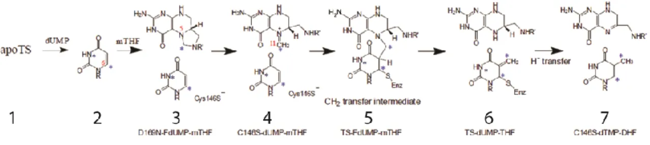
Figure 3. Breakdown of thymidylate synthase catalysis into seven trappable intermediates.
It was shown from kinetic studies that TS binds dUMP first, followed by mTHF(31). The rapid association of ligand to form a loosely bound ternary complex is followed by a slow step in which the cofactor’s imidazoline ring opens to produce the reactive iminium ion. This ring opening allows for covalent bond formation between cysteine 146 and dUMP C6 and, either simultaneously or subsequently, between dUMP C5 and the methylene group of mTHF. The methylene group is then transferred from mTHF to dUMP followed by hydride transfer to form dTMP and dihydrofolate. Escherichia coli TS, the isoform used in this work, and other isoforms of TS have been extensively studied by X-ray crystallography. There are currently 302 structures from 38 different organisms deposited in the Protein Data Bank (PDB), in which the enzyme has been crystallized with a variety of ligands and functionally relevant mutations. From these studies, the structural details of the reaction mechanism have been determined(29, 30, 32, 33). In all species, TS is an obligate dimer in which the active site is formed from residues in both subunits, at two equivalent interfaces on opposite sides of the molecule (Figure 4A). The dimer interface is comprised of two opposed six-stranded β-sheets, from which three strands form a 90° kink to form one lip of the active site (Figure 4A). dUMP binds to an essentially preformed active site flush with the three strands. dUMP binding is facilitated by hydrogen bonds to the sidechains of R126’, R166, S167, N177, H207, Y209, and R21 and R127’ which switch to different sidechain rotamers to contribute two hydrogen bonds to the phosphate of dUMP. dUMP binding is
8
maintained throughout the reaction by subtle conformational changes. mTHF then binds to the TS-dUMP complex at a conserved surface above the pyrimidine ring of dUMP (Figure 4A).
Upon cofactor binding, the four C-terminal residues close down over the active site (a shift of ~ 5 Å) and the side chains of W83 and L143 adopt different rotamer conformations to pack against the cofactor ring. The strain and/or chemical features of this conformational adjustment cause the cofactor ring to open and reorient from an extended to a folded
conformation (Figure 4D). This conformation positions the PABA moiety of the cofactor such that it is parallel and overlaps the pyrimidine ring of dUMP. This overlapping ring forms an extensive binding interface that makes the cofactor bind tighter to the TS-dUMP complex and positions the dUMP and mTHF for covalent adduct formation through C146. A crystal structure of L. casei TS suggests that initial cofactor binding utilizes a non-productive binding site prior to ring opening(34). Productive binding of mTHF in its solution conformation is sterically blocked by the protein, so mTHF initially binds such that its pterin ring is slightly out of plane with dUMP (Figure 4D). This orientation positions E58 close enough to assist in protonation of N10 of the imidazolidine ring which necessarily accompanies ring opening. Opening of the
9
accompany mTHF binding(34). This suggests that the C-terminus is flexible in pre-catalytic intermediates and that the C-terminal conformational change serves to occlude water from the binding site and enhance the reaction rate. A crystal of a mutant lacking the C-terminal valine, V262, showed a noncovalent ternary complex with an open ring can be formed without closure of the C-terminus over the active site(36). In this complex both dUMP and mTHF are in different orientations than in the productive ternary complex suggesting that C-terminal closing, and the other associated conformational changes are required to reorient the ligands but not to form the reactive intermediates.
It is interesting to note that after methylene and hydride transfer to form dTMP the enzyme is still in the closed conformation. A comparison of the ternary and product
complexes(37) shows a conserved water molecule in the ternary complex that is displaced in the product complex due to the steric clash with the methyl group of dTMP. The displacement of this conserved water by dTMP may serve as a mechanism for substrate selectivity and product release. However, there are no structural clues concerning the mechanism of opening to allow for product release.
Overall, dUMP and mTHF bind to an open, highly conserved, binding pocket which closes down progressively and helps to reorient the ligands. Apart from the large motions of the C-terminus to close the active site cavity, the majority of the conformational changes are
10
11
1.3 Allostery and dynamics in bacterial thymidylate synthase
The structural underpinnings of TS function provide evidence for dynamics as an
important modulator of function. The extensive crystal structures of TS show mostly subtle, local segmental conformational adjustments at various stages of catalysis. It is possible that the
12
Overall, very little is known about the role of dynamics in these structural rearrangements or in facilitating ligand orientation, but the wealth of mechanistic information from
crystallographic and other studies suggests that dynamics might play important roles in structural modulation of TS(30).
The other key functional aspect of TS that may have dynamic implications is the
functional allostery of this enzyme. Functional allostery in TS has been implicated for substrate and cofactor binding, and at the level of catalysis, most notably via half-the-sites reactivity(41, 42). The degree of ligand binding cooperativity is unclear as there is no consistency in measured binding stoichiometry for dUMP, FdUMP, or various folates in multiple TS species(43-48). The main concern is that binding measurements, which tend to show a stoichiometry of 1:1, contradict the structural evidence from crystallography, symmetrical dimer with full ligand occupancy in both subunits. The other complication with negative cooperativity of ligand binding is the apparent lack of conformational change upon binding of dUMP(49), a requirement for the classical allosteric models. Thus, it is hard to reconcile negative cooperativity for dUMP binding with the fact that the binding site is pre-formed in the apo enzyme (and undergoes no significant conformational change upon binding), from crystal structures. Thermodynamic and kinetic studies of nucleotide binding have shown a binding stoichiometry of one molecule of dUMP per dimer(31, 46, 47, 50) or of one high affinity site and one low affinity site(43, 51). However for FdUMP binding the reported stoichiometry is two molecules per dimer(46, 52). This is
13
concentrations the major peak shifts to a higher temperature which could be due to both subunits being saturated with dUMP or that complete saturation of one site makes both subunits more symmetric. Thermal unfolding in the presence of FdUMP also showed two peaks, but at lower temperatures and closer together so FdUMP has less of a stabilizing effect. In addition to nucleotide binding, folate binding has also been shown to be negatively cooperative, sometimes(41, 43, 45). A crystal structure of TS from Pneumocystis carinii with dUMP and a cofactor analog, CB3717, showed two molecules of dUMP bound but one molecule of cofactor analog(41). This structure showed that covalent adduct formation in one subunit causes a slight conformational change in the other subunit that prevents cofactor binding. The slight asymmetry between the two subunits in this structure suggests a potential structural mechanism for negative cooperativity. However, much of the negative cooperativity is shown for cofactor analogs or mutants, as well as different cooperativity in different species, and so the extent of cooperativity for substrate and cofactor binding remains unclear.
14
indicating that in L. casei both subunits contribute to catalysis(54). Greene et. al then showed that catalytically active heterodimers could be formed by combining E. coli and L. casei mutants, using the same mutations as before. These cross-species heterodimers were again shown to have half the activity of wild-type TS, suggesting that for L. casei TS both active sites contribute to catalysis(55). On the other hand, studies of heterodimers of E. coli TS(42, 56) showed that complete wild-type activity could be restored with a heterodimer of C146W and R126E. These findings show that while E. coli TS demonstrates half-the-sites activity, L. casei does not appear to which points to potentially interesting differences in species-specific allostery in TS.
The complexities of substrate and cofactor binding as well as the half-the-sites activity make TS an interesting enzyme to study cooperativity, catalytic allostery, and potentially how functional allostery is different in different species while the overall structure and sequences are highly conserved. This sets up the possibility that the differences in allostery could be due to differences in dynamics as a result of key insertions or sequence differences in distal sites or that are not involved in ligand binding or catalysis.
1.4 Human thymidylate synthase and drug resistance
15
Human TS is overall structurally similar to bacterial TS but has a few key differences that are hypothesized to play key roles in hTS activity and drug resistance. hTS has three major structural differences; a 29 residue extension of the N-terminus (not visible in crystal structures), a 12 residue insertion at position 114, and an 8 residue insertion at position 145 (human
numbering) (Figure 5A). hTS has also been shown to have more conformational flexibility in its apo state than bacterial TS isoforms. The most important aspect of this flexibility is the
conformational switching of the active site loop, 181-197. This loop can adopt an active
conformation in which the catalytic cysteine 195 points “in” (same conformation as in E. coli) or an inactive conformation in which C195 is rotated 180° and points “out” (Figure 5B)(57-60)
. It is thought that these two conformations are in equilibrium in the apo state and it has been shown that different conditions can favor one conformation or the other. From crystallization and fluorescence studies it was shown that under physiological conditions the inactive conformation is favored and that dUMP binding shifts the equilibrium towards the active conformation(59, 60). Importantly, phosphate binds weakly to hTS and stabilizes the inactive conformation and then dUMP binding displaces the phosphate and favors the active state. The inactive to active
transition has also been linked to the flexibility of loop 108-129, adjacent to the first insertion. In crystal structures of the inactive conformation this loop is somewhat disordered, as evidenced by high B-factors, but in the active conformation it is ordered(58, 59, 61). The active-inactive
16
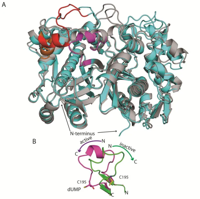
Figure 5. Structure of human thymidylate synthase. A) Comparison of E. coli (gray) and human (cyan) TS. In human the two inserts are shown in red, the active site loop (181-197) is shown in purple and loop 108-129 is shown in orange. The N-terminus extension is indicated but is missing from crystal structures. B) Active (purple) and inactive (green) conformations of loop 181-197. Arrows indicate the direction from N to C in each state. with a hydrophobic pocket near the active site(63), and an N-terminal deletion mutant was
17
suggests a new possibility for drug development in finding ways to target loop 108-129 or the N-terminus to stabilize the inactive conformation.
Drug resistance in hTS, particularly to 5-FU, is primarily mediated by increased
18
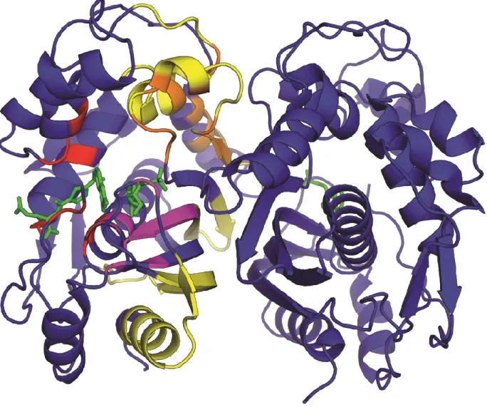
Figure 6. Regions in human TS involved in binding. Dimeric hTS is shown in blue. Residues involved in mRNA binding are shown in yellow, dUMP binding are shown in purple, cofactor binding are shown in red, and LR peptide binding are shown in orange.
1.5 Synopsis of this work
19
major focus is providing a detailed characterization of ligand binding events to conclusively demonstrate dUMP and mTHF binding cooperativity (or lack thereof). It is interesting that TS has been reported to bind dUMP with negative cooperativity but there is no evidence, from crystal structures, of dUMP-induced conformational change. Cooperativity in the absence of conformational change suggests changes in enzyme dynamics as a result of dUMP binding and so NMR will serve as a powerful tool to monitor motions on multiple timescales and potentially allow us to identify relevant motions that may be involved in cooperativity of ligand binding. My other, broad interest is to develop a general understanding of how subunits communicate across long distances. Crystal structures provide a wealth of information about the endpoints of
20
21
CHAPTER 2: STUDIES OF LIGAND BINDING COOPERATIVITY1
2.1 Introduction
Thymidylate synthase (TSase) catalyzes the synthesis of the sole source of dTMP in organisms ranging from viruses to humans(26). The mechanism involves reductive methylation of the substrate, dUMP, using a cofactor, N5,N10-methylene-5,6,7,8-tetrahydrofolate (CH2H4fol), as
both a methylene and hydride donor(23). Given its key role in DNA synthesis and cell division, TSase is an attractive drug target for treating microbial infection and cancer. As such, it has been highly scrutinized in terms of its structure and catalytic mechanism. TSase is a dimeric enzyme with two active sites, and one often cited feature is that there is allostery between the two sites, which are separated by ~30 Å. Among these reports are that TSase is a half-the-sites reactive enzyme(42, 73), the enzyme binds to only a single molecule of substrate(31), or binds it with negative cooperativity(48), and the enzyme binds to only a single molecule of cofactor(41, 43), or binds it with negative cooperativity(45). Contrary to these reports are the x-ray models of TSase, which for the case of the E. coli enzyme, have yet to capture singly bound forms. Rather, structures show symmetrical subunits with full occupancy of both active sites. These data, coupled with an NMR spectrum of substrate analog and cofactor-saturated TSase clearly showing binding to both subunits(74), are inconsistent with extreme negative cooperativity.
However, the question of cooperativity remains open because there has yet to be a rigorous study of the binding events in this key enzyme. To settle this, we measured the thermodynamics of
1
This chapter contains work previously published in Journal of the American Chemical Society. The original citation is as follows: Sapienza PJ, Falk BT, & Lee AL (2015) Bacterial Thymidylate Synthase Binds Two Molecules of Substrate and Cofactor without Cooperativity. Journal of the American Chemical Society
22
binding of substrate and cofactor to both sites of E. coli TSase. We employed isothermal titration calorimetry (ITC), which is exquisitely sensitive to strength, heat, and stoichiometry of binding, to provide the firstdetailed thermodynamic picture of the TSase-dUMP interaction. We show that E. coli TSase binds two molecules of dUMP, and unexpectedly, that both the free and singly bound forms have the same affinity for substrate. Further, our analysis highlights the challenges with analyzing multisite binding data in that very small errors in ITC cell concentration can lead to dramatically different pictures of cooperativity. Only by measuring titrations at multiple conditions and by including cell concentration as a fitted parameter were we able to obtain accurate binding parameters. For the case of cofactor binding, where heat of covalent bond formation can complicate interpretation of ITC data, we used NMR spectroscopy to directly quantify populations of all states over the course of a titration with a substrate analog and cofactor. This is a powerful approach as it provides a rare(75, 76) opportunity to monitor microscopic states in a multi-binding site system. The data show both sites are similar with respect to formation of the ternary complex, demonstrating that allostery is minimal for the two binding steps of the reaction cycle.
2.2 Materials and Methods
Expression and purification
E. coli TSase was cloned into pET21a (Addgene) and then transformed into BL21 (DE3) rne131- cells. For unlabeled ITC preparations, cells were grown in 3 mL LB media for 8 hours at 37 °C. The cells were then added to 50 mL LB and grown overnight at 37 °C. The 50 mL culture was then added to 1 L LB and grown to OD600 of 0.8 at which point 0.75 mM IPTG was
23
as described(77) with some modifications. After ammonium sulfate precipitation and DEAE chromatography, the TSase containing DEAE fractions were run over a Superdex G75 size exclusion column equilibrated with NMR buffer (25 mM NaPO4, 165 mM NaCl, 1 mM EDTA, 5
mM DTT, and 0.02% NaN3 at pH 7.5). G75 fractions containing pure TSase as determined by
SDS-PAGE were pooled. For NMR resonance assignments, deuterons were fully back
exchanged to protons as described(74). TSase activity was assayed as described(78) to determine functionality. The concentration of the TSase dimer was determined using ε280= 103,820 L•mol-1
•cm-1
. dUMP and 5F-dUMP were from Sigma and their concentrations determined using ε262=
9,660 L•mol-1•cm-1
. CH2H4Fol was purchased from Merck & Cie (Switzerland) and its concentration determined using ε290 = 32,000 L•mol-1•cm-1.
ITC
TSase was aliquoted into 10 mg samples and buffer exchanged into the appropriate ITC buffer (25 mM Sodium Phosphate, 1 mM EDTA, 2 mM TCEP, pH 7.5 at 5, 10, 15, 25, or 30 °C ) over a Superdex G25 size exclusion column. The protein samples were concentrated to 100, 200, or 300 µM as determined by UV spectrophotometry. For titrations with multiple protein
concentrations, the high concentration protein stock was aliquoted and diluted appropriately to a lower concentration. The samples were centrifuged at 11,000 RPM for one minute to remove any aggregates and the concentrations were again measured before loading into the ITC. Dry dUMP was dissolved in the appropriate ITC buffer to a concentration of 20, 30, or 40 molar excess of protein concentration. The concentration of the dUMP stock was then measured
24
mM sodium phosphate, HEPES, Tris, or TES at pH 7.5 at 25 °C with ionic strength fixed to 0.1 M with NaCl. ITC experiments were conducted on a MicroCal AutoITC 200. All experiments had an initial 0.2 µL injection followed by 19 2 µL injections.
ITC Binding Models
Single set of identical sites
( 1 )
Where Qk is the total heat after k injections, V0 is the cell volume, and Mt and Xt are the bulk
protein and ligand concentrations, respectively, in the cell volume. The heat associated with injection k, qk, is the difference in total heat between injections k and k-1, after applying a heat
correction for the heat contribution of the excluded volume v:
( 2 )
General binding model
To account for non-equivalent binding affinities and/or non-equivalent ΔH° of interaction with the two sites, we employed a general model based on the binding polynomial formalism(75). The binding polynomial, P, for two site binding is:
( 3 )
where [X] is the free ligand concentration and βi represent overall association constants, which
are related to the step-wise association constants by the following:
( 4 )
25
In the general model, the total accumulated heat after injection k, Qk, is:
( 6 )
and the heat associated with injection k, qk, is:
( 7 )
Modified general binding model
Because the general model assumes integral values of stoichiometry, errors in the cell concentration can have significant effects on the fitted parameters (Table 1). Thus, our modified general model includes cell concentration as a fitted parameter. In our case, the fitted [cell] was always less (by ~10%) than that determined by UV spectroscopy and this correction was the same for series of experiments that used the same enzyme preparation. Lastly, it is important to note that K1 and K2 do not distinguish which sites are occupied, but simply the total number of
sites that are occupied, and as such are phenomenological binding affinities. Thus the fitted values for K1 and K2 must be corrected for statistical degeneracy (the first ligand has more empty
sites to bind than the second), to yield intrinsic binding affinities, which are reported in this work. The relationship between the phenomenological and intrinsic binding constants is given by:
( 8 )
The 1H-15N TROSY HSQC of apo TSase supports use of intrinsic binding constants in the case of dUMP binding to the free enzyme because the spectrum yields a single set of
26
Figure 7. 1H-15N TROSY HSQC of the Apo state of TSase shows subunit symmetry. Apo spectra shows a single set of resonances (1 for each backbone amide), which is consistent with a symmetrical homodimer. This supports our treatment that the first dUMP molecule encounters a pair of degenerate sites.
All three ITC fitting models were implemented using custom MATLAB scripts in which the following error function vector, f, was passed to lsqnonlin, with the algorithm set to
27
( 9 )
where the number of elements in k is equal to the number of injections in single or globally fit datasets and σ is the error in the heat of injection, which was ~ 0.2 μcal based on average values of residuals found for the best fitting modified general model.
Assessing cooperativity
For a given protein with n binding sites, a set of n-1 cooperativity parameters, ρi, can be
calculated from the phenomenological binding constants(75).
( 10 )
In general ρ<1 suggests negative cooperativity or nonidentical binding sites, ρ=1 it suggests no cooperativity, and ρ>1 suggests positive cooperativity or nonidentical binding sites. For a system with two binding sites ρ is given by:
( 11 )
Proton linkage
The observed ΔH from a titration, ΔHo
, is linearly related to the number of protons exchanged between the buffer and the protein, n, and the heat of buffer ionization, ΔH°b, with
intercept equal to the intrinsic enthalpy of ligand binding in a buffer with no heat of ionization, ΔH°i(79).
28
NMR resonance assignments, titrations, and fitting
Apo resonance assignments were completed using a series of six TROSY triple resonance experiments as described previously(74). The 5F-dUMP-CH2H4Fol di-ligand titration was
monitored by 1H-15N TROSY HSQC spectra. Data were acquired at 25 °C on a 600 MHz Bruker Avance III spectrometer equipped with a TCI cryogenic probehead. Datasets were recorded with (150, 1024) complex points, acquisition times of (82 ms, 85 ms) in (t1, t2), 8 scans per complex
t1 point, and a recycle delay of 2 s. Non-uniform sampling was used in t1 with 50% sampling
density. Spectra were reconstructed using iterative soft thresholding(80) and we conducted tests to show that non-uniform sampling had no effect on relative resonance intensities (Figure 8).
29
sampling density, which is the same scheme used to collect data in Figure 14and Figure 16 ) in t1. Resonance intensities were measured and compared in the figure above showing that NUS does not affect relative intensities under these conditions.
In this titration, resonances are in slow exchange. For a subset of amino acids, peak positions of the singly bound states are different than the free and doubly bound state, which allowed us to quantify the populations of all species. These residues yield a quartet of resonances at intermediate titration points: one for the free enzyme, one for the doubly bound enzyme, one for the free subunit of the singly bound enzyme and one for the bound subunit of the doubly bound enzyme (Figure 14A and B). To determine di-ligand association constants, resonance intensities were measured in NMRViewJ(81). To normalize the populations on a per-mole of dimer basis, the free and doubly bound intensities were divided by two. Further, because there are significant structural differences between the free and bound enzyme, different relaxation properties could influence the intensities coming from the singly bound species. To account for this, the resonances from the singly bound forms were normalized to their “like” symmetrical forms. Specifically, the free subunit of the singly bound species was scaled to the initial point of the titration and the resonance from the bound subunit of the singly bound species was scaled to the saturated, doubly bound point of the titration. Explicitly stated:
I0 = Intensity from the apo state
I0,1 = Intensity from the free subunit of the singly bound state
I1,0 = Intensity from the bound subunit of the singly bound state
I2 = Intensity from the doubly bound state
F0 = I0/ I0,initial
30
Where F values are population fractions, I0,initial is the intensity from the first (100% free) point of the titration, I2,final is the intensity from the last (100% bound) point of the titration, and the factor of 0.5 is present because two subunits contribute to the intensities of I0 and I2. Resonance
intensities were then fit to the general binding polynomial for two site binding ( 3 ) using custom MATLAB scripts where the fraction of free, singly bound, and doubly bound enzymes are expressed by equations ( 13 )( 14 ) and ( 15 ) respectively.
( 13 )
( 14 )
( 15 )
2.3 dUMP binding cooperativity
Given the general interest in the phenomenon of allostery and the question of dUMP binding in TSase, we set out to probe the binding thermodynamics of this dimeric system.
Thermodynamics of substrate binding was measured by ITC at 25 °C (Figure 9A and Figure 10).
Figure 9 ITC measurement of dUMP binding to TSase. Conditions are 290 μM TSase in the cell and 6 mM
dUMP in the syringe, both in 25 mM NaPO4, 1 mM EDTA, and 2 mM TCEP, pH 7.5. (A, B) Fits are shown for dUMP titrations using models for one-site binding (red line against closed circles), general two-site binding (blue line against closed circles), and modified general binding (black lines against open circles); insets show residuals for one-site binding (red circles), general two-site binding (blue squares), and modified general binding (black
31
versus T yields ΔC°P=-157 cal/mol K and ΔC°P=-183 cal/mol K for binding to free and singly bound TSase,
respectively. Errors in parameters were determined from Monte Carlo simulations and the error bars are smaller than the points. Values for fitted parameters from the modified general model are shown in Table 2.
Figure 10 Global fits of paired titrations with multiple protein or multiple dUMP concentrations at 25 °C to the modified general binding model. A) Multiple protein concentrations, 206 (○) and 98 µM (●), were titrated with 4.37 mM and 1.99 mM dUMP, respectively, giving a globally fit ρ=1.1. B) Multiple dUMP concentrations, 4 mM (○) and 3 mM (●), were titrated into 105.9 µM protein, giving a globally fit ρ=1.07.
The data fit well to a single site model with a stoichiometry (n) of 1.8 (Figure 9A and Table 1), but based on reports of cooperativity in this(31) and other forms(48) of the enzyme, the data were also fit to a general model that can accommodate differences in affinities and heats between the two binding events. The general model fit to intrinsic KD,1 of ~4 µM and KD,2 of ~
20 μM, for a ρ-value (Ratio of KA2/KA1) of 0.22 (Figure 9A and Table 1). This suggests negative
cooperativity, but a comparison of reduced Χ2 indicates the single model is a better fit to the data (Figure 9A, (Χ2 values for all models are compared in Table 1). Even though the single sites model gives the superior fit, both models make assumptions that might not be accurate for this system. The main assumption with the single sites model is that each of the n sites has the same K and ΔH° and it could be the case that K1≠K2 and/or ΔH°1≠ΔH°2. The potential pitfall of the
general model is that all of the calculated and fitted parameters are dependent on a fixed n (n=2 in the case of TSase). The fact that the best fitting single site model gave a non-integral
32
taken into account by the general multi-site model within the Origin ITC package, could lead to erroneous fits.
Table 1. Goodness of fit parameters from fitting the ITC temperature series to three modelsa in 25 mM NaPO4, pH 7.5.
Single Site Model General Model Modified General Model
T (° C) n b ρ
b ρ b
5 1.47 277 0.62 ± 0.05 26 0.80 ± 0.06 9
10 1.66 259 0.40 ± 0.04 179 0.82 ± 0.1 17
15 1.68 78 0.29 ± 0.06 285 0.91 ± 0.04 37
25 1.80 28 0.22 ± 0.03 486 0.63 ± 0.01 15
25c 1.69 123 0.32 ± 0.07 1327 1.1 ± 0.01 24
25d 1.83 98 0.55 ± 0.08 105 1.07 ± 0.01 98
30 1.89 70 0.48 ± 0.07 93 0.93 ± 0.03 72
To overcome these issues, the data were fit using a general binding model that included cell concentration as a fitted parameter. Treating protein concentration as an adjustable parameter is reasonable given the possibility that the active fraction of TSase is less than 100%, and
potential discrepancy between actual and theoretical extinction coefficients. Further, fitting protein concentration within the general model was employed previously with other multi-binding site systems(82, 83). This is a rigorous fitting approach because the only assumption is the total number of binding sites. The assumption is justified here by the x-ray model of the E. coli TSase-dUMP complex in which both sites have full occupancy(49).Fits to this modified general model (Figure 9A) gives ρ≈1, a lower reduced Χ2 than either the single or unmodified general
a
See Section 2.1 for model descriptions.
b
Goodness of fit parameter, , where υ is the number of degrees of freedom (υ = N – n – 1, N is number of observations and n is the number of fitted parameters), and σ is the error in the heat of injection.
c
Global fit of two datasets: first with [TSase] = 98 μM, [dUMP] = 1.99 mM; second with [TSase] = 206 μM, [dUMP] = 4.37 mM. See Figure 10Afor fit to modified general model.
d
33
models (Table 1), and a fitted protein concentration 10% lower than that measured by UV spectroscopy. To ensure that the improved Χ2 associated with the modified general model is not simply the result of over-fitting, we doubled the ratio of observables to fitted parameters by performing global fits to paired titrations with either two cell or syringe concentrations. This approach was shown previously to break degeneracies and increase robustness of fitted ITC parameters(84). Global fits to the paired titrations described above yield ρ≈1 (Figure 10 and Table 1) in support of using the modified general model and the conclusion that binding affinities are similar. This analysis underscores the importance of accounting for inaccuracies in ITC cell concentration as errors of even 10% here can lead to a misinterpretation of up to 4-fold negative cooperativity when binding sites are truly identical (Table 1).
Because the heat capacity change upon binding is a sensitive probe of changes in
structure and dynamics upon binding(85), we looked at dUMP binding at additional temperatures. The data fit poorly to the single site model at some temperatures other than 25 °C (Figure 9B, Figure 11, and Table 1), indicating that either cooperativity is temperature dependent, or that ΔH°1 and ΔH°2 are not equivalent at all temperatures. The data were then fit to the modified
general model which, for cases where ΔH°1 ≠ ΔH°2, fits significantly better than either the single
or un-modified general models (Figure 9B, Table 1, Figure 11). Data for all five temperatures showed both active sites have nearly equivalent binding affinity (Figure 9C, Table 1, and Table 2) and all datasets required a similar correction to enzyme concentration, which is expected if the enzyme originates from the same preparation. In contrast to binding affinities, ΔH° for the two binding events diverge as a function of temperature (Figure 9C and Table 2).
34
Figure 11 Single site and modified general fits to temperature titration. Comparison of the best fits to the single site (--,○) and modified general (˗˗, ●) models at multiple temperatures. Insets compare the residuals for each model. Values for fitted parameters are shown in Table 1. A) 10 °C B) 15 °C C) 30 °C. Note that example fits for 5 and 25 °C are in Figure 9. Fitted parameters for the entire temperature series are in Table 2.
The slope of ΔH° versus T yields a ΔC°P of -157 ± 1 cal/mol•K for site 1 and -183 ± 2
35
Table 2 Thermodynamic parametersa for binding of dUMP to TSase at pH 7.5 in 25 mM phosphate buffer.
Temperature (°C) K1 x10 4
(M-1) K2 x10 4
(M-1) ΔH°1 (kcal/mol)
ΔH°2 (kcal/mol)
ρ
5 7.7 ± 0.8 6.2 ± 1.0 -1.2 ± 0.01 -0.58 ± 0.01 0.80 ± 0.06 10 6.8 ± 1.0 5.3 ± 1.2 -2.0 ± 0.02 -1.5 ± 0.02 0.82 ± 0.1 15 6.1 ± 0.2 5.6 ± 0.2 -2.6 ± 0.01 -2.3 ± 0.04 0.91 ± 0.04 25b 6.0 ± 0.1 5.9 ± 0.1 -4.5 ± 0.01 -4.4 ± 0.02 0.98 ± 0.08 30 3.8 ± 0.1 3.5 ± 0.1 -5.1 ± 0.02 -5.2 ± 0.05 0.93 ± 0.03
To determine if the differences in ΔH° were the result of proton exchange with the solution upon dUMP binding, titrations were conducted in a series of four buffers with different heats of buffer ionization, ΔHb. The chosen buffers were phosphate, HEPES, TES, and Tris in
order of increasing ΔHb(86). The measured ΔH° from ITC is linked to the heat of ionization of the
buffer (ΔH°b) and is related to the number of protons exchanged during binding(79).
Figure 12Minimal proton linkage accompanies dUMP binding to TSase. Modified general model was used to fit ITC experiments in multiple buffers with different enthalpies of ionization (ΔH°b). Buffer color key in (C) applies to all panels. In panels A, B, and D, sites 1 (2) are represented by filled (open) bars/symbols. Slopes of lines in panel D give number of protons (n) linked to binding. Site 1 (●) fits to n of -0.09 ± 0.02 protons and ΔHi of -4.8 ± 0.17 kcal/mol. Site 2 (○) fits to n of -0.06 ± 0.03 protons and ΔHi of -4.5 ± 0.19 kcal/mol. Example fits are given in Figure 13, with values in Table 3.
a
Parameters are from individual fits to the modified general binding model and Monte Carlo error estimation. K1 and K2 are the intrinsic binding constants.
b
36
The data were fit to the general binding model with adjustable protein concentration, and fit with ρ ≈ 1 for all buffers (Figure 12, Figure 13, and Table 3), a convergence which further supports this fitting model.
Figure 13 dUMP titrations in multiple buffers fitted with the modified general model. Data were collected on protein samples, at 100 or 200 µM in each buffer, originating from a single purification. Buffers used were 25 mM sodium phosphate, HEPES, TES, and Tris pH 7.5 at 25 °C all with ionic strength fixed to 0.1 M with NaCl. Values for fitted parameters are shown in Table 3. A) 100 µM TSase in phosphate with 1.93 mM dUMP. B) 200 µM TSase in HEPES with 4.19 mM dUMP. C) 200 µM TSase in TES with 4.24 mM dUMP. D) 105 µM TSase in Tris with 2.18 mM dUMP.
37
slopes of ΔH° versus ΔH°b (Figure 12D) indicate that less than 0.1 mol of H+ are taken up by the
protein upon dUMP binding and H+ linkage does not play a large role in dUMP binding.
Table 3 Thermodynamic parameters for binding of dUMP to TSase in multiple buffers to assess proton linkage.
Buffera K1 x104 (M-1)b K2 x104(M-1)b ΔH°1 (kcal/mol) ΔH°2 (kcal/mol) ΔH°b (kcal/mol) ρ
Phosphatec 6.1 ± 0.12 6.5 ± 0.14 -4.9 ± 0.02 -4.7 ± 0.08 0.86 1.07 ± 0.04 HEPES 17.9 ± 0.26 18.9 ± 0.28 -5.5 ± 0.01 -4.8 ± 0.02 4.875 1.06 ± 0.03 TES 13.8 ± 0.30 16.3 ± 0.35 -5.3 ± 0.02 -4.7 ± 0.04 7.679 1.17 ± 0.07 Tris 15.7 ± 0.38 17.2 ± 0.42 -5.8 ± 0.02 -5.3 ± 0.06 11.34 1.09 ± 0.05
2.4 Cofactor binding cooperativity
It is possible that cooperative effects alternatively reside at the cofactor binding step. Substrate binding results in only modest conformational changes in TSase(30). Addition of
cofactor or cofactor analog causes more dramatic rearrangements in the local binding site(30) that could influence the neighboring subunit. This, coupled with reports of cofactor analog binding cooperativity in human(45) and bacterial TSase(41, 43) prompted us to further investigate cofactor binding. We chose to monitor binding of the substrate analog, 5F-dUMP, and the biological cofactor, CH2H4Fol by NMR titration. In the resulting ternary complex, C146 makes a covalent
bond to 5F-dUMP, which is covalently attached via a methylene bridge to the cofactor(87); the covalent attachments make it such that the two small molecules can be treated as a single “di-ligand”. Importantly, the di-ligand is considered a mechanism-based inhibitor as these covalent bonds are formed during the normal reaction cycle(23). Therefore, the complex is an excellent model for cooperativity in ternary complex formation during catalysis. Further, structures of this complex are isosteric with other complexes involving dUMP and cofactor analogs(88, 89). Lastly, a Conditions are 25 mM buffer, 1 mM EDTA, 2 mM TCEP, with ionic strength of 0.1 M NaCl, pH 7.5 and 25 °C. b Intrinsic binding constants from fits to the modified general model.
c
38
the stability of the complex yields high quality NMR spectra with resonances that are in slow-exchange on the NMR timescale, facilitating the quantification of all species.
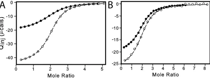
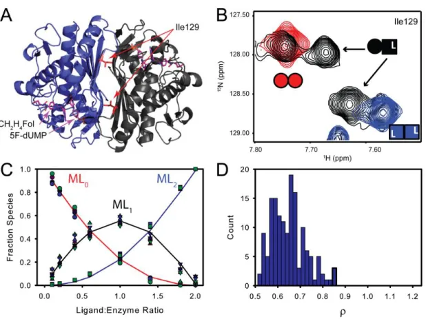
Titrations of TSase with the di-ligand were monitored by TROSY-1H-15H HSQC spectra. A subset of residues at the dimer interface yielded two resonances corresponding to the singly bound state that are distinct from symmetrical free and doubly bound resonances (Figure 14A and B).
Figure 14 Both TS active sites have similar affinity for the 5F-dUMP-CH2H4Fol “di-ligand”. A&B) In NMR spectra, at intermediate titration points (black, (B)), resonances from a subset of residues near the dimer interface (e.g. Ile 129 in (A)) have chemical shifts from the singly bound state that are different from the free (red, Panel (B)) and doubly bound states (blue (B)). For these residues there are four resonances total at intermediate titration points as the singly bound state produces two peaks: one from the free subunit and one from the bound subunit (B). C) Global fit of peak intensities from the four resonances having all three states resolved in NMR spectra. Circles and squares represent free and doubly bound data, respectively. Upward triangles are from the free subunit of singly bound species and downward tringles are from the bound subunit. D) Histogram of ρ (K2/K1 ratio) from fits of 150 Monte Carlo simulated datasets.
39
residues were fit to the two-site binding polynomial. At the limit of stoichiometric binding, which is observed in this case, we cannot determine the absolute binding affinities. However, differences in the relative binding affinities between the two sites are readily apparent (Figure 15).
Figure 15 Relative but not absolute binding affinities can be measured under the stoichiometric binding regime. Simulated data using the two site binding polynomial (equation ( 3 )) at the three conditions listed above (intrinsic binding constants). ML0, ML1, and ML2 represent the free, singly bound, and doubly bound species, respectively. The simulations demonstrate that for tight binding, which is expected for the covalent di-ligand used in this work, different strengths of binding where K1 = K2 cannot be differentiated (black and red-dashed lines). However, the shapes of the curves are dramatically different for K1 ≠ K2 (blue lines) vs. K1 = K2 (black and red-dashed lines). Thus, cooperative vs non-cooperative binding are readily distinguished from one another.
In the case of di-ligand binding to TSase, the ρ-values range from 0.55 to 0.90 for fits of single residue data (Figure 16 and
40
Table 4), which indicates a slight degree of negative cooperativity with a maximum magnitude of less than two-fold.
Table 4 Relative binding constants for TSase 5F-dUMP-CH2H4Fol di-ligand binding from NMR titration.
Residue ρ
Gln33 0.88 ± 0.067
Ile129 0.90 ± 0.12
Asn134 0.57 ± 0.080
Unassigned Trp Indole 0.77 ± 0.14
41
Figure 16 Local fits of 5F-dUMP-CH2H4Fol di-ligand titration data. Each row represents data from a single residue. In the first column, spectra from apo (red), saturated (blue), and titration intermediate point (black) are shown. Fits are in the second column. Note consistent color scheme in the first and second columns and that there are two observations for the singly bound species: one from the free subunit and one from the bound subunit (see
Methods). The final column shows ρ values (K2/K1) from fits to 150 Monte Carlo simulated
datasets where the error is based on the average residuals from fit to experimental data. Mean and standard deviations of ρ from the local and global fits are shown in
Table 4.
2.5 Conclusions
The data presented herein unequivocally show that substrate binds to the free and singly bound forms of E. coli TSase with similar affinity. This finding contrasts with a previous
42
measured by ITC. It is noteworthy that while dUMP binding affinities are the same in both the free and singly bound enzyme, ΔH°1 and ΔH°2 are not equivalent at some temperatures and in
some buffers. This phenomenon, in which binding is similar at the level of ΔG°, but different based on ΔH° and TΔS°, was termed “silent allosteric coupling”(90)
. The recent linkage between binding and re-distribution of side-chain dynamics(91-94) and the connection between side-chain dynamics and conformational entropy(92, 95, 96) suggests this type of coupling is wide-spread.
We also show by NMR, that the ternary complex is formed with nearly equal probability in both subunits which disagrees with an ITC study showing roughly one molecule binds to dimeric E. coli TSase(43). It is difficult to compare our data with the published ITC study because the fitting models are not described in detail and our work provides a dramatic example of how choice of fitting model can affect interpretation of ITC in multi-binding site systems. However, our data are in agreement with crystal structures(49, 87, 89, 97) and NMR spectra(74) that show two molecules of cofactor are bound. Interestingly, binding the first di-ligand does elicit chemical shift changes in the empty subunit (Figure 14B), leading us to conclude that the effects of