The role of NF-κB in BCR-ABL-driven oncogenic transformation and in hematopoiesis
Sarah J. Stein
A dissertation submitted to the faculty of the University of North Carolina at Chapel Hill in partial fulfillment of the requirements for the degree of Doctor of Philosophy in the
Department of Biology
Chapel Hill 2012
Approved by:
Dr. Albert Baldwin
Dr. Gregory Coperhaver Dr. Ian Davis
Dr. Charles Perou
ABSTRACT
SARAH J. STEIN: The role of NF-κB in BCR-ABL-driven oncogenic transformation and in hematopoiesis
(Under the direction of Dr. Albert Baldwin)
Chronic myeloid leukemia is a malignant clonal disorder of hematopoietic stem cells
that results in increased and deregulated growth of myeloid cells. About 95% of CML cases
arise from the formation of the Philadelphia (Ph) chromosome, resulting in the expression of
BCR-ABL, an oncoprotein with constitutive tyrosine kinase activity that drives oncogenesis.
Current therapeutics for CML includes the BCR-ABL inhibitor, imatinib, which results in
molecular remission in only 4% of patients. Drug resistance also occurs in CML through
point mutations in BCR-ABL itself, or through the upregulation of secondary signaling
pathways. Therefore, insight into novel therapeutic targets is needed for the treatment of
CML. Previous work has shown that BCR-ABL expression induces the activation of a
dimeric transcription factor, NF-κB, and that its activity is required for efficient cellular
transformation by BCR-ABL. Here, we show that BCR-ABL utilizes NF-κB for survival of
transformed cells, and for transformation to initiate to disease in vivo. Due to the role of
NF-κB activity in the development and function of numerous hematopoietic cell types, we also investigate the role of classical NF-κB subunit, p65, in normal hematopoiesis. Results
indicate that the loss of p65 in the hematopoietic compartment leads to defects in
hematopoiesis due to loss of hematopoietic stem cell homeostasis and the activation of
of hematopoiesis. These data indicate that therapeutics involving the inhibition of NF-κB,
DEDICATION
TABLE OF CONTENTS
LIST OF TABLES ... viii
LIST OF FIGURES ... ix
LIST OF ABBREVIATIONS ... xiii
CHAPTER I. INTRODUCTION ... 1
1.1 Summary ... 1
1.2 NF-κB signaling in inflammation and disease ... 2
1.3 Hematopoiesis ... 5
1.4 Mouse models of NF-κB deletion ... 10
1.5 NF-κB in hematopoiesis ... 13
1.6 Chronic myeloid leukemia ... 15
1.6.1 Drug resistance in chronic myeloid leukemia ... 16
1.6.2 Leukemia stem cells ... 18
1.6.3 NF-κB in chronic myeloid leukemia ... 19
1.7 Conclusions ... 20
II. IKKβ INHIBITION INDUCES CELL DEATH IN IMATINIB-RESISTANT AND T315I DASATINIB
RESISTANT BCR-ABL+ CELLS ... 27
2.1 Abstract ... 27
2.2 Introduction ... 28
2.3 Materials and Methods ... 31
2.4 Results ... 33
2.5 Discussion ... 38
References ... 47
III. NF-κB SUPPRESSES ROS LEVELS IN BCR-ABL+ CELLS TO PREVENT ACTIVATION OF JNK AND CELL DEATH ... 50
3.1 Abstract ... 50
3.2 Introduction ... 51
3.3 Material and Methods ... 54
3.4 Results ... 56
3.5 Discussion ... 62
References ... 75
IV. INVESTIGATION OF NF-κB AS A THERAPEUTIC TARGET IN CHRONIC MYELOID LEUKEMIA ... 79
4.1 Abstract ... 79
4.2 Introduction ... 80
4.3 Material and Methods ... 82
4.4 Results ... 84
4.5 Discussion and Future Direction ... 86
V. THE ROLE OF THE NF-κB SUBUNIT p65 IN HEMATOPOIESIS ... 97
5.1 Abstract ... 97
5.2 Introduction ... 98
5.3 Material and Methods ... 100
5.4 Results ... 103
5.5 Discussion and Future Directions ... 110
References ... 129
VI. COMPENSATORY JAK/STAT SIGNALING IN THE ABSENCE OF THE NF-κB SUBUNIT p65 ... 133
6.1 Abstract ... 133
6.2 Introduction ... 134
6.3 Material and Methods ... 137
6.4 Results ... 139
6.5 Discussion and Future Directions ... 142
References ... 153
VII. CONCLUSIONS & FUTURE DIRECTIONS ... 156
7.1 Conclusions ... 156
LIST OF TABLES
Table
1.1 Mouse models of NF-κB deletion ... 11 5.1 Specific genes affected by the loss of p65 in HSCs ... 123 Supplementary table
LIST OF FIGURES
Chapter I
1.1 The classical and alternative NF-κB signaling pathways. ... 3
1.2 Model of hematopoietic development hierarchy ... 8
Chapter II
2.1 IKKβ inhibitor CpA blocks phosphorylation of IκBα
in BCR-ABL-expressing cells. ... 41
2.2 IKKβ inhibition suppresses growth/viability of BCR-ABL+ cells ... 42
2.3 WeHi-conditioned media rescues BCR-ABL+ cell susceptibility to tyrosine kinase inhibitors but not to IKKβ inhibition. Inhibition of BCR-ABL kinase activity
promotes rescue by WeHi media. ... 43
2.4 BCR-ABL+ cells undergo cell death in response to
tyrosine kinase inhibitors or IKKβ inhibition ... 44
2.5 BCR-ABL+ cells exhibit G0-G1 accumulation in response
to Imatinib or Dasatinib treatment, but not IKKβ inhibition ... 45
2.6 Cells expressing tyrosine kinase inhibitor resistant BCR-ABL
mutants are susceptible to IKKβ inhibition ... 46
Chapter III
3.1 Inhibition of IKKβ causes apoptosis in BCR-ABL-expressing
myeloid cells. ... 66
3.2 NF-κB activity is required for survival of BCR-ABL-expressing cells ... 67
3.3 Inhibition of NF-κB causes an increase in intracellular
ROS in cells expressing BCR-ABL ... 67
3.4 IKKβ inhibition downstream of BCR-ABL induces JNK
activation and apoptosis ... 68
3.5 NF-κB regulates antioxidant gene expression in
3.6 JNK activation in the absence of NF-κB activity leads to
apoptosis in BCR-ABL-expressing cells ... 70 3.7 Antioxidants rescue BCR-ABL-expressing cells from death
when NF-κB is inhibited ... 71 3.8 NF-κB activity is required for the survival of
BCR-ABL-expressing cells through the regulation of ROS
and JNK ... 72 Chapter IV
4.1 The leukemia stem cell hypothesis ... 90 4.2 Characterization of p65 expression in p65hem-/- mice. ... 90 4.3 p65 expression is require for BCR-ABL-expressing HSPCs
to give rise to disease in vivo ... 91 4.4 p65 expression is required for efficient BCR-ABL-driven
transformation of HSPCs ... 92 4.5 IKKβ inhibition effectively impedes colony formation by
BCR-ABL-expressing HSPCs in vitro ... 93 4.6 Experiment design for the genetic deletion of p65 in animals with CML ... 94 4.7 Experiment design to determine the efficacy of compound A
treatment of CML stem cells in vivo ... 99 Chapter V
5.1 Characterization of p65 hematopoietic null mice ... 116 5.2 p65 deletion results in deregulation of hematopoietic stem
and progenitor homeostasis ... 117 5.3 Loss of p65 results in the accumulation of hematopoietic
stem cells in the spleen ... 118 5.4 p65 is not required for self-renew or differentiation of HSPCs ... 119 5.5 Deletion of p65 greatly reduces the repopulation ability of
5.6 The accumulation of hematopoietic stem cells in the absence
of p65 is cell intrinsic ... 121
5.7 p65 is an important regulator of gene expression in HSCs ... 122
Chapter VI 6.1 The JAK/STAT signaling pathway ... 146
6.2 Characterization of p65 hematopoietic null mice ... 146
6.3 Deletion of p65 in vivo causes splenomegaly and granulocytosis ... 147
6.4 IKKβ inhibition results in splenomegaly and granulocytosis ... 148
6.5 Loss of p65 results in phosphorylation of Stat3 and alteration of Jak2 ... 149
6.6 p65 null mouse embryonic fibroblasts display alteration of Jak2 ... 150
6.7 Model of the Jak2 cleavage hypothesis ... 151
6.8 Jak2 alteration may be due to cleavage by an unknown protease ... 151
6.9 Jak2 localization is altered in p65-/- mEFs ... 152
Supplemental Figures
3.1 IKKβ inhibition induces JNK activation and apoptosis
in Ba/F3 cells expressing p210 BCR-ABL ... 73 3.2 Antioxidant treatment rescues Ba/F3 cells expressing
LIST OF ABBREVIATIONS ABL Abelson kinase
B6 C57BL/6J “Black 6” mouse strain
BCR Breakpoint cluster region
CLP Common lymphoid progenitor
CML Chronic myeloid leukemia
CMP Common myeloid progenitor
CpA Compound A, a small molecule inhibitor of IKKβ
Cre Cre recombinase enzyme
EdU 5-ethynyl-2´-deoxyuridine, a thymidine analog which is incorporated into DNA during active DNA synthesis
G-CSF Granulocyte colony stimulating factors
GFP Green fluorescent protein
GM-CSF Granulocyte/monocyte colony stimulating factor
GMP Granulocyte/monocyte precursor
HSC Hematopoietic stem cell
HSPC Hematopoietic stem and progenitor cell
IκB Inhibitor of κB
IKK IκB kinase
IL-3 Interleukin 3
IL-6 Interleukin 6
JAK Janus kinase
lin Lineage
LPS lipopolysacchride
LSK Lineage-Sca-1+c-kit+ fraction of bone marrow cells
LT-HSC Long-term hematopoietic stem cell
mEF Mouse embryonic fibroblast
MEP Megakaryocyte/erythrocyte progenitor
MG132 Carbobenzoxy-L-leucyl-L-leucyl-L-leucinal, a proteasome inhibtor
MPP Multi-potent progenitor
p65hem-/- Mice in which the NF-κB subunit p65 has been deleted from the hematopoietic compartment
RBC Red blood cell
ROS Reactive oxygen species
SCF Stem cell factor
SH2 Src-homology domain
SLAM Signaling lymphocyte activation molecule; family of surface molecules that can be used to identify hematopoietic stem and progenitor cells
ST-HSC Short-term hematopoietic stem cell
STAT Signal Transducers and Activators of Transcription
TAD Transcriptional activation domain
CHAPTER I INTRODUCTION
1.1 Summary
Chronic myeloid leukemia (CML) is initiated by the expression of BCR-ABL, an oncoprotein with constitutive tyrosine kinase activity that drives oncogenesis. Current therapeutics for CML include the BCR-ABL inhibitor, imatinib, which results in complete hematologic response in 98% (Druker et al., 2006), but molecular remission in only 4%, of patients (Hughes et al., 2003). Drug resistance also occurs in CML through point mutations in BCR-ABL itself, or through the upregulation of secondary signaling pathways. Therefore, insight into novel therapeutic targets is needed for the treatment of CML. Previous work has shown that BCR-ABL expression induces the activation of a dimeric transcription factor, NF-κB, and that its activity is required for efficient cellular transformation by BCR-ABL (Reuther et al., 1998). Here, we show that BCR-ABL utilizes NF-κB for survival of transformed cells, and for transformation to initiate to disease in vivo.
Due to the role of NF-κB activity in the development and function of numerous hematopoietic cell types ((Bottero et al., 2006; Gerondakis et al., 1999); see Table 1), we also investigate the role of classical NF-κB subunit, p65, in normal hematopoiesis. We show that loss of p65 in the hematopoietic compartment of animals leads to skewed cell
distribution, extramedullary hematopoiesis, and activation of alternative signaling pathways. p65 null hematopoietic stem cells (HSCs) show defects in repopulation ability, due to
in proliferation to increase proliferative capacity. Together, these data show that p65 is an important regulator of hematopoiesis.
1.2 NF-κB signaling in inflammation and disease
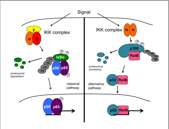
NF-κB is an inducible family of dimeric transcription factors comprised of five family members: p65 (RelA), RelB, c-Rel, p50/p105 (NF-κB1) and p52/p100 (NF-κB2). These proteins share a conserved N-terminal Rel homology domain, which controls DNA binding, dimerization and interaction with inhibitory IκB proteins. NF-κB is held in an inactive state by IκB. NF-κB is typically activated through one of two distinct pathways. In the classical pathway, the p50/p65 heterodimer is induced through the activation of the IκB kinase (IKK) complex, which contains two catalytic subunits, IKKα and IKKβ, and a
regulatory subunit, IKKγ. IKK phosphorylates IκBα, causing it to become ubiquitinated and subsequently degraded, allowing NF-κB to translocate to the nucleus. In the alternative pathway, IKKα homodimers are activated and subsequently phosphorylate p100. This results in the proteolytic processing of p100 to p52 and allows p52/RelB to translocate to the
nucleus. p65, RelB and c-rel each contain a transactivation domain (TAD) near the C-terminus, which facilitates the recruitment of coactivators. p52 and p50 lack a TAD and binding of homodimers to promoter regions can lead to repression of gene expression
(Hayden and Ghosh, 2004). Once in the nucleus, NF-κB is known to regulate the expression of a variety of genes, including those encoding cytokines and cytokine receptors,
Figure 1.1. The classical (left) and alternative (right) NF-κB signaling pathways.
Multiple signaling pathways lead to the activation of NF-κB. Generally, these
pathways activate the IKK complex, resulting in degradation of IκB or modification of p100, allowing NF-κB to enter the nucleus and regulate gene expression. In response to
inflammation, NF-κB is known to promote cell survival through the transcription of antiapoptotic genes, such as the cIAPs, which directly inhibit caspases, and genes that
indirectly inhibit apoptosis through the regulation of other signaling pathways. For example, the TNFα signaling pathway results in rapid activation of NF-κB through the classical pathway upon binding of TNFα to its receptor. NF-κB then inhibits the activation of c-jun N-terminal kinase (JNK), which can promote apoptosis in response to prolonged activation (Hayden and Ghosh, 2004). Furthermore, NF-κB is also important for regulating the level of
γ α β
IKK complex
p50 p65 IκBα p65 p50 Signal classical pathway P P proteosomal
degradation P
reactive oxygen species (ROS) in cells exposed to TNFα through the upregulation of antioxidant genes (Kamata et al., 2005; Tang et al., 2001).
In cancer, NF-κB is known to promote oncogenesis through the upregulation of genes with antiapoptotic, pro-metastastic, proliferative and invasive properties. Due to its
protumorigenic properties, NF-κB is often constitutively active in cancer, in both the tumors themselves, and in the tumor microenvironment. NF-κB also plays an important role in oncogenic transformation, the process by which a normal cell becomes tumorigenic. Oncogenes such as Ras and BCR-ABL require NF-κB for efficient transformation (Finco et al., 1997; Reuther et al., 1998) through the inhibition of apoptosis induced by events leading to transformation (Mayo et al., 1997).
In tumor cells, NF-κB promotes key oncogenic factors, including proliferation, metastasis, and evasion of apoptosis. Direct regulation of proliferative genes, such as cyclin D1, by NF-κB is known to promote proliferation. Expression of antiapoptotic factors such as Bcl-xL and Bcl-2, and inactivation of proapoptotic factors such as Foxo3a promote survival of tumor cells. Tumor progression is promoted through the expression of proangiogenic genes VEGF, and prometastastic genes MMP-2 and MMP-9 (Bassères and Baldwin, 2006). NF-κB is active in cancers of many different organs, including breast, prostate, lung,
pancreas, colon, brain, and in hematopoietic tumors including chronic myeloid leukemia, acute lymphoblastic leukemia, acute myeloid leukemia, and lymphoma (Bassères and Baldwin, 2006; Baldwin, 2001). In the microenvironment, immune cells are known to exhibit activate NF-κB to increase production and excretion of proinflammatory cytokines, such as IL-6, to activate protumorigenic pathways and to block antitumor
proinflammatory cytokines and reduced tumor formation in vivo (Greten et al., 2004). Breast epithelial cells transformed with mutant PI3K display activation of NF-κB and upregulate inflammatory cytokines. Conditioned media experiments show that these cytokines are sufficient to activate STAT3 in both monocytes and normal epithelial cells, mainly through an IL-6-dependent manner (Hutti et al., 2012).
Due to the role of NF-κB in tumorigenesis, NF-κB is an attractive target for novel therapeutics in cancer. Several IKKβ inhibitors are available to determine the efficacy of IKK/NF-κB inhibition in various types of cancer. For example, treatment of lung tumor cells ex vivo with an IKKβ inhibitor reduces NF-κB activation {Basseres:2010ks}, and treatment of animals with mutant Kras-driven lung tumors resulted in tumor regression and prolonged survival (Xue et al., 2011). However, IKKβ inhibition also leads to lymphocyte toxicity (Nagashima et al., 2006) and granulocytosis (Greten et al., 2007) in healthy animals. Further studies are required to determine the effects of IKKβ inhibition in other in vivo cancer
models, and if the benefits out weigh the side effects.
1.3 Hematopoiesis
The principle characteristics of stem cells include the ability to self-renew and to differentiate into a variety of cell types. Self-renewal is the process by which cells can self-perpetuate through numerous cycles of cell division while maintaining an undifferentiated state (Morrison et al., 1995; Bryder et al., 2006). HSCs can self-renew and support hematopoiesis throughout the lifetime of the recipient. Self-renewal is a key aspect in radioprotection of lethally irradiated animals transplanted with HSCs. Another key aspect of radioprotection is the ability to reconstitute required hematopoietic cells through
differentiation. HSCs are multipotent, defined by their ability to differentiate into cells of both the myeloid and lymphoid lineages, and can reconstitute all hematopoietic lineages in lethally irradiated transplant recipients (Morrison et al., 1995; Bryder et al., 2006).
HSCs first differentiate into multipotent progenitors (MPP), which have low self-renewal capacity, but retain multipotency. MPPs can differentiate into all hematopoietic cell types, and are positive for Flk2/Flt-3 expression (Boyer et al., 2011). These cells can then differentiate into common lymphoid (CLP) or common myeloid (CMP) progenitors. CLPs can differentiate further into pro-lymphocytes, while CMPs can become either
megakaryocyte/erythrocyte progenitors (MEP), which give rise to erythrocytes and platelets, or granulocyte/monocyte progenitors (GMP), which give rise to granulocytes and
Figure 1.2. Model of hematopoietic development hierarchy.
Adapted from “Hematopoietic Stem Cells: The Paradigmatic Tissue-Specific Stem Cell” by D. Bryder, D.J. Rossi, and I.L.Weissman, 2006, The American Journal of Pathology, 169, p.339. Copyright 2006 by American Society of
Investigative Pathology. Adapted with permission.
segregated to support these functions, and is an ongoing area of research. Recently, mesenchymal cells (MSC) have been implicated as players in stem cell niche. Stromal MSCs express transcripts of HSC maintenance factors, including CXCL12 and stem cell factor (SCF). Deletion of niche-associated MSCs results in
mobilization of approximately 50% of HSCs to the spleen, and greatly reduced homing of transplanted HSCs to the niche in recipients lacking MSCs (Ehninger and Trumpp, 2011).
MSCs can give rise to osteoblasts, which line the bones at the endosteum, the first location proposed as the HSC niche. HSCs have been identified at the
1.4 Mouse models of NF-κB deletion
Deletion of NF-κB subunits and upstream regulators in vivo has effects on development and survival of various cell types. Deletion of the NF-κB subunit p65, or p65 and c-rel, as well as the deletion of IKKβ result in liver apoptosis induced by TNFα, ultimately leading to embryonic lethality (Beg and Baltimore, 1996;
Grossmann et al., 1999; Li et al., 1999). Reconstitution of lethally irradiated
Table 1: Mouse models of NF-κB deletion
Gene(s) Protein(s) Phenotype Source
rela p65 (RelA) Embryonic lethal (Beg and Baltimore,
1996) Liver apoptosis
TNF sensitivity
*Defects in lymphocyte proliferation
(Grossmann et al., 1999)
c-rel c-Rel Impaired lymphocyte activation (Köntgen et al., 1995; Grumont et al., 1998) relb RelB Inflammatory infiltrates of organs (Weih et al., 1995)
Impaired antigen presentation by
dendritic cells (Burkly et al., 1995) (Weih et al., 1995) Granulocytosis
Skin inflammation (Freyschmidt et al., 2007)
nfkb1 p105 Abnormal B cell response (Sha et al., 1995) Resistance to arthritis (Campbell et al.,
2000)
Neural degeneration (Lu et al., 2006) nfkb2 p100 Abnormal splenic architecture (Caamaño et al.,
1998)
(Franzoso et al., 1998)
B cell reduction/impaired response
Defective T-cell response nfkb2/RelB p52/RelB HSC defects
Granulocyctosis
Death 3-10 months post-natal
(Zhao et al., 2012)
nfkb1/relb p105/RelB Lethal at 3-4 weeks post-natal
Organ inflammation (Weih et al., 1997) nfkb1/nfkb2 p105/p100 Growth retardation (Iotsova et al., 1997)
(Franzoso et al., 1997)
Craniofacial abnormalities due to bone thickening
Block in B cell development Block in osteoclast development Abnormal splenic architecture Impaired macrophage function
nfkb1/c-rel p105/c-Rel Decreased humoral immunity (Pohl et al., 2002) Deregulated cell cycle of B and T
c-rel/rela c-Rel/p65
Embryonic lethal (Grossmann et al., 1999) Liver apoptosis
Defective erythrocyte differentiation
*Anemia
*Granulocytosis
ikba IκBα Lethal at neonatal day 7-10 (Beg et al., 1995) Inflammatory dermatitis (Klement et al., 1996) Granulocytosis
ikkb IKKβ Embryonic lethal (Li et al., 1999)
Liver apoptosis TNF sensitivity
#Granulocytosis (Greten et al., 2007) #Splenomegaly
ikka/ikkb IKKα/I KKβ
Embryonic lethal (Li et al., 2000) Liver apoptosis
TNF sensitivity Neural defects
ikkg IKKγ Embryonic lethal (Rudolph et al., 2000)
Liver apoptosis TNF sensitivity
Skim inflammation and
hyperkeratosis (Makris et al., 2000) *Phenotype in recipients reconstituted with fetal liver cells
1.5 NF-κB in hematopoiesis
The NF-κB family of transcription factors is involved in the expression of a variety of genes involved in inflammation, immune response, and survival (Bassères and Baldwin, 2006). Therefore, it is not surprising that NF-κB could play a role in hematopoietic cell fate and function. Mouse models have been utilized to determine the effects of NF-κB pathway manipulation in lineage-restricted progenitors, while effects of NF-κB deletion in
hematopoietic stem and early progenitors has largely gone untouched.
Little evidence exists describing a requirement for classical NF-κB signaling in early hematopoiesis. Mice engrafted with fetal liver cells from p65-/- animals develop all lineages of hematopoietic cells and appear to be relatively normal (Alcamo et al., 2002; Igarashi et al., 2006). However, animals engrafted with c-rel-/- p65-/- fetal liver cells display decreased engraftment efficiency. Defects in short-term reconstitution have also been suggested, although death of recipients is more likely induced by severe anemia and granulocytosis than engraftment failure (Grossmann et al., 1999).
Recent evidence has shown involvement of alternative NF-κB signaling in
hematopoiesis. The subunits p52 and RelB are shown to be important for self-renewal of hematopoietic stem cells. Animals null of RelB/p52 show a severe decrease in the number of functional HSCs in their bone marrow. Although the number of HSPCs increased by
studied. The data indicate that RelB/p52 activation is important for the maintenance of stromal cells in the bone marrow to maintain HSC quiescence (Zhao et al., 2012).
NF-κB is an important regulator of myeloid cell development. Hematopoietic conditional IKKβ deletion animals develop granulocytosis and splenomegaly, and are more susceptible to toxic shock induced by exposure to LPS (Greten et al., 2004). Animals treated with ML120B, an IKKβ inhibitor, also develop mild granulocytosis (Nagashima et al., 2006). Granulocytosis in these animals has been attributed to a cytokine feedback loop, where
TNFα-dependent apoptosis of myeloid progenitors leads to the release of IL-1β, which promotes the Th17 polarization of peripheral CD4+ T cells. These cells release IL-17, inducing the production of GM-CSF and subsequent proliferation of myeloid cells (Mankan et al., 2011). The role of NF-κB in this feedback loop was not examined.
Mice deficient for IκBα die at postnatal day 7-10 due to severe inflammatory dermatitis and granulocytosis. These animals display an increase in CFU-GEMM,
suggesting that NF-κB contributes to the differentiation of these lineages (Gerondakis et al., 1999; Bottero et al., 2006). Mice engrafted with fetal liver cells from c-rel-/- rela-/- animals also display deregulated granulocyte proliferation (Grossmann et al., 1999). Conditional deletion of TAK1, an upstream activator of NF-κB, also results in granulocytosis and
splenomegaly (Alagbala Ajibade et al., 2012). Taken together, these studies suggest that NF-κB is an important regulator of myeloid cell development.
response upon antigen challenge (Gerondakis et al., 1999). T cells in animals lacking various NF-κB subunits display decreased proliferation, defects in differentiation at later stages, and a controversial role in cell survival during negative selection (Bottero et al., 2006). Defects in lymphocyte development and function due to loss of specific subunits are outlined in Table 1.
Taken together, data collected from various genetic model of NF-κB deletion show that each subunit plays an important role in development or immune response. The most severe phenotypes, including embryonic lethality, and notable defects in immune response occur in animals lacking p65/RelA and RelB, two of the three subunits that possess
transactivation domains, and are required to drive transcription of target genes involved in survival and immune response. Interestingly, loss of c-rel does not result in a severe phenotype, which may be due to compensation by other subunits. These data indicate that therapeutics involving the inhibition of NF-κB should be used with caution.
1.6 Chronic myeloid leukemia
pathway, PI3K/Akt and Stat5 (Diaz-Blanco et al., 2007), affecting the growth and differentiation of cells.
CML is characterized by distinct clinical phases. During the chronic phase of the disease, mature granulocytes are still produced, but patients have an increased number of myeloid progenitor cells in the peripheral blood. As the disease progresses, patients enter accelerated phase followed by blast crisis, where hematopoietic differentiation is inhibited and immature blasts accumulate in the bone marrow, spilling into the circulation (Melo and Barnes, 2007). Current treatment options for CML include the BCR-ABL tyrosine kinase inhibitor, imatinib, as well as second-generation inhibitors such as dasatinib. These
inhibitors work by blocking the binding of ATP to the BCR-ABL tyrosine kinase, inhibiting its ability to phosphorylate downstream protein targets. While imatinib results in complete hematologic and cytogenetic responses in 98% and 87% of patients, respectively (Druker et al., 2006), only 4% of patients entered complete molecular remission, defined by the disappearance of BCR-ABL transcripts using quantitative PCR (Hughes et al., 2003). Patients must continue imatinib treatment indefinitely to keep the disease in remission. However, the effectiveness of imatinib decreases greatly in patients in the accelerated or blast-crisis phases of the disease (Druker et al., 2006).
1.6.1 Drug resistance in chronic myeloid leukemia
tyrosine kinase inhibitors, including dasatinib, have eluded most imatinib resistant mutations with the exception of the T315I mutation, which renders cells resistant to all available BCR-ABL tyrosine kinase inhibitors. Dasatinib is also only effective in approximately 50% of patients in accelerated phase, and 30% of patients in blast-crisis (Cortes et al., 2007). This suggests that mechanisms beyond BCR-ABL point mutations can also lead to tyrosine kinase inhibitor resistance.
One such mutation is the overexpression and increased activity of the tyrosine kinase Lyn. Lyn is a Src family kinase member present in myeloid and B cells. It is essential in pre-B cell receptor-mediated NF-κB activation and B cell development (Saijo et al., 2003). However, in samples collected from imatinib resistant patients, Lyn remains phosphorylated and active even under increasing doses of imatinib. Persistent activation of Lyn kinase is associated with BCR-ABL mutation negative, Imatinib-resistance in cell lines and patient samples (Donato et al., 2003). Imatinib-resistant K562 cells, derived from a cell line isolated from a patient in blast crisis, have increased Lyn protein levels and activity and partially rely on Lyn for survival. Cells isolated from imatinib-resistant patients also show increased activation of Lyn (Donato et al., 2003; Wu et al., 2008). Upregulation of Lyn also occurs in lymphomas. Plasmablastic lymphomas show increased Lyn activity. Interestingly,
lymphoma growth is attenuated by administration of an IKK inhibitor in mice. Furthermore, inhibition of Lyn decreases NF-κB activity and proliferation in a lymphoma cell line
1.6.2 Leukemia stem cells
In CML and other cancers, a primitive population of cells, known as cancer stem cells, have escaped the normal control of self-renewal, resulting in the propagation of cancer (Kavalerchik et al., 2008). Hematopoietic development in CML is organized into a hierarchy that resembles that of normal hematopoiesis. The production of leukemic progeny is fueled by the presence of leukemic cells with self-renewing properties, known as leukemia stem cells (Ren, 2005). These cells initiate the chronic phase of the disease as a result of
acquisition of the Philadelphia chromosome. However, BCR-ABL is not sufficient for the generation of leukemia stems cells involved in blastic transformation. Mice that receive BCR-ABL-tranduced bone marrow cells aquire a myeloproliferative disorder that is similar to chronic phase CML, but rarely enter blast crisis (Daley et al., 1990; Pear et al., 1998). Other mutations may be necessary to facilitate leukemogenesis. For example,
overexpression of HOXA9 and MYC, inhibition of proapoptotic transcription factors like JunB and secondary chromosomal translocations such as NUP98-HOXA9 have been implicated in the transition to blast crisis (Ren, 2005; Jamieson, 2008).
CML stem cells have increased BCR-ABL kinase activity, even in the presence of imatinib. In fact, human CML stem cells, defined as CD34+ CD38-, are resistant to tyrosine kinase inhibitors regardless of their mutation status (Copland et al., 2006). Patients
1.6.3 NF-κB in chronic myeloid leukemia
1.7 Conclusions
Drug resistance through mutations and upregulation of secondary signaling pathways, as well as the maintenance of leukemia stem cells, is an ongoing clinical problem in the treatment of patients with chronic myeloid leukemia. Novel therapeutic targets that are able to target cells refractory to current therapies are needed. NF-κB is a promising target for the treatment of CML in the clinic given that BCR-ABL-expressing cells require NF-κB activity for survival (Duncan et al., 2008; Cilloni et al., 2006). Further experiments are required to determine the efficacy of IKKβ inhibition in an in vivo model of CML to account for
potential issues due to cues from the microenvironment and drug metabolism. Toxicity due to IKKβ inhibition is also a concern (Nagashima et al., 2006; Greten et al., 2007; Mankan et al., 2011). More insight into the effects of long-term NF-κB inhibition on normal
References:
Alagbala Ajibade A, Wang Q, Cui J, Zou J, Xia X, Wang M, et al. (2012). TAK1 Negatively Regulates NF-κB and p38 MAP Kinase Activation in Gr-1(+)CD11b(+) Neutrophils.
Immunity.
Alcamo E, Hacohen N, Schulte LC, Rennert PD, Hynes RO, and Baltimore D. (2002). Requirement for the NF-kappaB family member RelA in the development of secondary lymphoid organs. The Journal of experimental medicine 195: 233–244.
Baldwin AS. (2001). Control of oncogenesis and cancer therapy resistance by the transcription factor NF-kappaB. The Journal of clinical investigation 107: 241–246. Bassères DS, and Baldwin AS. (2006). Nuclear factor-kappaB and inhibitor of kappaB kinase pathways in oncogenic initiation and progression. Oncogene 25: 6817–6830.
Beg A, and Baltimore D. (1996). An essential role for NF-kappaB in preventing TNF-alpha-induced cell death. Science 274: 782–784.
Beg AA, Sha WC, Bronson RT, and Baltimore D. (1995). Constitutive NF-kappa B
activation, enhanced granulopoiesis, and neonatal lethality in I kappa B alpha-deficient mice. Genes & development 9: 2736–2746.
Bottero V, Withoff S, and Verma IM. (2006). NF-kappaB and the regulation of hematopoiesis. Cell Death Differ. 13: 785–797.
Boyer SW, Schroeder AV, Smith-Berdan S, and Forsberg EC. (2011). All hematopoietic cells develop from hematopoietic stem cells through Flk2/Flt3-positive progenitor cells. Cell Stem Cell 9: 64–73.
Braun T, Carvalho G, Fabre C, Grosjean J, Fenaux P, and Kroemer G. (2006). Targeting NF-kappaB in hematologic malignancies. Cell Death Differ. 13: 748–758.
Bryder D, Rossi DJ, and Weissman IL. (2006). Hematopoietic stem cells: the paradigmatic tissue-specific stem cell. Am. J. Pathol. 169: 338–346.
Burkly L, Hession C, Ogata L, Reilly C, Marconi LA, Olson D, et al. (1995). Expression of relB is required for the development of thymic medulla and dendritic cells. Nature 373: 531– 536.
Caamaño JH, Rizzo CA, Durham SK, Barton DS, Raventós-Suárez C, Snapper CM, et al. (1998). Nuclear factor (NF)-kappa B2 (p100/p52) is required for normal splenic
microarchitecture and B cell-mediated immune responses. The Journal of experimental medicine 187: 185–196.
Christensen JL, and Weissman IL. (2001). Flk-2 is a marker in hematopoietic stem cell differentiation: a simple method to isolate long-term stem cells. Proc Natl Acad Sci USA 98: 14541.
Cilloni D, Messa F, Arruga F, Defilippi I, Morotti A, Messa E, et al. (2006). The NF-kappaB pathway blockade by the IKK inhibitor PS1145 can overcome imatinib resistance. Leukemia 20: 61–67.
Copland M, Hamilton A, Elrick LJ, Baird JW, Allan EK, Jordanides N, et al. (2006). Dasatinib (BMS-354825) targets an earlier progenitor population than imatinib in primary CML but does not eliminate the quiescent fraction. Blood 107: 4532–4539.
Cortes J, Rousselot P, Kim DW, Ritchie E, Hamerschlak N, Coutre S, et al. (2007). Dasatinib induces complete hematologic and cytogenetic responses in patients with imatinib-resistant or -intolerant chronic myeloid leukemia in blast crisis. Blood 109: 3207–3213.
Daley GQ, Van Etten RA, and Baltimore D. (1990). Induction of chronic myelogenous leukemia in mice by the P210bcr/abl gene of the Philadelphia chromosome. Science 247: 824–830.
Diaz-Blanco E, Bruns I, Neumann F, Fischer JC, Graef T, Rosskopf M, et al. (2007). Molecular signature of CD34(+) hematopoietic stem and progenitor cells of patients with CML in chronic phase. Leukemia 21: 494–504.
Donato NJ, Wu JY, Stapley J, Gallick G, Lin H, Arlinghaus R, et al. (2003). BCR-ABL independence and LYN kinase overexpression in chronic myelogenous leukemia cells selected for resistance to STI571. Blood 101: 690–698.
Druker BJ, (null), O'Brien SG, Gathmann I, Kantarjian H, Gattermann N, et al. (2006). Five-year follow-up of patients receiving imatinib for chronic myeloid leukemia. The New
England journal of medicine 355: 2408–2417.
Duncan EA, Goetz CA, Stein SJ, Mayo KJ, Skaggs BJ, Ziegelbauer K, et al. (2008). IkappaB kinase beta inhibition induces cell death in Imatinib-resistant and T315I Dasatinib-resistant BCR-ABL+ cells. Molecular cancer therapeutics 7: 391–397.
Ehninger A, and Trumpp A. (2011). The bone marrow stem cell niche grows up:
mesenchymal stem cells and macrophages move in. The Journal of experimental medicine 208: 421–428.
Finco TS, Westwick JK, Norris JL, Beg AA, Der CJ, and Baldwin AS. (1997). Oncogenic Ha-Ras-induced signaling activates NF-kappaB transcriptional activity, which is required for cellular transformation. The Journal of biological chemistry 272: 24113–24116.
Franzoso G, Carlson L, Xing L, Poljak L, Shores EW, Brown KD, et al. (1997). Requirement for NF-kappaB in osteoclast and B-cell development. Genes & development 11: 3482–3496. Freyschmidt E-J, Mathias CB, MacArthur DH, Laouar A, Narasimhaswamy M, Weih F, et al. (2007). Skin inflammation in RelB(-/-) mice leads to defective immunity and impaired
clearance of vaccinia virus. J. Allergy Clin. Immunol. 119: 671–679.
Gerondakis S, Grossmann M, Nakamura Y, Pohl T, and Grumont R. (1999). Genetic approaches in mice to understand Rel/NF-kappaB and IkappaB function: transgenics and knockouts. Oncogene 18: 6888–6895.
Graham SM, Jørgensen HG, Allan E, Pearson C, Alcorn MJ, Richmond L, et al. (2002). Primitive, quiescent, Philadelphia-positive stem cells from patients with chronic myeloid leukemia are insensitive to STI571 in vitro. Blood 99: 319–325.
Greten FR, Arkan MC, Bollrath J, Hsu L-C, Goode J, Miething C, et al. (2007). NF-kappaB is a negative regulator of IL-1beta secretion as revealed by genetic and pharmacological inhibition of IKKbeta. Cell 130: 918–931.
Greten FR, Eckmann L, Greten TF, Park JM, Li Z-W, Egan LJ, et al. (2004). IKKbeta links inflammation and tumorigenesis in a mouse model of colitis-associated cancer. Cell 118: 285–296.
Grossmann M, Metcalf D, Merryfull J, Beg A, Baltimore D, and Gerondakis S. (1999). The combined absence of the transcription factors Rel and RelA leads to multiple hemopoietic cell defects. Proc Natl Acad Sci USA 96: 11848–11853.
Grumont RJ, Rourke IJ, O'Reilly LA, Strasser A, Miyake K, Sha W, et al. (1998). B lymphocytes differentially use the Rel and nuclear factor kappaB1 (NF-kappaB1) transcription factors to regulate cell cycle progression and apoptosis in quiescent and mitogen-activated cells. The Journal of experimental medicine 187: 663–674.
Hamdane M, David-Cordonnier MH, and D'Halluin JC. (1997). Activation of p65 kappaB protein by p210BCR-ABL in a myeloid cell line (P210BCR-ABL activates p65 NF-kappaB). Oncogene 15: 2267–2275.
Hayden MS, and Ghosh S. (2004). Signaling to NF-kappaB. Genes & development 18: 2195– 2224.
Hughes TP, Kaeda J, and Branford S. (2003). Frequency of Major Molecular Responses to Imatinib or Interferon Alfa plus Cytarabine in Newly Diagnosed Chronic Myeloid Leukemia — NEJM. … England Journal of ….
dispensable for normal lymphocyte development in bone marrow but required for protection of progenitors from TNFalpha. Int. Immunol. 18: 653–659.
Iotsova V, Caamaño J, Loy J, Yang Y, Lewin A, and Bravo R. (1997). Osteopetrosis in mice lacking NF-kappaB1 and NF-kappaB2. Nat Med 3: 1285–1289.
Jamieson CH. (2008). Chronic myeloid leukemia stem cells. Hematology / the Education Program of the American Society of Hematology.American Society of Hematology.Education Program 2008: 436–442.
Kamata H, Honda S, Maeda S, Chang L, Hirata H, and Karin M. (2005). Reactive oxygen species promote TNFalpha-induced death and sustained JNK activation by inhibiting MAP kinase phosphatases. Cell 120: 649–661.
Kavalerchik E, Goff D, and Jamieson CH. (2008). Chronic myeloid leukemia stem cells. Journal of clinical oncology : official journal of the American Society of Clinical Oncology 26: 2911–2915.
Klement JF, Rice NR, Car BD, Abbondanzo SJ, Powers GD, Bhatt PH, et al. (1996). IkappaBalpha deficiency results in a sustained NF-kappaB response and severe widespread dermatitis in mice. Molecular and cellular biology 16: 2341–2349.
Köntgen F, Grumont RJ, Strasser A, Metcalf D, Li R, Tarlinton D, et al. (1995). Mice lacking the c-rel proto-oncogene exhibit defects in lymphocyte proliferation, humoral immunity, and interleukin-2 expression. Genes & development 9: 1965–1977.
Li Q, Estepa G, Memet S, Israel A, and Verma IM. (2000). Complete lack of NF-kappaB activity in IKK1 and IKK2 double-deficient mice: additional defect in neurulation. Genes & development 14: 1729–1733.
Li Q, Van Antwerp D, Mercurio F, Lee KF, and Verma IM. (1999). Severe liver degeneration in mice lacking the IkappaB kinase 2 gene. Science 284: 321–325.
Lounnas N, Frelin C, Gonthier N, Colosetti P, Sirvent A, Cassuto J-P, et al. (2009). NF-kappaB inhibition triggers death of imatinib-sensitive and imatinib-resistant chronic myeloid leukemia cells including T315I Bcr-Abl mutants. Int. J. Cancer 125: 308–317.
Lu Z-Y, Yu SP, Wei J-F, and Wei L. (2006). Age-related neural degeneration in nuclear-factor kappaB p50 knockout mice. Neuroscience 139: 965–978.
Makris C, Godfrey VL, Krähn-Senftleben G, Takahashi T, Roberts JL, Schwarz T, et al. (2000). Female mice heterozygous for IKK gamma/NEMO deficiencies develop a
dermatopathy similar to the human X-linked disorder incontinentia pigmenti. Molecular cell 5: 969–979.
Mayo MW, Wang CY, Cogswell PC, Rogers-Graham KS, Lowe SW, Der CJ, et al. (1997). Requirement of NF-kappaB activation to suppress p53-independent apoptosis induced by oncogenic Ras. Science 278: 1812–1815.
Melo JV, and Barnes DJ. (2007). Chronic myeloid leukaemia as a model of disease evolution in human cancer. Nature reviews.Cancer 7: 441–453.
Morrison SJ, Uchida N, and Weissman IL. (1995). The biology of hematopoietic stem cells. Annu. Rev. Cell Dev. Biol. 11: 35–71.
Nagashima K, Sasseville VG, Wen D, Bielecki A, Yang H, Simpson C, et al. (2006). Rapid TNFR1-dependent lymphocyte depletion in vivo with a selective chemical inhibitor of IKKbeta. Blood 107: 4266–4273.
Nawata R, Yujiri T, Nakamura Y, Ariyoshi K, Takahashi T, Sato Y, et al. (2003). MEK kinase 1 mediates the antiapoptotic effect of the Bcr-Abl oncogene through NF-kappaB activation. Oncogene 22: 7774–7780.
O'hare T, Eide CA, and Deininger MW. (2007). Bcr-Abl kinase domain mutations, drug resistance, and the road to a cure for chronic myeloid leukemia. Blood 110: 2242–2249. Passegué E, Wagers AJ, Giuriato S, Anderson WC, and Weissman IL. (2005). Global analysis of proliferation and cell cycle gene expression in the regulation of hematopoietic stem and progenitor cell fates. The Journal of experimental medicine 202: 1599–1611. Pear WS, Miller JP, Xu L, Pui JC, Soffer B, Quackenbush RC, et al. (1998). Efficient and rapid induction of a chronic myelogenous leukemia-like myeloproliferative disease in mice receiving P210 bcr/abl-transduced bone marrow. Blood 92: 3780–3792.
Pohl T, Gugasyan R, Grumont RJ, Strasser A, Metcalf D, Tarlinton D, et al. (2002). The combined absence of NF-kappa B1 and c-Rel reveals that overlapping roles for these transcription factors in the B cell lineage are restricted to the activation and function of mature cells. Proc Natl Acad Sci USA 99: 4514–4519.
Prakash O, Swamy OR, Peng X, Tang Z-Y, Li L, Larson JE, et al. (2005). Activation of Src kinase Lyn by the Kaposi sarcoma-associated herpesvirus K1 protein: implications for lymphomagenesis. Blood 105: 3987–3994.
Ren R. (2005). Mechanisms of BCR-ABL in the pathogenesis of chronic myelogenous leukaemia. Nature reviews.Cancer 5: 172–183.
Reuther JY, Reuther GW, Cortez D, Pendergast AM, and Baldwin AS. (1998). A requirement for NF-kappaB activation in Bcr-Abl-mediated transformation. Genes & development 12: 968–981.
Rudolph D, Yeh WC, Wakeham A, Rudolph B, Nallainathan D, Potter J, et al. (2000). Severe liver degeneration and lack of NF-kappaB activation in NEMO/IKKgamma-deficient mice. Genes & development 14: 854–862.
Saijo K, Schmedt C, Su I-H, Karasuyama H, Lowell CA, Reth M, et al. (2003). Essential role of Src-family protein tyrosine kinases in NF-kappaB activation during B cell development. Nat Immunol 4: 274–279.
Sawyers CL. (1999). Chronic myeloid leukemia. The New England journal of medicine 340: 1330–1340.
Sha WC, Liou HC, Tuomanen EI, and Baltimore D. (1995). Targeted disruption of the p50 subunit of NF-kappa B leads to multifocal defects in immune responses. Cell 80: 321–330. Tang G, Minemoto Y, Dibling B, Purcell NH, Li Z, Karin M, et al. (2001). Inhibition of JNK activation through NF-kappaB target genes. Nature 414: 313–317.
Weih F, Carrasco D, Durham SK, Barton DS, Rizzo CA, Ryseck RP, et al. (1995).
Multiorgan inflammation and hematopoietic abnormalities in mice with a targeted disruption of RelB, a member of the NF-kappa B/Rel family. Cell 80: 331–340.
Weih F, Durham SK, Barton DS, Sha WC, Baltimore D, and Bravo R. (1997). p50-NF-kappaB complexes partially compensate for the absence of RelB: severely increased
pathology in p50(-/-)relB(-/-) double-knockout mice. The Journal of experimental medicine 185: 1359–1370.
Wilson A, and Trumpp A. (2006). Bone-marrow haematopoietic-stem-cell niches. Nat Rev Immunol 6: 93–106.
Wu J, Meng F, Kong L-Y, Peng Z, Ying Y, Bornmann WG, et al. (2008). Association between imatinib-resistant BCR-ABL mutation-negative leukemia and persistent activation of LYN kinase. Journal of the National Cancer Institute 100: 926–939.
Xue W, Meylan E, Oliver TG, Feldser DM, Winslow MM, Bronson R, et al. (2011). Response and resistance to NF-κB inhibitors in mouse models of lung adenocarcinoma. Cancer Discov 1: 236–247.
CHAPTER II
IKKβ INHIBITION INDUCES CELL DEATH IN IMATINIB-RESISTANT AND T315I DASATINIB-RESISTANT BCR-ABL+ CELLS
This chapter has been adapted from: Duncan EA*, Goetz CA* and Stein SJ* et al. IKKβ Inhibition Induces Cell Death in Imatinib-Resistant and T315I Dasatinib-Resistant BCR-ABL+ Cells. Molecular Cancer Therapeutics 2007. 7 (2): 391-397.
*These authors contributed equally to this work.
2.1 Abstract
Chronic myeloid leukemia (CML) is a malignant disease of the hematopoietic stem cell compartment, and is driven by expression of the BCR-ABL fusion protein. Expression of BCR-ABL allows myeloid cells to grow in the absence of the growth factors IL-3 and GM-CSF. The tyrosine kinase activity of BCR-ABL constitutively activates signaling pathways associated with Ras and its downstream effectors, and within the Jak/STAT pathway.
Additionally, we previously reported that BCR-ABL activates the transcription factor NF-κB
in a manner dependent on Ras and that inhibition of NF-κB by expression of a modified form
of IκBα blocked BCR-ABL-driven tumor growth in a xenograft model. Here we show that a
highly specific inhibitor of IKKβ, a key upstream regulator of the NF-κB pathway, induces
growth suppression and death in cells expressing wild-type, imatinib-resistant, or the T315I imatinib/dasatinib-resistant forms of BCR-ABL. Cell cycle parameters were not affected by
this compound. These data indicate that blockage of BCR-ABL-induced NF-κB activation
via IKKβ inhibition represents a potential new approach for treatment of imatinib- or
2.2 Introduction
The t(9;22)(q34;q11) chromosomal translocation is the most frequent cytogenetic abnormality found in human leukemias where it can be detected in approximately 95% of patients with chronic myelogenous leukemia (CML) and in 30 to 40% of pre-B and acute lymphoblastic leukemia (ALL) (Kurzrock et al., 1998; Gleissner et al., 2002; Westbrook et al., 1992). This translocation results in the fusion of the BCR and ABL genes, leading to the expression of a BCR-ABL fusion protein with constitutively active ABL tyrosine kinase activity (Kurzrock et al., 1998; Rosenberg et al., 1988). BCR-ABL-induced signaling is known to activate Ras-dependent signaling, PI3K/Akt, and the Jak/STAT pathway
(Deininger et al., 2000). Additionally, BCR-ABL activates the transcription factor NF-κB, at
least partly in a manner dependent on Ras (Reuther et al., 1998). Suppression of NF-κB
activation by expression of the so-called super-repressor form of IκBα blocked BCR-ABL-dependent xenograft tumor formation (Reuther et al., 1998). Others have also observed that
NF-κB is activated by BCR-ABL in manner dependent on Ras. Furthermore, that study
reported constitutive NF-κB DNA binding activity in CML blasts (Kirchner et al., 2003).
The NF-κB/Rel family of transcription factors is comprised of homo- and
heterodimers of p65/RelA, c-Rel, RelB, NF-κB1/p50 or NF-κB/p52. The IkB family of
inhibitory proteins, which functions to promote cytoplasmic accumulation of NF-kB dimers
and to prevent DNA binding activity, negatively regulates NF-kB. NF-κB is activated
downstream of signals elicited by inflammatory cytokines such as TNF and by bacterial products such as LPS. The primary mechanism for activation of NF-κB involves the
accumulation of NF-κB dimers in the nucleus. IKK is comprised of two catalytic subunits,
IKKα and IKKβ, and a regulatory subunit, IKKγ/NEMO. IKKβ is described as the dominant
catalytic subunit in the classical pathway downstream of TNF, and IKKα is involved in a
pathway associated with activation of the so-called alternative pathway, which activates
p52/RelB heterodimers (Hayden and Ghosh, 2004). NF-κB is strongly associated with oncogenesis where it is often important for cell proliferation, suppression of apoptosis, and
invasion/metastasis (Basseres and Baldwin, 2006; Karin, 2006). NF-κ is activated in several
hematologic malignancies, including CML (see above), acute myelogenous leukemia (AML), acute lymphoblastic leukemia, and Hodgkins disease (Guzman et al., 2001; Bueso-Ramos et al., 2004; Baumgartner et al., 2002; Kordes et al., 2000; Mathas et al., 2005). Recently inhibitors of IKKβ have been used to suppress growth of AML cells (Frelin et al., 2005) and a subset of diffuse large B-cell lymphoma (Lam et al., 2005).
The hypothesis that CML can be treated by selective inhibition of BCR-ABL has now been demonstrated by the generation of Abl tyrosine kinase inhibitors with positive results in clinical trials of these compounds (Druker et al., 2001). Phase 2 studies on the use of
Nardi et al., 2004). Dasatinib, a second-generation Abl inhibitor, is effective for most Imatinib-resistant CML (Shah et al., 2004; Talpaz et al., 2006). However, dasatinib resistance is generated from mutations occurring directly in the drug-binding site on Abl (Azam et al., 2003). One of these mutations, T315I, generates resistance to imatinib, dasatinib, and nilotinib (Talpaz et al., 2006; Weisberg et al., 2005). Imatinib resistance can also be generated by poorly described mechanisms. In this regard, IKK inhibition blocked growth of imatinib-resistant cells (K562 and KCL) that do not express BCR-ABL point mutants (Cilloni et al., 2006).
Here we have tested whether inhibition of the NF-κB/IKK pathway is effective at suppressing growth of cells expressing point mutants in BCR-ABL that lead to imatinib or
dasatinib resistance. Our results show that a highly selective IKKβ inhibitor, shown to be
effective at blocking inflammatory progression in vivo (compound A [hereafter CpA]; Ziegelbauer et al., 2005), strongly suppresses growth/viability of cells expressing either wild-type or mutant versions of BCR-ABL, including the dasatinib resistant T315I mutation.
Experimental results suggest that the involvement of NF-κB/IKK downstream of BCR-ABL
is distinct from the NF-κB activation response shown to be associated with WeHi media
2.3 Materials and Methods
Cells and Reagents. Ba/F3 cell lines expressing BCR-ABL mutants were developed in the
Sawyers lab (Skaggs et al., 2006). Ba/F3 and 32D parental cells were maintained in RPMI
with 10% FBS, 10 % conditioned WeHi media (as a source of IL-3), 100 µg/ml penicillin
and 100 µg/ml streptomycin (Sigma Chemical Co.). Ba/F3 and 32D cells expressing
BCR-ABL oncoproteins (see Reuther et al., 1998; gift of Dr. A.M. Pendergast) were maintained
without conditioned WeHi media. All cell lines were grown at 5% CO2, 37 °C. Antibodies for c-abl and cleaved Caspase-3 were obtained from Cell Signaling. Antibodies to
proliferating cell nuclear antigen (PCNA) and β-Tubulin were obtained from Santa Cruz.
Cell growth/viability. Cell viability after treatment was measured by spectrophotometric
monitoring of MTS reduction utilizing the CytoquantAQ assay system (Promega). After optimization of cell number an equal number of cells were seeded in a 96-well plate. Inhibitors or DMSO (as indicated in the figures) was added to each well and the cells were
incubated at 37oC overnight. 20 µl of CytoquantAQ reagent was added to each well, including the blank wells, and the mixture was incubated for 1 hour at 37oC. Absorbance was read at 490nm.
Apoptosis Assay. Apoptosis and cell death were assessed by staining with Propidium Iodide (BD Biosciences) and APC-labeled Annexin V (BD Biosciences) according to the
manufacturer’s protocol. After stimulation with the inhibitors described above, the cells
were washed twice in ice-cold 1x PBS. They were then resuspended in 100 µl of 1x Binding
buffer (BD Biosciences) and 2.5 µl of Propidium Iodide (PI) and Annexin V (AV) were
added to the cells. The cells were incubated at room temperature for 15 minutes, and 250 µl
of 1x Binding buffer was subsequently added to stop the reaction. The cells were analyzed
immediately on a CyAn flow cytometer (Dako). Cell death was characterized by the level of
AV and PI staining where apoptotic cells are AV+, PI- and dead cells are AV+, PI+.
Cell Cycle Analysis. Cell cycle analysis was performed by staining with Propidium Iodide (BD Biosciences) according to the manufacturer’s protocol. After stimulation for 24 hrs with
the inhibitors described above, 32D and 32D/p185 cells were fixed in ice-cold 70% ethanol
(added dropwise while vortexing) for 1 hour at 4°C. The cells were then centrifuged (400 x
g for 5 minutes) and washed once in staining buffer (2% FBS, 1xPBS). After centrifugation,
the cells were resuspended in100 µl staining buffer. The cells were then treated with RNAse
A (Sigma; 1 µl/sample) and incubated for 30 minutes at 37°C. After RNase A incubation,
the cells were stained for 30 minutes at room temperature (protected from light) with 10 µg
PI in a final volume of 0.5 ml staining buffer. The samples were then analyzed by flow
cytometry at a flow rate <400 events/second. The percentage of cells in G0-G1, S phase, and
G2-M were calculated using ModFit (Verity Software House).
µg/lane) from the extracts were separated using NuPAGE Novex 4-12% Bis Tris gels
(Invitrogen) followed by immunoblotting with antibodies as indicated.
2.4 Results
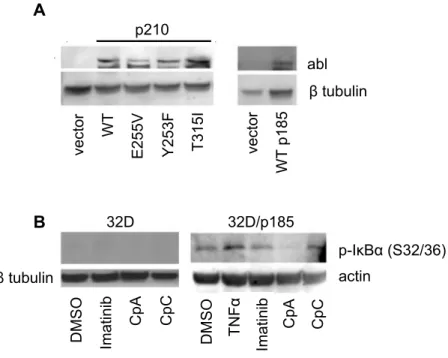
IKKβ inhibition decreases IκBα phosphorylation levels in BCR-ABL expressing cells.
Ba/F3 cells stably expressing p210 BCR-ABL as well as drug-resistant variants, and
32D cells expressing p185 BCR-ABL, were developed previously (see Reuther et al, 1998;
Skaggs et al., 2006). Immunoblotting confirms the expression of BCR-ABL in the different
cell lines (Figure 2.1A). As a measure of NF-κB activation downstream of BCR-ABL, we
analyzed phosphorylation of IκBα at ser32/36. Notably, phosphorylation of IκBα is
significantly higher in 32D/p185 cells as compared to the parental 32D cells (Figure 2.1B),
consistent with reports showing that BCR-ABL induces NF-κB activation (Reuther at al.,
1998; Kirchner et al., 2003). Additionally, this result demonstrates that IKK is activated
downstream of BCR-ABL since IKK phosphorylates IκBα at serines 32 and 36 (Hayden and
Ghosh, 2004). To analyze the effects of imatinib and a recently described inhibitor of IKKβ
(Cp A) on IκBα phosphorylation, 32D cells and those expressing the p185 form of
BCR-ABL were treated with DMSO, imatinib, the IKK inhibitor CpA, or a structurally inactive
form of compound A (CpC). Treatment with imatinib partly reduced while CpA, but not
CpC, significantly reduced IκBα phosphorylation in 32D/p185 cells (Figure 2.1B) indicating
that BCR-ABL activates IKK, which induces IκBα phosphorylation. Additionally, these
results show that CpA is an effective inhibitor of IKK/NF-κB activation downstream of
BCR-ABL expression induces susceptibility to IKKβ inhibition.
Previously we had shown that BCR-ABL induces strong NF-κB-dependent
transcriptional activity and that inhibition of NF-κB via IκBα expression blocked tumor
growth driven by BCR-ABL in a xenograft model (Reuther et al., 1998). Based on these
results, we asked whether IKKβ inhibitor CpA (1 µM) would affect the growth/viability of
32D cells or 32D/p185 cells as measured in an MTT assay. The effects of IKKβ inhibitor
CpA were compared to the effects of imatinib (1 µM) and dasatinib (5 nM), both with
well-established activity against BCR-ABL-expressing cells. Additionally, we included an IKKβ
inhibitor (CpB) with weaker activity than IKKβ inhibitor CpA and an inactive, related
compound (CpC). The results (Figure 2.2) demonstrate that IKKβ inhibitor CpA, at a dose
of 1 µM, is effective at suppressing cell growth/viability of 32D/p185 cells with minimal
effects on the parental 32D cells. As expected, imatinib and dasatinib each reduced cell
growth/viability of 32D/p185 cells. IKKβ inhibitor CpB reduced viability of 32D/p185 cells
but less effectively than IKKβ inhibitor CpA, consistent with its lower effectiveness as an
IKKβ inhibitor. The inactive IKKβ inhibitor compound did not affect cell growth/viability.
Similar results were observed with the proB Ba/F3 cell line expressing p210 BCR-ABL
(Figure 2.2B), although the IKKβ inhibitor reduced cell growth/viability of the parental
pro-B cells presumably indicating the involvement of NF-κB at some level in control of
proliferation or survival of these cells. Titration of IKKβ inhibitor CpA demonstrated
observable cell growth/viability effects around 100 nM, with an approximate 10-folder
stronger effect at that concentration, and at 1 µM and 10µM, in 32D/p185 cells as compared
WEHI media does not overcome the inhibitory effect of IKKβ blockade on 32D/p185 cell
viability.
Expression of BCR-ABL generates growth factor-independent growth of 32D
myeloid cells (Deininger et al., 2000). Addition of WEHI media (as a source of IL-3) to
BCR-ABL-expressing cells overcomes the inhibitory block on cell viability induced by
imatinib or dasatinib, essentially recreating a myeloid cell without BCR-ABL activity (Figure
2.3A). However, IKKβ inhibitor-treated 32D/p185 cells cannot be rescued with WEHI media
(Figure 2.3A).
WEHI-controlled growth/survival does not require IKKβ in myeloid cells where BCR-ABL
is inactive.
We then asked if 32D/p185 cells exposed to IKKβ inhibitor could be rescued with
WEHI media, or with imatinib and WEHI media. 32D/p185 cells were incubated in the
presence or absence of WEHI and/or imatinib for 24 hours. Subsequently, IKKβ inhibitor
Cp A (or C) was added for an additional 20 hours and cell viability was assessed. The results
of this experiment (Figure 2.3B) reveal that WEHI alone does not rescue the cells when IKK
is inhibited, consistent with the results shown in Figure 3A. However, WEHI media plus
imatinib rescues cell viability in 32D/p185 cells exposed to IKKβ inhibitor. This result shows
that growth factor-induced growth/survival of 32D cells (where BCR-ABL is inactivated or
IKKβ inhibition induces cell death in BCR-ABL expressing cells but does not affect cell
cycle parameters.
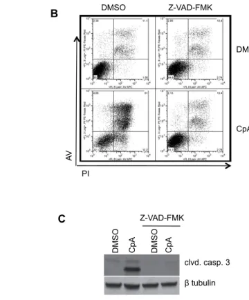
To analyze effects on 32D/p185 cell proliferation/viability induced by IKKβ inhibition, cells were treated with imatinib, dasatinib, or IKKβ inhibitor CpA (or inactive CpC) and were then assessed for cell death by staining with Annexin V (AV) and propidium
iodide (PI). The parental 32D cells exhibited no induction of apoptosis (AV+, PI-) with the
various drug treatments. However, a measurable, but relatively modest level early apoptosis
was induced in the 32D/p185 BCR-ABL-expressing cells with tyrosine kinase inhibitors or
with IKKβ inhibitor CpA (Figure 2.4A). These results are consistent with known effects of
BCR-ABL on blocking apoptosis in the transformation process (Kuroda et al., 2006). The
inactive IKKβ inhibitor (CpC) exhibited no effects on apoptosis and showed essentially the
same profile as DMSO treatment. Correspondingly, the drug treatments that induced
apoptosis also exhibited an increase in late apoptosis/necrosis (AV+, PI+). In order to further
examine an apoptotic response, immunoblotting was carried out to examine the level of
cleaved caspase-3 in these cells follow inhibitor treatment. Figure 3.4B shows that 32D/p185
cells treated with imatinib, dasatinib, or IKKβ inhibitor exhibit increased levels of cleaved caspase-3 relative to the DMSO control. This result suggests that the increased cell death
observed in these cells upon drug treatment is mediated at least partly via apoptosis. We note
that IKKβ inhibition induces less early apoptosis at the 24 hr time point as compared to BCR-ABL inhibition, but relatively equivalent levels of overall cell death (AV+, PI+).
Although there are elevated levels of apoptosis/cell death present in 32D/p185 cells treated
with the drugs listed above, it is also possible that these drugs are exhibiting effects via a cell
cells exhibit higher levels of G2-M and S-phase compartments as compared to parental 32D
cells (Figure 2.5). Experiments revealed that treatment with imatinib or dasatinib inhibits
cell cycle progression in 32D/p185 cells, with enhanced G0-G1 accumulation and reduced
numbers in S phase as compared to DMSO control. Treatment with IKKβ inhibitor CpA, or
with inactive CpC, did not significantly alter cell-cycle parameters. Importantly it was
shown that cell cycle progression is not abrogated in 32D cells treated with these compounds
(see Figure 2.5). Overall, these studies indicate that IKKβ inhibition of
BCR-ABL-expressing myeloid cells induces a cell death response that can be partly attributed to
apoptosis, but does not induce a cell-cycle block in a specific cell-cycle compartment. Future
studies will be required to determine if overall cell growth is blocked by IKKβ inhibition and
whether non-apoptotic forms of cell death contribute to the IKKβ inhibition response in
BCR-ABL+ cells.
IKKβ inhibition blocks viability in cells expressing wild-type, imatinib resistant, or the
T315I-dasatinib resistant forms of BCR-ABL.
Point mutations in the kinase domain of BCR-ABL produce resistance to imatinib and
promote oncogenicity (22-27). We analyzed Ba/F3 cells expressing wild-type p210 or
variants that are either imatinib resistant (Y253F and E255V) or both imatinib and dasatinib
resistant (T315I). Cells were exposed to imatinib, dasatinib, IKKβ inhibitor (CpA), or the
inactive structural variant of the IKKβ inhibitor compound (CpC). Consistent with
experiments shown above (Figure 2.2), each of the inhibitors strongly reduced the viability of
cells expressing wild-type BCR-ABL (Figure 2.6). As expected, cells expressing mutant
the T315I mutant exhibiting complete resistance to imatinib. Those cells expressing the
T315I mutation were also resistant to growth inhibition by dasatinib (Figure 2.6), as
expected. Interestingly, each of the mutant cell lines, including the T315I line, was
susceptible to IKKβ inhibition. The inactive IKKβ inhibitor (CpC) in DMSO showed no
inhibitory effects on any of the cells. These studies show that blocking NF-κB/IKK
suppresses viability of cells expressing all forms of BCR-ABL, including wild-type and
imatinib- and dasatinib-resistant variants, indicating that IKK/NF-κB is required for
growth/survival of BCR-ABL+ cells.
2.5 Discussion
The dramatic success of targeting BCR-ABL in CML has been dampened by the
emergence of imatinib and dasatinib resistant variants in the kinase domain. An alternative or
adjuvant approach is to target essential downstream regulators of BCR-ABL. Here we have
addressed the potential that inhibition of NF-κB and/or its upstream regulator IKK will
function to block growth/viability of cells expressing wild-type BCR-ABL or imatinib- or
dasatinib-resistant variants (Figure 2.1A). The IKKβ inhibitor, compound A, works to block
the phosphorylation of IκBα in myeloid cells expressing BCR-ABL (Figure 2.1B). Our
results show that IKKβ inhibition functions similarly to imatinib and to dasatinib in
suppressing growth of myeloid or pro-B cells expressing wild-type BCR-ABL or imatinib- or
dasatinib-resistant forms of BCR-ABL (Figure 2.2A and Figure 2.2B). More over,
IKKβ inhibition has a greater effect of cell viability of BCR-ABL-expressing cells than
IKKβ inhibitor-treated 32D/p185 cells cannot be rescued with IL-3-rich WEHI media
(Figure 2.3A). This result suggests one of the following possibilities: (i) WEHI media/IL-3
requires IKK/NF-κB to promote cell growth/survival, (ii) BCR-ABL-activated signaling
where IKKβ is inhibited interferes with the ability of IL-3 to induce cell survival, or (iii)
NF-κB activity promoted by growth factor support which involves Stat5 (Nakamura et al., 2002)
is not sufficient to replace the NF-κB/IKK activity suppressed by IKKβ inhibition in the
BCR-ABL-expressing cells. This latter hypothesis may be explained by different forms of
NF-κB activated by growth factors versus BCR-ABL, by different modifications to NF-κB in
response to growth factors or to BCR-ABL expression leading to distinct gene responses, or
by a function of IKK controlled by BCR-ABL that is not regulated by growth factors.
The viability of BCR-ABL-expressing cells treated with compound A and then
subsequently treated with imatinib was partially rescued (Figure 2.3B). This result indicates
that BCR-ABL requires IKKβ activity to promote cell viability. When BCR-ABL is blocked
by imatinib, the cells revert to a growth-factor-dependent status. IL-3 does not require IKKβ
to promote cell growth as shown by this assay. Thus the effect of IKKβ inhibition on
blocking rescue of growth/survival by exogenous growth factors is mediated through
BCR-ABL-controlled suppression of the growth factor response under these conditions and/or
through inhibition of an IKK/NF-κB function in BCR-ABL+ cells that cannot be
recapitulated by growth factors.
Treatment of BCR-ABL cells with compound A results in the induction of apoptosis,
as indicated by the cleavage of caspase 3 (Figure 2.4A and 2.4B), but does not impact cell
cycle status (Figure 2.5). These results indicate that NF-κB/IKK is essential for
inhibition on BCR-ABL+ cells relative to growth and survival. Interestingly, IKKβ
inhibition results in loss of cell viability of cells expressing imatinib- and dasatinib-resistant
BCR-ABL mutants, including the T315I mutation (Figure 2.6). These data indicate the
potential for the use of IKKβ inhibitors as stand-alone therapies for CML or in combination
with existing therapies. The results also indicate that IKKβ inhibition may be therapeutic in
Figure 2.1: IKKβ inhibitor CpA blocks phosphorylation of IκBα in
BCR-ABL-expressing cells. (A) Whole cell extracts were prepared from untreated Ba/F3 parental and BCR-ABL expressing cells (left), and 32D and 32D/p185 cells (right). Immunoblotting was performed with an antibody to ABL. (B) Cytoplasmic extracts were prepared from 32D and 32D/p185 BCR-ABL cells treated with vehicle (DMSO), 1 mM imatinib, 1 µM IKKβ inhibitor (CpA), or 1 µM inactive related compound (CpC). As a control, 32D/p185 cells were treated with TNF (5 ng/ml). Immunoblot analysis of was carried out using an antibody that recognizes IκBα phosphorylated on ser32/36. β-tubulin or actin antibodies were used to normalize loading. Data are representative of 3 experiments.
abl
β tubulin
ve
ct
or WT
E2
55
V
Y2
53
F
T315I vect
or
WT
p185
p210
D
MSO
Ima
tin
ib
CpA CpC
D
MSO TNF
α
Ima
tin
ib
CpA CpC
p-IκBα (S32/36) actin
β tubulin
32D 32D/p185
A