part of Epigenomics
Research Article 2017/02/289
3
2017
Aim: Sex-based differences in response to adverse prenatal environments and infant outcomes have been observed, yet the underlying mechanisms for this are unclear. The placental epigenome may be a driver of these differences. Methods: Placental DNA methylation was assessed at more than 480,000 CpG sites from male and female infants enrolled in the extremely low gestational age newborns cohort (ELGAN) and validated in a separate US-based cohort. The impact of gestational age on placental DNA methylation was further examined using the New Hampshire Birth Cohort Study for a total of n = 467 placentas. Results: A total of n = 2745 CpG sites, representing n = 587 genes, were identified as differentially methylated (p < 1 × 10⁻⁷). The majority (n = 582 or 99%) of these were conserved among the New Hampshire Birth Cohort. The identified genes encode proteins related to immune function, growth/transcription factor signaling and transport across cell membranes. Conclusion: These data highlight sex-dependent epigenetic patterning in the placenta and provide insight into differences in infant outcomes and responses to the perinatal environment.
First draft submitted: 30 September 2016; Accepted for publication: 10 January 2017; Published online: 17 February 2017
Keywords: CpG DNA methylation • epigenetics • placenta • sexual dimorphism
The placenta serves as the primary organ responsible for regulation of the prenatal environment critical for optimal fetal devel-opment. It is a nutrient transporter and produces vital hormones to maintain preg-nancy and support the fetus [1]. In contrast to these essential functions, the placenta also serves as a source of exposure for toxic sub-stances, dysregulated hormone signaling and immune-related proteins [2]. Such exposures including maternal stress, excess hormones, cytokines or environmental toxicants impact fetal health and influence later-life health outcomes [3–11].
An increasing body of literature supports sex-specific birth and later-life outcomes in relation to adverse in utero environments [12]. For example, sex-specific perinatal and later-life outcomes have been linked to toxic sub-stance exposure [5,6,13–16], maternal stress [7]
and maternal immune status [9,17,18]. We hypothesize that sex-dependent health out-comes in infants may be associated with sexual dimorphism of the placenta. In sup-port of this hypothesis, physiologic differ-ences between male and female placentas have been observed [19]. For example, pla-centas derived from pregnancies of males and females display variation in the abun-dance and type of glucocorticoid transporter proteins, the expression of hormones and the production of immune-related proteins including cytokines [12,19–21]. In addition, sex-based differences in response to prenatal stressors have been observed at the levels of the placental proteome, transcriptome and epigenome [3,7,14,20,22–27]. Thus, the sexu-ally dimorphic nature of the placenta could influence variation in toxicant transport and accumulation, hormone levels and immune
Sexual epigenetic dimorphism in the
human placenta: implications for
susceptibility during the prenatal period
Elizabeth Martin1, Lisa Smeester1, Paige A Bommarito1, Matthew R Grace2, Kim Boggess2, Karl Kuban3, Margaret R Karagas4, Carmen J Marsit5, T Michael O’Shea6 & Rebecca C Fry*,1
1Department of Environmental Sciences
& Engineering, Gillings School of Global Public Health, University of North Carolina, Chapel Hill, NC, USA
2Department of Obstetrics & Gynecology,
University of North Carolina School of Medicine, University of North Carolina, Chapel Hill, NC, USA
3Department of Pediatrics, Boston
Medical Center, Boston, MA, USA
4Department of Epidemiology, Geisel
School of Medicine at Dartmouth, Hanover, NH, USA
5Department of Environmental Health,
Rollins School of Public Health at Emory University, Atlanta, GA 30322, USA
6Department of Pediatrics, University
of North Carolina School of Medicine, University of North Carolina, Chapel Hill, NC, USA
response experienced by the fetus. While key physi-ological differences have been observed in male and female placentas, the underlying mechanisms are not well established.
Epigenetic regulation may underlie the physiologic differences observed between male and female pla-centas [12]. For instance, key differences in chromatin structure have been observed between male and female placentas, suggesting that there is a role for epigen-etic regulation in placental sexual dimorphism [12,21]. Understanding epigenetic regulation in the placenta is important given the modifiability of its epigenome in response to prenatal stressors, and its role as a mediator of the developmental origins of health and disease [2,20,21,28]. Additionally, because certain epig-enomic markers are stable over time [29], it is possible that changes to the fetal placenta methylome explain the sex-based differences in later-life disease risks.
To our knowledge, this study is among the first to address the gap in knowledge related to sexual dimor-phism of the placental DNA methylome. Here, we investigated whether DNA (CpG) methylation pat-terns in the placenta differed based on the sex of the fetus. Sex-based differences within the placenta were identified in a subset of subjects from the extremely low gestational age newborns (ELGAN) cohort [30–35], and validated in a replication cohort comprised of women recruited at the University of North Carolina (UNC) hospitals [36]. To confirm that gestational age was not a major driver of the differences in DNA methylation, all data were subsequently compared with CpG meth-ylation assessed in the New Hampshire Birth Cohort. The goal of these analyses is to provide key insights into whether sexual dimorphism of the placental meth-ylome may explain differential susceptibility to adverse prenatal environmental conditions.
Methods
ELGAN study subject recruitment & sample collection
ELGAN study recruitment has been previously described in detail [37]. In short, from 2002 to 2004, women giving birth at one of the 14 participating sites before 28 weeks gestation were asked to enroll in the ELGAN study. Their consent was provided either upon hospital admission prior to or shortly after delivery. All procedures were approved by the Institutional Review Board at each of the 14 participating study sites.
A total of 1506 infants and 1249 mothers enrolled in the ELGAN study. A subcohort of 84 mother-infant pairs, representative of the larger cohort, were selected for this analysis. As part of participation in ELGAN, women were asked to contribute their placentas. A description of the methodology that was used for
pla-cental collection is as follows: delivered placentas were placed in a sterile exam basin and transported to a sampling room, where they were biopsied under ster-ile conditions. At the midpoint of the longest distance between the cord insertion and the edge of the pla-cental disk, the amnion was pulled back using sterile technique to expose the chorion. Traction was applied to the chorion and the underlying trophoblast tissue and a piece of tissue was removed by cutting at the base of the section. The tissue was placed into a cryo vial and immediately immersed in the liquid nitro-gen. Specimens were stored at -80°C until laboratory processing [38].
The replication cohort included 40 subjects of women receiving obstetric care at UNC hospitals who consented to collection of placental samples at the time of birth. Subjects gave written informed consent prior to enrollment and participation in this study. A full-thickness placental biopsy was obtained after delivery, avoiding the periphery and areas of obvious infarction. Samples were flash frozen in liquid nitrogen, and stored at -70°C until analysis. This research was approved by the Institutional Review Board at the University of North Carolina (#11–2054).
Data from a third cohort comprising 343 placenta samples obtained from women and children involved in the New Hampshire Birth Cohort, an ongoing pro-spective pregnancy and birth cohort, were utilized as a contrast and validation of the results in a cohort with a more generalizable spread of gestational age. This cohort and the placental DNA methylation array data have been previously described [39]. Briefly, pregnant women whose primary source of residential water is a private well and who obtained their prenatal care at clinics in New Hampshire, were recruited into the study. Eligibility criteria were that women were cur-rently pregnant; 18–45 years old; received routine pre-natal care at one of the study clinics; used a private well that serves <15 households or 25 individuals at their residence; resided in the sample place since their last menstrual period; and did not plan to move prior to delivery. Like the replication cohort, a full-thickness placental biopsy was obtained after delivery, avoid-ing the periphery and areas of obvious infarction, and placed immediately in RNA later for at least 48 h, then removed from the fixative and stored at -80°C until laboratory processing.
DNA extraction & assessment of DNA methylation
(PBS) to remove any residual blood, and homogenized in Buffer RLT™ with β-mercaptoethanol (Qiagen, CA, USA). DNA and RNA sequences >18 nucleotides in length were collected using the AllPrep DNA/RNA/ miRNA Universal Kit (Qiagen, CA, USA), accord-ing to manufacturer’s instructions. CpG methyla-tion was assessed using the Illumina HumanMethyl-ation450 BeadChip© array (Illumina, Inc., CA, USA).
This platform assesses the DNA methylation levels of 486,428 individual probes at single nucleotide resolu-tion. Isolated DNA was first bisulfite converted using the EZ DNA methylation kit (Zymo Research, CA, USA) and converted DNA was then hybridized onto the array. The DNA methylation data were collected at Expression Analysis, Inc. (NC, USA; www.expression-analysis.com). Methylation levels were calculated and expressed as β values (β = intensity of the methylated allele [M]/intensity of the unmethylated allele [U] + intensity of the methylated allele [M] + 100).
Statistical analysis
The array signal data were processed using R software (version 3.0.3). For each set of samples, batch effect was evaluated using principle component analysis and was determined to not be a significant source of variation. Any methylation β-values with a detection p-value above 0.01 were considered unreliable and were removed from analysis. β-mixture quantile normaliza-tion was performed using the WateRmelon package (version 1.11.0) in R [40]. Following normalization, probes containing a single nucleotide polymorphism in the assayed CpG dinucleotide, as well as those for which two or more single nucleotide polymorphisms were located in the probe sequence were removed. Last, probes on the Y chromosome were removed from this study as they were not comparable between male and female placental samples [41,42]. A total of n = 377,673 probes, representing 20,442 genes, remained for analy-sis. β-differences were calculated for differentially methylated probes by subtracting the median female
β-value from the median male β-value. To identify covariates, a directed acyclic graph approach was used. No factors were identified that influenced both the sex of the infant and the methylome. For this reason, p-values were calculated from t-statistics. Multiple testing correction of the p-values was performed using the Bonferroni correction (p < 1 × 10⁻⁷) as well as a
q-value correction, using the Partek Genomic Suite (MO, USA).
To replicate these findings, CpG methylation data were analyzed in a separate cohort based at UNC hos-pitals using the HumanMethylation450 BeadChip©
(Illumina, CA, USA). Data were processed and ana-lyzed according to the protocol described above,
how-ever data for only those CpG locations which were sta-tistically significant in the ELGAN dataset were tested (n = 4371). This increased the Bonferroni corrected p-value threshold to 1 × 10⁻⁵. In addition to the
valida-tion performed, t-statistics and a multiple test-corrected q-value were calculated for all probes.
Given the known impact of gestational age on the placental DNA methylome [43,44], and that the two previously described cohorts utilized in this study were of lower gestational age, data from the New Hampshire birth cohort [39] were also assessed (n = 343 placen-tas). In addition, we also performed a second analysis where linear regression modeling was used to control for gestational age in each of the cohorts.
Transcription factor binding site analysis of validated gene sets
To characterize transcription factor binding site pat-terns, two separate analyses were conducted. First, enriched transcription factor binding site motifs were identified using Gene Set Enrichment Analysis (GSEA) [45]. This analysis relies on a Kolmogorov– Smirnov like statistic of an enrichment score to cal-culate p-values. Subsequently, the p-values are false discovery rate corrected by calculating a normalized enrichment score. Second, identified transcription actors were validated with the Genomatix Com-mon Transcription Factor Binding Site module using methods as previously described [46].
Gene set-based analysis
In addition to transcription factor binding site analy-sis, we analyzed whether the differentially methylated probes in the placenta were enriched for specific bio-logical functions. Four gene sets were analyzed where gene content was established using www.uniprot.org. Specifically, four gene sets were tested: transport-related genes (n = 3704), immune-transport-related genes (n = 1622), inflammation-related genes (n = 410) and growth/transcription-related genes (n = 2030). A χ2
differen-tially expressed in male and female placentas [47]. For these analyses, relaxed statistical significance was used to define differentially methylated probes (p < 0.05; q < 0.1) [47], to enable comparability to the previously published work.
Results
Study subject characteristics
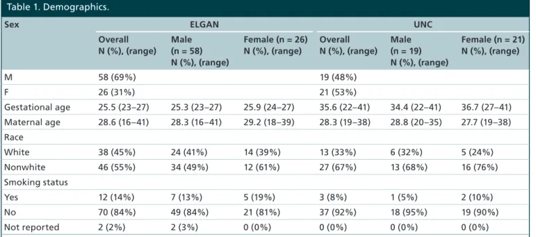
In the ELGAN cohort subset, 58 (69%) were male and 26 (31%) were female. In the UNC cohort, 19 (48%) were male and 21 (53%) were female (Table 1). For both cohorts, none of the demographic variables differed between males and females. These variables included average maternal age, gestational age, parity, smoking status and race. Demographic characteristics for the New Hampshire Birth Cohort Study have been previously published [39].
Identification & replication of differentially methylated probes in ELGAN
In the ELGAN cohort, we analyzed CpG meth-ylation differences between male and female infants for 377,673 CpG probes representing 20,442 genes across 84 placentas. For this analysis, statistical sig-nificance was set at the Bonferroni-corrected p-value threshold of p < 1 × 10⁻⁷. A total of 4371 probes,
representing 714 genes were found to be differen-tially methylated between male and female placentas (Figure 1, Supplementary Table 1). The vast majority (97.9%) of these probes (n = 4280, 666 genes) were located on the X chromosome (Table 2). Only 91 dif-ferentially methylated probes (2.1%), representing 48 genes, were located in autosomal regions (Table 2).
When comparing probes that were differentially meth-ylated between males and females, the greatest absolute
β-difference displayed was 44.3% and the smallest was 2.5% (Supplementary Table 1). The majority of probes (99.2%) were greater than 5%, a level that has been previously used as a threshold while identifying sex-based differences [41]. Interestingly, and in contrast to our a priori hypothesis, 52.5% (n = 2296) probes were hypermethylated in males as compared with females (Table 2). Hypermethylation of probes in males relative to females occurred on both autosomal chromosomes (n = 73, 82.4%) and the X chromosome (n = 2223, 51.9%) (Table 2).
To establish whether CpG methylation levels in the ELGAN subjects replicated in a separate cohort, we ana-lyzed placental CpG methylation data from a cohort of 40 pregnant women from the UNC hospital. A total of 2745 probes, representing 587 genes, replicated across both cohorts as differentially methylated between male and female placentas (Figure 2, Supplementary Table 1).
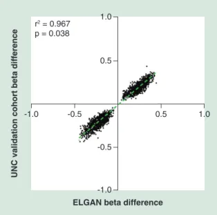
As noted within the ELGAN subcohort, the vast majority of probes 99.2% (n = 2724, 574 genes) were located on the X chromosome, and only 21 probes (13 genes) occurred on autosomal chromosomes (Table 2, Table 3). Again we observed similar patterns of hyper-methylation where 51.3% (n = 1407) probes were hypermethylated in males as compared with female placentas, and this pattern was true for probes on the X chromosome (n = 1394, 51.2%) (Table 2). Addi-tionally, across cohorts changes in the magnitude and direction of methylation were highly consistent as rep-resented by the strong correlation between β-values (r2
= 0.95; p < 0.0001) (Figure 3).
Table 1. Demographics.
Sex ELGAN UNC
Overall N (%), (range)
Male (n = 58) N (%), (range)
Female (n = 26) N (%), (range)
Overall N (%), (range)
Male (n = 19) N (%), (range)
Female (n = 21) N (%), (range)
M 58 (69%) 19 (48%)
F 26 (31%) 21 (53%)
Gestational age 25.5 (23–27) 25.3 (23–27) 25.9 (24–27) 35.6 (22–41) 34.4 (22–41) 36.7 (27–41)
Maternal age 28.6 (16–41) 28.3 (16–41) 29.2 (18–39) 28.3 (19–38) 28.8 (20–35) 27.7 (19–38)
Race
White 38 (45%) 24 (41%) 14 (39%) 13 (33%) 6 (32%) 5 (24%)
Nonwhite 46 (55%) 34 (49%) 12 (61%) 27 (67%) 13 (68%) 16 (76%)
Smoking status
Yes 12 (14%) 7 (13%) 5 (19%) 3 (8%) 1 (5%) 2 (10%)
No 70 (84%) 49 (84%) 21 (81%) 37 (92%) 18 (95%) 19 (90%)
Not reported 2 (2%) 2 (3%) 0 (0%) 0 (0%) 0 (0%) 0 (0%)
Figure 1. Manhattan plot displaying all CpG probes tested for the analysis of sex-dependent DNA methylation in the placenta. Probes are organized according to chromosomal positions. Probes in black and gray represent probes tested. Probes above the red line were those that were statistically significant. Probes in green are those that were validated in the replication cohort.
1 2 3 4 5 6 7 8 9 10 12 14 16 18 20 23
0 10 20 30 40 50
-Lo
g10
(
p
)
Chromosome
Among the replicated sexually dimorphic probes in all cohorts (n = 2745 probes, 587 genes), the major-ity of autosomal probes were located in the gene body (38.1%) and were hypermethylated. Interestingly, no sexually dimorphic probes were found within the TSS200 or 1st Exon (Figure 4). The majority of sexu-ally dimorphic probes identified in the gene body and the 3′UTR were hypermethylated, with the TSS1500 and unspecified region showing higher proportions of hypomethylation (Figure 4). Of the sexually dimor-phic probes on the X chromosome, the highest pro-portion were identified in the gene body (21.9%), and the majority of these were hypermethylated in males relative to females (Figure 4). Additionally, only the TSS200, 5′UTR and 1st Exon displayed higher proportions of hypomethylation (Figure 4).
X-inactivation has been previously cited as a poten-tial mechanism which can account for many of the differences for the sexually dimorphic methylation changes observed on the X chromosome [26,42]. For this reason, the data were compared with genes that escape X-inactivation [49] and pseudo-autosomal genes [50]. Interestingly, a subset of the genes (n = 10) are known to escape X-inactivation (n = 10, 2%), and one XG blood group (one, namely XG, was a member of the human pseudoautosome (Supplementary Table 2).
Due to concerns about the potential for gestational age to impact CpG methylation, these sites were fur-ther tested in a cohort of 343 women recruited from the New Hampshire Birth cohort. In support of the primary data, we found replication of n = 2662 probes and n = 582 genes (99% of validated probes
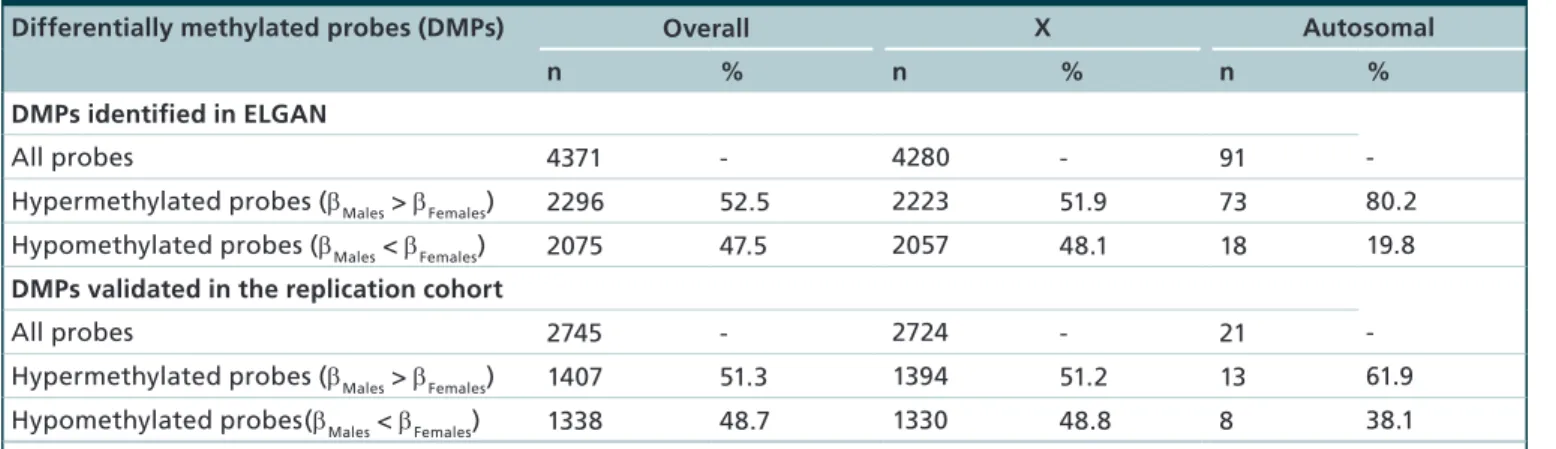
Table 2. Comparison of methylation levels among sex-dependent differentially methylated probes.
Differentially methylated probes (DMPs) Overall X Autosomal
n % n % n %
DMPs identified in ELGAN
All probes 4371 - 4280 - 91
-Hypermethylated probes (βMales > βFemales) 2296 52.5 2223 51.9 73 80.2
Hypomethylated probes (βMales < βFemales) 2075 47.5 2057 48.1 18 19.8
DMPs validated in the replication cohort
All probes 2745 - 2724 - 21
-Hypermethylated probes (βMales > βFemales) 1407 51.3 1394 51.2 13 61.9
Hypomethylated probes(βMales < βFemales) 1338 48.7 1330 48.8 8 38.1
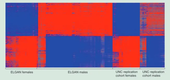
Figure 2. Heatmap displaying the 2745 validated sexually dimorphic probes. Red represents increased methylation in males relative to females and blue represents decreased methylation in males relative to females.
ELGAN: Extremely low gestational age newborn; UNC: University of North Carolina.
ELGAN females ELGAN males UNC replication
cohort females
UNC replication cohort males
and genes). Importantly, replication occurred for all probes across the studies that passed p-value detection. Similar results were observed when gestational age was included in the model where n = 2561 probes and n = 568 genes (replication of 92% of probes and genes). These data were not considered during downstream analyses, but can be found in the Supplementary Materials (Supplementary Tables 1 & 2).
Transcription factor binding site analysis
In addition to characterization of the replicated probes/ genes by region and methylation status, transcription factor binding site enrichment analysis was performed on the promoter regions of differentially methylated genes. An analysis of hypermethylated probes revealed an enrichment for binding sites for NFATC (GSEA p = 1.07 × 10⁻²⁶, Genomatix p = 2.23 × 10⁻⁴), and PAX4
(GSEA p = 1.39 × 10⁻¹⁷, Genomatix p = 9.62 × 10 -81). Among hypomethylated probes, binding sites for
MAZ (GSEA p = 3.68 × 10⁻²⁷, Genomatix p = 3.56 ×
10-131) and FOXO4/MLLT7 (GSEA p = 4.54 × 10⁻²⁰,
Genomatix p = 2.41 ×10-37) were enriched. SP1 was
significant in both the hypermethylated (GSEA p = 1.6 × 10⁻¹⁷, Genomatix p = 2.29 × 10-49) and
hypomethyl-ated (GSEA p = 2.29 × 10⁻³⁰, Genomatix p = 7.82 ×
10-48) gene sets.
Differentially methylated gene sets associated with response to the prenatal environment and sex-based differential gene expression
In addition to the transcription factor binding site analysis, we analyzed whether the differenitally
meth-ylated probes in the placenta were enriched for spe-cific biological functions, namely transport, immune response, inflammation and growth/transcription fac-tors. For this analysis, probes that were differentially methylated (p < 0.05 and q < 0.1) in both cohorts were considered (n = 4900 probes, n = 761 genes) (Supplementary Table 2). Using a Yates corrected χ2
test it was determined that immune proteins (n = 31, p = 0.0337), transporters (n = 119, p = 0.0233) and growth/transcription factors (n = 66, p = 0.0001) were enriched (Supplementary Table 2). Only inflammation proteins were not enriched (n = 12, p = 0.5623). Signifi-cantly enriched immune-related genes were involved in primary immunodeficiency and Toll-like receptor sig-naling, such as TLR7 and TLR8. The inflammatory-response genes included NOD-like receptor signaling pathways. Growth/transcription factors related genes were associated with neurotrophin and mTOR signal-ing pathways. Last, transporters tended to be involved in neuroactive ligand–receptor interaction and cal-cium signaling pathways, such as voltage-dependent anion-selective channel protein 1.
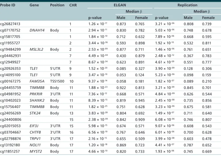
over-Table 3. Autosomal sex-dependent differentially methylated probes (n = 21 probes, 13 gene).
Probe ID Gene Position CHR ELGAN Replication
Median β Median β
p-value Male Female p-value Male Female
cg26827413 1 1.26 × 10⁻¹⁷ 0.873 0.765 3.21 × 10-06 0.808 0.739
cg07170752 DNAH14 Body 1 2.94 × 10⁻¹¹ 0.830 0.782 5.03 × 10-06 0.748 0.678
cg15817705 1 1.84 × 10⁻¹⁰ 0.712 0.632 7.89 × 10-06 0.668 0.595
cg11955727 2 3.44 × 10-36 0.593 0.898 1.92 × 10⁻¹⁰ 0.532 0.811
cg19484299 MSL3L2 Body 2 2.53 × 10-09 0.877 0.711 1.46 × 10-06 0.761 0.651
cg04462931 7 4.49 × 10-44 0.430 0.879 2.48 × 10⁻¹² 0.367 0.783
cg12949927 7 8.67 × 10⁻³⁴ 0.623 0.891 4.61 × 10-09 0.551 0.777
cg20926353 TLE1 5′UTR 9 1.52 × 10⁻²⁰ 0.085 0.327 3.90 × 10-09 0.128 0.306
cg14095100 TLE1 5′UTR 9 3.47 × 10⁻¹⁰ 0.053 0.124 5.23 × 10-06 0.098 0.159
cg00167275 FAM35A TSS1500 10 9.37 × 10⁻¹⁴ 0.058 0.181 1.82 × 10-07 0.089 0.210
cg04455759 TIMM8B Body 11 1.88 × 10⁻²³ 0.922 0.813 3.21 × 10-09 0.845 0.701
cg04981952 PRKRIR 3′UTR 11 7.36 × 10⁻¹² 0.668 0.571 4.84 × 10-09 0.626 0.544
cg10402023 SHANK2 Body 11 8.39 × 10⁻¹⁵ 0.819 0.945 2.45 × 10-08 0.735 0.856
cg15756407 TIMM8B Body 11 1.82 × 10⁻²⁵ 0.751 0.628 3.23 × 10-06 0.675 0.581
cg24056269 STK24 Body 13 3.83 × 10⁻²⁸ 0.804 0.692 1.49 × 10-07 0.711 0.640
cg24400806 15 2.38 × 10-09 0.842 0.909 6.08 × 10-06 0.746 0.807
cg02015053 EIF3J 3′UTR 15 5.98 × 10⁻¹⁵ 0.674 0.571 9.07 × 10-06 0.608 0.543
cg03704667 CHTF8 3′UTR 16 6.56 × 10⁻¹⁹ 0.767 0.646 6.01 × 10-06 0.700 0.628
cg02798874 TRPV1 3′UTR 17 2.16 × 10⁻¹⁵ 0.655 0.509 3.99 × 10-08 0.603 0.478
cg13192180 NOL11 Body 17 1.20 × 10⁻¹⁸ 0.869 0.723 4.41 × 10-07 0.787 0.652
cg11851257 MYST2 Body 17 4.66 × 10⁻¹⁰ 0.820 0.733 1.93 × 10-06 0.745 0.669
CHR: Chromosome; ELGAN: Extremely low gestational age newborns.
lap between studies was statistically significant (p < 0.0001). This suggests that DNA methylation may play a key role in the regulation of gene expression linked to sexual dimorphism.
Discussion
Fetal sex is known to influence susceptibility to the prenatal environment and can influence differential biological signaling within the placenta [3,5–7,12–15,23– 25,27,41]. Therefore, we set out to identify sex-based dif-ferences in the placental DNA methylomes in three independent US-based birth cohorts. We identified a total of 587 genes (2745 probes), which were differ-entially methylated (p < 1 × 10⁻⁷) in male placentas as
compared with female placentas in two cohorts. We further validated these probes in a third US cohort of normal gestational age and found that 99.2% of probes replicated. The majority of these probes were located on the X-chromosome, but a subset (n = 21 probes, 13 genes) were found on autosomal chromosomes. Many of the identified genes are involved in the transport and
transcriptional control of the immune response. Inter-estingly, some of the genes identified here have been previously shown to be sexually dimorphic at the level of the placental transcriptome. Overall, our findings demonstrate that there is a sexual epigenetic dimor-phism of the placental DNA methylome.
Figure 3. Comparison of sex-dependent beta differences between the extremely low gestational age newborns (X-axis) and University of North Carolina replication cohort (Y-axis) for all 2745 validated probes. Autosomal probes are highlighted in gray, while those found on the X-chromosome are represented in black. ELGAN: Extremely low gestational age newborn; UNC: University of North Carolina.
ELGAN beta difference
UNC validation cohort beta difference
r2 = 0.967
p = 0.038
-0.5
-1.0 -0.5
-1.0 0.5 1.0
0.5 1.0
the X-chromosome would have higher levels of meth-ylation in females relative to males. Strong support for these data come from their reproducibility across three independent cohorts. In addition, other studies have identified some sites on the X chromosome that display higher levels of methylation in males rela-tive to females [41,51]. While it may be intuirela-tive that X-inactivation would likely present as a pattern of hypermethylation in females as compared with male placentas, prior research has shown that gene silenc-ing on the X-chromosome is dependent on gene region and in some cases hypomethylation-associated silenc-ing has been observed [49,52,53]. Taken together, these data support that the functional impact of methyla-tion on X-chromosome inactivamethyla-tion exhibits posimethyla-tional dependencies, similar to those observed previously [54].
The sexually dimorphic genes were enriched for proteins involved in immune function, micronutrient transport and transcription/growth factors [12,20,21]. Specifically, immune-related TLR7 was shown to be hypermethylated in males relative to females and TLR8 was hypomethylated in males as compared with females. These immune-related differences between the male and female placental methylomes may con-tribute to differential susceptibility to environmen-tal exposures between the sexes [8]. For example, an enhanced capability for transporting toxicants across the placenta in males during the prenatal period may be accounted for by differences in activation of the
pla-cental transporter voltage-dependent anion-selective channel protein 1, a major calcium transport chan-nel [55] critical for fetal development. This protein, which normally enhances placental micronutrient delivery [56], can be hijacked by toxicants to cross cell membranes [57] during the prenatal period.
One possible mechanism underlying gene-specific genome-wide patterns of DNA methylation is the transcription factor occupancy theory [46]. Briefly, this theory posits that gene-specific methylation is influ-enced by transcription factor binding that either pre-vents or provides access to the DNA sequence for the DNA methylation machinery. Supporting this theory, we identified that binding sites for several transcrip-tion factors were significantly enriched in the hyper- and hypomethylated genes identified in this analysis. For instance, binding sites for NFATC were enriched in the promoter regions of the hypomethylated genes. Interestingly, in a separate study of placental sexually dimorphic gene expression, binding sites for NFATC were similarly enriched among the identified genes [47]. Additionally, binding sites for MAZ were identified to be enriched among the hypomethylated genes. MAZ is of interest as it has been previously identified as a key transcription factor associated with differential methylation patterns observed in response to a diverse suite of environmental contaminants [46]. Last, SP1, a transcription factor with enriched binding sites among both the hyper- and hypomethylated gene sets, is a known target of environmental contaminants, specifi-cally toxic metals [58]. These transcription factors may play a role in impacting CpG methylation as well as responding to adverse conditions experienced during the prenatal environment.
Several factors should be considered in interpreting the results of this study. While we identified and validated a set of sexually dimorphic CpG sites across three cohorts, gene expression was not assessed in these samples. To address this, the sexually dimorphic placental methylome data were integrated with an existing genomic dataset in order to establish functional epigenetics. Future research should integrate data CpG methylation, as well as mRNA and protein expression. A potential confounder to these analyses is gestational age [43,44]. To address this, we integrated CpG methylation across three cohorts with varying gestational age, and found strong conserva-tion. Last, future research should characterize X-inactive and X-active methylation patterns, through sequencing, to allow for male-female X-chromosome comparisons. These additional data would enhance the understanding of functional differences between methylation between males and females.
meth-Figure 4. Distribution of sex-dependent differential methylation by region for all probes.(A) displays probe locations for overall, hypermethylated and hypomethylated autosomal probes. (B) displays probe locations for overall, hypermethylated and hypomethylated X-chromosome probes.
Overall (n = 21)
Overall (n = 2,724)
Hypermethylated (n = 13) Hypomethylated (n = 8)
Direction of methylation
Hypermethylated (n = 1,394) Hypomethylated (n = 1,330)
Direction of methylation Autosomal chromosome probes (n = 21)
Percent per region (%)
Percent per region (%)
Validated X chromosome probes Unspecified 3’ UTR 1st exon 5’ UTR Body TSS200 TSS1500
0 50 100
0 50 100
ylome. The epigenetic dimorphism observed could result in the sex-dependent transport of toxicants, nutrients and signaling molecules across the placenta, thereby resulting in a sex-dependent response of the fetus. Furthermore, it is possible that these differences
Financial & competing interests disclosure
This research was supported by grants from the NIH in-cluding the Environmental Influences on Child Health Out-comes (ECHO) award (1U2COD023375, UG33OD023348 and 1UG3OD023275), the National Institute of Environ-mental Health Sciences (P42-ES007126, T32-ES007018, P42-ES007373, P01-ES022832) and the National Institute of Neurological Disorders and Stroke (5U01NS040069 and 2R01NS040069). Further support was provided by the Wake
Forest School of Medicine Innovation Pilot Grant, the Harold M and Mary Earnhardt Eagle Endowed Fund for Pediatric and Neonatal Research, and the EPA (RD83544201). The authors have no other relevant affiliations or financial involvement with any organization or entity with a financial interest in or fi-nancial conflict with the subject matter or materials discussed in the manuscript apart from those disclosed.
No writing assistance was utilized in the production of this manuscript.
Executive summary
• The placenta provides an interface between the maternal and fetal compartments transporting nutrients, signaling molecules and toxic substances between mother and fetus.
• Differences in CpG methylation between male and female placentas may provide mechanistic understanding for sex-based differences observed, following adverse exposures during the prenatal period.
• To investigate sex-based differences in the epigenome of the placenta, we analyzed DNA methylation in relation to fetal sex using genome-wide techniques comparing data from three separate US-based cohorts. • Methylation at 2745 probe (n = 587 genes) was identified and replicated, with enrichment of binding
sites for transcription factors previously related to sexually dimorphic gene expression and environmental contaminants.
• Genes with sex-based differential methylation in the placenta enrich for functions related to response to environmental exposures including: cellular transport, immune response and growth/transcription factors. • Within the placental DNA methylome, differences in the regulation of genes related to crucial biological
functions may account for sex-specific effects of adverse in utero environments.
References
1 Godfrey KM. The role of the placenta in fetal
programming-a review. Placenta 23(Suppl. A), S20–S27 (2002).
2 Burton GJ, Fowden AL, Thornburg KL. Placental
origins of chronic disease. Physiol. Rev. 96(4), 1509–1565 (2016).
3 Broberg K, Ahmed S, Engstrom K et al. Arsenic exposure
in early pregnancy alters genome-wide DNA methylation in cord blood, particularly in boys. J. Dev. Orig. Health Dis. 5(4), 288–298 (2014).
4 Cao J, Rebuli ME, Rogers J et al. Prenatal bisphenol A
exposure alters sex-specific estrogen receptor expression in the neonatal rat hypothalamus and amygdala. Toxicol. Sci. 133(1), 157–173 (2013).
5 Kippler M, Wagatsuma Y, Rahman A et al. Environmental
exposure to arsenic and cadmium during pregnancy and fetal size: a longitudinal study in rural Bangladesh. Reprod.
Toxicol. 34(4), 504–511 (2012).
6 Llop S, Guxens M, Murcia M et al. Prenatal exposure to
mercury and infant neurodevelopment in a multicenter cohort in Spain: study of potential modifiers. Am. J.
Epidemiol. 175(5), 451–465 (2012).
7 Ostlund BD, Conradt E, Crowell SE, Tyrka AR, Marsit CJ,
Lester BM. Prenatal stress, fearfulness, and the epigenome: exploratory analysis of sex differences in dna methylation of the glucocorticoid receptor gene. Front. Behav. Neurosci. 10, 147 (2016).
8 Winans B, Humble MC, Lawrence BP. Environmental
toxicants and the developing immune system: a missing
link in the global battle against infectious disease? Reprod.
Toxicol. 31(3), 327–336 (2011).
9 Parker-Athill EC, Tan J. Maternal immune activation and
autism spectrum disorder: interleukin-6 signaling as a key mechanistic pathway. Neurosignals 18(2), 113–128 (2010).
10 Veru F, Laplante DP, Luheshi G, King S. Prenatal maternal
stress exposure and immune function in the offspring. Stress 17(2), 133–148 (2014).
11 De Escobar GM, Obregon MJ, Del Rey FE. Maternal
thyroid hormones early in pregnancy and fetal brain development. Best Pract. Res. Clin. Endocrinol. Metab. 18(2), 225–248 (2004).
12 Gabory A, Roseboom TJ, Moore T, Moore LG, Junien C.
Placental contribution to the origins of sexual dimorphism in health and diseases: sex chromosomes and epigenetics. Biol.
Sex Differ. 4(1), 5 (2013).
13 Gilbert-Diamond D, Emond JA, Baker ER, Korrick SA,
Karagas MR. Relation between in utero arsenic exposure and birth outcomes in a cohort of mothers and their newborns from New Hampshire. Environ. Health Perspect. 124(8), 1299–1307 (2016).
14 Laine JE, Bailey KA, Rubio-Andrade M et al. Maternal
arsenic exposure, arsenic methylation efficiency, and birth outcomes in the Biomarkers of Exposure to ARsenic (BEAR) pregnancy cohort in Mexico. Environ. Health Perspect. 123(2), 186–192 (2015).
15 Xu L, Yokoyama K, Tian Y et al. Decrease in birth weight
16 Tan J, Loganath A, Chong YS, Obbard JP. Exposure to persistent organic pollutants in utero and related maternal characteristics on birth outcomes: a multivariate data analysis approach. Chemosphere 74(3), 428–433 (2009).
17 Hodyl NA, Stark MJ, Osei-Kumah A, Clifton VL. Prenatal
programming of the innate immune response following in utero exposure to inflammation: a sexually dimorphic process? Expert Rev. Clin. Immunol. 7(5), 579–592 (2011).
18 Wang XY, Hagberg H, Nie CX, Zhu CL, Ikeda T,
Mallard C. Dual role of intrauterine immune challenge on neonatal and adult brain vulnerability to hypoxia-ischemia.
J. Neuropathol. Exp. Neurol. 66(6), 552–561 (2007).
19 Rosenfeld CS. Sex-specific placental responses in fetal
development. Endocrinology 156(10), 3422–3434 (2015).
20 Januar V, Desoye G, Novakovic B, Cvitic S, Saffery R.
Epigenetic regulation of human placental function and pregnancy outcome: considerations for causal inference. Am.
J. Obstet. Gynecol. 213(Suppl. 4), S182–S196 (2015).
21 Tarrade A, Panchenko P, Junien C, Gabory A. Placental
contribution to nutritional programming of health and diseases: epigenetics and sexual dimorphism. J. Exp. Biol. 218(Pt 1), 50–58 (2015).
22 De Coster S, Van Leeuwen DM, Jennen DG et al.
Gender-specific transcriptomic response to environmental exposure in Flemish adults. Environ. Mol. Mutagen. 54(7), 574–588 (2013).
23 Filis P, Nagrath N, Fraser M et al. Maternal smoking
dysregulates protein expression in second trimester human fetal livers in a sex-specific manner. J. Clin. Endocrinol.
Metab. 100(6), E861–E870 (2015).
24 Drake AJ, O’shaughnessy PJ, Bhattacharya S et al.
In utero exposure to cigarette chemicals induces sex-specific disruption of one-carbon metabolism and DNA methylation in the human fetal liver. BMC Med. 13, 18 (2015).
25 Hansen S, Strom M, Olsen SF et al. Prenatal exposure
to persistent organic pollutants and offspring allergic sensitization and lung function at 20 years of age. Clin. Exp.
Allergy 46(2), 329–336 (2016).
26 Mccarthy NS, Melton PE, Cadby G et al. Meta-analysis
of human methylation data for evidence of sex-specific autosomal patterns. BMC Genomics 15, 981 (2014).
27 Virani S, Rentschler KM, Nishijo M et al. DNA methylation
is differentially associated with environmental cadmium exposure based on sex and smoking status. Chemosphere 145, 284–290 (2016).
28 Bronson SL, Bale TL. The placenta as a mediator of
stress effects on neurodevelopmental reprogramming.
Neuropsychopharmacology 41(1), 207–218 (2016).
29 Skinner MK. Environmental epigenetic transgenerational
inheritance and somatic epigenetic mitotic stability.
Epigenetics 6(7), 838–842 (2011).
30 Leviton A, Allred EN, Fichorova RN, Kuban KC, Michael
O’shea T, Dammann O. Systemic inflammation on postnatal days 21 and 28 and indicators of brain dysfunction 2 years later among children born before the 28th week of gestation.
Early Hum. Dev. 93, 25–32 (2016).
31 Laughon M, Bose C, Allred EN et al. Patterns of blood
protein concentrations of ELGANs classified by three patterns of respiratory disease in the first 2 postnatal weeks.
Pediatr. Res. 70(3), 292–296 (2011).
32 O’shea TM, Shah B, Allred EN et al.
Inflammation-initiating illnesses, inflammation-related proteins, and cognitive impairment in extremely preterm infants. Brain
Behav. Immun. 29, 104–112 (2013).
33 O’shea TM, Allred EN, Kuban KC et al. Elevated
concentrations of inflammation-related proteins in postnatal blood predict severe developmental delay at 2 years of age in extremely preterm infants. J. Pediatr. 160(3), 395–401 (2012).
34 Hecht JL, Fichorova RN, Tang VF, Allred EN, Mcelrath
TF, Leviton A. Relationship between neonatal blood protein concentrations and placenta histologic characteristics in extremely low GA newborns. Pediatr. Res. 69(1), 68–73 (2011).
35 Mcelrath TF, Allred EN, Van Marter L, Fichorova RN,
Leviton A. Perinatal systemic inflammatory responses of growth-restricted preterm newborns. Acta Paediatr. 102(10), e439–e442 (2013).
36 Martin E, Ray PD, Smeester L, Grace MR, Boggess K,
Fry RC. Epigenetics and preeclampsia: defining functional epimutations in the preeclamptic placenta related to the TGF-beta pathway. PLoS ONE 10(10), e0141294 (2015).
37 O’shea TM, Allred EN, Dammann O et al. The ELGAN
study of the brain and related disorders in extremely low gestational age newborns. Early Hum. Dev. 85(11), 719–725 (2009).
38 Onderdonk AB, Hecht JL, Mcelrath TF, Delaney ML,
Allred EN, Leviton A. Colonization of second-trimester placenta parenchyma. Am. J. Obstet. Gynecol. 199(1), 52e51–52e10 (2008).
39 Green BB, Karagas MR, Punshon T et al. Epigenome-wide
assessment of DNA methylation in the placenta and arsenic exposure in the New Hampshire Birth Cohort Study (USA).
Environ. Health Perspect. 124(8), 1253–1260 (2016).
40 Morris TJ, Beck S. Analysis pipelines and packages for
Infinium HumanMethylation450 BeadChip (450k) data.
Methods 72, 3–8 (2015).
41 Hall E, Volkov P, Dayeh T et al. Sex differences in the
genome-wide DNA methylation pattern and impact on gene expression, microRNA levels and insulin secretion in human pancreatic islets. Genome Biol. 15(12), 522 (2014).
42 Yousefi P, Huen K, Dave V, Barcellos L, Eskenazi B, Holland
N. Sex differences in DNA methylation assessed by 450 K BeadChip in newborns. BMC Genomics 16, 911 (2015).
43 Novakovic B, Yuen RK, Gordon L et al. Evidence for
widespread changes in promoter methylation profile in human placenta in response to increasing gestational age and environmental/stochastic factors. BMC Genomics 12, 529 (2011).
44 Hillman SL, Finer S, Smart MC et al. Novel DNA
45 Subramanian A, Tamayo P, Mootha VK et al. Gene set enrichment analysis: a knowledge-based approach for interpreting genome-wide expression profiles. Proc. Natl
Acad. Sci. USA 102(43), 15545–15550 (2005).
46 Martin EM, Fry RC. A cross-study analysis of prenatal
exposures to environmental contaminants and the epigenome: support for stress-responsive transcription factor occupancy as a mediator of gene-specific CpG methylation patterning. Environ. Epigenet. 2(1), dvv011 (2016).
47 Buckberry S, Bianco-Miotto T, Bent SJ, Dekker GA,
Roberts CT. Integrative transcriptome meta-analysis reveals widespread sex-biased gene expression at the human fetal– maternal interface. Mol. Hum. Reprod. 20(8), 810–819 (2014).
48 Singmann P, Shem-Tov D, Wahl S et al. Characterization of
whole-genome autosomal differences of DNA methylation between men and women. Epigenet. Chromatin 8, 43 (2015).
49 Cotton AM, Price EM, Jones MJ, Balaton BP, Kobor
MS, Brown CJ. Landscape of DNA methylation on the X chromosome reflects CpG density, functional chromatin state and X-chromosome inactivation. Hum. Mol. Genet. 24(6), 1528–1539 (2015).
50 Helena Mangs A, Morris BJ. The human pseudoautosomal
region (PAR): origin, function and future. Curr. Genomics 8(2), 129–136 (2007).
51 Liu J, Morgan M, Hutchison K, Calhoun VD. A study of
the influence of sex on genome wide methylation. PLoS ONE 5(4), e10028 (2010).
52 Sharp AJ, Stathaki E, Migliavacca E et al. DNA methylation
profiles of human active and inactive X chromosomes.
Genome Res. 21(10), 1592–1600 (2011).
53 Yasukochi Y, Maruyama O, Mahajan MC et al. X
chromosome-wide analyses of genomic DNA methylation states and gene expression in male and female neutrophils.
Proc. Natl Acad. Sci. USA 107(8), 3704–3709 (2010).
54 Rojas D, Rager JE, Smeester L et al. Prenatal arsenic
exposure and the epigenome: identifying sites of 5-methylcytosine alterations that predict functional changes in gene expression in newborn cord blood and subsequent birth outcomes. Toxicol. Sci. 143(1), 97–106 (2015).
55 Gincel D, Zaid H, Shoshan-Barmatz V. Calcium binding
and translocation by the voltage-dependent anion channel: a possible regulatory mechanism in mitochondrial function.
Biochem. J. 358(Pt 1), 147–155 (2001).
56 Baczyk D, Kingdom JC, Uhlen P. Calcium signaling in
placenta. Cell Calcium 49(5), 350–356 (2011).
57 Atchison WD. Effects of toxic environmental
contaminants on voltage-gated calcium channel function: from past to present. J. Bioenerg. Biomembr. 35(6), 507–532 (2003).
58 Zawia NH, Sharan R, Brydie M, Oyama T, Crumpton