Determining the Tissue-Specific Importance of Centrosomes and the Spindle Assembly Checkpoint in Mitotic Fidelity
By
John Cuningham
Senior Honors Thesis Biology
The University of North Carolina at Chapel Hill
04/23/2015
Approved:
Abstract:
During mitosis, cells must accurately segregate chromosomes to daughter cells. This is accomplished by the mitotic spindle, which is primarily formed by a pair of centrosomes. However, acentrosomal cells can still build spindles. We therefore used developing Drosophila wings and brains to investigate the importance of centrosomes in vivo. In wing epithelia, we found centrosomes are important for mitotic spindle assembly, chromosome segregation, spindle orientation, and cell viability. The Spindle Assembly Checkpoint (SAC), which monitors
microtubule-kinetochore attachment, buffers acentrosomal wing cells, as loss of both centrosomes and the SAC leads to complete loss of wing epithelia. Interestingly, brain cells appear robust to centrosome loss, as no cell death was detected there. However, lack of centrosomes and the SAC dramatically perturbed brain development, including loss of neural stem cells. We are working to understand the basis for these phenotypes and the tissue-specific differences in the importance of centrosomes and the SAC.
Introduction:
2
by pericentriolar material (Walczak and Heald, 2008). Microtubules within the mitotic spindle form attachments to the kinetochores of sister chromatids and facilitate the forces that segregate them to each daughter cell. Failure to form a properly functioning mitotic spindle and attach sister chromatids to opposing spindle poles can result in unequal segregation of genetic material. Segregation errors can lead to aneuploidy (abnormal chromosome number), which is found in most cancers and believed to contribute to disease progression. The importance of accurate chromosome segregation for normal development, and the prevalence of chromosome missegregation in cancer cells, necessitates the study of components that help maintain the fidelity of mitosis (Hanahan and Weinberg, 2011).
With the seemingly crucial role of centrosomes as microtubule nucleators, it was surprising when flies mutant for the key centriolar protein, Sas-4, which lacked centrioles and thus did not form centrosomes, survived to adulthood. These flies only die due to a separate role of centrioles in cilia production and thus in the sensory nervous system in Drosophila (Basto et al., 2006). The ability of these flies, which die not because of the loss of the microtubule nucleating function of the centrosome but due to an independent function of centrioles, to
3
buffering acentrosomal cell division. Using the epithelial tissue of the wing imaginal disc from Drosophila larvae, we discovered that centrosome loss leads to increased cell death via
apoptosis, errors in chromosome segregation, and aneuploidy, indicating that centrosomes do in fact play an important role in the successful completion of mitosis. We also found that
acentrosomal epithelial cells relied heavily on the spindle assembly checkpoint (SAC), which monitors mitosis for correct microtubule-kinetochore attachments.
Our data from the wing disc show that centrosomes play a key role in mitosis by
promoting the fidelity of chromosome segregation. Chromosome mis-segregation can destabilize the genome and thus contribute to the progression of cancer, suggesting that centrosomes are important for the maintenance of epithelial tissues. However, cancer is a disease that occurs in many tissue types with different tumors having a diverse array of physical and molecular hallmarks that result in clinically significant heterogeneity across tumor types (Burrel et al. 2013). It is therefore important to understand how different tissues respond to mitotic
4
but it is not known how they contribute to this disease (Megraw et al. 2011). This further motivates our study of the contributions of centrosomes and the SAC to brain development.
Through our work we have shown that centrosomes play a key role in mitosis and that their absence increases the incidence of mitotic errors and reliance on compensatory
mechanisms. The study of centrosomes relates to human health by allowing analysis of the variable responses of different tissues to mitotic defects, which is needed to understand the highly diverse pathways to tumorigenesis, and by serving as a model for primary microcephaly.
The work done examining cell death, chromosomal instability, aneuploidy and the role of the SAC in the wing disc is part of a study that has been published: Poulton J., Cuningham J. and Peifer M., (2014) “Acentrosomeal Drosophila epithelial cells exhibit abnormal cell division, leading to apoptosis and compensatory proliferation.” Dev Cell., 30, 731-745. All work
presented was done as a collaboration with my research mentor, Dr. John Poulton.
Materials and Methods:
Drosophila Genetics
5
Gal4-UAS expression system (Phelps and Brand, 1998) to drive sas-4 RNAi in different
promoter-controlled regions of the disc. This RNAi system uses short double-stranded RNA that can be expressed in specific groups of cells (Orfanos, 2008). The sequence of this RNA can be strategically picked to match the sequence of the mRNA coding for the gene of interest so that base pairing will occur. This will result in a double-stranded RNA that will be recognized by the RNAi machinery and eliminated, thus depleting the mRNA of the targeted gene. Therefore when the sas-4 mRNA is targeted, centrosomes will be eliminated. To control where and when this sas-4 RNAi is expressed, we used promoter-controlled Gal-4 drivers to drive its expression in specific patterns in the developing wing disc (Hidalgo, 1998). The transcription factor Gal-4 binds to an upstream activation sequence (UAS), which in turn activates transcription of the transgene of interest. All fly stocks were maintained at 25oC.
Immunohistochemistry and Imaging
To examine the effects of the loss of centrosomes on cells of the wing discs and larval brains, we used antibodies to determine the expression of several key cell markers in wild-type and mutant tissue (either homozygous for the sas-4 mutation, or expressing sas-4 RNAi). Antibodies used include: Cleaved Caspase3 (Millipore) to label cell undergoing apoptosis, phosphorylated-Histone H3 (Millipore) to label mitotic staged condensed chromatin, and alpha-tubulin (Sigma) to label microtubules. Actin was labeled with Phalloidin (Molecular Probes). To perform
6
cell components, diluted in PBS. The tube with solution and larvae was allowed to fix on a nutator for 15 minutes. Then the fix was removed, and the remaining larvae were washed with three consecutive 1 mL aliquots of PBT (PBS plus 0.1% Triton) at 10 minutes per wash (each wash occurred on the nutator that continuously stirred the solution). Once the final PBT wash was removed, 1 mL of PBTG (PBT plus 0.3% Goat Serum) was added, and the solution was allowed to rest on the nutator for an hour. After this step was completed, 200 μL of a primary antibody, that would recognize a protein of interest, was added to the tube containing the prepared larvae and allowed to nutate in the 250C room overnight. The primary antibody was then pipetted out of the tube, the prepared larvae were washed three times in PBT, and a
fluorescent secondary antibody (Alexa) was added to the tube. This solution was allowed to stain for two hours. The stained larvae were washed twice (10 min each) in 1 mL of PBT followed by a 10 minute wash in 1mL PBS, and were then ready to undergo mounting. The larvae were pipetted out of the tube and placed onto a dissecting tray. The wing discs or brains were removed from each larva and collected while the remaining bodies were discarded. A pipette was again used to remove the discs from the dissecting solution and place them on a slide. Once the excess solution was removed from the slide Aquapolymount was used to cover the tissue, and a cover slip was placed over the sample. The slides were then imaged using confocal fluorescence microscopy, which revealed the expression levels and patterns of the different stained markers.
7
images in which cleaved Caspase-3 staining was applied to mark apoptotic cells in wing discs, the drawing tool was used to calculate the area of the Caspase-3 signal along with the total area of the pouch of the wing disc. These two areas could then be used to quantify the percentage of the wing disc undergoing apoptosis.
Karyotyping
To determine levels of aneuploidy present in different genotypes in both wing discs and larval brains, a chromosome squash procedure was employed. First, 3rd instar larvae were dissected and wing discs or brains extracted in 0.7% NaCl solution. The tissue was then pipetted onto a poly-lysine coated slide, excess NaCl was removed and the discs were incubated in 0.5% sodium citrate for 10 minutes. This solution was then removed and the discs were incubated in 45% acetic acid for 2 minutes and 60% acetic acid from 1 minute. A coverslip was placed over the slide and the slide was squashed between stacks of Kimwipes. The slide was then immersed in liquid nitrogen for 1 minute. Upon removal from the liquid nitrogen (-80 oC) the coverslip was flicked off, Vectashield (with DAPI) was pipetted onto the slide to label DNA, and another coverslip was placed on the slide. Squashes were analyzed using a 518 Zeiss Axiophot to determine the karyotype of cells from the indicated genotypes.
Results:
Centrosomes promote chromosome segregation and cell viability in the wing disc
8
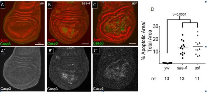
investigate the importance of centrosomes in somatic cell division by comparing the levels of apoptosis (programmed cell death) in wing discs homozygous for the sas-4 mutation to those in wild-type discs. To measure apoptosis, we used immunohistochemistry to label the cells with cleaved Caspase 3, a component of the apoptotic caspase cascade (Porter and Janicke, 1999). This revealed a significant increase in the amount of apoptosis occurring in the sas-4 mutant wing discs compared to the control discs (Figure 1).
With the observed increase in cell death in the sas-4 mutant wing discs we were motivated to look for errors in chromosome segregation in the acentrosomal cells that might contribute to the apoptotic phenotype of the mutants. We analyzed chromosome segregation by visualizing
chromosomes during anaphase, when they are moving away from the metaphase plate toward the spindle poles. In wild-type cells of the wing disc, chromosomes form an orderly arrangement as they segregate toward the spindle poles during anaphase. However, in sas-4 mutant cells,
Figure 1: Centrosome Loss Leads to Elevated Apoptosis: (A-C) Cleaved caspase 3(green) and actin (red) staining in the indicated genotypes (Note: yw represents wild type) (A’-C’). Cleaved caspase 3 staining shown in grayscale indicates increased apoptosis in acentrosomal mutants (B’,C’)
9
abnormalities such as lagging chromosomes and disorganized arrays of chromosomes are
observed at a significantly higher rate (data not shown). Lagging chromosomes indicate errors in chromosome segregation and chromosomes that are left behind during anaphase could become missegregated upon completion of mitosis, leading to aneuploidy. We therefore analyzed the karyotypes of cells from wild-type and sas-4 mutant wing discs to determine whether aneuploidy was present. Surprisingly, this analysis revealed no increase in aneuploidy in the acentrosomal cells. However, given the increase in cell death we observed in the mutant wing discs, we hypothesized that aneuploid cells may be eliminated from the tissue via apoptosis. To address this we sought to inhibit apoptosis using the baculovirus protein, p35, which has been shown to block apoptosis in Drosophila (Hay et. al., 1994). We drove the expression of this protein in the posterior half of wing discs using the engrailed promoter under the UAS-Gal4 system.
10 Genotype #
Normal #
Aneuploid %
Aneuploid
wild-type 137 5 3.5%
sas-4- 176 4 2.2%
+p35 145 6 4.0%
sas-4-,+p35 159 17 9.7% *
The Spindle Assembly Checkpoint facilitates acentrosomal cell division in the wing disc We were also interested in the effects of the loss of centrosomes on the timing of mitosis and spindle assembly. Using live imaging to measure the time between nuclear envelope break down and anaphase onset we found that this period of mitosis, which corresponds to the formation of a bipolar mitotic spindle, is prolonged in acentrosomal mutants compared to wild-type flies (data not shown). This led us to question what factors could be delaying mitosis in the mutant cells and how those factors are contributing to the efficacy of acentrosomal cell division. The SAC is known to operate during mitosis to delay anaphase onset until proper kinetochore-microtubule
B C
A
Figure 2: Aneuploidy Analyisis in the Wing Disc (A) Table showing the number of aneuploid and normal karyotypes observed in the indicated genotypes; * sas-4-,+p35 cells display a significant increase in aneuploidy relative to all other genotypes
(p<0.05). (B-C) DAPI stain is used to label chromosomes. (B) Example of a normal karyotype for Drosophila. (C)
11
12
Similarities and differences in acentrosomal cell division in the brain
In contrast to our work in the wing disc epithelium, previous studies have proposed that centrosome loss does not cause cell death in the developing fly brain, though it does result in moderate defects in asymmetric stem cell division (Basto et. al., 2006). To examine how centrosome loss affects mitotic fidelity in different tissues, we extended our analyses to the
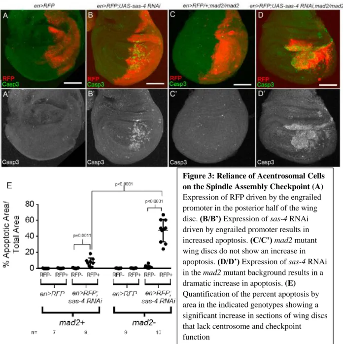
Figure 3: Reliance of Acentrosomal Cells on the Spindle Assembly Checkpoint (A)
Expression of RFP driven by the engrailed promoter in the posterior half of the wing disc. (B/B’) Expression of sas-4 RNAi driven by engrailed promoter results in increased apoptosis. (C/C’)mad2 mutant wing discs do not show an increase in apoptosis. (D/D’) Expression of sas-4 RNAi in the mad2 mutant background results in a dramatic increase in apoptosis. (E)
Quantification of the percent apoptosis by area in the indicated genotypes showing a significant increase in sections of wing discs that lack centrosome and checkpoint
13
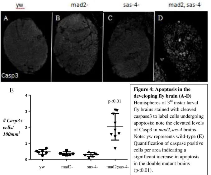
developing brain. As previously reported, and unlike in wing discs, we observed no significant increase in the amount of cell death in sas-4 mutant brains compared to wild-type (Figure 4).
0 1 2 3 4
D a t a 1
However, we know from our work in the wing disc that development is robust and compensatory mechanisms exist that can accommodate potential deleterious effects of centrosome loss on mitotic fidelity. We therefore again turned to the flies that were double mutant for sas-4 and the SAC component, mad2. Interestingly, we found that while larvae of this genotype completely lacked imaginal discs, they did form brains, though they were dramatically smaller than
yw mad2- sas-4- mad2;sas-4- p<0.01
# Casp3+ cells/ 100mm3
Figure 4: Apoptosis in the developing fly brain (A-D)
Hemispheres of 3rd instar larval fly brains stained with cleaved caspase3 to label cells undergoing apoptosis; note the elevated levels of Casp3 in mad2,sas-4 brains. Note: yw represents wild-type (E)
Quantification of caspase positive cells per area indicating a
significant increase in apoptosis in the double mutant brains (p<0.01).
14
type or single mutant brains, indicating significant developmental abnormalities in brains lacking both centrosomes and the SAC (Figure 5).
The source of many of the cells in the brain is the neuroblasts, which are effectively neural stem cells. The smaller brain size in the double mutants may thus be caused by a decreased number of neuroblasts. Therefore, to investigate the reduction in brain size in the double mutants and to
Figure 5: Loss of Centrosomes and SAC Results in Reduced Brain Size
(A-B) Images of brains from 3rd larval instars in the indicated genotypes with microtubules (green) and actin (red) showing tissue morphology. (yw=wild type)
(C) Quantification showing the significant decrease in brain size in the double mutants compared to both single mutants and wild type; single mutants not shown.
15
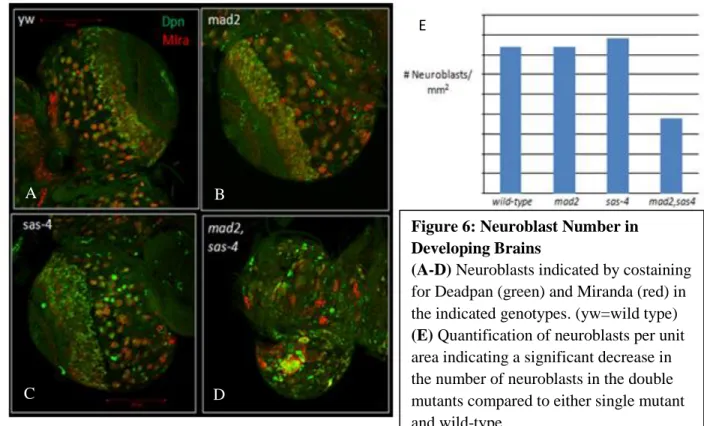
further analyze the effect of centrosome and SAC loss in the developing brain, we sought to characterize the number of neuroblasts present in the mad2;sas-4 double mutants compared to each of the single mutants and wild-type brains. To identify neuroblasts, we stained larval brains for two neuroblast-specific proteins: the transcription factor, Deadpan, and Miranda, a protein involved in asymmetric neuroblast division and cell fate determination. We quantified the number of neuroblasts in the mad2;sas-4 double mutant, as well as the sas-4 and mad2 single mutant and wild-type controls. This revealed a significant decrease in the number of neuroblasts in the double mutant when compared to either single mutant or wild-type brains (Figure 6). With the evidence of developmental defects in the brains of mad2;sas-4 double mutants we again sought to characterize cellular defects that could result from the loss of centrosome and SAC function. By analyzing cleaved Caspase 3 staining in the brains of the double mutants, we observed an increase in apoptosis compared to either single mutant, which did not display increased apoptosis (Figure 4). Thus, although brain cells appear refractory to the loss of
16
Finally, because we observed increased errors in chromosome segregation and aneuploidy in wing discs lacking centrosomes we sought to determine if similar errors would be present in mutant brains. Interestingly, neither the mad2 or sas-4 single mutants displayed a significant increase in aneuploidy. However, the double mutant showed a dramatic increase in aneuploidy with the majority of cells exhibiting abnormal chromosome number (Figure 7). Thus, while the brain has been shown to have a degree of tolerance for the loss of centrosomes, our data
demonstrate that the SAC plays a crucial role in facilitating acentrosomal cell division.
A B
C D
E
Figure 6: Neuroblast Number in Developing Brains (A-D) Neuroblasts indicated by costaining for Deadpan (green) and Miranda (red) in the indicated genotypes. (yw=wild type)
17
Genotype # Normal # Aneuploid Percent Aneuploid
wild-type 80 2 2.4%
mad2- 24 2 7.7%
sas-4- 86 9 9.5%
mad2;sas-4- 4 17 81%
Discussion:
With the presumed importance of centrosomes in nucleating a mitotic spindle and executing mitosis, it was surprising to learn that mutant flies lacking centrosomes survive to adulthood. This led to the interpretation that centrosomes are dispensable for mitosis. However, our data indicate that centrosomes promote spindle assembly, genome stability, and cell viability. The increase in cell death and the prevalence of errors in chromosome segregation in
acentrosomal cells are indicative of the challenges acentrosomal cells face when assembling a spindle. Multiple semi-redundant mechanisms exist in animal cells to ensure spindle assembly. Consistent with this, our data demonstrate that cells lacking functional centrosomes are able to form mitotic spindles and divide because of this robustness of mitosis. However, we did observe a significant decrease in the efficiency of spindle assembly in these mutants. Spindles required more time to assemble in the absence of centrosomes as indicated by the increase in the time between nuclear envelope breakdown and anaphase onset in mutant cells. Our data also indicate that acentrosomal cells are reliant on the SAC to facilitate the delay in mitosis until proper kinetochore attachments to microtubules are made. Although the ability of the SAC to delay
p<0.0001
18
mitosis has been well characterized, the reliance of acentrosomal cells on this cell cycle checkpoint reflects the importance of centrosomes in efficient spindle assembly.
The previously reported robustness of the brain to centrosome loss establishes this tissue as an interesting counter-model to wing disc epithelia, where we found centrosomes to be important in cell division and viability. Comparison of the findings from the brain and the wing disc facilitates an analysis of the abilities of different tissues to respond to mitotic perturbation via compensatory mechanisms. For example, while centrosome loss resulted in increased
19
have found that this tolerance can be partially attributed to the activity of cell cycle checkpoints, like the SAC, which are able to compensate for centrosome loss. Moving forward it would be interesting to investigate whether the dampened response of the larval brain to centrosome loss is due to an intrinsically greater reliance on centrosome independent pathways of microtubule nucleation, or if cell cycle checkpoints are better able to compensate for mitotic perturbation in this tissue than in the wing disc, or perhaps that brain cells are less likely to undergo apoptosis, possibly due to differences in expression or regulation of pro- or anti-apoptotic proteins.
With the phenotype of decreased brain size in the mad2;sas-4 double mutants in mind it is interesting to consider the disease of primary microcephaly in humans. Many of the mutations that have been linked to this disease occur in centrosomal proteins, but it is not known how these mutations contribute to disease development. The robustness of the developing fly brain to the loss of centrosomes alone is interesting in this context as it suggests that further perturbation of compensatory mechanisms could have an unappreciated role in the development of
microcephaly. The smaller brains of sas-4;mad2 double mutants display a decrease in neuroblast number indicating a significant developmental defect linked to the mitotic function of
centrosomes and the SAC. The presence of aneuploidy in the double mutants is significant when considering these flies as a model of this disease because mosaic variegated aneuploidy has been linked to microcephaly in humans suggesting that chromosomal instability and perturbations of the genome could have an underlying role in this disease (Hanks et al. 2004). Further
20 References:
Basto, R., Lau, J., Vinogradova, T., Gardiol, A., Woods, C.G., Khodjakov, A., Raff, J.W., (2006) “Flies without Centrioles.” Cell. Vol. 125, Issue 7, 1375-1386.
Burrel R. A., McGranahan N., Burlek J., Swanton C., (2013) “The causes and consequences of genetic heterogeneity in cancer evolution” Nature. Vol. 501, 338-345.
Clarke, P.R., and Zhang, C. (2008). Spatial and temporal coordination of mitosis by Ran GTPase. Nat. Rev. Mol. Cell Biol. 9, 464–477.
Foley, E.A., and Kapoor, T.M. (2013). “Microtubule attachment and spindle assembly checkpoint signalling at the kinetochore.” Nat. Rev. Mol. Cell Biol. 14, 25–37.
Goshima, G., Mayer, M., Zhang, N., Stuurman, N., and Vale, R.D. (2008). “Augmin: a protein complex required for centrosome-independent microtubule generation within the spindle.” J. Cell Biol. 181, 421–429.
Hanahan D., Weinberg R. A., (2011) “Hallmarks of Cancer: The Next Generation” Cell. 646-674 Hanks S., Coleman K., Reid S., Plaja A., Firth H., FitzPatrick D., Kidd A., Mehes K., Nash R.,
Robin N., Shannon N., Tolmie J., Swansbury J., Irrthum A., Douglas J., Rahman N., (2004) “Consititutional aneuploidy and cancer predisposition caused by biallelic mutations in BUB1B” Nat gen. Vol 36. Number 11, 1159- 1161.
Hay, B.A., Wolff, T., Rubin, G.M., (1994) “Expression of Baculovirus p35 Prevents Cell Death in Drosophila.” Development. Vol. 120, 2121-2129.
Hidalgo A., “Growth and Patterning from the Engrailed Interface.” (1998) The International Journal for Developemental Biology. Vol. 42, 317-324.
Kashio S., Obata F., Mintra M., (2014) “Interplay of cell proliferation and cell death in Drosophila tissue regeneration.” Develop Growth Differ. Vol. 56, 368-375.
Megraw, T.L., Sharkey, J.T., and Nowakowski, R.S. (2011). “Cdk5rap2 exposes the centrosomal root of microcephaly syndromes.” Trends Cell Biol. 21,470–480.
Orfanos, Z., “Transgenic tools for Drosophila muscle research”(2008) Journal of Muscle Research and Cell Motility. Vol. 29, Issue 6-8, 185-188.
21
Porter A.G. and Janicke R. U., “Emerging roles of Caspase-3 in apoptosis” (1999) Cell Death and Differentiation. 6, 99-104.