Address for correspondence Dr. Jasleen Kaur, Assistant Professor Department of Dermatology
D-Block, 5th Floor
Government Medical College & Hospital Chandigarh, India-160030
Original Article
Cutaneous features of chikungunya - A case series
from North India
Introduction
Chikungunya (CKG) is a mosquito-borne viral illness caused by CKG virus, which is an Arbovirus. Clinically, CKG presents as a self-limiting illness characterized by acute onset of high grade fever accompanied by severe arthralgia, myalgia, headache and various
mucocutaneous manifestations.1 The name is
derived from the Makonde language of Tanzania meaning "that which bends up" in reference to the contorted posture because of severe joint pain in this illness.2
The first outbreak of CKG was documented in Makonde, United Republic of Tanzania in 1952.2,3 The disease has been reported mainly
from Africa and different Asian countries cyclically, with an interepidemic interval of several years to decades.4 Although there is
evidence of persistence of the virus at a low level in the community; explosive outbreaks after periods of long quiescence is the usual epidemiologic pattern.5 This has been attributed
to various factors such as mutation in virus, loss of herd immunity, globalization and emergence of other vectors e.g. Aedes albopictus and A. aegyptii.6,7
In India, major epidemics of CKG were reported in 1963 in Kolkata. Thereafter, sporadic cases continued to be documented in various other states.8 National Vector Borne Disease Control Priyanka Sharma, Jasleen Kaur, Gurvinder P Thami
Department of Dermatology, Government Medical College & Hospital, Chandigarh, India.
Abstract
Objective To document cutaneous features of Chikungunya (CKG) in a recent outbreak of CKG in August 2016 in North India.Methods A total of 32 patients with cutaneous manifestations of suspected CKG were enrolled in the study.
Results A wide variety of cutaneous lesions were observed. Maculopapular rash was the most frequent cutaneous manifestation followed closely by hyperpigmentation. A typical nose pigmentation previously reported in CKG, was also noted in this study. In addition, diffuse facial pigmentation was another common pattern observed, predominantly in the females. Aphthous stomatitis and lymphedema were the other less frequent manifestations in this study. Vesiculobullous lesions were seen in infants. Exacerbation of underlying dermatoses was also noted.
Conclusion There is a wide spectrum of cutaneous manifestations in CKG besides the maculopapular rash, unlike most other viral infections. Some of the typical pigmentary patterns can be considered as markers for the diagnosis of CKG.
Key words
Program (NVBDCP) confirmed an outbreak of CKG in North India, with a peak in Delhi which commenced around August 2016 and lasted till the end of October affecting thousands of people.9 Here, we report the varied cutaneous
manifestations of CKG encountered during the outbreak in Chandigarh, India.
Methods
A total of 32 suspected cases of CKG during the two months of outbreak in September and October 2016, who presented with skin lesions to our dermatology outpatient department, were included. Suspect cases were fulfilling the "case definition" as per National Vector Borne Disease Control Programme (NVBDCP) 2016 guidelines
i.e. acute onset of fever, severe
arthralgia/arthritis, with or without skin rash and residing or having left an epidemic area 15 days prior to onset of symptoms. Capture IgM ELISA for CKG was done in all cases at the time of presentation. Skin biopsy of two cases was sent for histopathological examination. Patients were followed up for 8 weeks to study the evolution of lesions.
Results
Out of 32 patients, 17 were males and 15 were females (ratio of 1.13: 1). 60% of the patients were in the age group of 20-40 years with a range from 3 months to 69 years. The median age was 30 years.
Patients presented with a wide array of skin lesions, with many having more than one type of
lesions. Table 1 enumerates the various types of
lesions and their frequency. Maculopapular rash was the most frequent cutaneous manifestation followed closely by hyperpigmentation. In 12 (66.7%) patients, skin hyperpigmentation was preceded by maculopapular rash or transient erythema with pigmentary changes appearing
after 4-7 days of the subsidence of rash. However, in 6 (33.3%), pigmentation appeared
de novo after 7-10 days of the onset of fever. The pattern and extent of pigmentation varied from just localized to nose to generalized
involvement of the whole body (Table 2).
Histopathology from the pigmented lesions showed increase in epidermal basal layer
pigmentation and melanophages with
lymphocytic infiltration in the dermis. Serology for IgM antibodies for CKG was positive in 15 (46.8%) cases.
Discussion
CKG is a re-emerging viral illness with cyclical explosive outbreaks in different parts of the
world including Africa and South East Asia.5 A
variety of skin and mucosal manifestations have been documented in the cases of CKG infection in literature across the country and world.4,10-13 In
our study, the cutaneous manifestations were
Table 1 Various cutaneous lesions observed in patients of chikungunya (n=32).
Type of cutaneous lesion N (%)
Maculopapular rash 19 (59.4)
Hyperpigmentation 18 (56.3)
Vesicles and bullae 3 (9.4) Exacerbation of preexisting
dermatoses
4 (12.5) Aphthous stomatitis 2 (6.3)
Lymphedema 1 (3.1)
Table 2 Patterns of hyperpigmentation (n=18).
Pattern No. of
patients
Males Females Localized
pigmentation over nose
9 7 2
Freckle like (speckled) facial hyperpigmentation
5 0 5
Diffuse slate gray facial pigmentation
2 0 2
Generalized 1 1 0
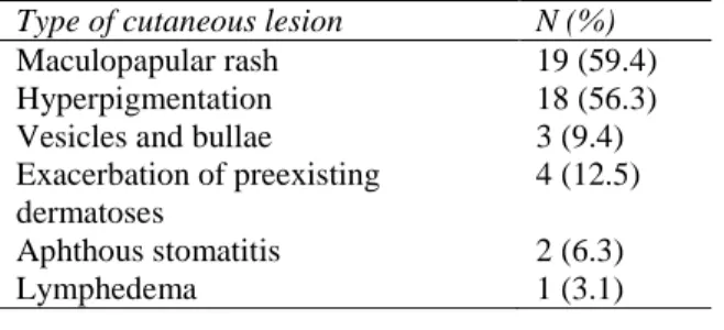
Figure 1 Nose pigmentation (Chik sign)
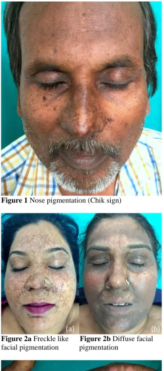
Figure 2a Freckle like Figure 2b Diffuse facial facial pigmentation pigmentation
Figure 3 Vesiculobullous lesions after rupture with denuded areas in an infant
noted in the acute phase of the illness. All age groups and both sexes were affected. Most common cutaneous manifestation observed in this study was maculopapular rash (59.4%), as has been reported in other studies too.10-12 The
rash appeared within 2-3 days of the onset of fever and subsided within first week in all the patients; earliest after 2 days and latest after 6 days of onset of fever, thus was abrupt in onset, short lasting and was associated with moderate to severe pruritus in most of the patients in this study (78.9%).
Hyperpigmentation, either de novo or as a
sequelae to other cutaneous lesions, was a prominent feature. The most common evolution of pigmentary change was following the generalized maculopapular rash, which might be because of increased intraepidermal melanin dispersion/retention triggered by the virus.4,13
Variety of patterns like nose only, diffuse slate-gray facial, freckle like, generalized and periorbital were noted as has been seen in previous studies also.5,11-13 In addition, the
pigmentation over nose which has been referred to as ‘Chik sign’ in a previous study11 was noted
predominantly in males (Figure 1) while
speckled and diffuse facial melanosis was noted predominantly in females (Figure 2 a and b). The reason for this variation could be the hormonal influences in females playing a role to
give more extensive pigmentation. The
occurrence of pigmentary changes in CKG and its variable patterns is peculiar and the exact pathogenesis is still not clear. Role of solar UV rays is postulated to play a role as it mainly affects the face in most of the patients. Histopathologic examination of hyperpigmented lesions has shown diffuse hypermelanosis of the entire epidermis with no melanin incontinence and a sparse perivascular lymphocytic infiltrate in the dermis in a previous study.4 Similar
study, but in addition melanophages were also seen in the dermis.
Superficial vesicobullous lesions were noted in two infants and one adult. There was difference in extent between the children and adults in the extent, with the vesicular lesions being widespread in the infants and only a few in the adult. In infants the clinical picture resembled toxic epidermal necrolysis or acrodermatitis enteropathica after the rupture of vesicles, which left superficial denuded areas of skin (Figure 3). Viral replication in the epidermis causing focal necrosis and ballooning degeneration has been attributed to be the cause of blistering in CKG.13
The vesicular lesions have been well
documented in infants,4,11,13 but the reports in
adults are not many. Vesicular lesions confined to hands and feet have been reported in two
adults in a study from South India.4
Exacerbation of existing dermatoses has been described in CKG.4,11 Exacerbation of psoriasis,
ichthyosis vulgaris, melasma and darkening of melanocytic nevi was observed in this study.
Serology was positive for IgM antibody in 46. 8% of cases in this study. It was negative in most of the cases presenting with maculopapular rash and early pigmentary changes. This could be because of absence of IgM antibodies against CKG in the early course of the disease. Positivity of IgM CKG antibodies has been reported in 13-97% of patients in other studies.10,11
The clinical differentiation of CKG from dengue is difficult since both share a similar geographical and clinical pattern leading to potential misdiagnosis of CKG in dengue endemic areas. Moreover, both pathogens (CKG and dengue virus) are transmitted by the same vector i.e. Aedes species mosquitoes, therefore epidemiology of both infections is temporally and spatially related.8
Presence of joint pain and joint swelling are more common in CKG while nausea/vomiting, retro-orbital pain, hemorrhagic manifestations, pleural effusion and ascites are more likely to occur in dengue. Characteristic cutaneous manifestation of dengue include generalized confluent erythema with petechiae and rounded islands of sparing - "white islands in a sea of red" while CKG presents as maculopapular rash,
skin hyperpigmentation characteristically
affecting nose in most of the patients. Vesicobullous lesions have been noted in infants with CKG fever.
Conclusion
CKG is a self-limiting re-emerging infection.
Unlike most viral infections, cutaneous
manifestations of CKG are not limited to just maculopapular rash; rather there is a wide spectrum of cutaneous lesions. Pigmentary changes, especially nose pigmentation also known as ‘Chik sign’, are peculiar to CKG. Diffuse facial pigmentation was the other common pattern noted predominantly in females in this study.
In conclusion, the characteristic cutaneous involvements in CKG fever play an important role in the clinical diagnosis and awareness about these should be there in all healthcare providers.
References
1. Mohan A. Chikungunya fever: clinical manifestations and management. Indian J Med Res. 2006;124:471-4.
2. Robinson MC. An epidemic of virus disease in Southern Province, Tanganyika Territory, in 1952-53 I: Clinical features. Trans R Soc Trop Med Hyg. 1955;49:28-32.
4. Inamadar A, Palit A, Sampagavi V, Raghunath S, Deshmukh NS. Cutaneous manifestations of chikungunya fever: observations made during a recent outbreak in south India. Int J Dermatol. 2008;47 :154-9.
5. World Health Organization, Regional Office for South-East Asia. Guidelines on Clinical Management of Chikungunya Fever. New Delhi: World Health Organization, Regional Office for South-East Asia; 2008.
6. Pialoux G, Gauzere BA, Jaureguiberry S, Strobel M. Chikungunya: an epidemic arbovirosis. Lancet Infect Dis. 2007;7 :319-27.
7. World Health Organization. Proposed case definition of chikungunya fever: WHO South East Asia Regional Office. Available at:
htttp://www.searo.who.int/LinkFiles/Chikun gunya_Def_Chikungunya_Fever.pdf [accessed on 2009 Sep. 21]
8. Cecilia D. Current status of dengue and chikungunya in India. WHO South East Asia J Public Health. 2014;3:22-6.
9. NVBDCP 2016.http://nvbdcp.gov.in/chik-cd.html Status as of Dec 2016. Accessed on Feb 16th, 2017.
10. Bandyopadhyay D, Ghosh SK. Mucocutaneous manifestations of Chikungunya fever. Indian J Dermatol. 2010;55:64-7.
11. Riyaz N, Riyaz A, Rahima, Abdul Latheef EN, Anitha PM, Aravindan KP et al. Cutaneous manifestations of chikungunya during a recent epidemic in Calicut, north Kerala, south India. Indian J Dermatol Venereol Leprol. 2010;76:671-6.
12. Bhat RM, Rai Y, Ramesh A, Sukumar D, Martis J, Kamath GH. Mucocutaneous manifestations of chikungunya fever: A study from an epidemic in coastal Karnataka. Indian J Dermatol. 2011;56 :290-4.
13. Seetharam KA, Sridevi K, Vidyasagar P. Cutaneous manifestations of chikungunya fever. Indian Pediatr. 2012;49:51-3.