Arrest of human mitochondrial RNA polymerase
transcription by the biological aldehyde adduct
of DNA, M
1
dG
Susan D. Cline
1,*, M. Fernanda Lodeiro
2, Lawrence J. Marnett
3, Craig E. Cameron
2and
Jamie J. Arnold
21
Division of Basic Medical Sciences, Mercer University School of Medicine, Mercer, GA 31207, 2
Department of Biochemistry and Molecular Biology, The Pennsylvania State University, University Park, PA 16802 and3A. B. Hancock Jr. Memorial Laboratory for Cancer Research, Department of Biochemistry, Vanderbilt University School of Medicine, Nashville, TN 37232, USA
Received March 25, 2010; Revised July 10, 2010; Accepted July 12, 2010
ABSTRACT
The biological aldehydes, malondialdehyde and base propenal, react with DNA to form a prevalent guanine adduct, M1dG. The exocyclic ring of M1dG opens to the acyclic N2-OPdG structure when paired with C but remains closed in single-stranded DNA or when mispaired with T. M1dG is a target of nucleotide excision repair (NER); however, NER is absent in mitochondria. Anin vitro transcrip-tion system with purified human mitochondrial RNA polymerase (POLRMT) and transcription factors, mtTFA and mtTFB2, was used to determine the effect of M1dG on POLRMT elongation. DNA templates contained a single adduct opposite either C or T downstream of either the light-strand (LSP) or heavy-strand (HSP1) promoter for POLRMT. M1dG in the transcribed strand arrested 60–90%
POLRMT elongation complexes with greater
arrest by the adduct when opposite T. POLRMT was more sensitive to N2-OPdG and M1dG after initiation at LSP, which suggests promoter-specific differences in the function of POLRMT complexes. A closed-ring analog of M1dG, PdG, blocked 95% of transcripts originating from either promoter regardless of base pairing, and the transcripts remained associated with POLRMT complexes after stalling at the adduct. This work suggests that persistent M1dG adducts in mitochondrial DNA hinder the transcription of mitochondrial genes.
INTRODUCTION
The mitochondria of eukaryotic cells each harbor multiple copies of a 16 569 bp circular chromosome that must be maintained for efficient cellular energy production (1,2). Electrons that escape from the transport chain in the inner membrane react with molecular oxygen and water to produce reactive oxygen species (ROS) in the matrix where mitochondrial DNA (mtDNA) resides (3). The ROS may damage mtDNA directly by oxidation of bases or the sugar backbone. Additionally, ROS oxidation of DNA and inner membrane lipids generates biological aldehydes, such as base propenals, malondialdehyde (MDA) and 4-hydroxynonenal, which react with nucleic acid bases (4). The base propenals generated from DNA sugar backbone oxidation appear to be the major endogenous source of the 3-(20-deoxy-b
-D
-erythro-pentofuranosyl)pyrimido[1,2-a]purin-10(3H)-one adduct of guanine known as M1dG (Figure 1) (5–8). M1dG has been reported to occur in human tissues at levels compar-able to the most abundant form of base oxidation, 8-oxodeoxyguanine, and to be twice as prevalent in mtDNA than in nuclear chromosomes (4,8–12). In eu-karyotic cell nuclei, excision repair pathways remove modified bases and maintain genetic integrity. Base excision repair (BER), which removes oxidized bases such as 8-oxodeoxyguanine and thymine glycol, also func-tions in mitochondria (13–15). Conversely, nucleotide excision repair (NER), the mechanism implicated in pro-cessing of M1dG, is absent in the organelle (16–18). The lack of NER and the presence of ROS in the mitochon-drial matrix may contribute synergistically to the persist-ence of M1dG in mtDNA.
*To whom correspondence should be addressed. Tel: +1 478 301 2231; Fax: +1 478 301 5487; Email: [email protected]
doi:10.1093/nar/gkq656
ßThe Author(s) 2010. Published by Oxford University Press.
The chemical properties of M1dG make this form of base damage particularly interesting with respect to its potential detriment to DNA replication and gene expres-sion. As seen in Figure 1, the external ring of M1dG is closed in single-stranded DNA or when mispaired with thymine, but opens to an acyclic N2 -(3-oxo-1-propenyl)-dG derivative (N2-OPdG), when correctly paired in DNA with cytosine (19,20). Therefore, a single aldehyde modi-fication of DNA to form M1dG may result in two struc-turally distinct adducts that must be negotiated by DNA and RNA polymerases and recognized by DNA repair pathways. A chemically stable M1dG analog, 1,N2 -propano-20-deoxyguanosine, called PdG, has been used
to elucidate differences in M1dG and N2-OPdG effects on prokaryotic and eukaryotic polymerases.
In bacteria and mammalian cells, M1dG causes a high rate of frameshifts and base substitutions during replica-tion (21–24). M1dG and PdG disrupt nucleotide insertion and translocation by the Klenow fragment of bacterial DNA polymerase I (25), the human BER polymerase, DNA pol b (26,27), and the human translesion DNA polymerase, pol Z (28,29). Recent in vitro transcription studies revealed a structure-dependent M1dG arrest of elongation by the T7 bacteriophage RNA polymerase (T7RNAP) and by mammalian RNA polymerase II (RNAP II) that suggests M1dG may also disrupt gene expression (30). Both polymerases exhibited a greater transcriptional arrest at M1dG opposite T; however, RNAP II elongation complexes were more sensitive than T7RNAP to both biological forms of the adduct. PdG, the analog of the closed-ring M1dG, completely blocked
RNAP II translocation regardless of the opposing base
(30). Human mitochondrial RNA polymerase
(POLRMT) is a single subunit enzyme homologous to T7RNAP; therefore, M1dG and its acyclicN2-OPdG con-formation may substantially hinder POLRMT transcript elongation. In addition, POLRMT, like RNAP II, requires the assistance of other proteins for promoter ini-tiation, a property that could influence POLRMT behavior upon encountering DNA damage (1,31–34). As POLRMT elongation remains largely uncharacterized, studies of POLRMT interaction with M1dG provide not only information about the biological impact of mtDNA damage on mitochondrial gene expression but also under-standing of basic POLRMT function.
POLRMT transcription initiates at different promoter sequences on the heavy (H) strand and the light (L) strand of the mitochondrial chromosome and produces polycistronic RNAs (1,31,35–37). The H strand contains genes for 12 of the 13 mtDNA-encoded proteins and the mitochondrial ribosomal RNA (rRNA) and transfer RNA (tRNA) required for translation of the proteins. The heavy strand promoter HSP1 is thought to primarily direct the synthesis of a transcript including 12S and 16S rRNA that terminates in the tRNALeugene (38), while a second promoter, HSP2, has been proposed to be the site for messenger RNA and tRNA production from the H strand (39). The L strand promoter, LSP, not only permits POLRMT transcription, but also controls POLRMT synthesis of an RNA primer for mtDNA rep-lication at the H strand origin (40–42). The nuclear-encoded proteins, mtTFA, mtTFB1 and mtTFB2, support promoter-directed transcription of mtDNA by POLRMT (1,2,31,43,44). The combination of mtTFA and mtTFB2 maximally stimulates POLRMT activity at LSP and HSP1in vitro, while the primary role of mtTFB1 appears to be as an rRNA methyltransferase that helps to link mtDNA transcription and translation (1,31,33,34, 45–47). Given the different roles of LSP and HSP1 in directing activities on the mitochondrial chromosome, DNA damage that interferes with POLRMT transcript elongation could hinder both mtDNA gene expression and replication.
To investigate the consequences of M1dG on transcript elongation by POLRMT and gain insight into POLRMT function, we employed an in vitro transcription system containing purified human POLRMT, transcription factors, mtTFA and mtTFB2, and DNA templates con-taining a site-specific M1dG or PdG in either the transcribed (Xts) or non-transcribed (Xnts) strand down-stream of LSP or HSP1 (Figure 2). M1dG was placed opposite either C or T to elucidate the structural effects ofN2-OPdG and M1dG, respectively, on POLRMT. The DNA sequence context of the adduct within the POLRMT templates was identical to that used in the previous studies of RNAP II and T7RNAP, allowing for a comparison of the elongation behavior of these polymer-ases. In light of the results showing substantial M1dG effects on T7RNAP and RNAP II transcription, we hypothesized that M1dG would impede POLRMT elong-ation in a structure-dependent manner suggesting that persistent M1dG adducts may disrupt the expression of Figure 1. Formation of M1dG by malondialdehyde (MDA) and base
propenal. The exocyclic adduct, M1dG, can react with the amino group
of a paired cytosine and form acyclicN2-OPdG. PdG is a stable analog
genes in mtDNA. As POLRMT operates from two dis-tinctly different promoters on the mitochondrial chromo-some, we believed that our system also would provide insight into potential promoter-dependent properties of POLRMT elongation, such as the response of POLRMT to mtDNA sequences that govern transcript length.
MATERIALS AND METHODS
Reagents
T4 polynucleotide kinase, T4 DNA polymerase and M13K07 helper phage were purchased from New England Biolabs (Beverly, MA, USA), and T4 DNA ligase was from Invitrogen (Carlsbad, CA, USA). The F0
Escherichia coli strain MV1184 was developed by J. Messing (Rutgers University, Piscataway, NJ, USA). Non-radioactive ribonucleoside and deoxyribonucleoside triphosphates were purchased from GE Healthcare Bio-sciences Corp (Piscataway, NJ, USA). Radionuclides,
[a-32P] UTP, [a-32P] CTP and [g-32P] ATP, were obtained from Perkin Elmer (Boston, MA, USA). Unmodified oligonucleotides were synthesized by Eurofins MWG Operon (Huntsville, AL, USA). Streptavidin-linked paramagnetic particles were from Promega (Madison, WI, USA).
Preparation of Oligonucleotides
Oligonucleotides of the 18-base sequence 50-ATC GGC
GCC GXC GGT GTG-30 containing either a single
M1dG or PdG at the position X were synthesized as pre-viously described (48), and their purity and presence of the adduct was verified by mass spectrometry. Samples of the oligonucleotides were 32P labeled and resolved by denaturing polyacrylamide gel electrophoresis (PAGE) to check for degradation that could affect construction of the plasmid templates below. The undamaged 18-mer (X = dG) and the oligonucleotides for both strands of LSP and HSP1 were prepared by Eurofins MWG Operon (Huntsville, AL, USA). The LSP and HSP1 se-quences were as follows: LSP nontranscribed strand,
50-AAT TCA TGT GTT AGT TGG GGG GTG ACT
GTT AAA AGT GCA TAC CGC CAA AAG ATA AAA TTT GAA ATC T-30; LSP transcribed strand,
30-GTA CAC AAT CAA CCC CCC ACT GAC AAT
TTT CAC GTA TGG CGG TTT TCT ATT TTA AAC TTT AGA GAT C-50; HSP1 nontranscribed strand,
50-AAT TCC GCT GCT AAC CCC ATA CCC CGA
ACC AAC CAA ACC CCA AAG ACA CCC CCC T-30; and HSP1 transcribed strand, 30-GGC GAC GAT
TGG GGT ATG GGG CTT GGT TGG TTT GGG GTT TCT GTG GGG GGA GAT C-50. These sequences were
designed according to studies of human mtDNA pro-moters by the Clayton group with modifications to facili-tate transcription template construction (37)
Construction of transcription templates
The plasmid transcription templates for POLRMT were constructed from plasmids used in previously published studies of RNAP II and T7RNAP transcriptional arrest (30). The identical sequence context for the adduct position (Figure 2,X) was maintained allowing functional comparisons of the three polymerases. In brief, a double-stranded oligonucleotide containing either the mitochondrial LSP or HSP1 sequence modified to create a ‘labeling cassette’ downstream of the promoter was ligated, at a 20:1 molar ratio of oligonucleotide to vector, into the EcoRI–XbaI cloning site replacing the T7RNAP promoter (30) (Figure 2). The constructs were transformed into the F0E. colistrain MV1184 for
replica-tion of the plus strand by the M13K07 phage. Plasmids containing dG, M1dG or PdG in either the transcribed or nontranscribed strand were prepared by T4 DNA poly-merase second-strand synthesis on a single-stranded plasmid using the appropriate 18-mer as a primer (49). For each synthesis, a control reaction without the 18-mer was performed to insure that no intact plasmid was generated from aberrant priming by the single-stranded DNA. Products were resolved by electrophoresis in TAE (40 mM Tris–acetate, 2 mM disodium EDTA, Figure 2. DNA transcription templates for POLRMT. The linear Xts
and Xnts DNA templates contain dG, M1dG or PdG (X) either in the
pH 8.5) on 1% agarose containing 0.3mg/ml ethidium bromide. Plasmids were excised, electroeluted from the gel using an Amicon Centrilutor (Millipore, Billerica, MA, USA), and concentrated by centrifugation. The con-structs were digested with HindIII, then gel purified and isolated as described earlier.
The 118-bp POLRMT transcription templates were constructed by equimolar annealing of 79-, 18- and 21-base transcribed strand oligonucleotides to a 118-21-base nontranscribed strand sequence followed by ligation with T4 DNA ligase for 12–16 h at 16C. The 18-mer
was the same PdG-adducted sequence utilized above, and the 21-mer comprising the 50 end of the transcribed
strand contained a 50 biotin. The ligation reactions were
extracted with 25:24:1 phenol/chloroform/isoamyl alcohol and filtered through G-25 then G-100 Sephadex to remove contaminates and unligated transcribed strand 18-mer and biotinylated 21-mer. The 118-bp templates were resolved on 3.5% agarose to ensure purity. The concentration of each template was determined by ultraviolet spectropho-tometry with a Shimadzu ND-1000 Nanodrop instrument (Columbia, MD, USA).
POLRMTin vitro transcription
Recombinant human POLRMT, mtTFA and mtTFB2 were purified fromE. coliby previously published proto-cols with modifications submitted separately for publica-tion (43,50,51). Proteins of comparable quality from these preparations can be obtained from Enzymax, LLC (Lexington, KY, USA), which has not provided funding for or influenced the research reported herein. For promoter binding of transcription factors, 100 nM mtTFA and 24 nM mtTFB2 were incubated at 32C for
5 min with 4 nM LSP or HSP templates in POLRMT tran-scription buffer (10 mM HEPES, pH 7.9, 10 mM MgCl2, 20 mM NaCl, 0.1mg/ml bovine serum albumin, 1 mM dithiothreitol). Reactions for LSP template binding con-tained 100mM ATP, 20mM GTP, 10mM UTP and 0.1mM [a-32P] UTP (3000 Ci/mmol) and those for HSP template binding contained 100mM ATP, 20mM GTP, 10mM CTP and 0.1mM [a-32P] CTP (3000 Ci/mmol). Addition of 20 nM POLRMT initiated transcription at each promoter and continued incubation at 32C allowed
POLRMT extension of transcripts to the end of the ‘labeling cassette’ in each template (Figure 2). By excluding the next ribonucleotide required for transcript extension (CTP for the LSP templates and UTP for the HSP1 templates), POLRMT complexes were stalled until ribonucleoside triphosphate (NTP) addition. After 5 min for LSP template reactions and 10 min for HSP template reactions, heparin was added at 50 ng/ml to inactivate un-initiated POLRMT, then elongation by the stalled POLRMT complexes was induced by increasing all four NTP concentrations to 400mM. The incubation times chosen for POLRMT initiation provided the maximal amount of cassette-arrested transcripts that remained competent for elongation following heparin and NTP addition. Control reactions confirming heparin inhibition of POLRMT re-initiation and an absence of additional 32
P incorporation during elongation are shown in
Supplementary Data (Figure S1). After elongation times up to 30 min at 32C, reactions were stopped with 0.3%
SDS/22.5 mM EDTA, proteins digested with 0.4 mg/ml proteinase K, and nucleic acids precipitated with ethanol by centrifugation. Samples were dried under vacuum, re-suspended in 80% formamide dye, and resolved by denaturing 5% polyacrylamide gel electrophoresis in TBE (89 mM Tris-borate, 1 mM EDTA, pH 8.0). The gels were dried, and then transcripts were detected by phosphorimaging analysis using a Storm 840 system with Image Quant software (GE Healthcare Bio-sciences Corp, Piscataway, NJ, USA).
POLRMT transcription complex stability
The stability of PdG-arrested POLRMT complexes was assessed by isolation of transcripts produced from a biotinylated 118-bp template linked to streptavidin-conjugated paramagnetic particles (SA-PMPs). Briefly, SA-PMPs were washed in cold POLRMT oligonucleotide transcription buffer (10 mM HEPES, pH 7.5, 10 mM MgCl2, 100 mM NaCl, 0.1mg/ml bovine serum albumin, 1 mM dithiothreitol) then resuspended at 6 mg/ml. For DNA binding to SA-PMPs, 150mg of SA-PMPs were mixed with 3.6 pmol biotinylated 118-bp template and incubated at 32C for 5 min. The buffer was removed
from the template-linked SA-PMPs followed immediately by addition of 8ml of transcription reaction mix contain-ing 360 nM mtTFA, 360 nM mtTFB2, 600mM ATP, 150mM GTP, 100mM UTP, 60mM CTP and 0.33mM [a-32P] UTP (3000 Ci/mmol) in POLRMT oligonucleotide transcription buffer. Samples were incubated at 32C for
2 min to allow transcription factor binding then POLRMT was added at 360 nM to initiate transcription in a total reaction volume of 10ml. After 2 min, 60 ng/ml heparin was added to prevent further initiation and incubation was continued at 32C for 1, 5 or 15 min when the SA-PMPs
were washed two times with 100ml 32C POLRMT
tran-scription buffer. Following buffer aspiration from the SA-PMPs, POLRMT complexes were denatured by heating at 80C in 80% formamide/50 mM EDTA
loading dye. Transcripts associated in stable complexes on the SA-PMPs-linked templates were resolved by 20% polyacrylamide gel electrophoresis in TBE and detected by phosphorimager analysis as above.
reaction samples, then proteins were detected in the gel by silver staining.
RESULTS
M1dG and PdG adducts block POLRMT transcription
from LSP
We have employed an experimental design used previously for assessing DNA damage arrest by T7RNAP and RNAP II to analyze the behavior of POLRMT at a site-specific M1dG adduct in the identical sequence context following initiation at either LSP or HSP1 (30,44). Beginning with an analysis of transcripts produced by POLRMT following LSP initiation, we have compared transcript arrest by the adduct and its PdG analog in each template as well as the extent of arrest after initiation at the two promoters. Transcripts from the LSP Xts and Xnts DNA transcription templates were32P labeled by radionucleotide incorporation along an 18-base sequence cassette downstream of the transcrip-tion start site for POLRMT (Figure 2). POLRMT was stalled at the end of the labeling cassette on each DNA molecule by exclusion of CTP. Following heparin addition, all four ribonucleotides were provided for tran-script elongation, which minimized further incorporation of 32P due to the presence of the excess cold NTP (see Supplementary Figure S1). By this method, the transcripts produced during POLRMT elongation were the result of a single promoter-initiated, transcription event on each active DNA template molecule. Full-length, run-off tran-scripts (RO) and transcripts resulting from arrest at the adduct position (X) were identified by comparison with 32
P-labeled, single-stranded DNA markers (Figure 3). The percentage of POLRMT transcriptional arrest by the adducts was determined by dividing the intensity of the signal corresponding to adduct arrested transcripts by the sum of the intensities of the arrested and run-off tran-scripts in each sample lane on the gels.
M1dG present in the transcribed strand of the LSP template arrested70% of transcripts when paired with C and>90% when paired with T (Figure 3A and C). The PdG analog of M1dG blocked around 95% of POLRMT elongation complexes regardless of base pairing, and neither adduct in the nontranscribed strand affected POLRMT transcription (Figure 3B). These results were consistent with the findings from T7RNAP and RNAP II experiments showing that the exocyclic M1dG is a stronger block to elongation than the acyclic N2-OPdG; however, POLRMT arrest by the adducts was more sub-stantial than that observed with the other polymerases (30). The mitochondrial polymerase was more sensitive to M1dG than the nuclear RNAP II (90% versus 70% arrest, respectively), and POLRMT was impeded to a much greater extent than the structurally similar T7 enzyme by both N2-OPdG and M1dG, suggesting that unique amino acid sequences in POLRMT or the associ-ation of transcription factors with POLRMT may impart functional differences to the POLRMT elongation complex (1). As POLRMT is capable of transcript elong-ation past the M1dG adduct, particularly its acyclic form
N2-OPdG, a timecourse was performed to ascertain how readily this bypass occurs.
POLRMT is capable of efficient M1dG bypass
To determine the efficiency of POLRMT elongation past M1dG and the relationship between POLRMT bypass and M1dG structure, transcripts generated from the LSP Xts templates were sampled at times up to 30 min following the restart of POLRMT elongation from the end of the labeling cassette (Figure 4). On templates containing M1dG opposite C, the appearance of POLRMT run-off transcripts coincided with transcripts from arrest at the adduct, indicating that N2-OPdG caused a transient block to POLRMT translocation (Figure 4A). On all tem-plates, the total amount of transcription (arrested plus run-off transcripts) reached a maximum within 1 min, and the proportion of run-off transcripts increased by Figure 3. POLRMT arrest at M1dG and PdG in LSP templates. The 32P-labeled transcripts from 30 min of elongation by POLRMT on LSP
Xts (A) and LSP Xnts (B) DNA templates containing dG (G), M1dG
(M) or PdG (P) paired with C (LSP Xts X:CandLSP Xtns X:C) or T (LSP Xts X:T) are shown. Adduct-arrested transcripts are indicated with X and run-off transcripts with RO. Numbers in the center indicate DNA marker lengths in lanes L100 and L10. (C) A graph presenting the averages of the percent of transcripts arrested at M1dG and PdG opposite C (white bars) or T (black bars) in at least
only 5% over the duration of the time course. Because total transcription did not increase over time, all POLRMT complexes stalled at the labeling cassette ap-parently restarted transcription simultaneously upon the addition of NTPs, and the majority of POLRMT complexes arrested at N2-OPdG may be unstable. This was supposed as the proportion of run-off transcripts did not significantly increase after the first minute.
Similar elongation kinetics were observed for bypass of M1dG:T (Figure 4B). As predicted from the 30-min elong-ation reactions above, the exocyclic M1dG was a stronger block thanN2-OPdG to transcription in the initial seconds and arrest at the adduct was sustained over time. Regardless of base pairing, PdG immediately halted nearly all POLRMT transcription (Figure 4A, PdG:C; PdG:T not shown), indicating that the chemical dynamics of the exocyclic ring has a significant influence on POLRMT templating and translocation. It is possible that POLRMT complexes that correctly insert cytosine opposite N2-OPdG or M1dG readily extend their tran-scripts, while complexes that are incapable of insertion or that incorporate another base quickly become unstable and abort transcription. Indeed, if the exocyclic ring is incapable of opening, as with the PdG adduct, base insertion by POLRMT is apparently prevented.
PdG-arrested POLRMT transcription complexes are stable
The arrested transcripts detected in the experiments above may be released during detergent denaturation of stable POLRMT complexes stalled at the adducts or may result from the dissociation of unstable transcription complexes soon after POLRMT encounters an adduct. As PdG induced the strongest POLRMT transcription arrest, biotinylated, oligonucleotide templates containing the
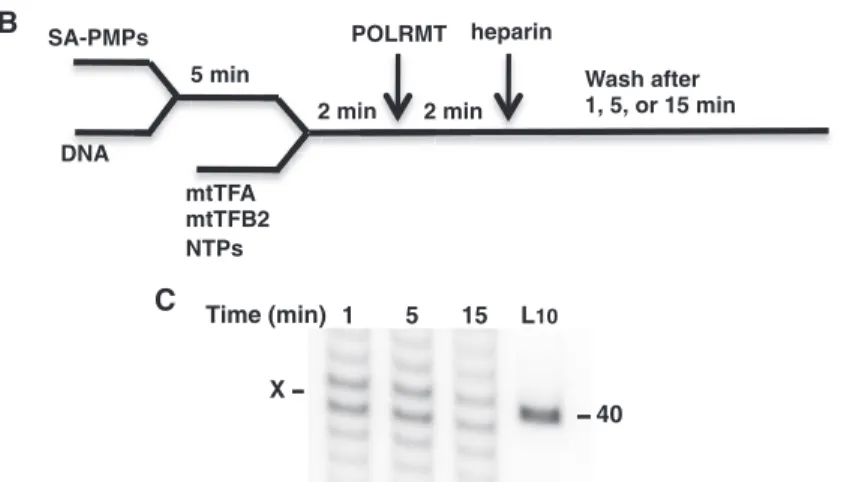
LSP and PdG adduct were utilized for in vitro analysis of arrested POLRMT complex stability (Figure 5). Transcripts corresponding to POLRMT arrest at the PdG adduct were observed up to 15 min after heparin addition and appear to reflect insertion up to and possibly opposite the adduct (Figure 5C). The arrested transcripts were absent in reactions containing a biotinylated, undamaged 118-bp template and isolation of the transcripts depended upon biotin linkage of the damaged template to the SA-PMPs (data not shown). The DNA marker (at 40 nt) served only to approximate the migration of the arrested transcripts, expected to be 35 or 36 nt, because mobility discrepancies between short DNA and RNA are known to occur. The transcripts produced by POLRMT complexes were formed prior to heparin addition and were released only when denatured with formamide after washes with copious amounts of buffer, indicating that they are a part of stable transcrip-tion complexes on the templates. Further evidence for arrested elongation complex stability was revealed when POLRMT was found in association with the PdG-containing biotinylated template following SA-PMP iso-lation from 1-min transcription reactions (data not shown).
While the time-course studies were conducted with an equimolar excess of NTPs to favor adduct bypass, the reactions to assess stability contained NTPs at a molar ratio of 6 ATP:1.5 GTP:1 UTP:0.6 CTP, which was based on a report of mitochondrial NTP levels in HeLa cells and intended to emulate biological conditions (52). The shorter oligonucleotide templates in this analysis also allowed for the detection of RNA products that may have resulted from templating by the adduct (Figure 5C,X) and suggest that further studies of insertion kinetics by POLRMT at M1dG and PdG are warranted.
Figure 4. POLRMT transcriptional bypass of M1dG or PdG following initiation at LSP. POLRMT damage-arrested (X) and full-length (RO)
transcripts from LSP Xts templates containing dG, M1dG, and PdG paired with C [dG:C, M1dG:C, andPdG:C, (A)] or with T [M1dG:T, (B)] are
POLRMT elongation complexes initiated from HSP1 are
less sensitive to M1dG and PdG
POLRMT molecules initiating transcription at LSP and HSP1 in mtDNA are responsive to different signals in the mitochondrial chromosome controlling the length of RNA transcripts (31,36–39). LSP directs transcription of both a polycistronic transcript, nearly the length of the chromosome, and a short RNA primer for mtDNA H strand replication. HSP1 initiates POLRMT transcription to produce an rRNA transcript that terminates at a sequence within the tRNALeu gene (38,39). Previous studies have characterized POLRMT activity at both pro-moters in the presence of mtTFA and mtTFB2 (33,44). Here, we have compared the response of POLRMT to M1dG and PdG following POLRMT initiation at HSP1 and LSP in ourin vitro templates (Figure 2).
An analysis of the transcripts from POLRMT elong-ation on the HSP Xts and Xnts templates are shown in Figure 6. As expected, POLRMT translocation was impeded to a greater extent by M1dG, which arrested nearly 80% of HSP1-initiated transcripts, while the acyclicN2-OPdG arrested60% of POLRMT transcripts (lanes M, panel A and panel C). PdG blocked nearly all transcript elongation (lanes P, panel A) regardless of base pairing, and no arrested transcripts were observed when the adducts were in the nontranscribed strand (panel B). Surprisingly, the amount of transcripts arrested by N2 -OPdG and M1dG was 10% lower from HSP templates than from LSP templates (Table 1), and POLRMT inter-action with the adducts when opposite T resulted in tran-scripts that appear to arrest immediately and to continue several bases past the adducts. It is unclear whether POLRMT response to the T-paired adducts relates to
properties imparted to the polymerase by HSP1 at initi-ation or to long-range DNA structural effects on HSP1 stemming from a distortion at the mispaired adducts that alter initiation site selection by POLRMT. Two tran-scripts of 315 and 340 bases in length were noted in the lanes for samples with the undamaged and M1dG tem-plates (Figure 6A, lanes G and M). These products likely resulted from stalling of POLRMT at pause sites in the sequence downstream of the adduct position and were not as apparent for POLRMT transcription of the identical sequences in the LSP templates (Figure 3). The distinctive POLRMT pausing after HSP1 and the differences in response to T-paired adducts in the HSP and LSP tem-plates may relate to recently reported differences in POLRMT initiation that could affect POLRMT inter-action with the template strand during elongation (44). Taken together, these findings indicate that POLRMT complexes on both strands of the mitochondrial chromo-some will respond to M1dG in a structure-dependent manner but that POLRMT transcribing the rRNA genes from HSP1 on the G-rich H strand may more readily bypass both forms of the endogenous adduct.
POLRMT elongation complexes initiated from HSP1 also
efficiently bypassN2-OPdG
To assess whether the kinetics for adduct bypass are promoter-dependent, POLRMT transcripts initiated at HSP1 were isolated at times up to 30 min following NTP stimulation of elongation (Figure 7). As observed with the LSP templates, all POLRMT molecules appeared to resume transcription concurrently upon NTP addition and to complete elongation within the initial minute. While PdG immediately blocked >95% of POLRMT Time (min) 1 5 15 L10
40 X
C
X
+1 +36
mtTFA site
A
SA-PMPs
DNA
5 min
mtTFA mtTFB2 NTPs
2 min POLRMT
2 min heparin
Wash after 1, 5, or 15 min B
transcripts, the acyclicN2-OPdG initially arrested around 60% of the transcripts, with run-off transcripts increasing slightly over the first 5 min. While POLRMT was less sen-sitive toN2-OPdG following HSP1 initiation, the rate at which bypass occurred did not differ significantly from that observed with LSP template. This indicates that neither the stability of POLRMT complexes arrested at these adducts nor the kinetics for POLRMT bypass of N2-OPdG is dependent on the promoter directing tran-scription. The findings from this in vitro system indicate that M1dG in mtDNA may cause stable arrest of POLRMT complexes thereby interfering with elongation from subsequent initiation events, and in essence, inactivating a polycistronic region of the mitochondrial chromosome.
DISCUSSION
In the nucleus, RNAP II serves as a sensitive DNA damage recognition factor for NER to increase the effi-ciency of repair in the transcribed relative to the nontranscribed strand of active genes (53). M1dG is a target of NER and inhibits transcription by mammalian
RNAP II and the bacteriophage T7RNAP (16,30). RNAP II arrest by M1dG may facilitate NER recognition and removal the adduct from nuclear DNA, but mitochondria have no known mechanism comparable to NER (16–18). Indeed, our current knowledge of mtDNA repair suggests that the arrest of POLRMT would have only deleterious consequences for mitochondrial function and cellular health. Since M1dG occurs in mitochondria at a 2-fold higher base frequency than in nuclear DNA, the mitochondrial chromosomes are highly susceptible to genetic instability due to the presence of this endogenous form of DNA damage (11).
Our study has provided insight into the ramifications of persistent M1dG for mtDNA gene expression through the use of a POLRMTin vitrotranscription system including templates with site-specific M1dG and PdG adducts and the mitochondrial transcription factors, mtTFA and mtTFB2. The DNA template sequence context of the adducts was identical to that in previous work characterizing the effects of M1dG and PdG on RNAP II and T7RNAP transcript elongation; therefore, the behavior of the three polymerases at the adducts could be compared for evident differences in their biochemical function (30).
As observed with T7RNAP and RNAP II, POLRMT arrest by M1dG was dependent upon the structure of the adduct such that the polymerase more readily bypassed the ring-openN2-OPdG (formed when M1dG is opposite C) than the exocyclic M1dG (when M1dG is opposite T) (Table 1). PdG, the closed-ring analog of M1dG, halted nearly all POLRMT elongation, suggesting that chemical dynamics of M1dG may allow the active site of POLRMT to accommodate templating and insertion of a ribonucleotide for bypass of the endogenous adduct (54). Nuclear magnetic resonance (NMR) studies have shown that the oxopropenyl group of N2-OPdG lies within the DNA minor groove and causes little distortion at the base pair (19), while PdG, the planar analog of the M1dG, participates in Hoogsteen pairing and rotates to a syn conformation at the glycosylic bond placing the exocyclic ring in the major groove (55). M1dG is predicted to cause base pair distortions similar to those observed in DNA containing PdG; however, the chemistry of the M1dG ring appears to determine the behavior of RNA polymer-ases attempting to ‘read’ the adduct during transcription. Figure 6. POLRMT arrest at M1dG and PdG in HSP templates. The
32P-labeled transcripts from (
A) HSP Xts templates (HSP Xts X:Cand
HSP Xts X:T) or (B) HSP Xnts X:C templates containing dG (G), M1dG (M) or PdG (P). Adduct-arrested (X) and run-off (RO)
tran-scripts are indicated. Numbers at the sides indicate DNA marker lengths in lanes L100and L10. (C) A graph presenting the averages of the percent of transcripts arrested at M1dG and PdG opposite C
(white bars) or T (black bars) for at least four independent experiments.
Table 1. POLRMT, RNAP II and T7RNAP transcriptional arrest induced by M1dG and PdG (30)
X:(C/T)a POLRMTb(%) RNAP II (%) T7RNAP (%)
N2-OPdG LSP 68 ± 5.4 60 27
(M1dG:C) HSP1 59.9 ± 3.6
M1dG LSP 92.1 ± 6.8 70 36
(M1dG:T) HSP1 82.7 ± 3.3
PdG LSP & HSP195 100 68
a
X = M1dG shown opposite either C or T; X = PdG results were
similar regardless of base pairing.
b
The observed differences in the arrest of T7RNAP, RNAP II and POLRMT by M1dG within an identical sequence context suggest that the polymerases have unique active site interactions with the transcribed strand and with the adduct itself during elongation.
The initiation and elongation mechanisms of T7RNAP and RNAP II have been characterized through biochem-ical and structural studies, but information about POLRMT is limited. Based on its sequence homology to T7RNAP, POLRMT has been presumed to operate much like the single-subunit bacteriophage enzyme (1,31). However, our results suggest that POLRMT elongation complexes have properties that cannot be predicted from structural alignments with the T7 enzyme. POLRMT arrest at N2-OPdG and M1dG was 2-fold greater than that observed for T7RNAP, indicating that nonhomologous active site or amino terminal sequence in POLRMT may affect function (Table 1). The amino terminal domain, which is not found in T7RNAP, may also help establish promoter-specific conformations of the POLRMT transcription complex through associations with transcription factors and the promoter. POLRMT displayed greater sensitivity to M1dG and N2-OPdG after initiation at LSP than at HSP1 (promoter differences forN2-OPdG, P= 0.002 and for M1dG,P= 0.018, in a two-tailed, unpairedt-test), and transcripts from possible pause sites were more apparent from transcription of the HSP templates (Figure 6). This finding indicates that promoter sequence may play a significant role in determining POLRMT interaction with the template during transcript elongation.
While the extent of POLRMT arrest at M1dG and N2-OPdG was promoter dependent, the ability of the enzyme to transcribe past the adducts was not. On both
LSP and HSP1 templates, POLRMT transcription reached maximal levels (assessed as the sum of all tran-scripts) within the first minute of elongation (Figures 4 and 7). The majority of N2-OPdG and M1dG bypass occurred during the first minute, with only a 5% increase in the proportion of run-off transcripts thereafter. This indicates that POLRMT base insertion opposite both ring structures of the adduct occurs readily in the first seconds of elongation, but POLRMT extension of tran-scripts may depend on the base inserted by enzyme. If arrested POLRMT complexes remained stable over the time course, a gradual increase in the run-off transcripts would have been expected due to delayed nucleotide in-sertion opposite the adduct and continued elongation by POLRMT. However, the arrested transcripts did not appear to lengthen past the adducts after the initial 5 min, suggesting that POLRMT dissociated from the template and released the truncated RNAs. These findings indicate that N2-OPdG and M1dG stall POLRMT and induce elongation complex instability if nucleotide insertion does not occur within a short time frame after POLRMT encounters the damage. Further studies are necessary to determine properties of POLRMT base insertion at M1dG and whether the result-ing transcripts are extended with different efficiencies by the enzyme.
Under conditions that approximate the NTP ratios present in mitochondria (52), POLRMT elongation complexes arrested at PdG remain stable for several minutes in vitro and possibly contain transcripts that include a nucleotide insertion opposite the adduct (Figure 5C). A detailed investigation of POLRMT inser-tion kinetics opposite M1dG will be necessary to assess the potential impact of the transcriptional arrest on mtDNA Figure 7. POLRMT transcriptional bypass of M1dG or PdG following initiation at HSP1. POLRMT damage-arrested (X) and full-length (RO)
transcripts from HSP Xts templates containing dG, M1dG and PdG paired with C (dG:C,M1dG:CorPdG:C) are shown at times following NTP
gene expression. Mitochondria may contain factors resembling nuclear elongation factor SII that could inter-vene to rescue stable complexes; however, such a factor or proteins constituting a repair mechanism to remove the guanine adducts have yet to be identified. Whether POLRMT complexes continue past M1dG or become unstable after damage arrest, essential transcripts will not be produced in a timely fashion resulting in a defi-ciency of mitochondrial RNAs and proteins, an imbalance in the transcription of the mtDNA strands, and perhaps a failure of mtDNA replication and mitochondrial biogenesis.
Only a few human mitochondrial proteins involved in POLRMT transcript initiation and elongation have been identified (31,56). A recent study identified several proteins associating with the yeast polymerase, Rpo41p, that may have human orthologs (57). The human POLRMT contains a unique pentatricopeptide repeat sequence in its amino terminal domain that could mediate specific interactions with the RNA transcript or with proteins that couple transcription and translation (56,58). Yet to be discovered factors in the organelle also could enhance POLRMT elongation complex stabil-ity, enable transcriptional bypass of DNA damage or facilitate mtDNA damage repair. While T7RNAP lacks associated proteins, RNAP II carries several factors during elongation and recruits additional proteins when arrested at pause sites or DNA damage (59,60). The previous study of M1dG arrest of RNAP II revealed that RNAP II complexes remain stable at PdG and are responsive to the elongation factor SII (59,61). While SII stimulates an RNAP II nuclease activity, an activity not associated with POLRMT, it is possible that a mitochon-drial factor may associate with arrested POLRMT elong-ation complexes to prevent dissocielong-ation or to promote backward translocation of POLRMT from a damaged base to permit mtDNA repair.
As no NER-like mechanism for M1dG repair has been identified in mitochondria, the findings presented here suggest that this endogenous form of DNA damage may significantly impair mitochondrial gene expression by arrest of POLRMT or by induction of transcriptional mu-tagenesis. Studies are underway to characterize M1dG effects on POLRMT nucleotide insertion kinetics and fidelity to better understand the biological impact of our findings. Certainly, this work has led us to consider whether the inability of mitochondria to repair M1dG and M1dG effects on mtDNA transcription contribute to the decline in cellular energy production observed in aging and diseased tissues.
SUPPLEMENTARY DATA
Supplementary Data are available at NAR Online.
FUNDING
The National Institutes of Health (GM087681 to S.D.C. and CA087819 to L.J.M); Mercer University Seed Grant (to S.D.C.); Berg Endowment of the Eberly
College of Science (to C.E.C.). Funding for open access charge: National Institutes of Health and National Institute of General Medical Sciences (grant GM087681).
Conflict of interest statement. Co-authors C.E.C. and J.J.A. receive royalties in connection with the distribution of human mitochondrial transcription proteins by Enzymax, Inc.
REFERENCES
1. Bonawitz,N.D., Clayton,D.A. and Shadel,G.S. (2006) Initiation and beyond: multiple functions of the human mitochondrial transcription machinery.Mol. Cell.,24, 813–825.
2. Falkenberg,M., Larsson,N.G. and Gustafsson,C.M. (2007) DNA replication and transcription in mammalian mitochondria.
Annu. Rev. Biochem.,76, 679–699.
3. St-Pierre,J., Buckingham,J.A., Roebuck,S.J. and Brand,M.D. (2002) Topology of superoxide production from different sites in the mitochondrial electron transport chain.J. Biol. Chem.,277, 44784–44790.
4. Marnett,L.J. (2002) Oxy radicals, lipid peroxidation and DNA damage.Toxicology,181–182, 219–222.
5. Plastaras,J.P., Riggins,J.N., Otteneder,M. and Marnett,L.J. (2000) Reactivity and mutagenicity of endogenous DNA
oxopropenylating agents: base propenals, malondialdehyde, and N(epsilon)-oxopropenyllysine.Chem. Res. Toxicol.,13, 1235–1242. 6. Dedon,P.C., Plastaras,J.P., Rouzer,C.A. and Marnett,L.J. (1998)
Indirect mutagenesis by oxidative DNA damage: formation of the pyrimidopurinone adduct of deoxyguanosine by base propenal.
Proc. Natl Acad. Sci. USA,95, 11113–11116.
7. Zhou,X., Taghizadeh,K. and Dedon,P.C. (2005) Chemical and biological evidence for base propenals as the major source of the endogenous M1dG adduct in cellular DNA.J. Biol. Chem.,280, 25377–25382.
8. Jeong,Y.C. and Swenberg,J.A. (2005) Formation of M1G-dR from endogenous and exogenous ROS-inducing chemicals.
Free Radic. Biol. Med., 39, 1021–1029.
9. Jeong,Y.C., Sangaiah,R., Nakamura,J., Pachkowski,B.F., Ranasinghe,A., Gold,A., Ball,L.M. and Swenberg,J.A. (2005) Analysis of M1G-dR in DNA by aldehyde reactive probe labeling and liquid chromatography tandem mass spectrometry.
Chem. Res. Toxicol.,18, 51–60.
10. Kadlubar,F.F., Anderson,K.E., Haussermann,S., Lang,N.P., Barone,G.W., Thompson,P.A., MacLeod,S.L., Chou,M.W., Mikhailova,M., Plastaras,J.et al. (1998) Comparison of DNA adduct levels associated with oxidative stress in human pancreas.
Mutat. Res.,405, 125–133.
11. Jeong,Y.C., Nakamura,J., Upton,P.B. and Swenberg,J.A. (2005) Pyrimido[1,2-a]-purin-10(3H)-one, M1G, is less prone to artifact than base oxidation.Nucleic Acids Res.,33, 6426–6434.
12. Mangal,D., Vudathala,D., Park,J.H., Lee,S.H., Penning,T.M. and Blair,I.A. (2009) Analysis of 7,8-dihydro-8-oxo-20-deoxyguanosine
in cellular DNA during oxidative stress.Chem. Res. Toxicol.,22, 788–797.
13. Karahalil,B., Hogue,B.A., de Souza-Pinto,N.C. and Bohr,V.A. (2002) Base excision repair capacity in mitochondria and nuclei: tissue-specific variations.FASEB J.,16, 1895–1902.
14. Szczesny,B., Tann,A.W., Longley,M.J., Copeland,W.C. and Mitra,S. (2008) Long patch base excision repair in mammalian mitochondrial genomes.J. Biol. Chem.,283, 26349–26356. 15. Liu,P., Qian,L., Sung,J.S., de Souza-Pinto,N.C., Zheng,L., Bogenhagen,D.F., Bohr,V.A., Wilson,D.M. III, Shen,B. and Demple,B. (2008) Removal of oxidative DNA damage via FEN1-dependent long-patch base excision repair in human cell mitochondria.Mol. Cell. Biol.,28, 4975–4987.
17. Clayton,D.A., Doda,J.N. and Friedberg,E.C. (1974) The absence of a pyrimidine dimer repair mechanism in mammalian mitochondria.Proc. Natl Acad. Sci. USA,71, 2777–2781. 18. Pascucci,B., Versteegh,A., van Hoffen,A., van Zeeland,A.A.,
Mullenders,L.H. and Dogliotti,E. (1997) DNA repair of UV photoproducts and mutagenesis in human mitochondrial DNA.
J. Mol. Biol.,273, 417–427.
19. Mao,H., Schnetz-Boutaud,N.C., Weisenseel,J.P., Marnett,L.J. and Stone,M.P. (1999) Duplex DNA catalyzes the chemical
rearrangement of a malondialdehyde deoxyguanosine adduct.
Proc. Natl Acad. Sci. USA,96, 6615–6620.
20. Szekely,J., Rizzo,C.J. and Marnett,L.J. (2008) Chemical properties of oxopropenyl adducts of purine and pyrimidine nucleosides and their reactivity toward amino acid cross-link formation.
J. Am. Chem. Soc.,130, 2195–2201.
21. Benamira,M., Johnson,K., Chaudhary,A., Bruner,K., Tibbetts,C. and Marnett,L.J. (1995) Induction of mutations by replication of malondialdehyde-modified M13 DNA in Escherichia coli: determination of the extent of DNA modification, genetic requirements for mutagenesis, and types of mutations induced.
Carcinogenesis,16, 93–99.
22. Riggins,J.N. and Marnett,L.J. (2001) Mutagenicity of the malondialdehyde oligomerization products 2-(30-oxo-10
-propenyl)-malondialdehyde and
2,4-dihydroxymethylene-3-(2,2-dimethoxyethyl)glutaraldehyde in Salmonella.Mutat. Res.,
497, 153–157.
23. VanderVeen,L.A., Hashim,M.F., Shyr,Y. and Marnett,L.J. (2003) Induction of frameshift and base pair substitution mutations by the major DNA adduct of the endogenous carcinogen
malondialdehyde.Proc. Natl Acad. Sci. USA,100, 14247–14252. 24. Niedernhofer,L.J., Daniels,J.S., Rouzer,C.A., Greene,R.E. and
Marnett,L.J. (2003) Malondialdehyde, a product of lipid peroxidation, is mutagenic in human cells.J. Biol. Chem.,278, 31426–31433.
25. Hashim,M.F., Riggins,J.N., Schnetz-Boutaud,N., Voehler,M., Stone,M.P. and Marnett,L.J. (2004) In vitro bypass of malondialdehyde-deoxyguanosine adducts: differential base selection during extension by the Klenow fragment of DNA polymerase I is the critical determinant of replication outcome.
Biochemistry,43, 11828–11835.
26. Sobol,R.W., Horton,J.K., Kuhn,R., Gu,H., Singhal,R.K., Prasad,R., Rajewsky,K. and Wilson,S.H. (1996) Requirement of mammalian DNA polymerase-beta in base-excision repair.Nature,
379, 183–186.
27. Hashim,M.F., Schnetz-Boutaud,N. and Marnett,L.J. (1997) Replication of template-primers containing
propanodeoxyguanosine by DNA polymerase beta. Induction of base pair substitution and frameshift mutations by template slippage and deoxynucleoside triphosphate stabilization.
J. Biol. Chem.,272, 20205–20212.
28. Washington,M.T., Johnson,R.E., Prakash,L. and Prakash,S. (2001) Accuracy of lesion bypass by yeast and human DNA polymerase eta.Proc. Natl Acad. Sci. USA,98, 8355–8360. 29. Stafford,J.B., Eoff,R.L., Kozekova,A., Rizzo,C.J.,
Guengerich,F.P. and Marnett,L.J. (2009) Translesion DNA synthesis by human DNA polymerase eta on templates containing a pyrimidopurinone deoxyguanosine adduct, 3-(20
- deoxy-beta-d-erythro-pentofuranosyl)pyrimido-[1,2-a]purin-10(3H)-one.Biochemistry,48, 471–480.
30. Cline,S.D., Riggins,J.N., Tornaletti,S., Marnett,L.J. and Hanawalt,P.C. (2004) Malondialdehyde adducts in DNA arrest transcription by T7 RNA polymerase and mammalian RNA polymerase II.Proc. Natl Acad. Sci. USA,101, 7275–7280. 31. Asin-Cayuela,J. and Gustafsson,C.M. (2007) Mitochondrial
transcription and its regulation in mammalian cells.
Trends Biochem. Sci.,32, 111–117.
32. Shutt,T.E. and Gray,M.W. (2006) Bacteriophage origins of mitochondrial replication and transcription proteins.
Trends Genet.,22, 90–95.
33. Falkenberg,M., Gaspari,M., Rantanen,A., Trifunovic,A., Larsson,N.G. and Gustafsson,C.M. (2002) Mitochondrial transcription factors B1 and B2 activate transcription of human mtDNA.Nat. Genet.,31, 289–294.
34. Fisher,R.P. and Clayton,D.A. (1985) A transcription factor required for promoter recognition by human mitochondrial RNA polymerase. Accurate initiation at the heavy- and light-strand promoters dissected and reconstituted in vitro.J. Biol. Chem.,
260, 11330–11338.
35. Shuey,D.J. and Attardi,G. (1985) Characterization of an RNA polymerase activity from HeLa cell mitochondria, which initiates transcription at the heavy strand rRNA promoter and the light strand promoter in human mitochondrial DNA.J. Biol. Chem.,
260, 1952–1958.
36. Montoya,J., Christianson,T., Levens,D., Rabinowitz,M. and Attardi,G. (1982) Identification of initiation sites for heavy-strand and light-strand transcription in human mitochondrial DNA.
Proc. Natl Acad. Sci. USA,79, 7195–7199.
37. Chang,D.D. and Clayton,D.A. (1984) Precise identification of individual promoters for transcription of each strand of human mitochondrial DNA.Cell,36, 635–643.
38. Kruse,B., Narasimhan,N. and Attardi,G. (1989) Termination of transcription in human mitochondria: identification and purification of a DNA binding protein factor that promotes termination.Cell,58, 391–397.
39. Montoya,J., Gaines,G.L. and Attardi,G. (1983) The pattern of transcription of the human mitochondrial rRNA genes reveals two overlapping transcription units.Cell,34, 151–159. 40. Chang,D.D. and Clayton,D.A. (1985) Priming of human
mitochondrial DNA replication occurs at the light-strand promoter.Proc. Natl Acad. Sci. USA,82, 351–355. 41. Chang,D.D., Hauswirth,W.W. and Clayton,D.A. (1985)
Replication priming and transcription initiate from precisely the same site in mouse mitochondrial DNA.EMBO J.,4, 1559–1567. 42. Lee,D.Y. and Clayton,D.A. (1998) Initiation of mitochondrial
DNA replication by transcription and R-loop processing.
J. Biol. Chem.,273, 30614–30621.
43. Sologub,M., Litonin,D., Anikin,M., Mustaev,A. and Temiakov,D. (2009) TFB2 is a transient component of the catalytic site of the human mitochondrial RNA polymerase.Cell,139, 934–944. 44. Lodeiro,M.F., Uchida,A.U., Arnold,J.J., Reynolds,S.L.,
Moustafa,I.M. and Cameron,C.E. (2010) Identification of multiple rate-limiting steps during the human mitochondrial transcription cycle in vitro.J. Biol. Chem.,285, 16387–16402.
45. Cotney,J. and Shadel,G.S. (2006) Evidence for an early gene duplication event in the evolution of the mitochondrial transcription factor B family and maintenance of rRNA methyltransferase activity in human mtTFB1 and mtTFB2.
J. Mol. Evol.,63, 707–717.
46. Cotney,J., Wang,Z. and Shadel,G.S. (2007) Relative abundance of the human mitochondrial transcription system and distinct roles for h-mtTFB1 and h-mtTFB2 in mitochondrial biogenesis and gene expression.Nucleic Acids Res.,35, 4042–4054.
47. Metodiev,M.D., Lesko,N., Park,C.B., Camara,Y., Shi,Y., Wibom,R., Hultenby,K., Gustafsson,C.M. and Larsson,N.G. (2009) Methylation of 12S rRNA is necessary for in vivo stability of the small subunit of the mammalian mitochondrial ribosome.
Cell Metab.,9, 386–397.
48. Schnetz-Boutaud,N.C., Mao,H., Stone,M.P. and Marnett,L.J. (2000) Synthesis of oligonucleotides containing the alkali-labile pyrimidopurinone adduct, M(1)G.Chem. Res. Toxicol.,13, 90–95. 49. Bregeon,D. and Doetsch,P.W. (2004) Reliable method for
generating double-stranded DNA vectors containing site-specific base modifications.Biotechniques,37, 760–762, 764, 766.
50. Dairaghi,D.J., Shadel,G.S. and Clayton,D.A. (1995) Addition of a 29 residue carboxyl-terminal tail converts a simple HMG box-containing protein into a transcriptional activator.
J. Mol. Biol.,249, 11–28.
51. Gangelhoff,T.A., Mungalachetty,P.S., Nix,J.C. and Churchill,M.E. (2009) Structural analysis and DNA binding of the HMG domains of the human mitochondrial transcription factor A.
Nucleic Acids Res.,37, 3153–3164.
52. Bestwick,R.K., Moffett,G.L. and Mathews,C.K. (1982) Selective expansion of mitochondrial nucleoside triphosphate pools in antimetabolite-treated HeLa cells.J. Biol. Chem.,257, 9300–9304. 53. Hanawalt,P.C. (2002) Subpathways of nucleotide excision repair
54. Riggins,J.N., Pratt,D.A., Voehler,M., Daniels,J.S. and Marnett,L.J. (2004) Kinetics and mechanism of the
general-acid-catalyzed ring-closure of the malondialdehyde-DNA adduct, N2-(3-oxo-1-propenyl)deoxyguanosine (N2OPdG-), to 3-(20
-Deoxy-beta-D-erythro-pentofuranosyl)pyrimido[1,2-alpha]purin- 10(3H)-one (M1dG).J. Am. Chem. Soc.,126, 10571–10581.
55. Singh,U.S., Moe,J.G., Reddy,G.R., Weisenseel,J.P., Marnett,L.J. and Stone,M.P. (1993) 1H NMR of an oligodeoxynucleotide containing a propanodeoxyguanosine adduct positioned in a (CG)3 frameshift hotspot of Salmonella typhimurium hisD3052: Hoogsteen base-pairing at pH 5.8.Chem. Res. Toxicol.,6, 825–836.
56. Wang,Z., Cotney,J. and Shadel,G.S. (2007) Human mitochondrial ribosomal protein MRPL12 interacts directly with mitochondrial RNA polymerase to modulate mitochondrial gene expression.
J. Biol. Chem.,282, 12610–12618.
57. Markov,D.A., Savkina,M., Anikin,M., Del Campo,M., Ecker,K., Lambowitz,A.M., De Gnore,J.P. and McAllister,W.T. (2009) Identification of proteins associated with the yeast mitochondrial
RNA polymerase by tandem affinity purification.Yeast,26, 423–440.
58. Rodeheffer,M.S., Boone,B.E., Bryan,A.C. and Shadel,G.S. (2001) Nam1p, a protein involved in RNA processing and translation, is coupled to transcription through an interaction with yeast mitochondrial RNA polymerase.J. Biol. Chem.,276, 8616–8622.
59. Reines,D. (1991) RNA polymerase II elongation complex. Elongation complexes purified using an anti-RNA antibody do not contain initiation factor alpha.J. Biol. Chem.,266, 10510–10517.
60. Charlet-Berguerand,N., Feuerhahn,S., Kong,S.E., Ziserman,H., Conaway,J.W., Conaway,R. and Egly,J.M. (2006) RNA polymerase II bypass of oxidative DNA damage is regulated by transcription elongation factors.EMBO J.,25, 5481–5491. 61. Reines,D., Chamberlin,M.J. and Kane,C.M. (1989) Transcription


![Figure 4. POLRMT transcriptional bypass of M 1 dG or PdG following initiation at LSP. POLRMT damage-arrested (X) and full-length (RO) transcripts from LSP Xts templates containing dG, M 1 dG, and PdG paired with C [dG:C, M 1 dG:C, and PdG:C, (A)] or with T](https://thumb-us.123doks.com/thumbv2/123dok_us/8209272.2176612/6.918.153.787.738.1045/polrmt-transcriptional-following-initiation-arrested-transcripts-templates-containing.webp)