CoV-2 RNA Polymerase in Mice
Graphical Abstract
Highlights
d
Remdesivir binding of active site of polymerase is conserved
across all human CoVs
d
Remdesivir inhibits SARS-CoV-2 in primary and continuous
human lung cell cultures
d
Remdesivir potency depends on cell-type-specific
metabolism to its active form
d
Therapeutic remdesivir reduces viral loads and improves
outcomes in mice
Authors
Andrea J. Pruijssers, Amelia S. George,
Alexandra Scha¨fer, ..., Ralph S. Baric,
Mark R. Denison, Timothy P. Sheahan
Correspondence
[email protected] (A.J.P.),
[email protected] (T.P.S.)
In Brief
SARS-CoV-2 causes severe lung disease
(COVID-19) in humans. Pruijssers et al.
demonstrate that the antiviral drug
remdesivir potently inhibits SARS-CoV-2
in human lung cell cultures. Therapeutic
treatment of infected mice with
remdesivir reduces viral loads and
improves clinical outcomes, further
supporting use of remdesivir for the
treatment of COVID-19.
Pruijssers et al., 2020, Cell Reports32, 107940 July 21, 2020ª2020 The Authors.
Article
Remdesivir Inhibits SARS-CoV-2 in Human Lung Cells
and Chimeric SARS-CoV Expressing
the SARS-CoV-2 RNA Polymerase in Mice
Andrea J. Pruijssers,1,2,7,*Amelia S. George,1,2Alexandra Scha¨fer,3Sarah R. Leist,3Lisa E. Gralinksi,3
Kenneth H. Dinnon III,3,4Boyd L. Yount,3Maria L. Agostini,1,2Laura J. Stevens,1,2James D. Chappell,1,2Xiaotao Lu,1,2
Tia M. Hughes,1,2Kendra Gully,3David R. Martinez,3Ariane J. Brown,3Rachel L. Graham,3Jason K. Perry,5
Venice Du Pont,5Jared Pitts,5Bin Ma,5Darius Babusis,5Eisuke Murakami,5Joy Y. Feng,5John P. Bilello,5
Danielle P. Porter,5Tomas Cihlar,5Ralph S. Baric,3,4Mark R. Denison,1,2,6and Timothy P. Sheahan3,*
1Department of Pediatrics, Vanderbilt University Medical Center, Nashville, TN 37232, USA 2Vanderbilt Institute for Infection, Immunology, and Inflammation, Nashville, TN 37232, USA
3Department of Epidemiology, University of North Carolina at Chapel Hill, Chapel Hill, NC 27599, USA
4Department of Microbiology and Immunology, University of North Carolina at Chapel Hill, Chapel Hill, NC 27599, USA 5Gilead Sciences, Inc., Foster City, CA 94404, USA
6Department of Pathology, Microbiology, and Immunology, Vanderbilt University Medical Center, Nashville, TN 37232, USA 7Lead Contact
*Correspondence:[email protected](A.J.P.),[email protected](T.P.S.)
https://doi.org/10.1016/j.celrep.2020.107940
SUMMARY
Severe acute respiratory syndrome coronavirus 2 (SARS-CoV-2) is the causative agent of the novel viral
dis-ease COVID-19. With no approved therapies, this pandemic illustrates the urgent need for broad-spectrum
antiviral countermeasures against SARS-CoV-2 and future emerging CoVs. We report that remdesivir
(RDV) potently inhibits SARS-CoV-2 replication in human lung cells and primary human airway epithelial
cul-tures (EC
50= 0.01
m
M). Weaker activity is observed in Vero E6 cells (EC
50= 1.65
m
M) because of their low
ca-pacity to metabolize RDV. To rapidly evaluate
in vivo
efficacy, we engineered a chimeric SARS-CoV encoding
the viral target of RDV, the RNA-dependent RNA polymerase of SARS-CoV-2. In mice infected with the
chimeric virus, therapeutic RDV administration diminishes lung viral load and improves pulmonary function
compared with vehicle-treated animals. These data demonstrate that RDV is potently active against
SARS-CoV-2
in vitro
and
in vivo
, supporting its further clinical testing for treatment of COVID-19.
INTRODUCTION
Coronaviruses (CoVs) are genetically diverse positive-sense RNA viruses that circulate in animals and humans. In the past 20 years, three new human CoVs have emerged: severe acute respiratory syndrome CoV (SARS-CoV) in 2002, Middle East res-piratory syndrome (MERS)-CoV in 2012, and SARS-CoV-2, the causative agent of the current CoV disease 2019 (COVID-19) pandemic (de Wit et al., 2016; Zhou et al., 2020b). Although four endemic human CoVs (HCoV-OC43, -229E, -NL63, and -HKU1) typically cause mild respiratory disease with common cold-like symptoms, SARS-CoV, MERS-CoV, and SARS-CoV-2 cause severe respiratory disease with respective mortality rates of 11% (Chan-Yeung and Xu, 2003), 35% (Arabi et al., 2017), and an estimated 3% (Chen, 2020). The development of effective broad-spectrum antiviral agents has been hampered by viral diversity, the capacity of CoVs to adaptively overcome negative selective pressure, and the ability to actively counteract drugs through the action of a proofreading exoribonuclease. We reported previously that remdesivir (RDV), a monophosphorami-date prodrug of theC-adenosine analog GS-441524, potently
RDV in established human and monkey cell lines because of their lower metabolic capacity to activate the compound. Mice in-fected with chimeric SARS-CoV encoding the SARS-CoV-2 RdRp and treated therapeutically with RDV show decreased viral loads in the lungs and increased pulmonary function. These data emphasize the potential of RDV as a promising countermeasure against the ongoing COVID-19 pandemic.
RESULTS
A Structural Model of RDV Incorporation by the SARS-CoV-2 Polymerase and Conservation of the Active Site across Human CoV
Drug function and performance are heavily influenced by micro-variation in target genes across virus families, biodistribution in the organism, and, importantly, host cell and tissue expression patterns that influence drug stability and metabolism. We previ-ously modeled RDV on a homology model of SARS-CoV-2 based on the cryo-EM structure of the SARS-CoV polymerase complex (Gordon et al., 2020b;Kirchdoerfer and Ward, 2019). Composed of nsp12, nsp7, and nsp8, the model was consistent with biochemical findings predicting efficient incorporation of RDV-TP into the growing RNA strand and provided an explana-tion for the observed delayed chain terminaexplana-tion after incorpora-tion of three addiincorpora-tional nucleotides. We have since refined this model using the recently released cryoelectron microscopy (cryo-EM) structure of the SARS-CoV-2 polymerase complex (Gao et al., 2020). The major qualitative change is a more com-plete picture of the N-terminal NiRAN (nidovirus RdRp-associ-ated nucleotidyltransferase) domain of nsp12, which was not resolved in the SARS-CoV structure. The current model of the pre-incorporation state, with RDV-TP, RNA primer and template strands, and catalytic metals was well-optimized with a series of constrained energy minimizations and conformational searches, as described previously (Figure 1A). Bound to the two catalytic Mg2+ions, RDV-TP is coordinated by two basic residues (R553 and R555). The ribose 20OH forms hydrogen bonds to T680 and N691, and the 10CN resides in a shallow pocket formed by T687 and A688 (Figure 1B). The interaction with T680 distin-guishes CoVs from other structurally related families, including noroviruses, picornaviruses, and flaviviruses. Although key
resi-dues, including D623, S682, and N691, are conserved across all of these virus families and have been shown to govern posi-tioning of the nucleoside triphosphate (NTP) into the active site, the role of T680 appears to be novel. While writing this manuscript, another model of RDV-TP in the SARS-CoV-2 active site (Shannon et al., 2020) was published that predicts a role for S682 as well. The position of T680 relative to N691 strongly im-plies that it will contribute to recognition of the ribose 20OH, likely diminishing the role of S682 as a result, consistent with earlier predictions (Kirchdoerfer and Ward, 2019). Furthermore, while this manuscript was under review, a cryo-EM ternary structure of the complex with RDV was released (Yin et al., 2020). The new structure (PDB: 7BV2) aligned with the model with a root-mean-square deviation (RMSD) of 0.91 A˚, with an RMSD of 1.66 A˚ for the double-stranded RNA (dsRNA) less the template overhang. Even though the new structure captures the post-incorporation/pre-translocation state, and our model depicts the pre-incorporation state, the new structure predicts no inter-action between S682 and the incorporated RDV and, thus, con-firms its diminished role. This contrasts with structures that cap-ture this same post-incorporation state of the poliovirus polymerase, in which the homologous serine residue is seen to directly hydrogen-bond to the 20OH of the incorporated nucleo-tide (Gong and Peersen, 2010). The new structure also reinforces our previous prediction (Gordon et al., 2020b) that S861 is the residue most likely responsible for the delayed chain termination caused by RDV incorporation.
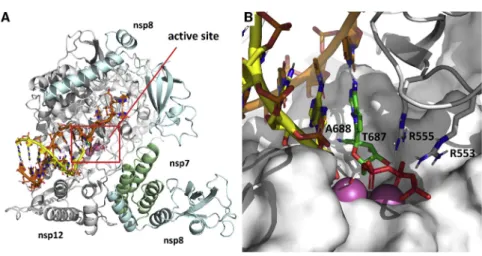
Modeling of the RDV resistance mutations identified in MHV (Agostini et al., 2018) onto the homologous residues V557 and F480 in the SARS-CoV-2 RdRp structure reveals that V557L shifts the position of the template base, which, in turn, shifts the positioning of the incoming NTP (Figures S1A and S1B). This will affect RDV activity in that it alters the position of the 10CN in the pocket. Because the model predicts no direct inter-action of F480 with the NTP, primer, or template, the effect of the F480L mutation is more difficult to discern. The F480L mutation could potentially induce a subtle change to the 10CN binding pocket (Figures S1C and S1D). Alignment of nsp12 sequences from SARS-CoV-2 used in other studies of RDV shows complete conservation of nsp12 nucleotide sequences, predicting compa-rable antiviral activity of RDV against these isolates (Figure S2). Figure 1. Modeling of RDV onto the SARS-CoV-2 RdRp Structure
(A) Model of the SARS-CoV-2 polymerization complex in its elongating state. The model was based on the cryo-EM apo structures of SARS-CoV (http://www.rcsb.org/structure/6NUR) and SARS-CoV-2 (http://www.rcsb.org/structure/ 6M71). The active site is boxed in red.
(B) Enlarged view of the active site, depicting RDV pre-incorporation. The 10CN substituent sits in a shallow pocket formed by residues T687 and A688. Bound to the two catalytic Mg2+
ions (pink), the TP is coordinated by two basic residues (R553 and R555).
We next modeled the active sites of the six other human CoVs SARS-CoV (Figure S3A), MERS-CoV (Figure S3B), HCoV-OC43 (Figure S3C), HCoV-229E (Figure S3D), HCoV-NL63 (Figure S3E), and HCoV-HKU1 (Figure S3F). The models show that SARS-CoV-2 is identical to SARS-CoV out to a radius of 18 A˚ from the active site. Differences detected on the periphery of the active site of MERS-CoV and HCoV-OC43, -229E, -NL63, and -HKU1 correspond to residues that do not directly interact with RDV-TP. Together, these data demonstrate high structural con-servation of the RdRp active site interacting with RDV-TP across all seven known human CoV strains.
RDV and GS-441524 Potently Inhibit SARS-CoV-2 Replication
RDV and its parent nucleoside analog GS-441524 inhibit CoVs and multiple other viruses (Agostini et al., 2018; Cho et al., 2012; Lo et al., 2017; Sheahan et al., 2017; Warren et al., 2016). Previous reports (Choy et al., 2020;Wang et al., 2020; Runfeng et al., 2020) suggest that RDV inhibits SARS-CoV-2, but comparative studies of anti-SARS-CoV-2 activity using authentic compounds in physiologically relevant cell lines are lacking. We first compared SARS-CoV-2 replication in estab-lished cell lines to determine which cell types could potentially be suitable for studying RDV efficacy against SARS-CoV-2. Viral yields were determined 24, 48, and 72 h post-infection (hpi) in Vero E6, Vero CCL-81 (Vero), Huh7, and Calu3 2B4 cells (Yoshi-kawa et al., 2010;Figure 2A). Vero E6 and Vero cells supported the highest levels of SARS-CoV-2 replication, consistent with a previous study (Harcourt et al., 2020). Maximum yields were de-tected at 48 hpi in Vero E6 cells (>6 logs at MOI = 0.1 and 0.01 PFU/cell), 24 hpi in Vero cells infected at MOI = 0.1 PFU (>5 logs), 48 hpi in Vero cells infected at MOI = 0.01 PFU/cell (>5 logs), 72 hpi in Calu3 2B4 cells (>4 logs at MOI = 0.1 PFU/cell), and 48 hpi in Huh7 cells (>4 logs at MOI = 0.1 PFU/cell and <2 logs at MOI = 0.01 PFU/cell). These results indicate that Vero E6, Vero, and Calu3 2B4 cells support varying levels of SARS-CoV-2 replication and that the cell type should be chosen for a given study depending on the study goals.
To determine whether RDV and GS-441524 inhibit SARS-CoV-2 replication in established cell lines, Calu3 2B4 human lung adenocarcinoma cells and Vero E6 African green monkey kidney cells were infected with the SARS-CoV-2 clinical isolate 2019-nCoV/USA-WA1/2020 and treated with a range of RDV or GS-441524 concentrations. Supernatants were harvested at time points corresponding to peak viral replication for each cell type, and infectious viral titers and viral genome copy numbers in the supernatant were quantified by plaque assay and qRT-PCR, respectively. RDV and GS-441524 potently inhibited SARS-CoV-2 replication in a dose-dependent manner in both cell types (Figure 2;Table 1). In Calu3 2B4 cells, both com-pounds displayed dose-dependent inhibition of viral replication, as determined by plaque assay (Figure 2B) and qRT-PCR (Fig-ure 2C). RDV inhibited SARS-CoV-2 with an EC50 value of 0.28mM and a 90% maximal effective concentration (EC90) value of 2.48mM. The parent compound GS-441524 displayed similar potency: an EC50value of 0.62mM and EC90value of 1.34mM (Figure 2D;Table 1). EC50values determined by quantification of viral genome copies showed a similar trend (Figure 2E;Table
1). Both compounds also displayed dose-dependent inhibition of viral replication in Vero E6 cells, as determined by infectious viral titers and genome copy numbers (Figure 2F). RDV inhibited SARS-CoV-2 with an EC50value of 1.65mM and EC90value of 2.40mM. However, in this cell type, GS-441524 was more potent (EC50= 0.47mM and EC90= 0.71mM) than RDV, as determined by infectious viral titers (Figure 2G;Table 1) and genome copy numbers (Figure 2H;Table 1). Thus, RDV inhibits SARS-CoV-2 more potently in Calu3 2B4 than in Vero E6 cells, and the relative potencies of the prodrug and parent nucleoside are cell type dependent.
RDV Is a Highly Potent Antiviral Inhibitor of SARS-CoV-2 in Primary HAE Cultures
SARS-CoV-2 is known to replicate in the upper and lower airways in humans (Adachi et al., 2020;Wo¨lfel et al., 2020). In addition, we have shown recently that SARS-CoV-2 replicates in human primary cells from the nasal to alveolar epithelium (Hou et al., 2020). To demonstrate the antiviral activity of RDV against SARS-CoV-2 in a human primary epithelial culture sys-tem, we performed antiviral assays in HAE cultures, which are grown on air-liquid interface and recapitulate the cellular complexity and physiology of the human conducting airway (Sims et al., 2005). HAE cultures are the model system in which we have amassed data for many other enzootic, emerging, and endemic CoVs, allowing comparison of SARS-CoV-2 data with previous reports (Sheahan et al., 2017,2020a). In RDV-treated, SARS-CoV-2-infected HAE cultures, we observed a dose-dependent reduction in infectious virus production with more than 100-fold inhibition at the highest tested concentration (Fig-ure 3A). Importantly, RDV demonstrates potent antiviral activity with EC50values of 0.0010 and 0.009mM in two independent ex-periments (Figure 3B). Similar to previously reported studies, RDV did not cause cytotoxicity in HAE cultures across the dose range where we see potent antiviral effects (Figure S4; Sheahan et al., 2017). Together, these data demonstrate that RDV is potently antiviral against SARS-CoV-2 in primary human lung cultures with a selectivity index of more than 1,000.
Antiviral Activities of RDV and GS-441524 Correlate with RDV-TP Metabolite Levels
B C
E D
F G
H I
A Figure 2. The Prodrug Remdesivir (RDV) and
Parent Nucleoside GS-441524 (’524) Potently Inhibit SARS-CoV-2 Replication
(A) Vero E6, Vero CCL-81 (Vero), Huh7, and Calu3 2B4 cells were infected with an MOI of 0.01 and/or 0.1 PFU/cell SARS-CoV-2 (2019-nCoV/USA-WA1/ 2020), and infectious viral titers were determined by plaque assay 0.5, 24, 48, and 72 h post-infec-tion (hpi). Viral yields were calculated by sub-tracting the average 0.5 h (post-adsorption, pre-incubation) titer from each subsequent time point. Data represent the average of three replicates from one experiment. Error bars indicate SD. B.D., below detection. Calu3 2B4 cells were infected with 0.1 PFU/cell SARS-CoV-2, and Vero cells were infected with 0.01 PFU/cell SARS-CoV-2 and treated with RDV, GS-441524 (’524), or DMSO only (control) in cell culture medium. Supernatants were collected 48 hpi (Vero E6 cells) or 72 hpi (Calu3 2B4 cells).
(B and C) Reduction of SARS-CoV-2 replication by RDV in Calu3 2B4 cells, as determined by infec-tious viral titer (B) and qRT-PCR (C).
(D) Percent inhibition of SARS-CoV-2 replication by RDV and GS-441524 in Calu3 2B4 cells, as determined by infectious viral titer (RDV: EC50=
0.28 mM, EC90= 2.48mM; GS-441524: EC50=
0.62mM, EC90= 1.34mM). No significant
cytotox-icity of either compound was detected in Calu3 cells.
(E) Percent inhibition of SARS-CoV-2 replication by RDV and GS-441524 in Calu3 2B4 cells, as determined by qRT-qPCR (RDV: EC50= 0.60mM,
EC90= 1.28mM; GS-441524: EC50= 1.09mM,
EC90= 1.37mM).
(F and G) Reduction of SARS-CoV-2 replication by RDV in Vero E6 cells, as determined by infectious viral titer (F) and qRT-PCR (G).
(H) Percent inhibition of SARS-CoV-2 replication by RDV and GS-441524 in Vero E6 cells, as determined by infectious viral titer (RDV: EC50=
1.65 mM, EC90= 2.40mM; GS-441524: EC50=
0.47mM, EC90= 0.71mM). No significant
cytotox-icity of either compound was detected in Vero E6 cells.
(I) Percent inhibition of SARS-CoV-2 replication by RDV and GS-441524 in Vero E6 cells, as deter-mined by qRT-PCR (RDV: EC50= 1.49mM, EC90=
3.03 mM; GS-441524: EC50= 0.47mM, EC90=
0.80mM).
HAE cultures, we used cells from two independent donors with similar demographic profiles. RDV-TP was formed efficiently in both donor cultures following incubation with RDV, with a differ-ence of less than 3-fold from each other. The lowest levels of RDV-TP were observed following RDV treatment of Vero E6 cells and were approximately 4- and 20-fold lower than those observed in Calu3 2B4 and HAE cultures, respectively. Incuba-tion of Vero E6 cells with GS-441524 yielded 3.5-fold higher RDV-TP levels compared with incubation with RDV, correspond-ing to higher antiviral potency of GS-441524 relative to RDV (Table S1). In conclusion, the RDV-TP levels in the different cell types directly correlated with the antiviral potency of RDV against SARS-CoV-2. HAE cultures produced substantially higher levels of RDV-TP, which translated into markedly more potent antiviral activity of RDV (Table 1). Importantly, the meta-bolism of RDV in Vero E6 cells appeared to be less efficient, particularly in comparison with the HAE cultures, indicating that Vero E6 cells might not be an appropriate cell type to char-acterize the antiviral activity of RDV and, potentially, also other nucleotide prodrug-based antiviral agents.
RDV Is Active against the SARS-CoV-2 RdRpIn Vivo
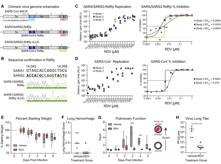
SARS-CoV-2 does not bind the murine ortholog of the human en-try receptor (i.e., mouse angiotensin-converting enzyme-2 [mACE2]) to enter cells (Zhou et al., 2020b). To rapidly assess the therapeutic efficacy of RDV against SARS-CoV-2in vivo, we constructed a chimeric mouse-adapted SARS-CoV variant encoding the target of RDV antiviral activity, the RdRp, of SARS-CoV-2 (SARS/SARS2-RdRp) (Figure 5A). Although other chimeric replicase open reading frame (ORF) recombinant CoVs have been shown to be viable (Stobart et al., 2013), this is the first demonstration that the RdRp from a related but different CoV can support efficient replication of another. After recovery and sequence confirmation (Figure 5B) of recombinant
chimeric viruses with and without a nanoluciferase reporter, we compared SARS-CoV and SARS/SARS2-RdRp replication and sensitivity to RDV in Huh7 cells. Replication of both viruses was inhibited similarly in a dose-dependent manner by RDV (SARS-CoV mean EC50= 0.007mM, SARS/SARS2-RdRp mean EC50= 0.003 mM) (Figures 5C and 5D). We then determined the therapeutic efficacy of RDV against the SARS/SARS2-RdRp in mouse models employed for previous studies of RDV (Sheahan et al., 2017). Mice produce a serum esterase absent in humans, carboxyl esterase 1c (Ces1c), which dramatically re-duces the half-life of RDV. Thus, to mirror pharmacokinetics observed in humans, mouse studies with RDV must be per-formed in transgenic C57BL/6Ces1c/mice (Sheahan et al., 2017). We infected female C57BL/6 Ces1c/ mice with 103 PFU SARS/SARS2-RdRp and initiated subcutaneous treatment with 25 mg/kg RDV BID 1 day post-infection (dpi). This regimen was continued until study termination. Although weight loss and lung hemorrhage did not differ significantly between vehicle-and RDV-treated animals (Figures 5E vehicle-and 5F), we found differ-ences in pulmonary function, as measured by whole-body plethysmography (WBP) between RDV- and vehicle-treated an-imals. The WBP metric, PenH, is a surrogate marker of pulmo-nary obstruction (Menachery et al., 2015a). Therapeutic RDV significantly ameliorated the loss of pulmonary function observed in the vehicle-treated group (Figure 5G). Importantly, RDV treatment dramatically reduced the lung viral load (Fig-ure 5H). Taken together, these data demonstrate that therapeu-tically administered RDV can reduce virus replication and improve pulmonary function in an ongoing infection with a chimeric SARS-CoV/SARS-CoV-2 virus encoding the RdRp target of RDV.
DISCUSSION
The COVID-19 pandemic has gravely illustrated the need for countermeasures against emerging epidemic and pandemic CoVs. Broad-spectrum antiviral drugs, antibodies, and vaccines are needed to combat the current pandemic and those that will emerge in the future. RDV shows potent activity against an array of genetically diverse CoVs as well as against unrelated emerging viruses like Ebola (Agostini et al., 2018;Brown et al., 2019;Sheahan et al., 2017,2020a;Warren et al., 2016). In this study, we demonstrate that RDV and its parent nucleoside GS-441524 are active against SARS-CoV-2 in the physiologically relevant Calu3 2B4 cell line and that RDV has strong antiviral ac-tivity in primary human airway cultures. The potency of RDV was directly related to the intracellular concentration of the pharma-cologically active TP metabolite, which was markedly higher in primary HAE cultures compared with human lung cells (Calu3 2B4) and monkey kidney cells (Vero E6). Our data are consistent with recent studies demonstrating important contributions of natural variation in host- and tissue-specific gene expression patterns and microbiome-specific contributions to drug meta-bolism, stability, and bioavailability in different tissues (Eriksson, 2013;Koczor et al., 2012). Modeling of RDV-TP onto the SARS-CoV-2 RdRp revealed that the positioning of RDV-TP into the active site closely resembled that of the cognate natural sub-strate ATP, consistent with efficient incorporation into RNA Table 1. Cell-Specific SARS-CoV-2 Potency and Metabolism
Analysis
RDV GS-441524a
Vero E6 Calu3
2B4 HAEb Vero E6
Calu3 2B4 Plaque assay
EC50(mM)
1.65 0.28 0.010 0.47 0.62
Genome copy EC50(mM)
1.49 0.60 n.d. 0.47 1.09
RDV-TP at 24 hc
(pmol/million cells) 0.50± 0.15d 2.17± 0.14e 10.6± 5.3b 1.78± 0.68d 0.85± 0.16e
See alsoTable S1andFigure S5.
aGS-441524 was not tested for antiviral potency or RDV-TP levels in HAE
cultures.
bHAE antiviral potencies and RDV-TP levels were determined
indepen-dently in differentiated cultures from two donors. RDV-TP levels in HAE cultures are presented as the mean±SD of quadruplicate technical rep-licates from two donors.
cIndividual analyte data are presented inTables S1andS2.
dValues represent mean±SD from four independent experiments, each
performed with duplicate samples.
e
during replication of the viral genome. RDV decreased viral loads and improved lung function in mice infected with the SARS/ SARS2-RdRp chimeric virus when treated at 1 dpi. This is the first rigorous demonstration of potent inhibition of SARS-CoV-2 in continuous and primary human lung cultures and the first study suggesting efficacy of RDV against SARS-CoV-2 in mice.
Previous studies of RDV anti-SARS-CoV-2 activity reported EC50 values of 0.77 mM as determined by quantification of genome copy number (Wang et al., 2020), 23.2mM as deter-mined by 50% tissue infectious dose (TCID50), 26.9mM as deter-mined by RNA copy number (Choy et al., 2020), and 0.65mM as determined by cytopathic effect (CPE) (Runfeng et al., 2020), all in Vero E6 cells. The potency of RDV in Vero E6 cells (EC50, 1.65mM) observed in our study is comparable with values re-ported byWang et al. (2020)andRunfeng et al. (2020)but greater than reported byChoy et al. (2020). Sequence comparison of the nsp12 from the Seattle, Washington isolate used in this study versus SARS-CoV-2 isolates used in the previously mentioned studies assessing RDV potency did not reveal consensus changes in the nsp12 sequence, suggesting that any isolate-specific variation in RDV sensitivity is not likely to be due to dif-ferences in the RDV-TP interaction with the RdRp. Therefore, the differences in EC50values might be partially explained by intrinsic differences in SARS-CoV-2 virus isolates, quantification methods, Vero cell lineages, and assay conditions, such as incu-bation period and virus input.
Although Vero E6 cells support robust replication of SARS-CoV-2, as illustrated here and elsewhere, our study emphasized that caution should be exercised when interpreting nucleoside prodrug potency experiments performed using Vero cell line-ages. Nucleoside analog potency is dependent on metabolism into the active form. In contrast to the nucleoside GS-441524, RDV is a monophosphoramidate prodrug with moieties that mask the negative charges of its monophosphate group, which enhances its cellular uptake. Intracellularly, RDV is then rapidly metabolized to its monophosphate, which is efficiently con-verted to the active TP (Mehellou et al., 2018). Consistent with previous reports for SARS-CoV and MERS-CoV (Agostini et al., 2018), RDV showed a similar potency of inhibition against SARS-CoV-2 over GS-441524 in Calu3 2B4 cells. In contrast, RDV was 2-fold less potent than GS-441524 in Vero E6 cells. The relative potency of the two compounds was directly linked to intracellular concentration of the active TP metabolite, sug-gesting an altered uptake and/or intracellular metabolism of RDV, consistent with a previous report describing inefficient metabolism of the nucleotide prodrug sofosbuvir in Vero cells (Mumtaz et al., 2017). Drug potency in Vero E6 was similar whether quantified by infectious virus or genome copy number. In Calu3 2B4 cells, the potency determined by qRT-PCR was about 2-fold lower than when quantified by plaque assay. It is possible that qRT-PCR, which was developed to detect nucleo-capsid (N) RNA, also detects packaged subgenomic RNAs and defective genomes in addition to full-length genomes, resulting
A B Figure 3. RDV Is Potently Antiviral against
SARS-CoV-2 in Primary Human Airway Epithelial (HAE) Cultures
HAE cultures were infected with a SARS-CoV-2 clinical isolate (2019-nCoV/USA-WA1/2020) at an MOI of 0.5 PFU/cell for 2 h, and then the virus was removed and cultures were washed 3 times, fol-lowed by incubation at 37C for 48 h.
(A) SARS-CoV-2 infectious virus production in two independent studies. The virus was titered via pla-que assay in apical washes 48 hpi. Each symbol represents the titer from a single culture, and a line is drawn at the mean.
(B) Percent inhibition generated from the titer data in (A).
See alsoFigure S4.
Figure 4. RDV-TP Levels in Vero E6, Calu3, and HAE Cultures
in underestimation of the reduction in infectious titer. Notably, the potency of RDV against CoV encoding the SARS-CoV-2 RdRp in Huh7 cells was more than 100-fold higher than that of RDV againstbona fideSARS-CoV-2 in Huh7 and Calu3 2B4 cells. This difference could be due to infectivity, which is driven by the SARS-CoV instead of the SARS-CoV-2 spike pro-tein. In addition, SARS-CoV infects Huh7 cells at low frequency at the MOI used in this study and does not appear to spread
throughout the culture over the course of the experiment. The number of Huh7 cells in which the virus replicates is relatively lower compared with Calu3 2B4 cells, possibly enhancing the overall antiviral effect of RDV in Huh7 compared with Calu3 2B4 cells. Interestingly, the antiviral potency of RDV against SARS-CoV-2 in HAE cultures was comparable with SARS-CoV and MERS-CoV (Sheahan et al., 2017), which is consistent with the high conservation of the RdRp active site across these
C
B A
D
E F G H
Figure 5. RDV Is Active against the SARS-CoV-2 RdRpIn Vivo
The activity of RDV against the SARS-CoV-2 RdRp was evaluated using a chimeric SARS-CoV encoding the SARS-CoV-2 RdRp (SARS/SARS2-RdRp). (A) Schematic of the recombinant SARS-CoV mouse-adapted MA15 strain chimeric virus genomes generated for these studies. SARS/SARS2-RdRp and SARS/ SARS2-RdRp-nanoluciferase (nLUC) were constructed by exchanging the SARS-CoV MA15 RdRp with the SARS-CoV-2 RdRp. ORF7 is replaced by nLUC in SARS2-RdRp-nLUC.
(B) The presence of the SARS-CoV-2 RdRp was confirmed by Sanger sequencing in stocks of both recombinant chimeric viruses. Alignment of a stretch of nucleotides from the SARS-CoV and SARS-CoV-2 RdRp highlighting nucleotides that differ between the two strains is shown in boldface. These SARS-CoV-2 RdRp-specific nucleotides are present in both chimeric viruses used in this study, as determined by Sanger sequencing and shown in the histogram. (C) SARS/SARS2-RdRp-nLUC replication in Huh7 cells in the presence of RDV (left) and associated percent inhibition (right).
(D) SARS-CoV replication in Huh7 cells in the presence of RDV (left) and associated percent inhibition (right). (E) Percent starting weight of 17-week-old femaleCes1c/mice infected intranasally with 13103
PFU of SARS/SARS2-RdRp and treated subcutaneously with 25 mg/kg RDV or vehicle 1 day post-infection (dpi) and twice daily thereafter.
(F) Lung hemorrhage at 5 dpi.
(G) Pulmonary function by WBP. The PenH metric shown is a surrogate marker of pulmonary obstruction. p < 0.0001 as determined by two-way ANOVA with Sidak’s multiple comparisons test.
(H) Lung titer at 5 dpi as measured by plaque assay. p = 0.0012 by Mann-Whitney test. In (E) and (G), boxes encompass the 25th
–75th
different CoVs. Together, these results emphasize the need for careful selection and use of cell types and methods to study the potency and efficacy of nucleoside analogs.
The target of RDV antiviral activity is the viral RdRp. To mirror the pharmacokinetic exposures observed in humans, RDV studies in mice must be performed inCes1c/animals (Shea-han et al., 2017). In addition, SARS-CoV-2 does not readily infect wild-type mice because of incompatibilities between the virus spike and the murine ACE2 ortholog, which serves as the SARS-CoV-2 entry receptor (Wan et al., 2020). Breeding of a double-transgenic (hACE2,Ces1c/) mouse for use in RDV ef-ficacy studies is ongoing. To rapidly assess the therapeutic effi-cacy of RDV against SARS-CoV-2, we constructed SARS/ SARS2-RdRp. Virus entry, tropism, and pathogenesis of this chimeric virus are driven by the parental mouse-adapted CoV virus. Similar to our previous studies with SARS-CoV (Sheahan et al., 2017), we now show that therapeutic administration of RDV 1 dpi can reduce the viral load and improve pulmonary function in mice. The kinetics of SARS-CoV replication and disease are notably compressed in mice compared with humans, where the virus titer peaks 10– 15 days after onset of symptoms (Hung et al., 2004; Peiris et al., 2004). In comparison, initial reports suggest that SARS-CoV-2 replication peaks around 5–6 days after symptom onset, just prior to onset of dyspnea (Pan et al., 2020;Zhou et al., 2020a). Importantly, a recent preprint described the therapeutic efficacy of RDV against SARS-CoV-2 in rhesus macaques, where RDV treatment reduced respiratory pathology and viral loads in bronchoalveolar lavage fluid, consistent with our study with a chimeric virus in mice (Williamson et al., 2020). Prior to the emergence of pandemic SARS-CoV-2, RDV was evaluated in phase 1 clinical trials as well as phase 2 randomized controlled trials to treat acute Ebola virus disease in the Democratic Repub-lic of Congo (DRC), and human safety data are available (Mu-langu et al., 2019). Thus, our preclinical development of RDV supported immediate compassionate use of RDV for severely ill COVID-19 patients with promising early results (Grein et al., 2020). Furthermore, a preliminary report of a large-scale, dou-ble-blind, randomized, placebo-controlled trial in hospitalized adults suggests that RDV shortened the time to recovery (Beigel et al., 2020). This and other ongoing phase III randomized controlled trials for treatment of patients with COVID-19 will ulti-mately determine efficacy, safety, and optimal dosing of RDV in patients with different stages of COVID-19.
Despite worldwide drug discovery efforts and over 300 active clinical evaluations of potential treatments, no effective counter-measure currently exists to combat COVID-19 (Sanders et al., 2020) or likely future CoV pandemics. Large-scale deployment of antiviral monotherapies creates a high risk for emergence of drug resistance. Our previous work demonstrates that CoV resis-tance to RDV is generated slowly and conferred by two mutations in the RdRp. In addition, RDV-resistant CoVs exhibit reduced replication capacity and are also more sensitive to another potently active nucleoside analog inhibitor,b-D-N 4-hydroxycyti-dine (NHC; EIDD-1931/2801) (Agostini et al., 2019; Sheahan et al., 2020b). Therapies combining direct-acting antivirals (DAAs), such as RDV and EIDD-2801, along with other DAAs, such as antibodies and protease inhibitors that target different
stages of the viral replication cycle, could be considered for coun-teracting resistance if it emerges in patients treated with antiviral monotherapy. In addition, attention should be given to combining DAAs with anti-inflammatory drugs to potentially extend the treat-ment window during which DAAs can improve outcomes. With zo-onotic relatives of SARS-CoV, SARS-CoV-2, and MERS-CoV continuing to circulate in bat species, more outbreaks of novel CoVs are expected (Menachery et al., 2015b,2016). Identification and evaluation of broadly efficacious, robust anti-CoV therapies are thus urgently needed at present and in the future.
STAR+METHODS
Detailed methods are provided in the online version of this paper and include the following:
d KEY RESOURCES TABLE
d RESOURCE AVAILABILITY
B Lead Contact
B Materials Availability
B Data and Code Availability
d EXPERIMENTAL MODEL AND SUBJECT DETAILS
B Cells, viruses, and compounds d METHOD DETAILS
B Modeling
B Sequence alignments
B Replication in different cell types
B Antiviral activity assays
B Cytotoxicity Assays
B Quantification of infectious viral titer
B Quantification of viral RNA copy number
B In vitrometabolism of RDV and GS-441524
B Formulations forin vivostudies
B In vivoefficacy studies
d QUANTIFICATION AND STATISTICAL ANALYSIS
B Mathematical and statistical analyses
SUPPLEMENTAL INFORMATION
Supplemental Information can be found online athttps://doi.org/10.1016/j. celrep.2020.107940.
ACKNOWLEDGMENTS
grant management teams for administrative support of our research operations.
AUTHOR CONTRIBUTIONS
A.J.P. and T.P.S. conceived, designed, and performed experiments and man-agement and coordinated responsibility for research activity planning and execution. A.J.P., J.K.P., J.P.B., and T.P.S. wrote the manuscript. A.S.G., A.S., S.R.L., K.H.D., B.L.Y., M.L.A., L.J.S., J.D.C., X.L., T.M.H., K.G., D.R.M., A.J.B., R.L.G., J.K.P., V.D.P., J.P., B.M., D.B., and E.M. performed ex-periments. T.P.S., D.R.M., R.S.B., A.J.P., J.D.C., and M.R.D. secured funding. D.P.P. and T.C. provided reagents. J.D.C., J.Y.F., J.P.B., D.P.P., T.C., R.S.B., and M.R.D. edited the manuscript and provided expertise and feedback.
DECLARATION OF INTERESTS
The authors affiliated with Gilead Sciences, Inc. are employees of the company and may own company stock.
Received: April 21, 2020 Revised: June 2, 2020 Accepted: June 30, 2020 Published: July 7, 2020
REFERENCES
Adachi, T., Chong, J.-M., Nakajima, N., Sano, M., Yamazaki, J., Miyamoto, I., Nishioka, H., Akita, H., Sato, Y., Kataoka, M., et al. (2020). Clinicopathologic and Immunohistochemical Findings from Autopsy of Patient with COVID-19, Japan. Emerg. Infect. Dis.26.
Agostini, M.L., Andres, E.L., Sims, A.C., Graham, R.L., Sheahan, T.P., Lu, X., Smith, E.C., Case, J.B., Feng, J.Y., Jordan, R., et al. (2018). Coronavirus Sus-ceptibility to the Antiviral Remdesivir (GS-5734) Is Mediated by the Viral Poly-merase and the Proofreading Exoribonuclease. MBio9, e00221, e18.
Agostini, M.L., Pruijssers, A.J., Chappell, J.D., Gribble, J., Lu, X., Andres, E.L., Bluemling, G.R., Lockwood, M.A., Sheahan, T.P., Sims, A.C., et al. (2019). Small-Molecule Antiviral b-d-N4
-Hydroxycytidine Inhibits a Proofreading-Intact Coronavirus with a High Genetic Barrier to Resistance. J. Virol.93, e01348-19.
Appleby, T.C., Perry, J.K., Murakami, E., Barauskas, O., Feng, J., Cho, A., Fox, D., 3rd, Wetmore, D.R., McGrath, M.E., Ray, A.S., et al. (2015). Viral replication. Structural basis for RNA replication by the hepatitis C virus polymerase. Sci-ence347, 771–775.
Arabi, Y.M., Balkhy, H.H., Hayden, F.G., Bouchama, A., Luke, T., Baillie, J.K., Al-Omari, A., Hajeer, A.H., Senga, M., Denison, M.R., et al. (2017). Middle East Respiratory Syndrome. N. Engl. J. Med.376, 584–594.
Beigel, J.H., Tomashek, K.M., Dodd, L.E., Mehta, A.K., Zingman, B.S., Kalil, A.C., Hohmann, E., Chu, H.Y., Luetkemeyer, A., Kline, S., et al. (2020). Remde-sivir for the Treatment of Covid-19 – Preliminary Report. N. Engl. J. Med. Pub-lished online May 22, 2020.https://doi.org/10.1056/NEJMoa2007764.
Bojkova, D., McGreig, J.E., McLaughlin, K.-M., Masterson, S.G., Widera, M., Kra¨hling, V., Ciesek, S., Wass, M.N., Michaelis, M., and Cinatl, J. (2020). SARS-CoV-2 and SARS-CoV differ in their cell tropism and drug sensitivity profiles. bioRxiv.https://doi.org/10.1101/2020.04.03.024257.
Brown, A.J., Won, J.J., Graham, R.L., Dinnon, K.H., 3rd, Sims, A.C., Feng, J.Y., Cihlar, T., Denison, M.R., Baric, R.S., and Sheahan, T.P. (2019). Broad spec-trum antiviral remdesivir inhibits human endemic and zoonotic deltacoronavi-ruses with a highly divergent RNA dependent RNA polymerase. Antiviral Res. 169, 104541.
Chan-Yeung, M., and Xu, R.-H. (2003). SARS: epidemiology. Respirology8 (Suppl), S9–S14.
Chen, J. (2020). Pathogenicity and transmissibility of 2019-nCoV-A quick over-view and comparison with other emerging viruses. Microbes Infect.22, 69–71.
Cho, A., Saunders, O.L., Butler, T., Zhang, L., Xu, J., Vela, J.E., Feng, J.Y., Ray, A.S., and Kim, C.U. (2012). Synthesis and antiviral activity of a series of 10
-substituted 4-aza-7,9-dideazaadenosine C-nucleosides. Bioorg. Med. Chem. Lett.22, 2705–2707.
Choy, K.-T., Wong, A.Y., Kaewpreedee, P., Sia, S.-F., Chen, D., Hui, K.P.Y., Chu, D.K.W., Chan, M.C.W., Cheung, P.P., Huang, X., et al. (2020). Remdesi-vir, lopinaRemdesi-vir, emetine, and homoharringtonine inhibit SARS-CoV-2 replication in vitro. Antiviral Res.178, 104786.
de Wit, E., van Doremalen, N., Falzarano, D., and Munster, V.J. (2016). SARS and MERS: recent insights into emerging coronaviruses. Nat. Rev. Microbiol. 14, 523–534.
de Wit, E., Feldmann, F., Cronin, J., Jordan, R., Okumura, A., Thomas, T., Scott, D., Cihlar, T., and Feldmann, H. (2020). Prophylactic and therapeutic re-mdesivir (GS-5734) treatment in the rhesus macaque model of MERS-CoV infection. Proc. Natl. Acad. Sci. USA117, 6771–6776.
Elbe, S., and Buckland-Merrett, G. (2017). Data, disease and diplomacy: GI-SAID’s innovative contribution to global health. Glob. Chall.1, 33–46.
Eriksson, S. (2013). Is the expression of deoxynucleoside kinases and 50 -nu-cleotidases in animal tissues related to the biological effects of nucleoside an-alogs? Curr. Med. Chem.20, 4241–4248.
Fulcher, M.L., Gabriel, S., Burns, K.A., Yankaskas, J.R., and Randell, S.H. (2005). Well-differentiated human airway epithelial cell cultures. Methods Mol. Med.107, 183–206.
Gao, Y., Yan, L., Huang, Y., Liu, F., Zhao, Y., Cao, L., Wang, T., Sun, Q., Ming, Z., Zhang, L., et al. (2020). Structure of the RNA-dependent RNA polymerase from COVID-19 virus. Science368, 779–782.
Gong, P., and Peersen, O.B. (2010). Structural basis for active site closure by the poliovirus RNA-dependent RNA polymerase. Proc. Natl. Acad. Sci. USA 107, 22505–22510.
Gordon, C.J., Tchesnokov, E.P., Woolner, E., Perry, J.K., Feng, J.Y., Porter, D.P., and Go¨tte, M. (2020a). Remdesivir is a direct-acting antiviral that inhibits RNA-dependent RNA polymerase from severe acute respiratory syndrome co-ronavirus 2 with high potency. J. Biol. Chem.295, 6785–6797.
Gordon, C.J., Tchesnokov, E.P., Feng, J.Y., Porter, D.P., and Go¨tte, M. (2020b). The antiviral compound remdesivir potently inhibits RNA-dependent RNA polymerase from Middle East respiratory syndrome coronavirus. J. Biol. Chem.295, 4773–4779.
Grein, J., Ohmagari, N., Shin, D., Diaz, G., Asperges, E., Castagna, A., Feldt, T., Green, G., Green, M.L., Lescure, F.-X., et al. (2020). Compassionate Use of Re-mdesivir for Patients with Severe Covid-19. N. Engl. J. Med.382, 2327–2336.
Harcourt, J., Tamin, A., Lu, X., Kamili, S., Sakthivel, S.K., Murray, J., Queen, K., Tao, Y., Paden, C.R., Zhang, J., et al. (2020). Severe Acute Respiratory Syn-drome Coronavirus 2 from Patient with 2019 Novel Coronavirus Disease, United States. Emerg. Infect. Dis.26(6).
Holshue, M.L., DeBolt, C., Lindquist, S., Lofy, K.H., Wiesman, J., Bruce, H., Spitters, C., Ericson, K., Wilkerson, S., Tural, A., et al.; Washington State 2019-nCoV Case Investigation Team (2020). First Case of 2019 Novel Corona-virus in the United States. N. Engl. J. Med.382, 929–936.
Hou, Y.J., Okuda, K., Edwards, C.E., Martinez, D.R., Asakura, T., Dinnon, K.H., Kato, T., Lee, R.E., Yount, B.L., Mascenik, T.M., et al. (2020). SARS-CoV-2 Reverse Genetics Reveals a Variable Infection Gradient in the Respiratory Tract. Cell, Published online May 27, 2020. https://doi.org/10.1016/j.cell. 2020.05.042.
Hung, I.F.N., Cheng, V.C.C., Wu, A.K.L., Tang, B.S.F., Chan, K.H., Chu, C.M., Wong, M.M.L., Hui, W.T., Poon, L.L.M., Tse, D.M.W., et al. (2004). Viral loads in clinical specimens and SARS manifestations. Emerg. Infect. Dis.10, 1550– 1557.
Jeon, S., Ko, M., Lee, J., Choi, I., Byun, S.Y., Park, S., Shum, D., and Kim, S. (2020). Identification of antiviral drug candidates against SARS-CoV-2 from FDA-approved drugs. Antimicrob. Agents Chemother.64, e00819-20.
Kirchdoerfer, R.N., and Ward, A.B. (2019). Structure of the SARS-CoV nsp12 polymerase bound to nsp7 and nsp8 co-factors. Nat. Commun.10, 2342.
Lo, M.K., Jordan, R., Arvey, A., Sudhamsu, J., Shrivastava-Ranjan, P., Hotard, A.L., Flint, M., McMullan, L.K., Siegel, D., Clarke, M.O., et al. (2017). GS-5734 and its parent nucleoside analog inhibit Filo-, Pneumo-, and Paramyxoviruses. Sci. Rep.7, 43395.
Mehellou, Y., Rattan, H.S., and Balzarini, J. (2018). The ProTide Prodrug Tech-nology: From the Concept to the Clinic. J. Med. Chem.61, 2211–2226.
Menachery, V.D., Gralinski, L.E., Baric, R.S., and Ferris, M.T. (2015a). New Metrics for Evaluating Viral Respiratory Pathogenesis. PLoS ONE 10, e0131451.
Menachery, V.D., Yount, B.L., Jr., Debbink, K., Agnihothram, S., Gralinski, L.E., Plante, J.A., Graham, R.L., Scobey, T., Ge, X.-Y., Donaldson, E.F., et al. (2015b). A SARS-like cluster of circulating bat coronaviruses shows po-tential for human emergence. Nat. Med.21, 1508–1513.
Menachery, V.D., Yount, B.L., Jr., Sims, A.C., Debbink, K., Agnihothram, S.S., Gralinski, L.E., Graham, R.L., Scobey, T., Plante, J.A., Royal, S.R., et al. (2016). SARS-like WIV1-CoV poised for human emergence. Proc. Natl. Acad. Sci. USA113, 3048–3053.
Mulangu, S., Dodd, L.E., Davey, R.T., Jr., Tshiani Mbaya, O., Proschan, M., Mukadi, D., Lusakibanza Manzo, M., Nzolo, D., Tshomba Oloma, A., Ibanda, A., et al.; PALM Writing Group; PALM Consortium Study Team (2019). A Ran-domized, Controlled Trial of Ebola Virus Disease Therapeutics. N. Engl. J. Med. 381, 2293–2303.
Mumtaz, N., Jimmerson, L.C., Bushman, L.R., Kiser, J.J., Aron, G., Reusken, C.B.E.M., Koopmans, M.P.G., and van Kampen, J.J.A. (2017). Cell-line depen-dent antiviral activity of sofosbuvir against Zika virus. Antiviral Res.146, 161–163.
Pan, Y., Zhang, D., Yang, P., Poon, L.L.M., and Wang, Q. (2020). Viral load of SARS-CoV-2 in clinical samples. Lancet Infect. Dis.20, 411–412.
Peiris, J.S.M., Guan, Y., and Yuen, K.Y. (2004). Severe acute respiratory syn-drome. Nat. Med.10(12, Suppl), S88–S97.
Runfeng, L., Yunlong, H., Jicheng, H., Weiqi, P., Qinhai, M., Yongxia, S., Chu-fang, L., Jin, Z., Zhenhua, J., Haiming, J., et al. (2020). Lianhuaqingwen exerts anti-viral and anti-inflammatory activity against novel coronavirus (SARS-CoV-2). Pharmacol. Res.156, 104761.
Sanders, J.M., Monogue, M.L., Jodlowski, T.Z., and Cutrell, J.B. (2020). Phar-macologic Treatments for Coronavirus Disease 2019 (COVID-19): A Review. JAMA, Published online April 13, 2020.https://doi.org/10.1001/jama.2020. 6019.
Scobey, T., Yount, B.L., Sims, A.C., Donaldson, E.F., Agnihothram, S.S., Men-achery, V.D., Graham, R.L., Swanstrom, J., Bove, P.F., Kim, J.D., et al. (2013). Reverse genetics with a full-length infectious cDNA of the Middle East respira-tory syndrome coronavirus. Proc. Natl. Acad. Sci. USA110, 16157–16162.
Shannon, A., Le, N.T.-T., Selisko, B., Eydoux, C., Alvarez, K., Guillemot, J.-C., Decroly, E., Peersen, O., Ferron, F., and Canard, B. (2020). Remdesivir and SARS-CoV-2: Structural requirements at both nsp12 RdRp and nsp14 Exonu-clease active-sites. Antiviral Res.178, 104793.
Sheahan, T.P., Sims, A.C., Graham, R.L., Menachery, V.D., Gralinski, L.E., Case, J.B., Leist, S.R., Pyrc, K., Feng, J.Y., Trantcheva, I., et al. (2017). Broad-spectrum antiviral GS-5734 inhibits both epidemic and zoonotic coro-naviruses. Sci. Transl. Med.9, eaal3653.
Sheahan, T.P., Sims, A.C., Leist, S.R., Scha¨fer, A., Won, J., Brown, A.J., Mont-gomery, S.A., Hogg, A., Babusis, D., Clarke, M.O., et al. (2020a). Comparative therapeutic efficacy of remdesivir and combination lopinavir, ritonavir, and interferon beta against MERS-CoV. Nat. Commun.11, 222.
Sheahan, T.P., Sims, A.C., Zhou, S., Graham, R.L., Pruijssers, A.J., Agostini, M.L., Leist, S.R., Scha¨fer, A., Dinnon, K.H., 3rd, Stevens, L.J., et al. (2020b). An orally bioavailable broad-spectrum antiviral inhibits SARS-CoV-2 in human airway epithelial cell cultures and multiple coronaviruses in mice. Sci. Transl. Med.12, eabb5883.
Shu, Y., and McCauley, J. (2017). GISAID: Global initiative on sharing all influ-enza data - from vision to reality. Euro Surveill.22, 30494.
Sims, A.C., Baric, R.S., Yount, B., Burkett, S.E., Collins, P.L., and Pickles, R.J. (2005). Severe acute respiratory syndrome coronavirus infection of human cili-ated airway epithelia: role of cilicili-ated cells in viral spread in the conducting air-ways of the lungs. J. Virol.79, 15511–15524.
Stobart, C.C., Sexton, N.R., Munjal, H., Lu, X., Molland, K.L., Tomar, S., Mese-car, A.D., and Denison, M.R. (2013). Chimeric exchange of coronavirus nsp5 proteases (3CLpro) identifies common and divergent regulatory determinants of protease activity. J. Virol.87, 12611–12618.
Wan, Y., Shang, J., Graham, R., Baric, R.S., and Li, F. (2020). Receptor Recog-nition by the Novel Coronavirus from Wuhan: an Analysis Based on Decade-Long Structural Studies of SARS Coronavirus. J. Virol.94, e00127-20.
Wang, M., Cao, R., Zhang, L., Yang, X., Liu, J., Xu, M., Shi, Z., Hu, Z., Zhong, W., and Xiao, G. (2020). Remdesivir and chloroquine effectively inhibit the recently emerged novel coronavirus (2019-nCoV) in vitro. Cell Res. 30, 269–271.
Warren, T.K., Jordan, R., Lo, M.K., Ray, A.S., Mackman, R.L., Soloveva, V., Siegel, D., Perron, M., Bannister, R., Hui, H.C., et al. (2016). Therapeutic effi-cacy of the small molecule GS-5734 against Ebola virus in rhesus monkeys. Nature531, 381–385.
Williamson, B.N., Feldmann, F., Schwarz, B., Meade-White, K., Porter, D.P., Schulz, J., van Doremalen, N., Leighton, I., Yinda, C.K., Pe´rez-Pe´rez, L., et al. (2020). Clinical benefit of remdesivir in rhesus macaques infected with SARS-CoV-2. bioRxiv.https://doi.org/10.1101/2020.04.15.043166.
Wo¨lfel, R., Corman, V.M., Guggemos, W., Seilmaier, M., Zange, S., Muller,€ M.A., Niemeyer, D., Jones, T.C., Vollmar, P., Rothe, C., et al. (2020). Virological assessment of hospitalized patients with COVID-2019. Nature581, 465–469.
Yin, W., Mao, C., Luan, X., Shen, D.-D., Shen, Q., Su, H., Wang, X., Zhou, F., Zhao, W., Gao, M., et al. (2020). Structural basis for inhibition of the RNA-dependent RNA polymerase from SARS-CoV-2 by remdesivir. Science368, 1499–1504.
Yoshikawa, T., Hill, T.E., Yoshikawa, N., Popov, V.L., Galindo, C.L., Garner, H.R., Peters, C.J., and Tseng, C.-T.K. (2010). Dynamic innate immune re-sponses of human bronchial epithelial cells to severe acute respiratory syn-drome-associated coronavirus infection. PLoS ONE5, e8729.
Yount, B., Curtis, K.M., Fritz, E.A., Hensley, L.E., Jahrling, P.B., Prentice, E., Denison, M.R., Geisbert, T.W., and Baric, R.S. (2003). Reverse genetics with a full-length infectious cDNA of severe acute respiratory syndrome coronavi-rus. Proc. Natl. Acad. Sci. USA100, 12995–13000.
Zhou, F., Yu, T., Du, R., Fan, G., Liu, Y., Liu, Z., Xiang, J., Wang, Y., Song, B., Gu, X., et al. (2020a). Clinical course and risk factors for mortality of adult inpa-tients with COVID-19 in Wuhan, China: a retrospective cohort study. Lancet 395, 1054–1062.
STAR
+
METHODS
KEY RESOURCES TABLE
REAGENT or RESOURCE SOURCE IDENTIFIER Bacterial and Virus Strains
SARS-CoV-2 2019-nCoV/ USA-WA1/2020 clinical isolate
CDC Atlanta Accession number MN985325
SARS/SARS2-RdRp This paper N/A
SARS/SARS2-RdRp-nLUC This paper N/A Chemicals, Peptides, and Recombinant Proteins
Remdesivir Gilead Sciences GS-5734 (RDV)
GS-441524 Gilead Sciences GS-441524
Critical Commercial Assays
Nano-GloLuciferase Assay System Promega N1110 CellTiter-GloLuminescent
Cell Viability Assay
Promega G7570
TaqMan Fast Virus 1-Step Master Mix Life Technologies 4444436 mMESSAGE mMACHINE T7
Transcription Kit
ThermoFisher Scientific AM1344
Deposited Data
PDB SARS-CoV-2 RDV-TP This manuscript Mendeley datahttps://doi.org/10.17632/ x3sw5z2bg9.1
Experimental Models: Cell Lines
Vero ATCC CCL-81
Vero C1008 [Vero 76, clone E6, Vero E6] ATCC CRL-1586 Calu3 2B4 cells Chien-Te (Kent) Tseng, UTMB N/A
Huh7 Dr. Mark Heise, UNC N/A
Primary HAE cell cultures Tissue Procurement and Cell Culture Core Laboratory in the Marsico Lung Institute/ Cystic Fibrosis Research Center, UNC
N/A
Differentiated HAE cultures MatTek Corporation; Ashland, MA AIR-100 Experimental Models: Organisms/Strains
C57BL/6Ces1c/mice Jackson Labs Stock 014096 Oligonucleotides
N gene forward primer: 50 -GACCCCAAAATCAGCGAAAT
Integrated DNA Technologies 10006606
N gene reverse primer: 50 -TCTGGTTACTGCCAGTTGAATCTG
Integrated DNA Technologies 10006606
N gene probe: 50- FAM-ACCCC GCATTACGTTTGGTGGACC-BHQ1
Integrated DNA Technologies 10006606
T7 forward primer to createin vitro
transcription template 50- TAATACG ACTCACTATAGGGATGTCTGATA
This manuscript N/A
ATGGACCCCA
T7 reverse primer to createin vitro
transcription template: 50- TTAGG CCTGAGTTGAG
This manuscript N/A
TCAG
Recombinant DNA
SARS-CoV-2 N gene positive control plasmid
Integrated DNA Technologies 10006625
RESOURCE AVAILABILITY
Lead Contact
Further information and requests for resources and reagents should be directed to and will be fulfilled by the Lead Contact Andrea Pruijssers ([email protected]).
Materials Availability
Plasmids generated in this study are available upon request.
Data and Code Availability
The pdb file containing SARS-CoV-2 model showing RDV-TP in the active site has been deposited to Mendeley Data:https://doi.org/ 10.17632/x3sw5z2bg9.1.
EXPERIMENTAL MODEL AND SUBJECT DETAILS
Cells, viruses, and compounds
Vero (ATCC CCL-81) and Vero E6 (ATCC CRL-1586) cells were purchased from ATCC and cultured in DMEM supplemented with 10% fetal bovine serum (FBS) (GIBCO, ThermoFisher Scientific) or 10% FCS fetal clonal serum (FCS)(HyClone, GE Life Sciences), 100 U/ml penicillin and streptomycin (GIBCO, ThermoFisher Scientific), and 0.25mM amphotericin B (Corning). Human hepatoma (Huh7) cells were provided by Dr. Mark Heise at UNC Chapel Hill and grown in DMEM supplemented with 10% FBS (Hyclone) and 13antibiotic-antimycotic (GIBCO, ThermoFisher Scientific). Calu3 2B4 cells (Yoshikawa et al., 2010) were cultured in DMEM supplemented with 20% FBS, 100 U/mL penicillin and streptomycin, and 0.25mM amphotericin B.
Primary HAE cell cultures used in antiviral activity assays were obtained from the Tissue Procurement and Cell Culture Core Lab-oratory in the Marsico Lung Institute/Cystic Fibrosis Research Center at UNC. All assays in this report were performed using a single HAE cell donor. Human tracheobronchial epithelial cells provided by Dr. Scott Randell were obtained from airway specimens re-sected from patients undergoing surgery under University of North Carolina Institutional Review Board-approved protocols (#03-1396) by the Cystic Fibrosis Center Tissue Culture Core. Primary cells were expanded to generate passage 1 cells and passage 2 cells were plated at a density of 250,000 cells per well on Transwell-COL (12mm diameter) supports (Corning). Human airway epithe-lium cultures (HAE) were generated by provision of an air-liquid interface for 6 to 8 weeks to form well-differentiated, polarized cul-tures that resembledin vivopseudostratified mucociliary epithelium (Fulcher et al., 2005).
Clinical specimens of SARS-CoV-2 from a case-patient who acquired COVID-19 during travel to China and diagnosed in Wash-ington State, USA upon return were collected as described (Holshue et al., 2020). Virus isolation from this patient’s specimens was performed as described inHarcourt et al. (2020). The sequence is available through GenBank (accession number MN985325.1). A passage 3 stock of the SARS-CoV-2 Seattle isolate was obtained from the CDC and passed twice in Vero E6 cells to generate high-titer passage 5 stock for experiments described in this manuscript.
SARS-CoV expressing GFP (GFP replaces ORF7) was created from molecular cDNA clones as described (Scobey et al., 2013; Sims et al., 2005). To create SARS-CoV expressing nanoluciferase (nLUC), the gene for GFP was replaced with nLUC and isolated using our existing mouse adapted SARS-CoV (MA15) SARS-CoV Urbani molecular clone (Yount et al., 2003). A synthetic cDNA en-coding the SARS-CoV-2 RdRp (Integrated DNA Technologies) was cloned into SARS MA15 D fragment using StuI (50) and BsaI (30) via Gibson assembly. The resultant plasmids were sequence confirmed and then utilized to generate recombinant virus with or without nanoluciferase as described. Recombinant virus stocks were confirmed to harbor SARS-CoV-2 RdRp by Sanger sequencing.
Continued
REAGENT or RESOURCE SOURCE IDENTIFIER Software and Algorithms
Prime Schro¨dinger, LLC, New York, NY, 2020 https://www.schrodinger.com/prime
Macromodel Schro¨dinger, LLC, New York, NY, 2020 https://www.schrodinger.com/ macromodel
CLC Main Workbench 20 QIAGEN https://digitalinsights.qiagen.com/ products-overview/
discovery-insights-portfolio/ analysis-and-visualization/ qiagen-clc-genomics-workbench/
Prism 8 GraphPad https://www.graphpad.com/
Remdesivir (RDV; GS-5734) and GS-441524 were synthesized by the Department of Medicinal Chemistry, Gilead Sciences (Foster City, CA).
METHOD DETAILS
Modeling
A model of the elongating SARS-CoV-2 polymerase complex was generated based on a homology model which used the cryo-EM structure of apo SARS-CoV as a template (PDB: 6NUR;Kirchdoerfer and Ward, 2019) as described previously (Gordon et al., 2020b). RNA primer and template, a substrate ATP and two catalytic Mg2+ions were oriented in the structure based on alignment to a ternary X-ray structure of HCV NS5B (PDB: 4WTD;Appleby et al., 2015). The structure was then optimized with a series of constrained min-imizations. To this structure we aligned the new cryo-EM structure of the SARS-CoV-2 replication complex. As the SARS-CoV-2 structure does not significantly differ from the SARS-CoV structure, rather than a complete replacement of the model, we incorpo-rated only those residues that had not been resolved in the previous structure (residues 31-116). Additional optimization, particularly of the RNA in the exit channel, was done following the previously outlined procedures. After optimization of the ATP structure, RDV-TP was modeled into the active site and minimized. Models of the V557L and F480L mutants and the other coronavirus models re-ported here were generated based on this final model. All work was carried out using Prime and Macromodel (Schro¨dinger, LLC, New York, NY, 2020). 3D coordinates of the SARS-CoV-2/RDV-TP model are provided in the Supplementary Material.
Sequence alignments
Coronavirus nsp12 sequence alignment was generated using CLC Workbench (QIAGEN) from sequences downloaded from the NCBI website (Accession numbers MN985325.1 and MT123290.1) and from GISAID’s EpiFlu Database (Elbe and Buckland-Merrett, 2017;Shu and McCauley, 2017): Accession ID EPI_ISL_402124; virus name hCoV-19/Wuhan/WIV04/2019; Location: Asia / China / Hubei / Wuhan; Collection date 2019-12-30 Originating lab Wuhan Jinyintan Hospital; Submitting lab Wuhan Institute of Virology, Chinese Academy of Sciences; Authors Peng Zhou, Xing-Lou Yang, Ding-Yu Zhang, Lei Zhang, Yan Zhu, Hao-Rui Si, Zhengli Shi. Accession ID EPI_ISL_412028; virus name hCoV-19/Hong Kong/VM20001061/2020; Location Asia / Hong Kong; Collection date 2020-01-22; Originating lab Hong Kong Department of Health; Submitting lab: School of Public Health, The University of Hong Kong; Authors Dominic N.C. Tsang, Daniel K.W. Chu, Leo L.M. Poon, Malik Peiris.
Replication in different cell types
Vero E6, Vero, Calu3 2B4, and Huh7 cells were seeded in 24 well plates and allowed to adhere for 24 h. Cells were adsorbed with 100ml SARS-CoV-2 in gel saline for 30 minutes (min) at 37C with manual rocking every 10 min. Virus inoculum was removed, cells were washed in PBS, and 0.5 mL medium was added to each well. Supernatant was collected at 0, 24, 48, and 72 h post-infection, and infectious viral titer in supernatants was determined by plaque assay.
Antiviral activity assays
Vero E6 cells were seeded at 13105cells per well, and Calu3 2B4 cells were seeded at 23105cells per well in 24-well plates (Corn-ing). Cells were allowed to adhere for 16-24 h. Drugs were dissolved in DSMO and serially diluted in DMSO to achieve 1000x final concentration. Equal volumes of each 1000x concentration were further diluted 1000-fold in medium up to 2 h before start of the infection. Cells were adsorbed at MOI = 0.01 PFU/cell with SARS-CoV-2 in gel saline for 30 min at 37C. Plates were rocked manually to redistribute the inoculum every 10 minutes. Viral inoculum was removed, and cells were washed with pre-warmed PBS+/+ (Corn-ing) for 5 minutes. PBS+/+ was removed, and medium containing dilutions of RDV, GS-441524, or vehicle (DMSO) was added. Cells were incubated at 37C. At 48 (Vero E6) or 72 (Calu3 2B4) hpi, supernatants were harvested and processed for plaque assay and RT-qPCR.
Huh7 cells were plated at a density of 83104cells per well. Twenty-four hours later, fresh medium was added. Triplicate wells of cells were infected for 1 h at 37C with SARS1/SARS2-RdRp-nLUC or SARS-CoV-nLUC diluted 1:100 in culture medium. Virus was removed, cultures were rinsed once with medium, and fresh medium containing dilutions of RDV or vehicle (DMSO) was added. DMSO (0.05%) was constant in all conditions. At 48 hpi, virus replication was measured by nLUC assay (Promega) using a Spectra-Max plate reader (Molecular Devices).
Before infection, HAE cultures (approximately 13106cells per well) were washed with phosphate-buffered saline (PBS) and moved into air-liquid interface medium containing various doses of RDV ranging from 0.00260 to10 mM (final DMSO, < 0.05%). Cul-tures were infected with SARS-CoV-2 clinical isolate (2019-nCoV/USA-WA1/2020) at MOI = 0.5 PFU/cell for 2 h at 37C, after which virus was removed and cultures were washed three times with PBS, followed by incubation at 37C for 48 h. The apical surface of each culture was washed with PBS and collected for virus titration, measured as plaque-forming units (PFU) as previously described for SARS-CoV (Scobey et al., 2013;Sims et al., 2005).
Cytotoxicity Assays
(see ‘‘Antiviral activity assays’’). Cytotoxicity was determined using CellTiterGlo Cell Viability Assay (Promega) according to manu-facturer’s instructions at 48 h (Vero E6) or 72 h (Calu3). HAE cultures were treated with the same concentration range of drug in Trans-well plates (Corning). Cytotoxicity in HAE was previously determined by RT-qPCR of TRIzol-extracted RNA (Sheahan et al., 2017)
Quantification of infectious viral titer
Approximately 13106Vero E6 cells/well were seeded in 6-well tissue culture plates (Corning) and allowed to grow to confluence for 48 h. Medium was removed, and 200mL of 10-fold serial dilutions of virus-containing supernatants in gel saline were adsorbed in duplicate for 30 min at 37C. Plates were rocked manually to redistribute inoculum every 10 minutes. Cells were overlaid with a 1:1 mixture of 2x DMEM and 2% agar in ddH2O and incubated at 37C. Plaques were enumerated in unstained monolayers at 48-72 hpi using a light box.
Quantification of viral RNA copy number
Cell supernatants were harvested in TRIzol LS reagent (Invitrogen), and RNA was purified following phase separation by chloroform as recommended by the manufacturer. RNA in the aqueous phase was collected and further purified using PureLink RNA Mini Kits (Invitrogen) according to manufacturer’s protocol. Viral RNA was quantified by reverse-transcription quantitative PCR (RT-qPCR) on a StepOnePlus Real-Time PCR System (Applied Biosystems) using TaqMan Fast Virus 1-Step Master Mix chemistry (Applied Bio-systems). SARS-CoV-2 N gene RNA was amplified using forward (50-GACCCCAAAATCAGCGAAAT) and reverse (50 -TCTGGTTACTGCCAGTTGAATCTG) primers and probe (50- FAM-ACCCCGCATTACGTTTGGTGGACC-BHQ1) designed by the United States Centers for Disease Control and Prevention (oligonucleotides produced by IDT, cat# 10006606). RNA copy numbers were interpolated from a standard curve produced with serial 10-fold dilutions of N gene RNA. Briefly, SARS-CoV-2 N gene positive control plasmid (IDT, cat# 10006625) served as template to PCR-amplify a 1280 bp product using forward (50-TAATACGACTCAC TATAGGGATGTCTGATAATGGACCCCA) and reverse (50- TTAGGCCTGAGTTGAGTCAG) primers that appended a T7 RNA poly-merase promoter to the 50end of the complete N ORF. PCR product was column purified (Promega) for subsequentin vitro transcrip-tion of N RNA using mMESSAGE mMACHINE T7 Transcriptranscrip-tion Kit (ThermoFisher Scientific) according to manufacturer’s protocol. N RNA was purified using RNeasy mini kit (QIAGEN) according to manufacturer’s protocol, and copy number was calculated using SciencePrimer.comcop number calculator.
In vitrometabolism of RDV and GS-441524
Calu3 2B4 or Vero E6 cells were seeded in a 6-well plate at 8.03105or 3.5 X 10⁵cells/well, respectively. Twenty-four hours later, cell culture media was replaced with media containing 1mM RDV (GS-5734) or GS-441524 and incubated at 37C. Differentiated HAE cultures from two healthy donors (MatTek Corporation; Ashland, MA) were maintained with media replacement every other day for 1 week. The HAE donors were 56- and 62-year-old females of the same race. At the time of treatment, media was replaced on the basal side of the transwell HAE culture, while the apical surface media was replaced with 200mL media containing 1mM RDV. At 8, 24 and 48h post drug addition to all cultures, cells were washed 3 times with ice-cold tris-buffered saline, scraped into 0.5 mL ice-cold 70% methanol and stored at80C. Extracts were centrifuged at 15,000 x g for 15 minutes and supernatants were transferred to clean tubes for evaporation in a MiVac Duo concentrator (Genevac). Dried samples were reconstituted in mobile phase A containing 3 mM ammonium formate (pH 5) with 10 mM dimethylhexylamine (DMH) in water for analysis by LC-MS/MS, us-ing a multi-stage linear gradient from 10% to 50% acetonitrile in mobile phase A at a flow rate of 300mL/min. Analytes were separated using a 5032 mm, 2.5mm Luna C18(2) HST column (Phenomenex) connected to an LC-20ADXR (Shimadzu) ternary pump system and HTS PAL autosampler (LEAP Technologies). Detection was performed on a Qtrap 6500+ (AB Sciex) mass spectrometer oper-ating in positive ion and multiple reaction monitoring modes. Analytes were quantified using a 7-point standard curve ranging in con-centration from 0.156 to 40 pmol prepared in extracts from untreated cells. For normalization by cell number, multiple untreated Calu3 or Vero E6 culture wells were counted at each time point. HAE cells were counted at the 24-h time point and the counts for other time points were determined by normalized to endogenous ATP levels for accuracy.
Formulations forin vivostudies
RDV was solubilized at 2.5 mg/mL in vehicle containing 12% sulfobutylether-b-cyclodextrin sodium salt in water (with HCl/NaOH) at pH 5.0.
In vivoefficacy studies
All animal experiments were performed in accordance with the University of North Carolina at Chapel Hill Institutional Animal Care and Use Committee policies and guidelines. To achieve a pharmacokinetic profile similar to that observed in humans, we performed therapeutic efficacy studies inCes1c/mice (stock 014096, The Jackson Laboratory), which lack a serum esterase not present in
for lung hemorrhage, and the inferior right lobe was frozen at80C for viral titration via plaque assay on Vero E6 cells. Lung hem-orrhage is a gross pathological phenotype readily observed by the naked eye and driven by the degree of virus replication, where lung coloration changes from pink to dark red (Sheahan et al., 2017,2020a). For the plaque assay, 53105Vero E6 cells/well were seeded in 6-well plates. The following day, medium was removed, and monolayers were adsorbed at 37C for one h with serial dilutions of sample ranging from 101to 106. Cells were overlayed with 1X DMEM, 5% Fetal Clone 2 serum, 13antibiotic-antimycotic, 0.8% agarose. Viral plaques were enumerated three days later.
QUANTIFICATION AND STATISTICAL ANALYSIS
Mathematical and statistical analyses