Intermolecular Interactions within the Abundant DEAD-box
Protein Dhh1 Regulate Its Activity
in Vivo
*
□SReceived for publication, January 11, 2011, and in revised form, June 1, 2011Published, JBC Papers in Press, June 3, 2011, DOI 10.1074/jbc.M111.220251
Arnob Dutta‡§, Suting Zheng‡§, Deepti Jain‡§, Craig E. Cameron§, and Joseph C. Reese‡§1
From the‡Center for Eukaryotic Gene Regulation and§Department of Biochemistry and Molecular Biology, Penn State University, University Park, Pennsylvania 16802
Dhh1 is a highly conserved DEAD-box protein that has been implicated in many processes involved in mRNA regulation. At least some functions of Dhh1 may be carried out in cytoplasmic foci called processing bodies (P-bodies). Dhh1 was identified initially as a putative RNA helicase based solely on the presence of conserved helicase motifs found in the superfamily 2 (Sf2) of DEXD/H-box proteins. Although initial mutagenesis studies revealed that the signature DEAD-box motif is required for Dhh1 functionin vivo, enzymatic (ATPase or helicase) or ATP binding activities of Dhh1 or those of any its many higher eukaryotic orthologues have not been described. Here we pro-vide the first characterization of the biochemical activities of Dhh1. Dhh1 has weaker RNA-dependent ATPase activity than other well characterized DEAD-box helicases. We provide evi-dence that intermolecular interactions between the N- and C-terminal RecA-like helicase domains restrict its ATPase activity; mutation of residues mediating these interactions enhanced ATP hydrolysis. Interestingly, the interdomain inter-action mutant displayed enhanced mRNA turnover, RNA bind-ing, and recruitment into cytoplasmic foci in vivocompared with wild type Dhh1. Also, we demonstrate that the ATPase activity of Dhh1 is not required for it to be recruited into cyto-plasmic foci, but it regulates its association with RNAin vivo. We hypothesize that the activity of Dhh1 is restricted by inter-domain interactions, which can be regulated by cellular factors to impart stringent control over this very abundant RNA helicase.
The regulation of gene expression in eukaryotes is a com-plex series of events that is well coordinated by different cellular machineries. Central to this process is the regulation of mRNA metabolism. It begins with transcription followed by pre-mRNA processing in the nucleus, nucleo-cytoplasmic transport, translation, and finally, the controlled degrada-tion of mRNA.
mRNA degradation is one of the important means of mRNA quality control. In eukaryotic cells two major pathways have evolved for mRNA degradation; they are non-sense-mediated
decay, which is deadenylation-independent and the more ubiq-uitously used deadenylation-dependent pathway (1). Non-sense-mediated decay is the process of choice for quick and efficient removal of transcripts with non-sense codons, unspliced introns, or extended 3⬘-UTR (2). The deadenylation-dependent pathway begins with the removal of the 3⬘-poly(A) tail by 3⬘-5⬘exonucleases Ccr4 and Pop2 or poly(A) nuclease (3–7). This is followed by removal of the 5⬘-m7-guanosine triphosphate cap by the Dcp2/Dcp1 decapping complex (8, 9). The binding of Dcp1/Dcp2 to the 5⬘cap and decapping are enhanced by a complex of seven Lsm (like-Sm) proteins (Lsm1 through Lsm7), Pat1, Edc3, and Dhh1 (10 –17). The final step in the pathway involves degradation of the decapped and de-adenylated RNA by the 5⬘-3⬘exonuclease Xrn1 and by the exo-some that acts in the 3⬘-5⬘direction (16, 17). Proteins involved in mRNA turnover are localized to distinct foci in the cyto-plasm called processing bodies (P-bodies) or GW bodies (18 – 20). Although the formation of P-bodies have been initially associated with mRNA decay and storing translationally repressed RNA, recent studies have shown that they not termi-nal centers of RNA degradation, as a subset of translatiotermi-nally repressed RNA can reenter the active pool in response to cellu-lar signals (11, 21–23).
One of the factors important for mRNA degradation and translational repression in yeast is the abundant DEAD-box-containing protein, Dhh1. Dhh1 localizes to P-bodies and stim-ulates mRNA decapping, possibly by interacting with the decapping enzyme Dcp1p (11, 24, 25). Dhh1, along with its interacting partners Pat1 and Edc3, is also involved in transla-tional repression of mRNAs and has been shown to sequester RNAs into a non-translating pool that does not undergo degra-dation (22, 25, 26). Studies have linked Dhh1 to cell cycle regu-lation, where it is important for G1/S DNA-damage checkpoint recovery in Saccharomyces cerevisiae(27, 28). Recent studies have also shown that Dhh1 controls hyphal development (29). Thus, although not essential for normal cell growth, this pro-tein plays a number of important functions.
Dhh1 also interacts with the evolutionary conserved Ccr4-Not complex (30, 31). Multiple roles have been assigned to the Ccr4-Not complex in regulating the life of mRNAs (32–34). First identified as a complex regulating transcription initiation, it was later shown that the Ccr4 subunit is the major poly(A) deadenylase (5, 6). Although the later observations raised ques-tions about the function of Ccr4-Not in transcription, we recently confirmed that the Ccr4-Not complex interacts with elongating RNAPII and directly regulates transcription elonga-tion (35). It appears that the Ccr4-Not complex, and not Ccr4 *This work was supported, in whole or in part, by National Institutes of Health
Grant GM58672 (to J. C. R.).
□S The on-line version of this article (available at http://www.jbc.org) contains supplemental Figs. 1–3 and Table 1.
1To whom correspondence should be addressed: Center for Eukaryotic Gene Regulation, Dept. of Biochemistry and Molecular Biology, Penn State Uni-versity, 463A North Frear, University Park, PA 16802. Fax: 814-863-7024; E-mail: [email protected].
at University of North Carolina at Chapel Hill on April 29, 2020
http://www.jbc.org/
Downloaded from
at University of North Carolina at Chapel Hill on April 29, 2020
http://www.jbc.org/
Downloaded from
at University of North Carolina at Chapel Hill on April 29, 2020
http://www.jbc.org/
Downloaded from
at University of North Carolina at Chapel Hill on April 29, 2020
http://www.jbc.org/
Downloaded from
at University of North Carolina at Chapel Hill on April 29, 2020
http://www.jbc.org/
Downloaded from
at University of North Carolina at Chapel Hill on April 29, 2020
http://www.jbc.org/
Downloaded from
at University of North Carolina at Chapel Hill on April 29, 2020
http://www.jbc.org/
specifically, regulates mRNA decay because even the Not group of proteins has been found localized to P bodies and regulates mRNA half-lives (36). Thus, this complex provides a link to transcription in the nucleus and mRNA decay in the cytoplasm. Dhh1 shows exceptional sequence conservation. For exam-ple, the human orthologue of Dhh1, Ddx6 (rck/p54), bears a 69% identity and 83% similarity to Dhh1. Ddx6 can comple-ment the phenotypes of adhh1⌬mutant (27), suggesting they play similar roles in regulating cell function in their respective organisms. Dhh1 belongs to the superfamily 2 (Sf2) of DEXD/ H-box proteins (37). Sf2 helicases contain nine conserved heli-case motifs. DEXD/H-box proteins have been reported to be components of various cellular machineries that regulate mRNA metabolism and are involved in mRNA splicing, trans-port, translation, decay, ribosome biogenesis, and transcription in both eukaryotes and prokaryotes (38 – 41). Many of these functions have been attributed to the ability of these proteins to act as helicases and disrupt RNA secondary structure. In addi-tion, several DEXD/H-box proteins are shown to displace pro-tein factors from single-stranded RNA without duplex unwind-ing (42, 43). The crystal structure of the core domain of Dhh1 was recently solved, and its domain structure compares well with that of other DEAD-box helicases likeS. cerevisiaeeIF4A, MjDEAD,Drosophila melanogasterVasa, and hepatitis C virus NS3 (44). However, there are distinct differences in the relative orientation of the two globular (RecA-like) domains in Dhh1 compared with other DEXD/H proteins. It was proposed that interdomain interactions within Dhh1 might regulate its activ-ity (44). The structure of Dhh1 has been insightful in predicting domain orientations and key residues that may be involved in enzymatic function; however, enzymatic activities of Dhh1 have not been observed. Many questions remain as to how the various biochemical activities of Dhh1 regulate the multiple functions of this important helicase. Specifically, does the unusual conformation of the Dhh1 predicted from the crystal structure regulate its biochemical activities and its cellular functionsin vivo?
Here we provide the first analysis of Dhh1 ATPase and ATP binding activities and have identified key residues involved in ATP and RNA binding and ATP hydrolysis. Interestingly, Dhh1 has weaker ATPase activity compared with other DEAD-box helicases, and we present data arguing that the intermolecular interactions between the N- and C-terminal domains restrict its ATPase activity. Disruption of these interactions through mutagenesis increases ATP hydrolysis and enhances its mRNA metabolism and mRNA binding functionsin vivo. Based on our results, we hypothesize that Dhh1 activity is restricted by interdomain interactions, which can be regulated by cellular factors to impart stringent control over this very abundant RNA helicase.
EXPERIMENTAL PROCEDURES
Nucleotides—The 3⬘fluorescein-labeled oligonucleotides for RNA binding assays and RNA oligonucleotides for helicase assays were purchased from Dharmacon Research Dharmacon Research, Inc. (Lafayette, CO). DNA oligonucleotides used for mutagenesis were from Integrated DNA Technologies Inc. (Coralville, IA) or Invitrogen. [␥-32P]ATP (6000 Ci/mmol) was
from PerkinElmer Life Sciences. [␣-32P]ATP, [␣-32P]UTP, [␣-32P]CTP, [␣-32P]GTP, and [␣-32P]dATP (3000 Ci/mmol) were from PerkinElmer Life Sciences. Nucleotides were obtained from GE Healthcare. AMP-PNP,2 ADP, poly(U), poly(G), poly(C), and poly(A) were obtained from Sigma. RNA oligonucleotides were purified on denaturing 8Murea
polyacrylamide gels. Concentrations were determined by measuring the absorbance at 260 nm using a Nanodrop spec-trophotometer and using the appropriate calculated extinc-tion coefficient.
Expression and Purification of Recombinant Dhh1—The cod-ing sequence of DHH1 was amplified by PCR and cloned into pRSET-A (Invitrogen) to express the protein with a hexahisti-dine tag (His6) at the N terminus of the protein. This introduces an additional 37 amino acids, including the His6into the pro-tein. Mutant proteins were generated by PCR-generated site-directed mutagenesis. Dhh1 was expressed inEscherichia coli BL21-DE3 pLysS cells. Expression was induced with 1.0 mM
isopropyl 1-thio--D-galactopyranoside for 4 h at room tem-perature. The cells were harvested, and the cell pellet was resus-pended in 20 ml of buffer A (20 mMTris-HCl (pH 7.4), 500 mM
NaCl, 0.1% Triton X-100, and 10% glycerol) containing 100M
PMSF, pepstatin, leupeptin, and chymostatin (5g/ml each), 1 mMbenzamidine hydrochloride, and 10 mMimidazole. The cell
suspension was sonicated on ice using a Branson Sonifer. The resulting cell lysate was clarified by centrifugation at 16,000 rpm for 30 min. The supernatant was incubated with NTA-cobalt-agarose (Clontech, Mountain View, CA) for 1 h at 4 °C. The resin was packed into a column and washed with 50 ml of buffer A containing 10 mMimidazole followed by a wash with
Buffer A containing 20 mMimidazole. The protein was eluted
using 250 mMimidazole in buffer B (20 mMTris-HCl (pH 7.4),
250 mMNaCl, 0.1% Triton X-100, and 10% glycerol) containing
100MPMSF and 1 mMbenzamidine hydrochloride. The peak
fractions were pooled and dialyzed against 2 liters of buffer C (20 mMHEPES-NaOH (pH 7.4), 75 mMNaCl, 10% glycerol).
The dialyzed sample was passed over a SP-Sepharose column. The column was washed with 20 ml of buffer C followed by 10 ml of buffer D (20 mMHEPES-NaOH (pH 7.4), 150 mMNaCl,
10% glycerol). The protein was eluted using buffer E (20 mM
HEPES-NaOH (pH 7.4), 500 mMNaCl, 10% glycerol). The pure
fractions were pooled and dialyzed against 2 liters of buffer B containing 1 mMdithiothreitol, 0.5 mMEDTA. The protein was stored at⫺80 °C.
ATPase Assay—ATPase assays were typically performed at 30 °C in 25 mMMOPS (pH 7.0), 50 mMNaCl, 10 mMMgCl2, 4 MDhh1, 50MATP, 4Ci of [␥-32P]ATP, and 10g of
poly-nucleotide substrates (Sigma). Reactions were initiated by the addition of ATP and quenched with EDTA after 1 h. Specific concentrations of substrate or enzyme along with any devia-tions from the above are indicated in the legends to Figs. 1 and 2. 3l of the quenched reaction was spotted onto PEI-cellulose TLC plates (EM Science, Gibbstown, NJ). TLC plates were
2The abbreviations used are: AMP-PNP, adenosine 5⬘-(,␥ -imino)triphos-phate; mP, fluorescence polarization; SD, synthetic drop out; RIP, RNA immunoprecipitation; IP, immunoprecipitate; NTA, nickel-nitrilotriacetic acid; RFP, red fluorescent protein.
at University of North Carolina at Chapel Hill on April 29, 2020
http://www.jbc.org/
developed in a buffer containing 40 mMLiCl and 100 mMformic
acid, dried, and exposed to a PhosphorImager screen. TLC plates were visualized by using a PhosphorImager and quanti-fied using the ImageQuant software (GE Healthcare) to deter-mine the amount of ATP hydrolyzed to ADP and Pi. The pmol of ATP hydrolyzed by Dhh1 in 1 h were determined by calcu-lating the ratio of released Pito unhydrolyzed ATP.
Nucleotide Cross-linking—Dhh1 (4M) was incubated with
10Ci of␣-32P-labeled nucleotides in 25 m
MMOPS (pH 7.0),
50 mMNaCl, 10 mMMgCl2at room temperature for 10 min. For experiments to study competition with cold nucleotides, 100
MATP, GTP, CTP, UTP, dATP, or ADP were added before
cross-linking. The samples were then UV-cross-linked on ice (254 nm) for 6 min using a Stratalinker (Stratagene, La Jolla, CA). The samples were boiled in SDS-PAGE buffer and resolved in a 10% SDS-PAGE gel. The gels were stained with Coomassie Blue, dried and exposed to a PhosphorImager screen for detection. The gels were quantified using the ImageQuant software (GE Healthcare). The signals were cor-rected for the amount of protein in each sample.
Nucleic Acid Binding Assay—Experiments were performed using a Beacon fluorescence polarization system (GE Health-care). Assays were performed by mixing 0.1 nM3⬘
-fluorescein-labeled poly(U) with increasing amounts of Dhh1 in binding buffer (25 mMMOPS (pH 7), 2 mM-mercaptoethanol, 10 mM
MgCl2, and 50 mMNaCl) followed by monitoring the change in fluorescence polarization (mP). The volume of Dhh1 or buffer added to the binding reaction was equal to1⁄10the total reaction volume. All steps were performed in reduced light. To deter-mine the dissociation constant (Kd), mP was plotted as a func-tion of Dhh1 concentrafunc-tion, and the data were fit to a hyperbola using Kaleidagraph software (Synergy Software, Reading, PA).
mP⫽ mPmax[Dhh1]
Kd,app⫹[Dhh1]⫹
mP0 (Eq. 1)
The above equation represents the equation of a rectangular hyperbola, where mP is the observed millipolarization, mPmax is maximum polarization, mP0is polarization of RNA alone in the absence of Dhh1, andKd,appis the apparent dissociation constant.
Helicase Assay—Helicase assays were carried out as de-scribed in a previous publication (45). 5MRNA oligonucleo-tides were annealed in 25 mMMOPS (pH 7.0), 50 mMNaCl by
heating to 90 °C for 1 min and slowly cooling to room temper-ature using a Progene Thermocycler (Techne, Minneapolis, MN). The ratio of labeled to unlabeled strand was 1:1.2. All helicase assays were performed at 30 °C. 1MDhh1 was mixed
with 2 nM32P-labeled unwinding substrate in 25 mMMOPS (pH
7.0), 50 mMNaCl, and 1 mMEDTA. Reactions were initiated by
the addition of 10 mMATP, 10 mMMgCl2, and 100 nMtrapping strand (a 9-mer RNA that is complementary to the displaced strand) in 25 mMMOPS (pH 7.0), 50 mMNaCl. Reactions were
quenched by adding EDTA and SDS to a final concentration of 100 mMand 0.33%, respectively. Products were resolved on
native polyacrylamide gels.
Strains and Media—Strains used in this study are described in detail insupplemental Table 1). DHH1 containing 800 base
pairs (bp) upstream and 400 bp downstream of the open read-ing frame was cloned into the yeast centromeric vector pRS414 from the plasmid pRS426-DHH1(46). Mutants ofDHH1were generated by site-directed mutagenesis using the QuikChange method. Plasmids with wild type and mutantDHH1were trans-formed into strain YJR148 (27) and used in Figs. 4 and 5B. For the detailed analysis of some mutants, they were integrated into the genome. These mutants were cloned into the integrating vector pRS404, and the resulting plasmid was linearized by restriction endonuclease digestion and transformed into YJR148. Integration was confirmed by PCR and protein expres-sion by Western blotting. For spot growth assays, cultures were grown overnight in synthetic drop out (SD) medium lacking tryptophan and diluted to anA600⫽1.0. 3-Fold serial dilutions of the culture were spotted on to SD-tryptophan plates. Gene deletions were performed as previously described using a PCR-mediated gene disruption strategy (47). Integrated mutants were grown in 1% yeast extract, 2% peptone, 2% dextrose (unless indicated otherwise), and 20g/ml adenine sulfate.
RNA Isolation and Analysis of RNA by Northern Blotting— RNA isolation and northern blotting were carried out as previ-ously described (48). Total RNA (15g) was separated on 1% formaldehyde gel and transferred to nitrocellulose membrane (Hybond-N⫹; Amersham Biosciences) via capillary action. After UV-cross-linking and aⱖ4-h prehybridization at 65 °C, radioactively labeled gene-specific probes were added. Signal was detected using PhosphorImager screen, scanned with the Typhoon system, and quantified using ImageQuant software (GE Healthcare). scR1, an RNA polymerase III-transcribed gene, was used as a loading control.
RNA Immunoprecipitation (RIP)—RIP assays were based on a previous publication (49), with the changes described below. Yeast cultures (100 ml) were grown at 30 °C in synthetic drop-out medium lacking tryptophan and containing 4% dextrose to an absorbance (600 nm) of 0.8 and then shifted to rich media containing 1% yeast extract, 2% peptone, 4% dextrose, and 20
g/ml adenine sulfate and grown for 1 h at 30 °C. For the induc-tion of stress, cells were treated with 0.03% methyl methane sulfonate for 1 h before cross-linking. Cells were cross-linked with 1% (v/v) formaldehyde at room temperature for 15 min. Formaldehyde was quenched with glycine (125 mM). All buffers
were prepared in diethyl pyrocarbonate-treated water. Cells were resuspended in 500l of cold FA-lysis RIP buffer (50 mM
HEPES-KOH (pH 7.5), 150 mMNaCl, 1% Triton X-100, and
0.1% sodium deoxycholate supplemented with 2g/ml leupep-tin, 3g/ml aprotinin, 2g/ml pepstatin A, 1g/ml chymosta-tin, 1 mMbenzamidine-HCl, 0.5 mMPMSF). The cells were
distributed into two microcentrifuge tubes, and 300l of glass beads were added. Tubes were mixed at the highest speed on a vortex mixer for 45 min at 4 °C. The lysate was transferred to a 15-ml tube and brought up to a volume of 1.8 ml using FA-lysis RIP buffer. The lysate was sonicated in a Bioruptor sonicator (Diagenode, Philadelphia) for 2–30-s pulses and then trans-ferred back into microcentrifuge tubes. The lysates were clari-fied by two 30-min spins at 14,000 rpm at 4 °C. The protein concentration of the extracts ranged between 7 and 10 mg/ml. 500l of the whole cell extract was diluted with an equal vol-ume of FA-lysis RIP buffer, and MgCl2and CaCl2were added to
at University of North Carolina at Chapel Hill on April 29, 2020
http://www.jbc.org/
25 and 5 mM, respectively. RNase-free DNase I was added to 4
units per ml (Worthington, Lakewood, NJ), and the extract was incubated for 60 min at 30 °C. EDTA was added to 50 mM, and
the whole cell extract was cooled on ice and centrifuged at 14,000 rpm. An aliquot (100 l) was removed for the input sample. The remainder was added to tubes containing 20l of protein A-Sepharose beads (GE Healthcare) containing the equivalent of 2l of Dhh1 polyclonal antiserum. The anti-body was raised to full-length Dhh1 in rabbits. The beads were incubated overnight at 4 °C with agitation. The beads were washed 3 times with FA-lysis RIP buffer, twice using FA-wash buffer 2 (50 mMHEPES-KOH (pH 7.5), 1 mMEDTA, 1% Triton
X-100, 0.1% sodium deoxycholate, 0.5MNaCl, 0.5MPMSF, 1
mMbenzamidine-HCl), twice using FA-wash buffer 3 (0.25M
LiCl, 1% Nonidet P-40, 1% sodium deoxycholate, 1 mMEDTA, 10 mMTris-HCl (pH 8.0), 0.5M PMSF, 1 mM
benzamidine-HCl), and twice in TE buffer (10 mMTris-HCl, 1 mMEDTA). All
washes were performed with ice-cold cold buffers. The immune complexes were eluted off the beads for 20 min at 65 °C in 450l of elution buffer (25 mMTris-HCl (pH 7.5), 1 mM
EDTA, 0.2MNaCl, and 0.5% SDS). Proteinase K was added to
100g/ml, and the eluted material was incubated at 55 °C for 30 min and then 4 –5 h at 65 °C to reverse the cross-links. The RNA was purified by acid-phenol (pH 4.8)/chloroform (1:1) extraction and ethanol-precipitated in the presence of 20 g glycogen. The pellet was resuspended in diethyl pyrocarbonate-treated water and converted to cDNA using random 9-mer primers and avian myeloblastosis virus reverse transcriptase as recommended by the manufacturer (Promega, Madison, WI). Transcripts were detected using gene-specific primers by semi-quantitative PCR. The primers to the PYK1 ORF were described in previous publications (50, 51). PCR products were analyzed by electrophoresis in agarose gels and ethidium bro-mide staining, scanned with the Typhoon system (GE Health-care), and quantified by using ImageQuant software. Percent-age immunoprecipitate (IP) values were calculated using the following product: (IP signal/input signal)⫻100. Background was determined by conducting the RIP on ⌬dhh1cells, and
these values were subtracted from the specific signals.Error barsrepresent the S.E. of at least three repetitions.
Live Cell Imagining of Dhh1—Cells containing wild type or mutant DHH1-GFP integrated at its original genomic locus were transformed with a centromeric vector expressingDCP2 -RFP (pRP1186) (52). Yeast cultures were grown toA600of 0.4 – 0.6 in the appropriate synthetic dropout (SD) media. Cells were collected by brief centrifugation, resuspended in fresh synthetic dropout medium⫾2% glucose, incubated at room temperature for 15 min, and spun down. Cells were resuspended in1⁄20of the initial volume and spotted on slides. The slides were immedi-ately examined under a microscope at room temperature. All images were acquired using an Olympus Fluoview 1000 micro-scope system running FV1000 Fluoview software, with an Olympus 100⫻, oil-immersion 1.4 NA objective. Images were stored as 512⫻512 pixel files. All experimental images were captured asZ-stacks of 5– 8 images and compiled. ImageJ was used to measure the number and size of P bodies as described previously (52).
RESULTS
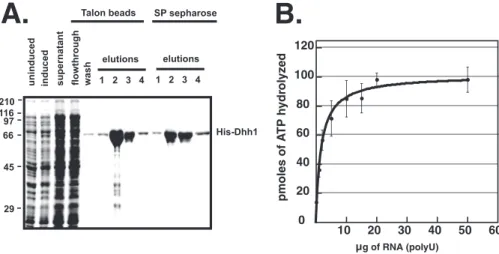
Dhh1 Has RNA-stimulated ATPase Activity—Recombinant His6-Dhh1 was expressed in E. coli, isolated on NTA-cobalt beads, and further purified by SP-Sepharose chromatography. The use of NTA-cobalt beads and ion-exchange chromatogra-phy was necessary to purify Dhh1 away from a contaminating E. coliprotein with nucleic acid-stimulated ATPase activity.3 The contaminating ATPase was also stimulated by DNA, indi-cating that it is not RNA-specific.3Fig. 1A, shows the purifica-tion of the protein through the SP column. ATPase assays were carried out at 30 °C with increasing amounts of poly(U) RNA. Dhh1 has very low ATPase activity in the absence of RNA, but a
⬃10-fold stimulation in activity was observed in the presence of RNA (Fig. 1B).
Although Dhh1 displayed RNA-dependent ATPase activity, the level of activity was significantly less than that reported for
3A. Dutta and J. C. Reese, unpublished information. 0
20 40 60 80 100 120
10 20 30 40 50 60
pmoles of A
TP h
ydr
ol
yz
ed
g of RNA (polyU)
µ
His-Dhh1
uninduced induced supernatant flowthrough wash
elutions elutions Talon beads SP sepharose
1 2 3 4 1 2 3 4
29 45 66 97 116 210
A.
B.
FIGURE 1.ATPase activity of Dhh1 is stimulated by RNA.A, purification of recombinant Dhh1 is shown. Recombinant His6-Dhh1 was purified on NTA-cobalt beads (Talon) followed by SP-Sepharose chromatography. An SDS-PAGE gel shows the different fractions during purification.E1-E4, elutions from NTA-cobalt beads or SP-Sepharose column.B, ATPase activity of Dhh1 is shown. An ATPase assay was performed at 30 °C for 60 min using 4MDhh1 and increasing amounts of poly(U) (0 –50g). Results are plotted as pmol of ATP hydrolyzed as a function of increasing poly(U) concentration. The data is shown as the average and S.E. of three experiments.
at University of North Carolina at Chapel Hill on April 29, 2020
http://www.jbc.org/
other DEXD/H-box proteins, such as eIF4A and Ded1 (39, 53, 54). Furthermore, a side by-side comparison of the ATPase activities of Dhh1 and the hepatitis C virus NS3 RNA helicase suggests that Dhh1 has weaker ATPase than better-studied DEAD-box proteins (supplemental Fig. 1). The difference in activity was unexpected because Dhh1 has all of the signature motifs and conserved catalytic residues found in other well studied DEXD/H-box proteins. We speculated that there may be biological significance for the weaker enzymatic activity and explored the possible causes. The crystal structure of the core domain of Dhh1 revealed extensive interdomain interactions between the C-terminal and N-terminal lobes of the protein, which positions Dhh1 into a closed conformation (44). Such interactions are lacking in eIF4A and mjDEAD, DEAD-box proteins with more robust ATPase activity than Dhh1 (see Ref. 44 andsupplemental Fig. 2). More specifically, Leu-342 and Thr-344 of motif V in the C-terminal domain of Dhh1 hydro-gen bond with Lys-91 of motif I in the N-terminal domain (Fig. 2A). Furthermore, the side chain of Arg-345 in motif V stacks
against that of Arg-89 in motif I and Leu-343 in motif V and make van der Waals interactions with Gly-93 and Thr-94 in motif I. Current models propose that binding of ATP and RNA by DEXD/H-box proteins results in a conformational change, bringing the C- and N-terminal domains closer, and rotation of these domains occurs during the ATP hydrolysis cycle (39, 55). Given the interactions between and N-terminal and C-terminal RecA-like domains observed in the crystal structure of the core domain Dhh1, it is possible that these interactions may hinder the movement of the two domains with respect to each other and limit ATPase activity. To test this hypothesis, we have mutated Thr-344 of motif V and Lys-91 of motif I to alanines to disrupt the hydrogen bonding between these residues. This is predicted to weaken the interdomain interactions and may increase the ATPase activity of the mutant. Furthermore, we analyzed a mutant in the first conserved aspartic acid in the “DEAD box” of Dhh1, D195A, as a control and to confirm that Dhh1 is abona fideDEAD-box ATPase. As expected, the level of ATPase activity of the D195A mutant was negligible, and the FIGURE 2.ATPase activity of Dhh1 is stimulated by the mutation of residues involved in interdomain interactions.A, the crystal structure of Dhh1 with the locations Lys-91 and Thr-344 indicated inredandblue, respectively. The structure was analyzed using PyMOL software (PDB code 1S2M).B, ATPase activity of wild type and mutant Dhh1 is shown. ATPase assays were carried at 30 °C for 60 min using 4Mprotein and increasing amounts of poly(U) (0 –50g). Results are plotted as pmol of ATP hydrolyzed as a function of increasing poly(U) concentration.C, RNA sequence requirement for ATPase activity is shown. ATPase activity was carried out using 4Mprotein and 10g of RNA indicated in the figure.D, nucleotide specificity of Dhh1 is shown. NTPase activity of wild type and mutant Dhh1 was carried at 30 °C for 60 min using 4Mprotein and 10g of poly(U) RNA. The data inpanels CandDrepresent an average and S.E. of three experiments.
at University of North Carolina at Chapel Hill on April 29, 2020
http://www.jbc.org/
little that was observed may originate from a trace amount of contaminatingE. coliATPase in our preparations (Fig. 2B). A double D195A/E196A mutant had a similar amount of activity as the single D195A mutant (not shown). Interestingly, muta-tion of both Lys-91 and Thr-344 to alanine residues resulted in a⬃2.5–3-fold increase in the ATPase activity over that of wild type Dhh1 (Fig. 2B). These results support the hypothesis that disrupting the contacts made between the N- and C-terminal RecA-like domains of Dhh1 would increase ATPase activity and suggest that the interdomain interactions may limit its abil-ity to hydrolyze ATP.
Next, we evaluated the RNA sequence and nucleotide speci-ficity requirements for the ATPase activity of Dhh1. The ATPase activities of wild type Dhh1 and the D195A and K91A/ T344A mutants were examined in the presence of poly(U), poly(A), poly(C), and poly(G) ribonucleic acids. As shown in Fig. 2C, poly(U), poly(A), and poly(C) stimulated the ATPase activity of Dhh1 to roughly the same degree, but poly(G) was significantly less effective. However, the ineffectiveness of poly(G) is most likely due to its propensity to form G-quartets in solution rather than reflect selectivity against G residues (56). Dhh1 Displays Specificity for Adenine-containing Nucleotides— Typical DEAD-box proteins display specificity for adenine-containing nucleotides and hydrolyze ribose and deoxyribose versions of adenine nucleotides equally well (39). To provide further evidence that Dhh1 is a typical DEAD-box ATPase, we examined the nucleotide specificity of the protein by measuring its ability to hydrolyze and bind to different nucleotides. The nucleotide specificity for hydrolysis was examined by conduct-ing NTPase assays in the presence of poly(U) RNA and 50M
cold nucleotide and a trace amount of corresponding␣-32 P-labeled versions. We analyzed the DEAD-box and interdomain interaction mutants (see above) in parallel as controls. As shown in Fig. 2D, wild type Dhh1 hydrolyzed ATP and dATP, but not GTP, CTP, or UTP. Similar to what was observed when ATP was used as a substrate, the K91A/T344A mutant dis-played enhanced hydrolysis of dATP compared with wild type protein but no hydrolysis of the other nucleotides. Thus, even though disrupting the interdomain interactions within Dhh1 enhanced its ATPase activity, these mutations did not alter the nucleotide specificity of the protein.
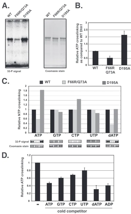
The nucleotide binding specificity of Dhh1 was examined by UV-cross-linking of radiolabeled nucleotides to the protein. The amount of NTP cross-linked was quantified, and the sig-nals for each sample were corrected for the amount of protein in the gel and normalized to the signal from wild type Dhh1. To distinguish signals attributed to nucleotide bindingversus non-specific cross-linking, we analyzed a mutant with substitutions in highly conserved residues within the Q-motif of Dhh1 (F66R/Q73A). The Q-motif, especially the conserved phenyla-lanine and glutamine residues, has been shown to participate in nucleotide binding in other DEAD-box proteins, and co-crystal structures of DEAD-box helicases with nucleotide analogs revealed that these residues make contact with the adenine base (57– 60). We used the D195A mutant as a control, as this mutant should bind ATP but not hydrolyze it. It has been reported that the analogous DEAD-box mutation in eIF4A enhanced nucleotide cross-linking, presumably by preventing
hydrolysis of the nucleotide (54, 61). If the cross-linking to Dhh1 is specific, we expect that cross-linking would be reduced and enhanced in the Q-motif and DEAD-box mutants, respec-tively. As shown in Fig. 3, AandB, radiolabeled ATP cross-linked to wild type Dhh1, and mutation of the Q-motif residues Phe-66 and Gln-73 resulted in a reduction in ATP cross-link-ing. We interpret the level of cross-linking remaining in this mutant to be background levels. In addition, as observed with eIF4A (54), the DEAD-box mutation (D195A) increased cross-linking of ATP to Dhh1⬃2-fold.
Next, we analyzed the cross-linking of different nucleotides to Dhh1 and its mutant derivatives. Because the level of back-ground cross-linking to the different nucleotides can vary depending on purity and the chemical nature of the nucleotide bases, we compared the cross-linking of the nucleotides to the wild type protein and the Q-domain mutant. Background cross-linking would be equal in both versions of Dhh1. Consis-tent with the data showing that Dhh1 can hydrolyze dATP, the cross-linking of this nucleotide was similar to that of ATP. In addition, cross-linking of dATP to the Q-motif mutant was reduced (Fig. 3C). On the other hand, although some incorpo-ration of radiolabeled CTP, GTP, and UTP into Dhh1 was observed, the levels were equal for the wild type protein and the two mutant derivatives (Fig. 3C). This suggests that the cross-linking of CTP, UTP, and GTP arises from the nonspecific interaction of these nucleotides with the protein and represents background. The failure to detect specific cross-linking of radiolabeled CTP, GTP, and UTP to Dhh1 is consistent with the NTPase assays showing that Dhh1 cannot hydrolyze these nucleotides (Fig. 2D).
Because the cross-linking efficiency of nucleotides differs, we addressed nucleotide specificity by competition analysis. Cross-linking was carried out with radiolabeled ATP and 200-fold cold competitor nucleotides. Excess cold ATP, dATP, and ADP competed equally well in the cross-linking assay, reducing the level to 40 –50% of that observed without competitor. The amount of cross-linking detected in the presence of excess ade-nine-containing nucleotides is approximately the same as that observed to the Q-motif mutant in the absence of competitor (Fig. 3D); thus, this likely represents background levels. GTP competed reasonably well but CTP and UTP less so. Even though some competition for binding was observed, Dhh1 was incapable of hydrolyzing these nucleotides (Fig. 2D). Overall, our analysis indicates that Dhh1 has a strong preference for adenine-containing nucleotides.
Dhh1 Binds RNA with High Affinity—Next, we sought to characterize the RNA binding properties of Dhh1. Dhh1 dis-plays RNA-dependent ATPase activity, and a previous report showed that it binds to poly(U) RNA using a semiquantitative, filter binding assay (44). The published account used only a fixed amount of RNA in the filter binding assays and did not measure the affinity or length requirement for binding. We used the highly sensitive and quantitative fluorescence polar-ization assay to measure the RNA binding affinity and length dependence. Dhh1 was titrated into a buffer containing a 3⬘ -fluorescein-labeled poly(U) oligomer, and mP was recorded. The dissociation constant (Kd) for the binding of Dhh1 to RNA
at University of North Carolina at Chapel Hill on April 29, 2020
http://www.jbc.org/
Coomasie stain
WT F66R/Q73A D195A
32-P signal Coomasie stain
WT F66R/Q73AD195A WT F66R/Q73AD195A
0 0.5 1 1.5 2 2.5 3
Relative A
T
P
crosslinking
as compared to WT Dhh1
Relative NTP
crosslinking
0 0.2 0.4 0.6 0.8 1 1.2 1.4 1.6 1.8 2
ATP
GTP
CTP
UTP
dATP
A.
B.
C.
WT F66R Q73A
D195A
0 0.2 0.4 0.6 0.8 1 1.2
ATP
GTP
CTP
UTP dATP ADP
D.
Relative A
T
P
crosslinking
-cold competitor
32-P signal
FIGURE 3.Analysis of UV-cross-linking of nucleotides to Dhh1.A, assays contained 4Mprotein and 10Ci of␣-32P-radiolabled nucleotide. Samples were cross-linked on ice (254-nm UV radiation) for 6 min and resolved in a 10% SDS-PAGE gel. The gel was stained with Coomassie Blue, dried, and exposed to a PhosphorImager screen. The32P signal is shown on theleft, and the Coomassie Blue-stained gel is shown on theright.B, quantification shown is of ATP cross-linking from three experiments. Data are expressed as relative ATP cross-linking compared with that of wild type Dhh1, which was set to 1.0. Cross-linking was corrected for the amount of total protein detected in the Coomassie Blue-stained gels.C, relative cross-linking of wild type and mutant proteins to NTPs is shown. In each case cross-linking to wild type Dhh1 is set to 1.D, nucleotide competition experiments are shown. Wild type Dhh1 was incubated with excess cold nucleotide indicated in the panel on thexaxis. Radiolabeled ATP was added, and cross-linking was carried out as described inpanel A.
at University of North Carolina at Chapel Hill on April 29, 2020
http://www.jbc.org/
was calculated by plotting mP as a function of Dhh1 concentra-tion and fitted to a rectangular hyperbola.
Dhh1 bound a 20-base polyuridine RNA (rU20) with high affinity, displaying aKdof⬃2 nM(Table 1). Thus, Dhh1 has a
very high affinity for RNA. Chenget al.(44) reported that the trypsin sensitivity of Dhh1 changed in the presence of ATP, suggesting that it undergoes a conformational change upon binding the nucleotide. The consequences of the conforma-tional change are not known, but it could affect RNA binding affinity by altering the positioning of the two lobes that form the RNA binding cleft. Thus, we examined if nucleotides affected RNA binding by conducting binding assays in the presence and absence of ATP, ADP, and AMP-PNP. No changes in affinity of Dhh1 for RNA were observed in the presence of ATP, and only a slight change was detected when ADP or AMP-PNP was included (Table 1). Therefore, even though there is evidence that ATP binding changes the conformation of Dhh1, this does not affect RNA binding affinity significantly.
The crystal structure of the core domain of Dhh1 was solved without RNA, but co-crystal structures of other DEXD/H-box proteins with RNA have been solved. We, therefore, used the crystal structure of D. melanogaster Vasa, a homologous DEAD-box protein, to conduct structure-guided mutagenesis to provide evidence that Dhh1binds RNA similarly. Vasa binds
⬃10 bases of RNA by bending it into a groove formed between the two globular domains of the protein (58). Importantly, the locations of the charged residues in Vasa that contact RNA are conserved in Dhh1 and are located in a cleft formed between the N- and C-terminal domains of the core of Dhh1 (44). To provide evidence that Dhh1 recognizes RNA through a similar mechanism, we analyzed the RNA length requirement and con-ducted mutagenesis of a subset of these conserved residues (see below). Fluorescence polarization experiments were conducted using poly(U) probes of differing lengths (rU5, rU7, rU8, rU10, rU12, and rU20). The data presented in Table 2 show that the affinity of Dhh1 for RNA is length-dependent and generally increased with increasing lengths of nucleic acid. Interestingly, there was a significant decrease in affinity when the length of RNA was reduced from 12 to 10 nucleotides (Table 2). The affinity dropped further when the RNA was shortened to eight nucleotides. Thus, the length requirement for RNA binding detected in the binding assays generally agrees with the mini-mal number of nucleotides contacting the protein in the co-crystal structures of analogous DEXD/H-box proteins and RNA (58, 62). However, Dhh1 must make contact with more than 10
nucleotides of RNA because increasing the number of nucleo-tides from 10 to 12 or 20 led to a further increase in affinity. Finally, we examined the binding of Dhh1 to a single-stranded deoxyribose nucleic acid probe. Dhh1 bound to a poly(dT20) probe with high affinity (⬃77 nM), but this is⬃30-fold lower
than the rU20 probe (Table 2). However, it is unclear if Dhh1 binds single-stranded DNAin vivo.
It was reported that mutation of basic residues lining the potential RNA binding cleft in Dhh1 reduced RNA binding (44). Specifically, mutation of 89 or Lys-91 in motif I, Arg-345 or Gly-346 in motif V, and Arg-370 in motif VI reduced RNA binding when assayed by a filter binding assay. We veri-fied and extended these studies by conducting mutagenesis of residues in Dhh1 implicated in RNA binding. Fluorescence polarization binding assays largely confirmed that mutation of the residues comprising the proposed RNA binding cleft reduced RNA binding. However, a quantitative measure ofKd values revealed that the effects of these mutations were not as severe as what was implied from the filter binding assay (44). Arginine 322 and serine 340 were also targeted for mutagenesis. These amino acids correspond to Arg-528 and Thr-546 of Vasa, which were shown to make direct contact with RNA in its co-crystal structure with RNA (58). Mutation of these residues individually did not affect RNA binding significantly (Table 3). However, when both residues were mutated to alanines, RNA binding affinity was reduced 4-fold compared with wild type protein (Kd⬃82 nMfor wild type proteinversus⬃320 nMfor the R322A/S340A mutant). Collectively, the length depen-dence for RNA binding and mutagenesis studies suggest that the RNA binding pocket of Dhh1 lies between the N- and C-ter-minal domains and that it binds RNA similar to the related DEAD-box protein Vasa.
We examined the ATPase activity of the RNA binding mutants. The assays were carried out with saturating amounts of RNA (20g) to diminish the contributions of reduced RNA binding on activity. All but one RNA binding mutant displayed ATPase activities similar to wild type Dhh1 (Table 3). The small changes we observed in most mutants were within experimen-tal error. However, the double R322A/S340A mutant showed no specific ATPase activity, hydrolyzing ATP to the same level as a DEAD-box (D195A) and ATP binding mutant (Q73A/ F66R). The reduction in ATPase activity cannot be explained by reduced ATP binding; the mutant cross-linked to ATP as well as wild type Dhh1 or by the reduced affinity for RNA. The latter point is supported by comparison of the two mutants with the
TABLE 1
Dhh1 binds RNAin vitrowith high affinity
Wild type Dhh1 or mutant proteins were titrated into a binding assay containing 0.1 nM3⬘fluorescein-labeled RNA or single-stranded DNA. Binding was measured by monitoring the change in mP. The data were fit to a hyperbola, and a dissociation constant was calculated from the curve. Dissociation constants for binding of Dhh1 to U20 (20-mer poly(U) containing a 3⬘fluorescein). Binding was carried out under different conditions; that is, the presence/absence of Mg2⫹and ATP. Values are
expressed as the average and S.E. of three experiments.
Conditions Dissociation constant
nM
Protein⫹MgCl2 1.8⫾0.4
Protein⫹MgCl2⫹ATP 2.2⫾0.8
Protein⫹MgCl2⫹ADP 3.9⫾0.5
Protein⫹MgCl2⫹AMP-PNP 3.6⫾0.4
TABLE 2
Length dependence of RNA binding
Binding assays were conducted as described in Table 1 and under “Experimental Procedures.” Dissociation constants for the binding of Dhh1 to sequences contain-ing different lengths of poly ribose uridine (rU) and polydeoxyribose thymidine (dT) nucleic acids are reported.
RNA length Dissociation constant
nM
rU5 968⫾317
rU7 577⫾112
rU8 405⫾54
rU10 81⫾8
rU12 5.4⫾0.5
rU20 2.7⫾0.6
dT20 77⫾9
at University of North Carolina at Chapel Hill on April 29, 2020
http://www.jbc.org/
greatest reduction in RNA binding. The R322A/S340A mutant displayed a slightly higher affinity for RNA than the R370A mutant, yet the R370A mutant showed robust ATPase activity when assayed in the presence of saturating amounts of RNA. This suggests that Arg-322 and Ser-340 have additional roles in ATP hydrolysis. Evidence for this possibility comes from stud-ies ofDrosophila DEAD-box protein Vasa. The orthologous residues in Vasa are important for RNA binding, ATPase, and helicase activities. The crystal structure of Vasa with RNA indi-cates that these residues are important for bending the RNA in the binding cleft. The bending of the RNA may be important for the ability of Vasa and Dhh1 to hydrolyze ATP; a similar model has been proposed by others when describing the mechanism of other DEXD/H helicases (38). Interestingly, the double mutant K91A/T344A mutant bound RNA somewhat better than the single K91A mutant. Although Lys-91 contributes to RNA binding via an interaction with the negatively charged RNA, the increased flexibility caused by disruption of the interdomain interactions in the K91A/T344A mutant may allow the RNA to make contacts with other residues in the presumed RNA bind-ing cleft, leadbind-ing to higher affinity for RNA.
Dhh1 Does Not Display Helicase Activity in Vitro—Our stud-ies thus far show that Dhh1, like many DEXD/H-box proteins, binds to RNA and hydrolyzes ATP in an RNA-dependent man-ner. We next sought to examine if Dhh1 has helicase activity. It cannot be taken for granted that Dhh1 displays helicase activity because many DEXD/H-box proteins do not display helicase activity. We used an assay to detect the unwinding of double-stranded RNA containing either a 5⬘or 3⬘overhang. Our pre-vious experiments have shown that Dhh1 can bind a 12-mer RNA with high affinity, so we designed duplex (8 base) sub-strates to incorporate a 12-nucleotide single-stranded RNA overhang. We titrated various concentrations of wild type Dhh1 (0.3, 0.6, and 1.0M) into the assay and carried out
exper-iments with 1MK91A/T344A and D195A/E196A mutants.
The K91A/T344A mutant was tested to determine whether enhancing ATPase activity could activate helicase activity. We failed to observe unwinding of short double-stranded RNAs with templates containing either a 5⬘or 3⬘overhang ( supple-mental Fig. 1). As a control we used the hepatitis C virus NS3
protein that unwinds double-stranded RNA with a 3⬘overhang. Under our reaction conditions 0.5 M NS3 specifically
unwound double-stranded RNA with a 3⬘overhang but not a substrate with a 5⬘ overhang, the same specificity that was described earlier (45) (supplemental Fig. 1). Because Dhh1 bound more strongly to a 20-mer RNA, we repeated the assay using substrate with a 20 nucleotide ssRNA overhang either at the 5⬘or 3⬘ends but failed to observe any Dhh1-dependent unwinding of substrates (not shown). Once again, hepatitis C virus NS3 unwound the substrate with a 20-mer 3⬘overhang (not shown). Thus, Dhh1 did not display helicase activity under the conditions used here.
Phenotypic Screening of Dhh1 Mutants—Biochemical analy-sis of Dhh1 has identified residues that are required for ATPase activity, RNA binding, and ATP binding. Earlier studies have shown that aDHH1null mutant is viable but displays sensitiv-ities to stress conditions including heat and DNA-damaging agents such as methyl methane sulfonate, hydroxyurea, and UV irradiation (27, 28, 30). Mutations to the DEAD-box or residues implicated in RNA binding could not complement the temper-ature sensitivity of adhh1⌬strain (44). We applied the same analysis to the mutants we characterized biochemically and examined their ability to complement the DNA damage sensi-tivity of the⌬dhh1strain. It is a formal possibility that the DNA damage resistance functions of Dhh1 may require different functional domains than those required for heat stress. Wild type Dhh1 or mutant proteins were expressed under the con-trol of its own promoter from low copy plasmids in adhh1⌬null strain. A schematic of the mutants in relation to the nine con-served functional motifs of Dhh1 is shown in Fig. 4A. Further-more, we verified by Western blotting that each of the Dhh1 mutants accumulated to levels equal to that of wild type Dhh1 (Fig. 4B). As a whole, the data show that mutating any of the conserved domains of Dhh1 resulted in DNA damage and heat stress sensitivity. Those showing the strongest phenotypes typ-ically displayed severely impaired ATPase activity. The R322A mutation, which reduced RNA binding and ATPase weakly, still showed sensitivity to stress conditions. However, reducing RNA binding by as much as 5-fold did not affect the stress resistance of the R370A mutant. This suggests that either the
TABLE 3
Comparison of biochemical activities of Dhh1 and its mutant derivatives
The mutants of Dhh1 are listed in the table. The mutants are shown in the table and are grouped according the contributions they make to the function of the protein, predicted by its comparison to analogous DEAD-box RNA helcases. ATPase activities were carried out as described before. Moles of ATP hydrolyzed by the proteins are shown. ATP binding was determined by UV-cross-linking assays, and the cross-linking of ATP to wild type Dhh1 was set to 1.0. Cross-linking of ATP by mutant proteins is expressed relative to that of wild type Dhh1. RNA binding was assayed using 0.1 nM3⬘fluorescein-labeled single-stranded 10-mer poly(U). Dissociation constants (nM) calculated from binding curves are tabulated. Errors represent the average and S.D. of at least three independent experiments. ND, not determined.
Activity Dhh1 ATP hydrolyzed Relative cross-linking to ATP Dissociation constant for rU10 binding
pmol nM
WT Dhh1 90⫾10 1 82⫾9
ATP binding Q73A 75⫾11 0.66⫾0.2 85⫾4
Q73A/F66R 42⫾14 0.51⫾0.1 91⫾12
ATP hydrolysis K96A 31⫾5 0.85⫾0.03 ND
D195A 35⫾4 2.15⫾0.3 120⫾20
RNA binding R322A 76⫾20 0.92⫾0.2 128⫾10
S340A 76⫾23 0.84⫾0.3 69⫾3
R322A/S340A 26⫾10 1.13⫾0.2 316⫾20
R370A 130⫾45 0.91⫾0.1 402⫾45
Interdomain interactions K91A 124⫾27 1.19⫾0.1 219⫾13
T344A 109⫾21 1.08⫾0.1 48⫾5
K91A/T344A 270⫾75 1.07⫾0.2 136⫾21
G346A 33⫾8 1.02⫾0.1 68⫾7
at University of North Carolina at Chapel Hill on April 29, 2020
http://www.jbc.org/
amount of RNA within the cell is saturating or, more likely, other factors in the cell compensate for its reduced affinity for RNA. This mutant did display a weak RNA turnover defect (see
below), however, suggesting that mutating this residue does have a subtle affect on Dhh1 function. The K91A/T344A and the K91A mutants, which displayed increased ATPase activity, FIGURE 4.Phenotypic analysis of Dhh1 mutants.A, a schematic shows the conserved domains in Dhh1 with the position of residues that were mutated. B, analysis of the expression of Dhh1 mutants is shown. Western blotting using anti-Dhh1 antibody is shown. The amount of TATA-binding protein (TBP) was used as a loading control.C, yeast cells expressing wild type or mutantDHH1were grown in SD-tryptophan, and 3-fold serial dilutions of cultures were spotted onto SD-tryptophan plates (control) or the same medium containing 75 mMhydroxyurea (HU) or 0.01% methyl methane sulfonate (MMS). UV sensitivity was measured by exposing a plate to 60 J/m2UV radiation. For testing heat sensitivity, plates were incubated at 37 °C. Control plates were incubated at 30 °C. The mutants were tested in two groups; a wild type strain and deletion mutant was analyzed in each group.
at University of North Carolina at Chapel Hill on April 29, 2020
http://www.jbc.org/
were not sensitive to stress. Interestingly, the K91A/T344A mutant showed slightly better growth at 37 °C than cells expressing wild type Dhh1 (Fig. 4C). The growth advantage of this mutant at 37 °C is modest but reproducible.
The biochemical assays used Dhh1 protein retaining the His6-tag and additional amino acids incorporated into the N terminus. In contrast, the genetic analysis was carried out in cells expressing the natural from of Dhh1. It is a formal possi-bility the His6tag changed the activity of the proteinin vitro. However, proteins generally tolerate tags in the N terminus, and the biochemical activities and the expected phenotypesin vivoof the Dhh1 mutants correlated very well (also see below). Mutation of the Functional Domains of Dhh1 Affect RNA Turnover—We isolated mutants with defects in the biochemi-cal activities of Dhh1, and next we examined the requirement for these functions in mRNA decay, recruitment into cytoplas-mic foci, and binding to mRNA in vivo. Represen-tative DHH1 mutants that displayed specific biochemical defects were chosen for further analyses. The effect of these mutations on RNA turnover was determined by measuring the levels ofGAL1mRNA over time after repressing transcription with dextrose.GAL1displayed an mRNA half-life of⬃11 min
in wild type cells, and deletingDHH1extended the half-life to 22 min (Fig. 5A); these values agree with previous reports (11). We observed that mutations in Dhh1 affecting ATP hydrolysis (D195A/E196A), RNA binding (R322A/S340A), and ATP bind-ing (F66R/Q73A) resulted in a doublbind-ing of half-life of theGAL1 mRNA, similar to that observed in thedhh1⌬mutant (Fig. 5A). On the other hand, the half-life ofGAL1mRNA in the K91A/ T344A mutant was reproducibly shorter than that measured in cells expressing wild type Dhh1, but the difference from the wild type value is within experimental error (not shown). This suggests that disrupting the interdomain interactions within Dhh1 and enhancing its ability to hydrolyze ATP increased its functionin vivo. To further substantiate our results showing that mutations in Dhh1 reduced mRNA turnoverin vivo, we studied the steady state levels ofEDC1mRNA, which is sensi-tive to the loss of Dhh1 (36, 44). Because monitoring steady state mRNA levels is more straightforward, more mutants were examined. Deletion ofDHH1caused a 2–2.5-fold increase in EDC1 mRNA (Fig. 5B and Ref 44). Here we observed that mutants that affect ATPase activity (D195A, D195A/E196A, G346A, and K96A), ATP binding (F66R/Q73A), or RNA bind-ing (R322A/S340A and R370A) led to an increased accumula-FIGURE 5.All activities of Dhh1 are required for mRNA decay.A, shown is a half-life estimation ofGAL1. Cells were grown overnight to anA600of 0.8 in 1% yeast extract, 2% peptone, and 20g/ml adenine sulfate supplemented with 2% galactose and then shifted to 4% dextrose-containing medium to repress GAL1transcription. Samples were collected at different time points. Blots were probed withGAL1and loading control,ScR1.GAL1signal was normalized to the amount ofScR1signal to correct for loading. To calculate half-life, a 0-min time point for each set was set to 100%, and the log of the amount of transcript was plotted as a function of time.B, shown is accumulation ofECD1mRNA, a known target of Dhh1. Northern blot analysis to detectEDC1andScR1(loading control) transcripts was carried out.EDC1signal was normalized to the amount ofScR1. Values were normalized to the wild type signal, which was set to 1.Barsrepresent the average and S.D. of at least three experiments.
at University of North Carolina at Chapel Hill on April 29, 2020
http://www.jbc.org/
tion ofEDC1mRNA, indicating impaired decay functions in these mutants (Fig. 5B). Interestingly, the steady state levels of EDC1mRNA were slightly reduced in mutants containing sub-stitutions in residues involved in forming the interdomain interactions (K91A and K91A/T344A) between the N- and C-terminal lobes. The reduction in mRNA may be the result of enhanced turnover ofEDC1mRNA. These results indicate that disrupting any of the biochemical activities of Dhh1 leads to defects in mRNA turnover and that the interdomain interac-tions within Dhh1 may limit its ability to contribute to the turn-over mRNAsin vivo(see below also).
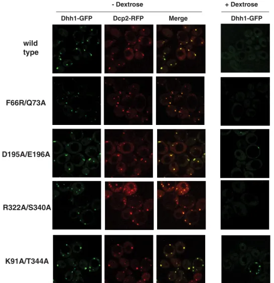
The Localization of Dhh1 into Cytoplasmic Foci Is Regulated by ATP and RNA Binding and Intermolecular Interactions— The role of Dhh1 in mRNA turnover and translational repres-sion has been attributed to its localization to cytoplasmic foci called P-bodies, where it interacts with components of the mRNA decapping machinery (Dcp1/Dcp2), Pat1, and Xrn1 (11). Dhh1 is localized throughout the cytoplasm in a punctate pattern and can be redistributed into larger discrete foci upon cell stress (25). It is not known which activities of Dhh1 are required for its localization to P-bodies. To address this ques-tion, we analyzed Dhh1 and its mutants’ ability to form P-bod-ies in resting and stressed cells. We constructed strains contain-ing C-terminal GFP-tagged Dhh1 at its genomic locus and transformed the wild type and mutant strains with a plasmid carrying RFP-tagged Dcp2, a reliable and easily visualized com-ponent of yeast P-bodies (52). Stress was induced by starving cells of dextrose, a condition that strongly and rapidly induces P-body formation (25, 52). The stressed or unstressed cells were immediately imaged using a confocal fluorescence microscope to visualize Dhh1- and Dcp2-containing foci (25). In addition to monitoring Dhh1 localization in cytoplasmic foci, we con-ducted RIP assays to monitor the association of Dhh1 with mRNAsin vivo. The procedure uses formaldehyde to rapidly cross-link cells in culture to preserve RNA-protein interactions and provides an effective method to measure the association of Dhh1 with mRNAs. In conjunction with monitoring Dhh1 localization, RIP can correlate mRNA binding with P-body for-mation in cells. Furthermore, as we will demonstrate by the correlation between Dhh1 cross-linking and the extent of foci formation in wild type and mutant cells, this method provides a more quantitative surrogate to measure the recruitment of Dhh1 into RNA-containing foci.
In wild type cells very few cells displayed Dhh1-containing foci under the resting condition (⫹dextrose), but the foci were increased in number and size when cells were deprived of dex-trose and the fraction of cells with foci increased (Fig. 6). The Dhh1 colocalized almost completely with the Dcp2-RFP, con-sistent with previous results showing that Dhh1 localizes to P-bodies in stressed cells (25) (Fig. 6). Correlating with the increase in Dhh1-containing foci in response to stress, there was an increase in the cross-linking of Dhh1 to a model mRNA, PYK1, in stressed cells (Fig. 7).
An analysis of the localization of Dhh1 mutants yielded inter-esting results. First, in the ATPase-defective mutant (D195A/ E196A), an increase in the size and number of Dhh1-containing foci was observed in resting cells (Fig. 6). Under stress, an increase in foci intensity was observed. The differences in foci
between this mutant and wild type cells were not obvious under stress conditions. However, the size and number of P-bodies may have been close to saturation under the stressed condition; therefore, the differences between the wild type and mutant are not easily observed. Dcp2-RFP fluorescence overlapped that of Dhh1, indicating that the ATPase activity of Dhh1 is not required for Dcp2 to shuttle into P-bodies. Examination of the association of the DEAD-box mutant with mRNA using RIP revealed an increase over wild type in resting cells, but even a more dramatic increase was observed in stressed cells. The cross-linking of the DEAD-box mutant was 2-fold higher than that observed in wild type cells. The increase in size and num-ber of foci and the elevated association of the mutant protein with mRNA suggest that the ATPase activity and mRNA decay functions of Dhh1 are not required for it to shuttle mRNAs into P-bodies. However, the failure to hydrolyze ATP may prevent the decay or release of the mRNAs and an accumulation of Dhh1 and mRNAs into P-bodies. This hypothesis is supported by the observation that impairing mRNA decay by deleting decapping enzyme Dcp1 or the exonuclease Xrn1 likewise led to an accumulation of Dhh1 into P-bodies in unstressed cells (25, 64).
The GFP signal of the Q-motif mutant is similar and may be slightly weaker than the wild type cells in the unstressed condi-tion. However, a decrease in the number and size of Dhh1-containing foci was observed when the cells were stressed by dextrose deprivation compared with wild type cells (Fig. 6). Calculation of the average size of mutant Dhh1-containing foci revealed that they are on average half the size of those observed in wild type cells (0.12versus0.25m for the wild type). The RIP experiments suggest that the association of the F66R/Q73A mutant was similar to that of wild type Dhh1 in resting cells but that the stress-induced increase in its association with mRNAs was nearly abolished in this mutant (Fig. 7). These results are a little surprising, as the Q-motif mutant displays no ATPase activity and impaired mRNA decay functions similar to the DEAD-box mutant, and yet its localization and RNA associa-tion patterns are different from the DEAD-box mutant. Thus, the localization and RIP studies suggest that ATP binding is required for Dhh1 to bind mRNA in vivoand transport into P-bodies. This suggests that ATP binding per se, and not hydrolysis specifically, plays a role in regulating Dhh1 (see “Discussion”).
Similar to the ATP binding mutant, the RNA binding mutant (R322A/S340A) also showed weaker background staining in unstressed cells and formed smaller foci when cells were stressed (Fig. 6). The foci formed by this mutant were also about half the size of those formed by wild type Dhh1 (0.14versus0.24 um). RNA-IP experiments revealed that this mutant displayed a 2-fold reduction in its cross-linking toPYK1mRNA both in the stressed and unstressed conditions (Fig. 7). These two pieces of data argue that the interaction of Dhh1 with RNA is required for its recruitment into P-bodies. Furthermore, because this mutant is defective for mRNA decay (Fig. 5), simply altering mRNA turnover and accumulating RNAs in the cell cannot cause the recruitment of Dhh1 into cytoplasmic foci. Further-more, the pattern of Dcp2 fluorescence in this mutant was
at University of North Carolina at Chapel Hill on April 29, 2020
http://www.jbc.org/
ilar to that of the wild type cells, indicating that reducing Dhh1 RNA binding does not significantly affect the recruitment of Dcp2 into P-bodies.
Mutating residues involved in the interdomain interactions enhanced ATPase activity, imparted a slight growth advantage to cells undergoing heat stress, and increased mRNA turnover wild
type
K91A/T344A F66R/Q73A
D195A/E196A
R322A/S340A
Dhh1-GFP Dcp2-RFP Merge Dhh1-GFP
- Dextrose + Dextrose
FIGURE 6.The localization of Dhh1 mutants to cytoplasmic foci requires residues involved in ATP and RNA binding.Dhh1-GFP and Dcp2-RFP were analyzed by confocal fluorescent microscopy. Log phase wild type and Dhh1 mutants expressing endogenous GFP- tagged Dhh1 protein and Dcp2 (pRP1186) were dextrose-deprived for 15 min, and live cell images were obtained. Dhh1-GFP, Dcp2-RFP, and merged images are shown in⫺dextrose conditions. Dhh1-GFP images are shown in⫹dextrose condition. Exposures displayed in the panels were chosen so that the outline of the cells can be seen.
FIGURE 7.RNA bindingin vivois affected by mutations in Dhh1.RIP analysis to studyin vivomRNA binding by Dhh1 is shown. Dhh1-cross-linked RNA was extracted and converted to cDNA. Binding of Dhh1 toPYK1mRNA was analyzed by PCR using primers to the ORF ofPYK1. Percent IP was calculated by correcting the IP RNA value for the input RNA value. The experiments shown in theleftandright panelswere conducted at different times; thus, the mutants in each panel should be compared with the wild type data conducted in parallel.
at University of North Carolina at Chapel Hill on April 29, 2020
http://www.jbc.org/
in vivo. These phenotypes suggest that interdomain interac-tions limit Dhh1 activityin vivo. Interestingly, we observed an increase in foci formation in a greater fraction of unstressed cells expressing the K91A/T344A mutant (Fig. 6). A slight increase in the number and intensity of P-bodies under stressed conditions was observed, but the increase was not robust. Sim-ilar to the DEAD-box mutant, foci formation may be saturated in cells under these conditions, and enhancement is not detected. The most striking result was observed in the RIP experiments. Disrupting interdomain interactions led an
⬃3-fold increase in the cross-linking of the mutant toPYK1 mRNA in unstressed cells, and the level increased further upon stress (Fig. 7). The increased association of the mutant with mRNA is not caused by differences in the expression of the mutant or an increase in its affinity for RNA. This mutant accu-mulates in the cells to the same level as wild type Dhh1 (Fig. 4B), and it has a slightly lower affinity for RNA (Table 3). These results argue that interactions between the N- and C-terminal RecA-like domains in the core of Dhh1 may play an important regulatory function in vivo, perhaps preventing Dhh1 from binding mRNAs and shuttling into P-bodies, thereby restrict-ing mRNA decay in unstressed cells.
DISCUSSION
Dhh1 Is a Bona Fide DEAD-box Protein with ATPase Activity— The classification of Dhh1 as a DEAD-box RNA helicase was based on the presence of well annotated motifs characteristic of this family of enzymes (37). It was surprising that a character-ization of the ATPase activity of Dhh1 or any of its orthologues has not been described. This is especially true given that the protein has been expressed in quantities sufficient for struc-tural studies, and a preliminary analysis of its RNA binding abilities was conducted (44). Comparison of the crystal struc-ture of Dhh1 to those of other RNA helicase revealed a distinct difference in the organization and interactions between the two N- and C-terminal lobes of the core domain. Significant inter-actions between residues in the N- and C-terminal domains were observed in the crystal structure Dhh1, which were not seen in other DEAD-box proteins whose structures have been solved (44). These interactions have the potential to “lock” Dhh1 into an inactive conformation. The prevailing hypothesis of how DEAD-box proteins hydrolyze ATP is that the binding of ATP and RNA to the protein results in a conformational change, bringing the N- and C-terminal domains together. ATP hydrolysis is coupled to the concomitant movement of these domains with respect to each other (55). The previous failures to demonstrate ATPase activity and the interdomain interac-tions detected in the crystal structure of Dhh1 raised the possi-bility that it is not a typical DEAD-box protein or that the gene evolved over time to lose its ATPase and helicase activities. Thus, it was important to show that Dhh1 is abona fide DEAD-box protein with ATPase activity.
A comparison of the ATPase activity of Dhh1 to that of other well characterized RNA helicases (53, 54, 65, 66) indicates that it is a weak ATPase. We surmised that the interdomain inter-actions could restrict the movement of the N- and C-terminal domains of Dhh1 and, hence, reduce its ATPase activity. Anal-ysis of a double K91A/T344A mutant and to a lesser extent the
single mutants showed that disrupting interdomain interac-tions increased ATPase activity. In addition to providing an explanation for the weak ATPase activity of the wild type pro-tein, this result also corroborates the existing models that pre-dict that the enzymatic activity of DEAD-box proteins requires the coordinated movement of the two RecA-like helicase domains. The ATPase activity of the interdomain interaction mutant was still less than that observed for more robust heli-cases. This is not surprising because there are multiple residues that form interdomain interactions between the two lobes of Dhh1 (44). Mutagenesis of more residues to further disrupt the interaction between the N and C lobes to increase activity to a higher level is not practical as some of the residues may play other roles in the function of the protein, such as RNA binding. Any gains in ATPase activity caused by disrupted interdomain interactions might be offset by reducing other activities required for ATPase activity.
Dhh1 Does Not Show Helicase Activity in Vitro—The classical definition of an RNA helicase is a protein that binds ATP and RNA, hydrolyzes ATP, and unwinds duplexed RNA. Our stud-ies thus far have confirmed all of these activitstud-ies for Dhh1, except for the helicase activity. In our hands Dhh1 could not unwind double-stranded RNA under multiple assay conditions and using different substrates (supplemental Fig. 1and data not shown). Our failure to detect helicase activity is not unusual. Many DEXD/H-box proteins have been classified as RNA heli-cases based on sequence and structural similarities to DNA helicases and their ability to hydrolyze ATP and bind RNA (39). However, only a subset of these proteins has been shown to unwind dsRNA in vitro (42, 43, 67, 68). It is clear that the ATPase activity is required for Dhh1 to carry out its functions in vivo, because mutants with substitutions in the DEAD-box or other residues required for ATP hydrolysis cannot restore the function of Dhh1 in stress resistance or mRNA decay. Like many other examples where helicase activity of a DEAD-box protein was not observed, it is unclear if this is due to technical limitations of the assay or that the protein uses the conforma-tional change associated with ATP hydrolysis to carry out other functions. For example, a growing number of helicases have been shown to disrupt or remodel RNA-protein interactions (69). Our mutational analysis and the work of others indicates that Dhh1 regulates the shuttling of mRNAs between the trans-latable pool and the non-transtrans-latable pool contained in cyto-plasmic foci (23, 26). Logically, this would require the assembly and disassembly of mRNAs into messenger ribonucleoprotein complexes. Because the transport of Dhh1 in and out of cyto-plasmic foci is regulated by its RNA binding and ATPase activ-ities, respectively, ATP hydrolysis may play a role in assembling and disassembling mRNP particles rather than unwind duplex RNA. Dhh1 orthologues may play similar functions. The Xeno-pusorthologue Xp54 has been shown to shuttle between the nucleus and cytoplasm in a developmentally regulated manner whereby it interacts with nascent transcripts in the nuclei of transcriptionally active oocytes and localizes to the cytoplasm in transcriptionally quiescent oocytes (70). Also, studies in Dro-sophila, trypanosomes and clam have shown that Dhh1 ortho-logues repress translation of maternal mRNAs during early development (71–73). An attractive model for the control of
at University of North Carolina at Chapel Hill on April 29, 2020
http://www.jbc.org/