Journal of Applied Food Technology
H o m e p a g e : h t t p s : / / e j o u r n a l 2 . u n d i p . a c . i d / i n d e x . p h p / j a f t
Review on Pathogenic Bacteria
Listeria monocytogenes
, the Detection and
the Sequencing Gene Methods Isolated from Meat Products
Bhakti Etza Setiani*, Yoyok Budi Pramono, Lutfi Purwitasari
Food Technology Department, Faculty of Animal and Agricultural Sciences, Diponegoro University, Indonesia
*Corresponding author ([email protected])
Abstract Received: 25 November 2019 Article information:
Accepted: 04 December 2019 Available online: 05 December 2019
Keywords:
Listeria monocytogenes, VIDAS-LDUO® method,
PCR-based method, conventional method, raw and processed meat
© 2019 Indonesian Food Technologists All rights reserved
This is an open access article under the CC BY-NC-ND license
doi: 10.17728/jaft.6460 A study was conducted to review on pathogenic bacteria Listeria
monocytogenes, the detection and the sequencing gene methods isolated from
meat products, compare selected methods that detect the presence of L.
monocytogenes in selected raw and processed meat products. L.
monocytogenes (originally named Bacterium monocytogenes) is a
gram-positive, non-sporeforming, highly mobile, rod-type, and facultative anaerobic bacterium species. It can grow under temperatures between -1.5 to 45°C and at pH range between 4.4 and 9.4, with the optimum pH of 7. Rapid methods (PCR based and VIDAS-LDUO®) detected L.monocytogenes faster than the conventional method. It was also gathered that Phenotypic identification and Genotypic identification were two types of confirmation test for L.
monocytogenes. L. monocytogenes can be found in raw meat and meat
product because of environmental contamination, cross contamination or error process.
Introduction
Listeria monocytogenes is a major food-borne
pathogen, recognized worldwide as one of the major concerns of food industries and the general public health. It is a ubiquitous microorganism commonly found in foods of animal origin (Schuchat et al., 1991). Consuming foods contaminated with L. monocytogenes, presents significant health risks to humans (Siegman-Igra et al., 2002). It can cause listeriosis which is relatively rare in humans. Severe cases of this infectious disease, however, are characterized by meningoencephalitis, abortion, and septicemia. There is 30% fatality rate for the infectious disease caused by listeriosis (Low and Donachie, 1997).
The United States Food and Drug Administration (USFDA) imposes a “zero tolerance” regulation to L.
monocytogenes in ‘ready-to-eat’ and processed foods,
which brought a great challenge to food processing industries (Farber and Peterkin, 1991; Johnson et al., 1990; Fuchs and Reilly, 1992; Embarek, 1994; Jeyasekaran et al., 1996).
Meat and meat products may serve as vectors for
pathogenic organisms and have frequently been contaminated with L. monocytogenes. Hence, L. monocytogenes can be found in a wide variety of raw and processed meat and meat products such as beef, pork, chicken, fermented sausages and minced meat (Rocourt and Cossart, 1997). Meat is a very rich medium for growth of microorganisms. It is mainly constituted of water, proteins, fats, oligonutrients and vitamins, especially the B group vitamins (Cheftel, 1995). The incidence of L. monocytogenes contamination in fresh meat may vary from 0 to 68%, while in processed meat products, including ready-to-eat food, contamination ranges from 8 to 92% (Johnson et al., 1990). According to the Community Summary Report on Trends and Sources Zoonoses in the EU in 2004, 2005, and 2006 the overall ranges of contamination of foods with L.
monocytogenes were (presence in 25 g): 0.48% in meat
products, 0.40% in poultry meat products, 0.3% in fish products (EFSA, 2007).
common agar media ;such as, tryptose agar, nutrient agar, and blood agar. However, isolation or re-isolation
of Listeria from inoculated or naturally contaminated food
and clinical specimens by use of nonselective media is often challenging. There are several conventional detection methods that have been developed such as direct plating, cold enrichment, selective enrichment such as adding antibiotic to selective media such as palcam (Marth and Ryser, 2007). Whereas rapid test methods consists of Vitek Immuno Diagnostic Assay System Listeria Duo (VIDAS-LDUO®) Method. The final phase of the analysis for L. monocytogenes is the confirmation of the identity of the genus and subsequent speciation of positive Listeria isolates. Phenotypic identification and Genotypic identification are two types of confirmation test for Listeria monocytogenes. It can be done by using some methods such as Molecular Characterization of Listeria monocytogenes by Polymerase Chain Reaction (PCR) and API kit. The objective of this work is to review on pathogenic bacteria
Listeria monocytogenes, the detection and the
sequencing gene methods isolated from meat products.
Genus Listeria
The genus Listeria is composed of six species namely: L. monocytogenes, L. innocua, L. seeligeri, L. welshimeri, L. ivanovii, L. murrayi (also called L. grayi) (Collins et al., 1991; Jones, 1992). The L. monocytogenes, L. ivanovii, L. seeligeri, L. welshimeri and L. innocua are more closely related to each other while L. grayi (or L. murrayi) is distantly related (Collins et al., 1991). However, only L. monocytogenes is commonly associated with human listeriosis. Listeriosis caused by L. ivanovii, L. welshimeri, or even L. seeligeri, is extremely rare in humans. The other two, L. innocua
and L. grayi are non-pathogenic. The universal
occurrence of L. monocytogenes in food (Marth and Ryser, 1999) and the risk of contracting food-borne listeriosis from L. monocytogenes have been thoroughly reviewed recently (SDA/FSIS, 1999).
All members of the genus Listeria, based on the explanation of Liu (2008) form regular, short rods, 0.4 to 0.5 μm by 1 to 2 μm with parallel sides and blunt ends. Usually they occur as single cells or in short chains. In older or rough cultures, 6 μm long filaments may develop. These bacteria give positive results upon gram staining. However; some cells, especially those from older cultures, lose their ability to retain the gram stain. All species of Listeria are motile with peritrichous flagella when cultured between 20° to 25°C but are non-motile at 37°C. They have aerobic and facultatively anaerobic mechanism and do not produce spores. Growth occurs between pH of 5.2 and 9 and at temperature ranging from less than 0° to 45°C; with optimal range between 30°C and 37°C. During growth, cytochromes are produced and glucose is processed through homo fermentative anaerobic catabolism into lactic acid, acetic acid, and other end products. Other sugars produce acids instead of gases. The five true Listeria species do not utilize mannitol or hydrolyze sodium hippurate. Only
L. grayi and L. murrayi hydrolyze sodium hippurate and
utilize mannitol. On nitrate reduction, only L. murrayi
reduces nitrate to nitrite (Weaver, 1989).
All Listeria species are morphologically similar on artificial media. After 24 to 48 hours on artificial media, Listeria colonies are about 0.5 to 1.5 mm in diameter, round, translucent, low convex with a smooth surface and entire margin, and non-pigmented with a crystalline central appearance. Colonies are bluish gray under normal illumination but have a blue-green sheen (Figure 1-left) under oblique light (Farber and Peterkin, 1991). The organisms have the ability to tolerate sodium chloride concentration of 5 to 25% and a variety of other toxic chemicals such as tellurite, acridine dyes, lithium chloride, nalidixic acid and cycloheximide. On blood agar plates, pathogenic Listeria species exhibit zones of hemolysis indicating the destruction of erythrocytes by toxic products of the organism. More so, they have the ability to survive in cells of the host’s immune system or in macrophages (Hass and Kreft, 1988; Schuchat et al., 1991).
The bacterial growth may be sticky when removed from agar surfaces, but it usually emulsifies easily and may leave a slight impression on the agar surface after removal. Older cultures (3 to 7 days) are larger, 3 to 5 mm in diameter, and have a more opaque appearance. Occasionally; rough colonial forms may develop with a sunken center (Liu, 2008).
Figure 1. Listeria monocytogenes cell (left) and colonies (right) (US Centers for Disease Control and Prevention, USDA and MERCK)
Listeria monocytogenes
Listeria monocytogenes (originally named
Bacterium monocytogenes) is a gram-positive,
non-sporeforming, highly mobile, rod-type, and facultative anaerobic bacterium species. It can grow under temperatures between -1.5 °C to 45 °C (Lake et al., 2002), and at pH range between 4.4 and 9.4, with the optimum pH of 7. At pH less than 4.4, the bacteria may be inactivated, depending on the acidulant and temperature of the environment. Organic acids, such as acetic acids, are more effective to inactivated bacteria than mineral acids such as hydrochloric acid. Inactivation is faster at higher temperatures. The organism generally grows well in meats with pH near or above 6.0 and poorly or not at all at pH below 5.0 (Glass and Doyle, 1989). Furthermore, it grows optimally under microaerophilic conditions but also grows aerobically and an-aerobically. It can also grow in relatively high concentration of CO2
(e.g. 30%). Growth is inhibited under 100% CO2
condition but it is not retarded by a 5 to 10% CO2
nitrite. The organism can tolerate sodium chloride at concentration of less than 11.5%. In terms of water activity (Aw), L. monocytogenes grows optimally above 0.97 like most bacteria. It can remain viable in dry environment for long periods of time.
L. monocytogenes can survive at freezing
temperatures and can be inactivated rapidly by exposing it to temperatures above 70°C for 10 seconds. Under certain conditions, it can be viable but non-culturable (VNC). Some recent evidences show that L.
monocytogenes may become VNC cells in dry
environments for a long period of time. It is inactivated on vegetables by lysozyme at 100 mg/kg, 0.2% sodium benzoate at pH 5, 0.25 to 0.3% sodium propionate at pH 5, but is less effective at lower temperatures, and 0.2 to 0.3% potassium sorbate at pH 5.0. L. monocytogenes is more sensitive to UV radiation than other gram-positive bacteria. D-values to kill L. monocytogenes range from 0.34 to 2 kGy depending on the type of food and the temperature. A dose of 3kGy does not remove L.
monocytogenes from vacuum-packed pork. In fish, the D
values are lower at around 0.2 to 0.3 kGy. (Lake et al., 2002).
Materials and Methods
Identification and Isolation
The classical approach to the identification of bacteria is cultural. This approach involves subjecting suspected samples to a series of tests designed to isolate and identify microorganisms possessing a profile of designation belonging to Listeria species at the strain and species levels. The common agar media for studying characterization of L.monocytogenes are tryptose agar, nutrient agar, and blood agar. However, isolation or re-isolation of Listeria from inoculated or naturally contaminated food and clinical specimens by use of nonselective media is often challenging. There are several conventional detection methods that have been developed since first discovered in 1926. Direct plating, cold enrichment, selective enrichment, and several rapid methods can be used in various combinations to detect
L. monocytogenes in food, clinical, and environmental
samples (Marth and Ryser, 2007).
Ancient researcher introduced the cold enrichment procedure at 4˚C in non-selective medium as an alternative method to isolate L. monocytogenes from highly contaminated samples (Gray et al., 1948). In 1950, the enrichment medium method was discovered. The enrichment medium contains inhibitory chemicals and antibiotics that enable isolation of Listeria species from specimens, whereas in the cold enrichment, the broth is non-selective (Klinger, 1988; Schuchat et al., 1991; Swaminathan and Feng, 1994).
The reagents commonly used to render liquid enrichment media selective for Listeria species include nalidixic acid, lithium chloride, acriflavine, glycine, anhydride, nitrofurazone, potassium thiocyanate and potassium tellurate (Klinger, 1988). However, in recent times, the commonly used selective enrichment broths are: (a) United States Food and Drug Administration (USFDA) – prepared medium from tryptic soy broth, acriflavine and nalidixic acid (Lovett, et al., 1987; Lovett,
1988); (b) United States Department of Agriculture – Food Safety and Inspection Service (USDA – FSIS) broth – containing esculin, acriflavine and nalidixic acid; (c) Oxford Listeria selective agar; and (d) Palcam broth agar (Curtis et al., 1989; Van-Netten et al., 1989).
McBride and Girrad (1960) developed the first selective plating agar medium for the cultivation of L.
monocytogenes and other Listeria species. Their
formulation contained blood, phenylethanol agar-base, lithium chloride and glycine. However, in 1988, Lovett modified this medium by excluding the blood component. This modification has favored isolation of L.
monocytogenes from foods.
Curtis et al. (1989) and Van-Natten et al. (1989) developed separately two differential Listeria media (Oxford Listeria agar and PALCAM agar), by incorporating differential agents into the media. On the PALCAM agar, Listeria colonies appear grey-green, approximately 2 mm in diameter and have black–sunken centers with a black–halo against cherry red background (Van-Natten, et al., 1989). L. monocytogenes and other
Listeria species colonies appear black on Oxford agar
with 1 mm to 3 mm in diameter after 24 to 48 hours and are surrounded by black halo against grey background of the medium (Curtis et al., 1989).
Enrichment Method for Isolation
A method being developed for the isolation of L.
monocytogenes and other Listeria species from
biological samples is by using cold enrichment at 4°C for several weeks in a suitable medium (Gray et al., 1948; Rodriguwez et al., 1984). However, due to the long incubation period, which is too time-consuming in emergency cases, more emphasis has been placed on effective direct enrichment. Buchanan (1990) suggested a modified alternative called the University of Vermont
Listeria enrichment broth. In this broth, after 24 hours of
sample incubation, there will be black precipitates showing aesculin utilization by the suspect organism. The broth would support the growth of L.monocytogenes at the optimum temperatures of 30 to 37˚C as well as at 4˚C, with a minor change in their fatty acid composition (Puttmann et al., 1993).
frequently present in semi-processed and processed foods or food-processing environments. The principle of enrichment at elevated temperatures (30°C to 37°C) is based on selective inhibition of indigenous microflora through addition of inhibitory agents while at the same time allowing unhindered growth of Listeria (Marth and Ryser, 2007).
VIDAS-LDUO® Method
The VIDAS (Vitek Immuno Diagnostic Assay System Listeria Duo) Listeria method screens for the presence of Listeria in dairy products, vegetables, seafoods, raw-processed meats and poultry. The method was validated by the Association Française de Normalisation (AFNOR) on June 17, 1994 as a rapid analysis method for all human food products. The method was also validated as Official Method No 999.06 by the Association of Analytical Chemists (AOAC) in May 1999. It is a kit for rapid phenotypic identification. VIDAS® Listeria Duo is an automated qualitative test used on VIDAS® instrument for simultaneous detection and differentiation of Listeria monocytogenes and
Listeria spp in food products and environmental
specimens. The VIDAS system uses the VIDAS-LDUO® assay, an enzyme-linked fluorescent immunoassay (ELFA) for the qualitative detection of L. monocytogenes and other Listeria spp. The internal surface of the Solid Phase Receptacle (SPR), a pipette tip-like disposable device, is pre-coated with anti-Listeria antibodies during production. The VIDAS-LDUO® assay configuration prevents non-specific reactions with the SPR. The reagent for an assay is held in a sealed multi-chambered strip. All assay steps are performed automatically and sequentially by the VIDAS instrument. The sample is inoculated into the reagent strip and cycled in and out of the SPR for a specific length of time. Listeria antigens present in the sample will bind to the anti-Listeria monoclonal antibodies, which are coated on the interior of the SPR. Unbound sample materials are washed away. Antibodies conjugated with alkaline phosphatase are then cycled in and out of the SPR and react with the
Listeria antigen-antibody complexes already adsorbed to
the SPR wall. Unbound conjugate is removed by washing (Warburton et al., 2003).
Principle. The Solid Phase Receptacle (SPR®) serves as the solid phase as well as the pipetting device. Anti-Listeriamonocytogenes and anti-Listeria antibodies are adsorbed on the interior surface of the SPR®. Reagents for the assay are ready-to-use and pre-dispensed in sealed reagent strips. All of the assay steps are performed automatically by the instrument. The reaction medium is cycled in and out of the SPR® several times. Part of the enrichment broth is dispensed into the reagent strip. The antigens present will bind to the antibodies coating the interior of the SPR®. Unbound sample components are then washed away.
Molecular Characterization by PCR
Due to the heightened worldwide interest in food-borne listeriosis, official protocols for the detection and isolation L. monocytogenes were established. Two protocols developed in the United States by the FDA and
USDA–FSIS have emerged as the “standard methods” to isolate L.monocytogenes from dairy foods, seafoods, vegetables, meat, poultry, and egg products. Despite the widespread use of these methods in the United States, Canada, and Western Europe, both procedures are still plagued with difficulties, including the inability to isolate Listeria from all positive samples and difficulties in recovering sub-lethally injured cells. In response to these concerns, the USDA–FSIS and FDA protocols have been modified to enhance their ability to recover injured
Listeria. Other official European agencies in cooperation
with the International Dairy Federation (IDF) have also developed similar protocols partially based on the current FDA methodology (Marth and Ryser, 2007).
To maintain a safe and wholesome food supply, food industries need to have access to rapid, reliable, and sensitive methods of detecting bacteria, including
Listeria spp. and L. monocytogenes. Other desirable
attributes for detection methods include simplicity, cost-effectiveness, quantitative capacity, and online or real-time capabilities. Polymerase Chain Reaction or PCR, antibody-based methods, and methods using more experimental technologies such as biosensors, chip-based system, and spectroscopy can offer some of these desirable attributes. Rapid methods are valuable tools for routine screening of foods and environmental samples but will not entirely replace standard methods in the near future.
At present, negative results by rapid methods are considered definitive, but positive results are viewed as presumptive and must be confirmed through culture. Changes in this outlook will require both advances in rapid detection technology and regulatory acceptance of alternative methods. Apart from the detection step itself, sample preparation and adequate sampling procedures remain key challenges in the development of effective rapid detection techniques. The sheer diversity of food matrices and the fact that Listeria may be present at very low levels are particularly daunting challenges. As a result, most rapid detection technologies still require a cultural enrichment step, although recent advances in immunological separation techniques have sought to circumvent this need. Data regarding the performance of rapid detection methods are most often collected on laboratory-grown cells cultured under optimal conditions.
Listeria present in foods or in the food-processing
environment may be subjected to a variety of stressful or injurious conditions, including heating, freezing, exposure to sanitizers, and high acid or salt concentrations. Rapid methods capable of discriminating between living, dead, and sub lethally injured cells are needed. Finally, the physiological effects of microbial inactivation technologies such as use of high pressure, pulsed electric fields, pulsed light, ohmic heating, or ozonolysis on bacterial cells have not been well characterized. Further studies should also address how the use of alternative processing methods may impact the ability of rapid methods to detect Listeria spp. in foods (Marth and Ryser, 2007).
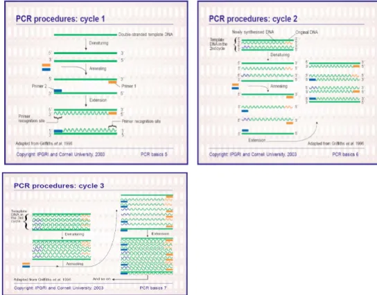
because it is quick, inexpensive and simple. It is based on the enzymatic amplification of a DNA fragment flanked by two oligonucleotide primers that hybridize to opposite strands of a target sequence. The primers are oriented with their 3’ends pointing towards each other. There are 30 repeated cycles of the following processes -denaturing of template; annealing of primers to their complementary sequences; and extension of the annealed primers by a deoxyribonucleic acid polymerase (Thermus aquaticus). This is accomplished by exposing the samples under various temperature conditions suitable for each process in a thermocycler machine. This results in the amplification of the segment flanked by the 5' ends of the PCR primers. The extension product of each primer further serves as template for other primers in the succeeding cycles. Each cycle essentially doubles the amount of DNA fragment produced in the previous cycle. This results in the exponential accumulation of the flanked fragments, up to several million-fold two to three hours.
The DNA sample is denatured by exposing it to a temperature of 95˚C for one minute; this process converts double-stranded DNA to single-stranded DNA. Annealing is then accomplished by dropping the temperature to between 45˚C and 60˚C for one minute. During this stage, one primer binds to one DNA strand and another binds to the complementary strand. The annealing sites of the primers are chosen so that the region of interest will be synthesized during extension. In extension, the DNA is extended through nucleotide addition from the primers by the action of DNA polymerase (Steffan and Atlas, 1991).
When the second cycle starts (Figure 2), there are effectively two types of template: (1) the original DNA strands; and (2) the newly synthesized DNA strands,
consisting of the target region and variable lengths of the flanking region at the 3' end. When the latter template is used in this cycle, only the target region is replicated. In the third cycle (Figure 4), the newly synthesized target region DNA (i.e. without flanking regions) also acts as template. The original DNA molecule is still present, and will be until the end of the reaction. However, after a few cycles, the newly synthesized DNA fragments quickly become the predominant template (Griffiths et al., 1996).
Figure 3. Exponential amplification of Polymerase Chain Reaction (Vierstraete, 1999).
triphosphate, resulting in the duplication of the target material. Denaturing the DNA product duplexes and repeating the process result in an exponential (Figure 3) increase in the amount of the target DNA (Taylor et al., 1991).
Confirmation Tests for Listeriamonocytogenes
Phenotypic identification. The final phase of the analysis for L. monocytogenes is the confirmation of the identity of the genus and subsequent speciation of positive Listeria isolates (Table 1). Most of the standard protocols dictate that at least five potential L.
monocytogenes colonies should be picked for
identification. To confirm the genus, several biochemical test parameters have been internationally accepted. These include aesculin hydrolysis on Oxford Listeria agar, Black halo-formation on Palcam agar and β-haemolysis on sheep blood agar (Curtis et al., 1989; Van Netten et al., 1989). Other common and quick tests for bacterial identification involve characteristics such as gram-positive coccoid to short rods, tumbling motility, catalase positive, oxidase-negative, and Voges-Proskaüer production from glucose. Beyond the simple tests outlined above, other suggested tests are tests for sugar utilization of xylose, mannitol, rhamnose, dextrose, esculin, α-methyl-D-mannoside, and D-xylose (Buchanan, 1990). Rapid methods based on morphological and biochemical characteristics have also been recently developed to specifically detect L.
Monocytogenes such as API Listeria kit. The comparison
of sugar fermentation is the principal work of this methods (Bille et al., 1992).
Genotypic identification. A genome is composed of double-stranded DNA sequences, which are built up of four basic types of nucleotides: guanine (G), cytosine (C), adenine (A), and thymine (T). The average content of G+C in the genome of Listeria is usually about 37%. This is clearly distinct from other bacteria species, such
as Eschericia coli, which has G+C content of about 50%.
Thus, the percentage G+C content can be exploited to identity species of Listeria in isolates and for differentiation between Listeria species (Liu, 2008). This method is more specific than biochemical and serological methods that are phenotype based as this procedure exploits differences among Listeria species at
the genetic level (Jeyaletchumi, 2010).
The 16S rRNA or 16S rDNA has been most frequently applied for bacterial characterization. With an approximate length of 1550 nucleotides, the 16S rRNA molecule contains alternating regions of conserved and heterogeneous sequences. The conserved region are useful as targets for phylogenetic determination of higher taxonomic orders of most bacteria, while (with detection of sequence identity over 97% in the 16S rRNA gene being regarded as the identical species) the regions with diverse sequences are valuable for characterizing isolates to the genus or species level (Liu, 2008). The obtained DNA sequences are confirmed with DNA sequence online databases, using BLAST (Basic Local Alignment Search Tool) search engine in NCBI (National Center for Biotechnology Information) at the online address (http://www.ncbi.nlm.nih.gov/blast/Blast.cgi).
L. monocytogenes and the Safety of Meat
An examination of retail meat slicers revealed a 13% contamination rate of L. monocytogenes. Such machines could be a source of cross-contamination. Factors, such as the development of proper methods to isolate Listeria spp. from samples containing a diverse microflora and a heightened concern about foodborne listeriosis, have led scientists to examine sausage, pâté, and other RTE meat products for Listeriae. Consequently, numerous worldwide surveys have been made since 1990 to determine the incidence of these organisms in such products (Marth and Ryser, 2007). While meat products, such as steaks or burgers, may be eaten partially cooked, normal butchery and manufacturing techniques cannot eliminate any pathogens present on meat. The minimum degree of control should ensure that the numbers and the incidence of infectious pathogens are not increased by slaughter, butchery and manufacture and that any cooking instructions provide a safe, good quality product (Brown, 2000).
Poultry products are probably more commonly contaminated with L. monocytogenes than beef (Lake et
al., 2002) even though the environment in which beef
cattle are reared has higher prevalence of this organism than that of the intensively reared broiler chicken because chickens may be exposed to greater risk of Table 1. Phenotypic reaction of Listeria species (Liu, 2008)
Invitro Character Species
L. monocytogenes L. ivanovii L. innocua L. welshimeri L. seeligeri L. grayi
Gram staining + + + + + +
Catalase test + + + + + +
β-Hemolysis on blood combining agar
+ ++ - - ± -
Lipase production + + - - + -
Amino acid peptidase activity - + + + + +
Aesculin hydrolysis + + + + + +
β-D glucosidase + + + + + +
Acid production form
D-mannitol - - - +
L-rhamnose + - + ± - ±
D-xylose - + - + + -
contamination from other carcasses and mechanical equipment during processing than beef. Fresh poultry meats generally have lower prevalence of L.
monocytogenes. When raw chicken carcasses are
packaged under aerobic or microaerophilic conditions, as is common in commercial practices, the number of L.
monocytogenes in the carcass will proliferate during
extended storage at 4˚C but growth of aerobic spoilage organisms will be strongly inhibited (FSIS.USDA, 2003).
A number of factors can cause or contribute to L.
monocytogenes contamination of RTE meat and poultry
products in a meat or poultry processing establishment. First, if the pathogen is already present in product ingredients, a processing error, such as incorrect formulation or inadequate processing time or temperature, can result in the production of products containing live organisms. Second, a product that has undergone a successful lethality treatment can be contaminated by biofilms on food-contact surfaces of equipment used for processing, handling, or packaging the product. The product can also be exposed to environmental contamination or cross-contamination in the post-lethality processing environment if precautions to protect the products during plant construction are not taken. Serious outbreaks of listeriosis have occurred because of the failure to take such precautions during facilities construction or remodeling (FSIS.USDA, 2003). Additional causes of contamination or cross contamination can be due to poor facilities design or plant equipment layout. Cross-contamination can occur if the flow paths of raw product and finished products cross or if vehicle or personnel traffic from outside the plant or from a raw-product area of the plant enters an area where exposed finished products are handled (Figure 4). Contamination or cross-contamination can also occur if processing equipment has not been designed for easy cleaning, or if equipment or facilities have hard-to-reach niches that can harbor L.
monocytogenes or other pathogens (FSIS.USDA, 2003).
Figure 4. Typical modular structure for estimating exposure to microbial hazards from meat products (FSIS.USDA, 2003).
Conclusion
L. monocytogenenes is a gram-positive,
non-sporeforming, highly mobile, rod-type, and facultative anaerobic bacterium species. It could grow under temperatures between –1.5 to 45°C and at pH range between 4.4 and 9.4, with the optimum pH of 7. There were two types of methods that could be used for isolating Listeria, conventional and rapid test methods. Rapid, PCR and VIDAS-LDUO® that could detect the presence of L.monocytogenes faster than conventional method. Phenotypic identification and Genotypic identification are two types of confirmation test for
Listeriamonocytogenes. Listeria monocytogenes can be
found in raw meat and meat product because of environmental contamination, cross contamination or error process.
References
Embarek, P. K.B. 1994. Presence, detection and growth
of Listeria monocytogenes in seafoods; a review.
International Journal of Food Microbiology 23(1): 17-34. DOI: 10.1016/0168-1605(94)90219-4. Bille, J., Catimel, B., Bannerman, E., Jacquet, C., Yersin,
M.N., Caniaux, I., Monget, D., Rocourt, J. 1992.
API Listeria, a new and promising one-day system
to identify Listeria isolates. Appl. Environ. Microbiol. 58(6): 1857–1860.
Brown, M. 2000. HACCP in the meat industry. Boca Raton, USA.
Buchanan, R.L. 1990. Advances in cultural methods for the detection of Listeria monocytogenes. In foodborne listeriosis. (Ed) Miller, L.A., Smith, L.J., Somkuti, A.G. 85 – 95. Elsevier, Amsterdam. Cheftel, J.C. 1995. Review: High pressure, microbial
inactivation and food preservation. Food Science and Technology International. 1(2-3): 75-90. DOI: 10.1177/108201329500100203
Collins, M.D., Wallbanks, S., Lane, D.J., Shah, J., Nietupski, R., Smida, J., Dorsch, M., Stackebrandt, E. 1991. Phylogenic analysis of the genus Listeria based on reverse transcriptase sequencing of 16S rRNA. Int. J. Syst. Bacteriol. 41(2) : 240–246. DOI: 10.1099/00207713-41-2-240.
Curtis, G.D.W., Mitchell, G.R., King, A.F., Griffin, E.J. 1989. A selective differential medium for the isolation of L. monocytogenes. Appl. Environ. Microbiol. 8(3): 95–98. DOI: 10.1111/j.1472-765X.1989.tb00231.x.
EFSA. 2007. Request for updating the former SCVPH opinion on Listeria monocytogenes risk related to ready-to-eat foods and scientific advice on different levels of Listeria monocytogenes in ready-to-eat foods and the related risk for human illness. http://www.efsa.europa.eu.
Farber, J.M., Peterkin, P.I. 1991. Listeria
monocytogenes, a foodborne pathogen.
Microbiological Review. 55(3): 476-511.
FDA/FSIS (U.S. Food and Drug Administration/USDA Food Safety and Inspection Agency). 2001. Draft assessment of the relative risk to public health from foodborne Listeria monocytogenes among selected categories of ready-to-eat foods. Center for food safety and applied nutrition (FDA) and food safety inspection service (USDA). Available at: www.foodsafety.gov/~dms/lmrisk.html. Report published September 2003 as: Quantitative assessment of the relative risk to public health from food-borne Listeria monocytogenes among selected categories of ready-to-eat foods. Available at: www.foodsafety.gov/~dms/lmr2-toc.html.
Fuchs, R.S.,Reilly, P.J.A. 1992. The incidence and significance of Listeria monocytogenes in seafoods. In quality assurance in the fish industry. Eds: H.H. Huss, M. Jakobsen and J. Liston. Elsevier Science Publishers, 217–230.
Glass, K.A., Doyle, M.P. 1989. Fate of Listeria
during refrigerated storange. Appl. Environ. Microbiol 55(6): 1565-1569.
Gray, M.L., Stafseth, H.J., Throp, F., Sholl, L.B.,Riley, W.F. 1948. A new technique for isolating listerellae from bovine brain. J. Bacteriol. 55(4): 471 – 476.
Griffiths, A.J.F., Miller, J.H., Suzuki, D.T., Lewontin, R.C., Gelbart, W.M. 1996. An introduction to genetic analysis (6th edition). W. H. Freeman and co., New York.
Hass, A., Kreft, J. 1988. Listeria – biotechnological aspect of a pathogenic microorganism. Inter. Industrial Biotech. 8: 17 – 32.
Jeyaletchumi, P., Tunung, R., Margaret, S.P., Son, R., Farinazleen M.G., Cheah, Y.K. 2010. Review article detection of Listeria monocytogenes in foods. International Food Research Journal 17: 1-11.
Jeyasekaran, G., Karunasagar, I., Karunasagar, I. 1996. Incidence of Listeria monocytogenes spp. in tropical fish. International Journal of Food Microbiology. 31(1-3): 333-340. DOI: 10.1016/0168-1605(96)00980-4
Johnson, J.L., Doyle, M.P., Cassens, R.G. 1990. Listeria
monocytogenes and other Listeria spp. in meat
and meat products. A review Journal of Food Protection. 53(1):81-91 DOI: 10.4315/0362-028X-53.1.81.
Jones, D. 1992. Current classification of the genus
Listeria. The 11th International symposium on
problems of listeriosis, Copenhagen: 7-8.
Klinger, J. D. 1988. Isolation of Listeria: A review of procedures and future prospects. Infection. 16(Suppl. 2):S98 – S105.
Lake, R.J., Hudson, J.A., Cressey, P., Nortje, G. 2002. Risk profile: Listeria monocytogenes in processed ready-to-eat meats. ESR Client Report FW0186. Christchurch: ESR.
Liu, D. 2008. Handbook of Listeria monocytogenes. CRC Press, New York.
Lovett, J. 1988. Isolation and identification of L.
monocytogenes in dairy products. J. Assoc. of
Annal. Chem. 71(3): 650 – 658.
Lovett, J., Francis, D.W., Hunt, J.M. 1987. Listeria
monocytogenes in raw milk: Detection, incidence
and pathogenicity. J. Food. Prot. 50(3):188 – 192. DOI: 10.4315/0362-028X-50.3.188.
Low, J.C., Donachie, W. 1997. A Review of Listeria
monocytogenes and listeriosis. The Veterinary
Journal. 153(1): 9-29. DO: 10.1016/S1090-0233(97)80005-6
Marth, E.H., Ryser, E.T. 1999. Listeria, listeriosis and food safety. 2nd edition. Marcel Dekker, New York.
Marth, E.H., Ryser, E.T. 2007. Listeria, listeriosis and food safety. 3rd edition. Boca Raton, New York.
DOI: 10.1201/9781420015188.
Mcbride, M.E., Girard, K.F. 1960. A selective medium for the isolation of L. monocytogenes from mixed bacterial populations. J. Lab. Clin. Med. 55(1): 153 – 157.
Puttmann, M., Ade, N., Hof, H. 1993. Dependence of fatty acid composition of Listeria species on
growth temperature. Res. Microbiol. 144(4): 279 – 283. DOI: 10.1016/0923-2508(93)90012-q. Rocourt, J., Cossart, P. 1997 Sadillah cv bu Heni.
Listeria monocytogenes p. 337-352. In M. P.
Doyle, L. R. Beuchat, and T. J. Montville (ed.), Food microbiology—fundamentals and frontiers. American Society for Microbiology Press, Washington, D.C.
Rodriguez, D.L., Fernandez, S.G., Garayzabal, F.F.J., Ferri, R.E. 1984. New methodology for the isolation of Listeria microorganisms from heavily contaminated environments. Appl. and Environmental Microbiol. 47(5): 1188 – 1190. Schuchat, A., Swaminathan, B., Broome, C.V. 1991.
Epidemiology of human listeriosis. Clinical Microbiology Review 4(2): 169-183. DOI: 10.1128/CMR.4.2.169.
SDA/FSIS. 1999. Isolation and identification of Listeria
monocytogenes from red meat, poultry, egg and
environmental samples. Ch. 8. Microbiology Laboratory Guidebook. 3rd Edition, Revision 2.
Siegman-Igra, Y., Levin, R., Weinberger, M., Golan, Y., Schwartz, D., Samra, Z., Konigsberger, H., Yinnon, A., Rahav, G., Keller, N., Bisharat, N., Karpuch, J., Finkelstein, R., Alkan, M., Landau, Z., Novikov, J., Hassin, D., Rudnicki, C., Kitzes, R., Ovadia, S., Shimoni, Z., Lang, R., Shohat, T. 2002. Listeria monocytogenes infection in Israel and review of cases worldwide. Emerging Infectious Disease 8(3): 305-310. DOI: 10.3201/eid0803.010195.
Steffan, J.R., Atlas, M.R. 1991. Polymerase chain reaction: Applications in envirometal microbiology. Annu. Rev. Microbiol. 45: 139 – 161. DOI: 10.1146/annurev.mi.45.100191.001033.
Taylor, G.R., Mcpherson, M.J., Quirke, P. 1991. PCR: a practical approach. Oxford University Press, New York.
Swaminathan, B., Feng, P. 1994. Rapid detection of foodborne pathogenic bacteria. Annu. Rev. Microbiol. 48: 404 – 426. DOI: 10.1146/annurev.mi.48.100194.002153.
Van–Netten, P., Perales, I., Van–De–Moosidijk, A., Curtis, D.G., Mossel, A.D. 1989. Liquid and selective differential media for the detection and enumeration of L. monocytogenes and other Listeria species. Int. J. Food Microbiol. 8(4): 299 – 316. DOI: 10.1016/0168-1605(89)90001-9. Vierstraete, A. 1999. Figure of exponential amplification.
http://users.ugent.be/~avierstr/principles/pcr.html. Warburton, D., Boville, A., Sewell, A. 2003. Detection of
Listeria spp. in foods and environmental samples
by The VIDAS Listeria Method, Bureau of Microbial Hazards, Food Directorate, Canada. Weaver, E.R. 1989. Morphological, physiological and
biochemical characterization of Listeria species. In: Isolation and identification of Listeria
monocytogenes (CDC Lab. Manual) ed: Jones, L.