ADRENOMEDULLIN AND RECEPTOR ACTIVITY MODIFYING PROTEIN 3 IN WOMEN’S HEALTH
Patricia Marie Lenhart
A dissertation submitted to the faculty of the University of North Carolina at Chapel Hill in partial fulfillment of the requirements for the degree of Doctor of Philosophy in the
Department of Cell and Molecular Physiology
Chapel Hill 2013
Approved by:
Kathleen Caron, PhD Scott Randell, PhD James Faber, PhD Carol Otey, PhD
ii
ABSTRACT
PATRICIA MARIE LENHART: Adrenomedullin and Receptor Activity Modifying Protein 3 in Women’s Health
(Under the direction of Kathleen Caron)
Adrenomedullin (AM) is a 52-amino acid peptide vasodilator that is elevated in the serum during normal human pregnancies but is often blunted in pregnancy complications including preeclampsia. One hallmark feature of preeclampsia is failure of uterine spiral arteries to remodel, a process that depends on uterine natural killer (uNK) cells. Our previous studies revealed that placentas lacking AM exhibit failed spiral artery remodeling and reduced maternal uNK cells. We sought to determine whether there are dosage-related effects of AM on the placenta through the use of a mouse model that overexpresses AM (Admhi/hi), and found that Admhi/hi placentas have a significant increase in uNK cell numbers. Furthermore, fetal AM dosage alters the immune milieu of the placenta, as numerous chemokines and cytokines are oppositely regulated in Admhi/hi versus Adm+/- placentas. We also investigated a variety of single nucleotide polymorphisms in the AM signaling system in a human population, and found that several polymorphisms in AM and its signaling components are significantly associated with adverse pregnancy outcomes. Together, these findings support the central role of AM in human pregnancy and pregnancy complications.
iii
iv
v
ACKNOWLEDGEMENTS
I would like to thank all of the members of the Caron Laboratory, my thesis committee, and especially my mentor, Dr. Kathleen Caron, for their innumerable contributions to my dissertation work.
For the work presented in Chapter 1, we thank past and current members of the Caron Laboratory for their helpful comments and discussions. This work was supported by NIH/NICHD HD060860, The March of Dimes Birth Defects Foundation and The Burroughs Wellcome Fund grants to K.M.C. and NIH/NHLBI F30 Fellowship HL104778 to P.M.L.
vi
For the work presented in Chapter 3, we thank all of the Pregnancy, Infection, and Nutrition (PIN) Study investigators that are not coauthors (David Savitz, John Thorp, Kelly Evenson, and Nancy Dole) for their help in obtaining funding and designing the study, and we would like to thank Kathryn Carrier for technical assistance. Additionally, we would like to thank the field collection staff for their dedication and hard work, and we would like to thank all the families involved in the PIN studies for their participation.
vii
TABLE OF CONTENTS
LIST OF TABLES ... ix
LIST OF FIGURES ... xi
LIST OF ABBREVIATIONS ... xii
Chapter 1: Adrenomedullin and Pregnancy: Perspectives from Animal Models to Humans... 1
Overview ... 1
The Calcitonin Gene-Related Peptide (CGRP) Family ... 1
AM and Normal Pregnancy ... 3
AM in Pregnancy Complications ... 8
Concluding Remarks ... 15
Tables ... 16
Figures... 18
References ... 21
Chapter 2: Adrenomedullin, Pregnancy, and Innate Immunity ... 30
Introduction ... 30
Materials & Methods ... 32
Results ... 33
Discussion ... 35
viii
Reference ... 40
Chapter 3: Adrenomedullin Signaling Pathway Polymorphisms and Adverse Pregnancy Outcomes ... 42
Overview ... 42
Introduction ... 43
Materials and Methods ... 44
Results ... 48
Comment ... 49
Tables ... 53
References ... 57
Chapter 4: Conclusions and Future Directions ... 61
Summary of Results ... 61
Current State of the Field - Adrenomedullin, Pregnancy, and Pregnancy Complications ... 62
Future Directions ... 66
Figures... 72
References ... 73
Chapter 5: The Biochemistry of Receptor Activity Modifying Proteins and their Receptor Partners ... 77
Introduction ... 77
The Discovery of the RAMPs ... 77
ix
The Pleiotropic Effects of RAMPs on GPCRs ... 80
Tables ... 84
Figures... 85
References ... 88
Chapter 6: G protein-coupled Receptor 30 Interacts with Receptor Activity Modifying Protein 3 and Confers Sex-Dependent Cardioprotection ... 90
Overview ... 90
Introduction ... 91
Materials & Methods ... 93
Results ... 98
Discussion ... 103
Figures... 107
References ... 114
Chapter 7: Conclusions and Future Directions ... 117
Summary of Results ... 117
Current State of the Field – GPR30, RAMP Biology, and Cardiovascular Disease 118 Future Directions ... 124
Figures... 133
References ... 137
x
LIST OF TABLES
Table 1-1: Adrenomedullin levels in pregnancy complications ... 16 Table 3-1: Study population... 53 Table 3-2: Outcomes of interest ... 54 Table 3-3: Associations between genetic variants in the adrenomedullin pathway
and pregnancy outcomes ... 55 Table 3-4: Summary of ADM, CALCRL, and CFH SNPs, their previously reported
xi
LIST OF FIGURES
Figure 1-1. Adrenomedullin expression during pregnancy ... 18
Figure 1-2. Clinically-relevant adrenomedullin (AM) polymorphisms ... 19
Figure 1-3. Outstanding questions about the role of adrenomedullin in pregnancy ... 20
Figure 2-1. Genetic over-expression of fetal Adm facilitates spiral artery remodeling and drives uNK recruitment to the decidua ... 37
Figure 2-2. AM dosage alters the cytokine and chemokine milieu of the placenta ... 38
Figure 2-3. Model of fetal-derived AM action at the maternal-fetal interface ... 39
Figure 4-1. The effects of AM dosage on placental MMPs and cathepsins ... 72
Figure 5-1. The RAMP/receptor paradigm for AM and CGRP signaling ... 85
Figure 5-2. BRET technique for the study of RAMP/receptor interactions ... 86
Figure 5-3. The pleiotropic effects of RAMPs on GPCRs ... 87
Figure 6-1. In vitro analysis of GPR30-RAMP3 protein-protein interaction ... 107
Figure 6-2. Localization of GPR30 and RAMP3 in vitro ... 108
Figure 6-3. Cardiac expression of Gpr30 and Ramp3 ... 109
Figure 6-4. Localization of GPR30 and RAMP3 in vivo ... 110
Figure 6-5. The effect of in vivo activation of GPR30 on cardiac fibrosis ... 111
Figure 6-6. The effect of in vivo activation of GPR30 on left ventricular hypertrophy ... 112
Figure 6-7. A model of GPR30- and RAMP3-mediated cardioprotection ... 113
Figure 7-1. GPR30 interaction with RAMP1-3 ... 133
Figure 7-2. GPR30/RAMP3 downstream signaling experiments ... 134
Figure 7-3. Proposed model for GPR30/RAMP3/NSF complex recycling ... 135
xii
LIST OF ABBREVIATIONS
ACOG – American College of Obstetricians and Gynecologists
Adm (mouse gene), ADM (human gene), AM (peptide) - adrenomedullin AMY - amylin
BMI – body mass index
BRET - bioluminescence resonance energy transfer
Calcrl (mouse gene), CALCRL (human gene), CLR (protein) - calcitonin receptor-like receptor
CFH - complement factor H
CGRP - calcitonin gene-related peptide CRF - corticotrophin releasing factor receptor CSE – cigarette smoke extract
CSR - calcium sensing receptor CT - calcitonin
CTR - calcitonin receptor DBP – diastolic blood pressure E2 - estradiol
EH - essential hypertension ERα - estrogen receptor α
GDM - gestational diabetes mellitus GLT - glucose loading test
xiii
hB2AR - human β2 adrenergic receptor hD1R - human dopamine 1 receptor HIF1α - hypoxia inducible factor 1α IMD - intermedin
IUGR - intrauterine growth restriction L-NAME - nitro-L-arginine methyl ester LV:BW - left ventricle to body weight ratio MMP- matrix metalloproteinase
MR-proADM - midregional proadrenomedullin NHERF1 - Na+/H+ exchanger regulatory factor-1 NSF - N-ethylmaleimide-sensitive factor
OGTT - oral glucose tolerance test Ovx - ovariectomized
P:C - plasma membrane to cytosolic ratio
PAMP - proadrenomedullin N-terminal 20 peptide PDZ - PSD-95/Discs-large/ZO-1
PE – preeclampsia
PIN – Pregnancy, Nutrition, and Infection
PPROM - preterm premature rupture of membranes PSD-95 - postsynaptic density 95
xiv
SecR - secretin receptor
Chapter 1: Adrenomedullin and Pregnancy: Perspectives from Animal Models to Humans1,2
Overview
A healthy pregnancy requires strict coordination of genetic, physiologic, and environmental factors. The relatively common incidence of infertility and pregnancy complications has resulted in increased interest in understanding the mechanisms that underlie normal versus abnormal pregnancy. The peptide hormone adrenomedullin (AM) has recently been the focus of some exciting breakthroughs in the pregnancy field. Supported by mechanistic studies in genetic animal models, there continues to be a growing body of evidence demonstrating the importance of AM protein levels in a variety of human pregnancy complications. With more extensive mechanistic studies and improved consistency in clinical measurements of AM, there is great potential for the development of AM as a clinically-relevant biomarker in pregnancy and pregnancy complications.
The Calcitonin Gene-Related Peptide (CGRP) Family
There are a multitude of genetic, physiologic, and environmental factors that must all work in perfect harmony throughout pregnancy to produce the so-called ‘‘miracle’’ that is a healthy full-term baby. Any aberration in this process may result in pregnancy complications,
1 Authors: Patricia M. Lenhart and Kathleen M. Caron
2 Reprinted with permission from 1. Lenhart PM, Caron KM (2012) Adrenomedullin and pregnancy:
2
which can include implantation failure, miscarriage, fetal growth restriction, gestational diabetes, preeclampsia (PE), and preterm birth. Given this complexity, there is currently a major interest and effort in the field to expand our understanding of the factors that contribute to healthy versus unhealthy pregnancies.
One of these active areas of study is the CGRP family and the critical roles these peptides play in female reproductive biology. The CGRP family is composed of five known peptides, CGRP, adrenomedullin (AM), calcitonin (CT), amylin (AMY), and intermedin/adrenomedullin 2 (IMD), that share a similar molecular structure and overlapping biological functions. The critical role of the CGRP family in sustaining life is suggested by the fact that these peptides are highly conserved throughout vertebrate evolution, with CGRP family genes dating as far back as the evolutionarily-distant teleost fish species [2]. The peptides of this family have little sequence homology but share similar secondary structures consisting of an amino acid ring structure formed by a single disulfide bond and a carboxyl terminus amidation [3-5]. CGRP family peptides are widely expressed in both peripheral tissues and the central nervous system and they are involved in diverse physiological functions, including vasodilation (AM, CGRP, IMD), angiogenesis (AM, CGRP), pain perception (CGRP), glucose metabolism (AMY), and bone mineral metabolism (CT) [6]. In addition to these previously known roles, emerging research implicates the CGRP family as having multiple essential roles in the establishment and maintenance of the healthy pregnancy. This review will focus on the CGRP family member AM and the many well-characterized and emerging roles it has in reproduction.
3
unique one, in which AM binds its G protein-coupled receptor, calcitonin receptor-like receptor (CLR), when the receptor is associated with receptor activity modifying protein 2 or 3 (RAMP2 or 3). The RAMPs dictate ligand binding specificity, therefore when CLR associates with RAMP1 this complex forms a receptor for the peptide CGRP rather than AM [8]. Other CGRP family members utilize different receptor and RAMP combinations. CT binds the calcitonin receptor without a RAMP present, but when RAMPs1, 2, or 3 associate with the calcitonin receptor it forms a receptor for AMY [3]. The receptor for IMD is not well characterized and is perhaps as yet unknown. This signaling paradigm adds a layer of complexity to interpreting experimental findings related to AM. The fact that CLR binds both CGRP and AM as ligands means that changes in CLR cannot always be extrapolated to indicate changes in AM signaling. Similarly, the RAMP family members interact with other receptors besides CLR [9] so RAMP alterations are also not necessarily AM-specific. AM signaling has been implicated in biological functions including cellular growth, regulation of blood pressure, protection from vascular hypertrophy and inflammation, inhibition of left ventricular hypertrophy and remodeling, stimulation of diuresis and natriuresis, and promotion of angiogenesis and lymphangiogenesis [10]. However, recent studies using genetic animal models add complimentary evidence of roles for AM in reproductive biology.
AM and Normal Pregnancy
Expression of AM
4
stromal macrophages [14], and trophoblast cells [15-18]. AM expression is regulated by multiple factors involved in the physiology of reproduction. Hypoxia, via hypoxia inducible factor 1 alpha (HIF-1α), potently upregulates AM expression in multiple tissue types in culture, including placental cytotrophoblast cells [19-21]. The regulation of AM by HIF-1α in hypoxia is of particular relevance to pregnancy because physiological hypoxia in the first trimester placenta is essential for normal trophoblast invasion and proper placental and embryonic development [22]. By contrast, the low oxygen tension that is necessary for first trimester development would be considered pathological in later pregnancy, and late pregnancy hypoxia is associated with complications including PE and intrauterine growth restriction (IUGR) [23]. Therefore, it is likely that both normal and abnormal levels of oxygen tension during pregnancy directly contribute to the expression and secretion of AM from reproductive tissues. However, the downstream physiological effects of hypoxia-induced AM expression in placental tissues have yet to be resolved.
5
infertility [26]. By genome-wide microarray analysis, RAMP3 was identified as one of the most potently estrogen-induced genes in the uterus [27]; thus, it is likely that AM signaling through a CLR/RAMP3 complex plays an important function in regulating some of the estrogenic effects of uterine receptivity and implantation.
The dynamic regulation of AM expression in female reproductive tissues results in significant changes in plasma AM levels during the course of human gestation. During normal pregnancy, plasma AM concentration increases steadily, reaching levels four to five times higher than the pre-pregnancy state by the third trimester [13,28-32]. Plasma AM levels rapidly drop back to pre-pregnancy values within 24 hours after delivery [13], which supports the notion that maternal plasma AM is derived largely from the placenta.
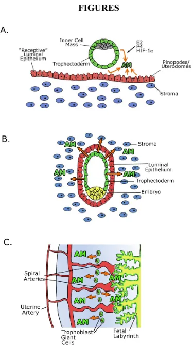
AM in fertility and implantation
6
stroma at the implantation site (Figure 1-1A,B) [35]. Therefore, the peptide is abundantly expressed throughout the female reproductive tract from the earliest stages of pregnancy.
Homozygous deletion of Adm results in embryonic lethality with abnormal development of the heart and lymphatic vascular system [36]. However, female mice heterozygous for Adm (50% AM expression) survive and are a very useful model for the study of haploinsufficiency of AM during pregnancy. Adm+/-female mice have a significantly reduced pregnancy success rate compared with wildtype females, despite the fact that ovulation and fertilization occurs normally in these mice. This reduced pregnancy rate persists even when wildtype blastocysts are transferred to the Adm+/- female, indicating that reduced maternal AM is responsible for the uterine receptivity defects in this model [37].
Furthermore, Adm+/- female mice have reduced numbers of uterine pinopodes (referred to as uterodomes in humans), which are plasma membrane extravasations of the uterine luminal epithelium that faithfully mark the window of uterine receptivity [35]. Even when implantation is successful in the Adm+/- female, fertility defects persist. The litter sizes of Adm+/- female mice are reduced when mated with wildtype males, whereas normal litter sizes are born to wildtype females mated with Adm+/- males. The implantation sites in pregnant Adm+/- females are abnormally spaced and overcrowded, resulting in increased rates of embryo loss and remarkable subfertility [37]. Therefore, even a modest 50% reduction in maternal expression of AM is sufficient to cause major implantation and fertility complications in genetic mouse models.
7
elevated AM levels in follicular fluid is associated with negative outcomes in in vitro
fertilization patients [38].
AM in placentation and maternal–fetal circulation
8
further evidence for a functional role for AM in maintaining vascular tone in pregnancy [44]. In women with unexplained recurrent pregnancy loss, high plasma AM was associated with increased uterine artery pulsatility index and an increased number of previous miscarriages, from which the authors suggest that increased AM may be acting in a compensatory role [45].
AM in Pregnancy Complications
Defects in the ability of trophectoderm cells to fully invade the maternal uterine wall and remodel vessels are thought to underlie many serious reproductive conditions [46,47]. Given that female mice heterozygous for Adm exhibit marked subfertility, and that homozygous deletion of Adm causes embryonic lethality, it is not surprising that altered AM expression has been associated with several of these pregnancy complications. Table 1-1 summarizes many of the clinical studies that have measured AM in pregnancy complications.
IUGR
Numerous rodent models have provided evidence for the necessity of AM in normal fetal growth. Yallampalli’s laboratory found that antagonism of AM during pregnancy resulted in IUGR, abnormal placental vascularization and increased fetal resorption in the rat [48,49]. Similar studies in the mouse have shown that Adm+/- mothers have a high rate of fetal growth restriction, which occurs in all fetal genotypes. The incidence of fetal growth restriction was highest among Adm-/- embryos, indicating that both maternal and fetal AM may contribute to normal fetal growth [37].
9
with reduced fetal growth [50,51], which could be a compensatory effect. However, a 2001 study by Yamashiro et al. and a more recent study by Akturk et al. showed no difference in AM concentrations between small for gestational age and appropriate for gestational age infants [52,53]. Based on animal studies, it is likely that altered AM levels may contribute to either the development of IUGR or the resulting adaptive compensation to other primary causes of IUGR. However, the inconsistencies between studies in the human population points to the necessity of further studies to determine with certainty how changes in AM levels may be involved in the pathogenesis of growth-restricted pregnancies.
AM in gestational diabetes
10
AM in PE
Based on the known roles for AM in trophoblast invasion and vascular adaptation to pregnancy, there is significant interest in determining whether changes in AM peptide or expression levels contributes to the pathogenesis of PE. Results from an experimental rat model of maternal hypertension further piqued this interest. In this model, whereby administration of the inhibitor of nitric oxide synthases, L-NAME (nitro-L-arginine methyl ester), during gestation results in hypertension and pup mortality; maternal infusion of AM attenuated the hypertensive phenotype [58].
11
a secondary effect in PE, it is clear that more controlled experiments to address the direction of the change and the exact role of AM in PE are needed.
AM and preterm birth
It has long been thought that altered AM levels may be present in cases of preterm birth. Human studies dating back over 10 years ago have suggested that AM protein may be elevated in patients with preterm birth. The Di Iorio laboratory has published several studies on this subject, finding increased amniotic AM levels in cases of preterm premature rupture of membranes (PPROM) [69], and increased amniotic fluid AM in patients with preterm labor [70]. Elevated second trimester amniotic fluid levels of AM have also been reported in patients that go on to preterm deliveries [51]. Glucocorticoids may be involved in the regulation of AM levels in pregnancy, as administration of betamethasone to stimulate fetal lung maturity in patients at risk for preterm birth resulted in significantly increased maternal and fetal plasma AM levels [71]. However, a 2009 study by Iavazzo et al. found that there was no change in AM levels in spontaneous preterm delivery or PPROM [72], suggesting that further studies may still be needed in this area.
12
immunity and host defense [76]. These include its six-member cysteine ring structure, characteristic of both human and murine beta-defensins [77] which allows these molecules to penetrate bacterial cell membranes, and the known anti-inflammatory effects of AM [78]. The potent bactericidal properties of AM [79] and its high level of expression in the skin, oral cavity cervical mucosa, fetal membranes, and breast milk [12,80] also support its role as a possible mediator of host defense. Future studies exploring a potential role for AM in providing antimicrobial effects in relation to preterm birth have the potential to be highly informative.
Caveats to current AM detection methods and midregional fragment of pro-adrenomedullin
(MR-proADM)
13
community acquired pneumonia [86], and heart failure [87,88], and demonstrates excellent clinical utility for MR-proADM as a diagnostic and prognostic biomarker.
Application of this new assay has been limited in the reproductive and pregnancy field so far, but to date two groups have measured MR-proADM levels in neonates. Miguel et al. established reference values for MR-proADM in umbilical cord blood of newborn infants [89], whereas Cao et al. found that serum levels of MR-proADM were significantly elevated by more than twofold in newborns with severe infection compared to those with mild or no infection [90]. Although this study was performed in newborns, it provides an interesting correlation with the hypothesis that AM may act as a regulator in host defense preventing infections during pregnancy, and suggests that this AM precursor could potentially be used as a laboratory marker of bacterial infection in neonates. Further studies utilizing the MR-proADM assay in the study of pregnancy have the potential to yield newly accurate and exciting results, improving our understanding of how AM levels change in healthy versus complicated pregnancies in humans and perhaps even validating MR-proADM as a potential biomarker for pregnancy-related diseases.
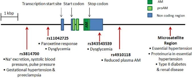
AM system polymorphisms
14
population, an ADM allele with 19 CA repeats was found with significantly increased frequency in individuals with essential hypertension (EH) compared to normotensive controls [91]. This same polymorphism has been associated with predisposition to proteinuria in EH and is found with greater frequency in type II diabetic patients in renal failure compared to controls or diabetic patients without nephropathy [92,93]. Specific single nucleotide polymorphisms (SNPs) in AM have also been associated with reduced systolic blood pressure, lower pulse pressure, and reduced urinary sodium excretion in a Chinese population [94], increased risk of developing dysglycemia in a Chinese population [95], and altered response to the antidepressant paroxetine [96]. A particular SNP in the AM receptor gene,
CALCRL, was found with increased frequency specifically among women with EH in a Japanese population [97], and other studies have found CALCRL SNPs associated with severe periodontitis [98] and acute primary angle closure glaucoma [99]. Recently, different
ADM haplotypes have been shown to correlate with changes in plasma AM levels [100], making the connection between polymorphisms in the ADM gene and level of expression.
15
they are associated with pregnancy complications is an emerging translational research target with great potential.
Concluding Remarks
16 TABLES
Table 1-1: Adrenomedullin levels in pregnancy complications Pregnancy
Complication
Sample Source
Sample
Type Assay
Change in AM
Levels Ref IUGR umbilical plasma protein AM commercial RIA ↑ 1.67X in IUGR 50 IUGR
maternal and cord venous
blood
AM
protein "non-commercial" RIA No change 53 Reduced Fetal
Growth amniotic fluid
AM protein
"non-commercial" RIA
AM levels inversely correlate with birth
weight and height
51
IUGR
umbilical and maternal
plasma
AM
protein HPLC No change 52
Gestational
Diabetes maternal plasma protein AM commercial RIA No change 54 Gestational Diabetes and Pregnant Women with T1DM maternal and fetal plasma, amniotic fluid AM
protein commercial RIA
↑ 1.36X in amniotic fluid of diabetic pregnancies; no change in maternal
or fetal plasma
55 Pregnant Women with T1DM maternal plasma AM protein "non-commercial"
RIA No change 56
Gestational
Diabetes maternal plasma protein AM "non-commercial" RIA No change 57
PE maternal
plasma
AM
protein commercial RIA ↓1.35X in PE 59
PE amniotic fluid, maternal and umbilical plasma AM
protein commercial RIA
↑2.25X in amniotic fluid and ↑2.13X in umbilical plasma in PE; no change in
maternal plasma
64
PE maternal plasma protein AM "non-commercial" RIA No change 13 PE maternal plasma protein AM commercial RIA ↑2.5X in PE 65 PE maternal plasma protein AM "non-commercial" RIA No change 43 PE placental villi AM
mRNA qRT-PCR
↓1.83X expression
17 PE cytotrophoblast purified
cultures AM protein, mRNA commercial RIA, Northern blot
↓2.91X mRNA and ↓5X protein in PE 63
PE choriodecidua, amnion, and placental tissue AM protein, mRNA commercial RIA, Northern blot ↑1.66-2.9X in choriodecidua, ↑1.63-2.26X in amnion; no change
in placental tissue
68
PE maternal plasma protein AM ELISA ↑1.3X in PE 32 PE maternal plasma protein AM HPLC ↓1.92X in PE 67 PE placental tissue AM
mRNA qRT-PCR
↓1.18X mRNA in
PE 62
PE maternal
plasma
AM
protein ELISA ↑1.07X in PE 66
Preterm Labor / Premature
Rupture of Membranes
amniotic fluid protein AM commercial RIA ↑1.75X in PROM 69
Preterm Labor maternal and fetal plasma, amniotic fluid
AM
protein commercial RIA
↑2.95X in amniotic fluid in preterm labor; no change in
maternal or fetal plasma
70
Preterm
Delivery amniotic fluid
AM protein
"non-commercial" RIA
↑ 1.40X in preterm delivery 51 Preterm Labor amniotic fluid AM
protein ELISA No change 72
18 FIGURES
Figure 1-1. Adrenomedullin expression during pregnancy
19
Figure 1-2. Clinically-relevant adrenomedullin (AM) polymorphisms
20
Figure 1-3. Outstanding questions about the role of adrenomedullin in pregnancy
21 REFERENCES
1. Lenhart PM, Caron KM (2012) Adrenomedullin and pregnancy: perspectives from animal models to humans. Trends Endocrinol Metab 23: 524-532.
2. Ogoshi M, Inoue K, Naruse K, Takei Y (2006) Evolutionary history of the calcitonin gene-related peptide family in vertebrates revealed by comparative genomic analyses. Peptides 27: 3154-3164.
3. Poyner DR, Sexton PM, Marshall I, Smith DM, Quirion R, et al. (2002) International Union of Pharmacology. XXXII. The mammalian calcitonin gene-related peptides, adrenomedullin, amylin, and calcitonin receptors. Pharmacol Rev 54: 233-246.
4. Takei Y, Inoue K, Ogoshi M, Kawahara T, Bannai H, et al. (2004) Identification of novel adrenomedullin in mammals: a potent cardiovascular and renal regulator. FEBS Lett 556: 53-58.
5. Roh J, Chang CL, Bhalla A, Klein C, Hsu SY (2004) Intermedin is a calcitonin/calcitonin gene-related peptide family peptide acting through the calcitonin receptor-like receptor/receptor activity-modifying protein receptor complexes. J Biol Chem 279: 7264-7274.
6. Muff R, Born W, Lutz TA, Fischer JA (2004) Biological importance of the peptides of the calcitonin family as revealed by disruption and transfer of corresponding genes. Peptides 25: 2027-2038.
7. Kitamura K, Kangawa K, Kawamoto M, Ichiki Y, Nakamura S, et al. (1993) Adrenomedullin: a novel hypotensive peptide isolated from human pheochromocytoma. Biochem Biophys Res Commun 192: 553-560.
8. McLatchie LM, Fraser NJ, Main MJ, Wise A, Brown J, et al. (1998) RAMPs regulate the transport and ligand specificity of the calcitonin-receptor-like receptor. Nature 393: 333-339.
9. Christopoulos A, Christopoulos G, Morfis M, Udawela M, Laburthe M, et al. (2003) Novel receptor partners and function of receptor activity-modifying proteins. J Biol Chem 278: 3293-3297.
22
11. Hague S, Zhang L, Oehler MK, Manek S, MacKenzie IZ, et al. (2000) Expression of the hypoxically regulated angiogenic factor adrenomedullin correlates with uterine leiomyoma vascular density. Clin Cancer Res 6: 2808-2814.
12. Trollmann R, Schoof E, Beinder E, Wenzel D, Rascher W, et al. (2002) Adrenomedullin gene expression in human placental tIssue and leukocytes: a potential marker of severe tIssue hypoxia in neonates with birth asphyxia. Eur J Endocrinol 147: 711-716. 13. Minegishi T, Nakamura M, Abe K, Tano M, Andoh A, et al. (1999) Adrenomedullin and atrial natriuretic peptide concentrations in normal pregnancy and pre-eclampsia. Mol Hum Reprod 5: 767-770.
14. Zhao Y, Hague S, Manek S, Zhang L, Bicknell R, et al. (1998) PCR display identifies tamoxifen induction of the novel angiogenic factor adrenomedullin by a non estrogenic mechanism in the human endometrium. Oncogene 16: 409-415.
15. Gratton RJ, Gluszynski M, Mazzuca DM, Nygard K, Han VK (2003) Adrenomedullin messenger ribonucleic acid expression in the placentae of normal and preeclamptic pregnancies. J Clin Endocrinol Metab 88: 6048-6055.
16. Marinoni E, Di Iorio R, Letizia C, Villaccio B, Scucchi L, et al. (1998) Immunoreactive adrenomedullin in human fetoplacental tissues. Am J Obstet Gynecol 179: 784-787. 17. Montuenga LM, Martinez A, Miller MJ, Unsworth EJ, Cuttitta F (1997) Expression of
adrenomedullin and its receptor during embryogenesis suggests autocrine or paracrine modes of action. Endocrinology 138: 440-451.
18. Yotsumoto S, Shimada T, Cui CY, Nakashima H, Fujiwara H, et al. (1998) Expression of adrenomedullin, a hypotensive peptide, in the trophoblast giant cells at the embryo implantation site in mouse. Dev Biol 203: 264-275.
19. Nakayama M, Takahashi K, Murakami O, Shirato K, Shibahara S (1998) Induction of adrenomedullin by hypoxia and cobalt chloride in human colorectal carcinoma cells. Biochem Biophys Res Commun 243: 514-517.
20. Cormier-Regard S, Nguyen SV, Claycomb WC (1998) Adrenomedullin gene expression is developmentally regulated and induced by hypoxia in rat ventricular cardiac myocytes. J Biol Chem 273: 17787-17792.
21. Marinoni E, Pacioni K, Sambuchini A, Moscarini M, Letizia C, et al. (2011) Regulation by hypoxia of adrenomedullin output and expression in human trophoblast cells. Eur J Obstet Gynecol Reprod Biol 154: 146-150.
23
23. Schneider H (2011) Oxygenation of the placental-fetal unit in humans. Respir Physiol Neurobiol 178: 51-58.
24. Watanabe H, Takahashi E, Kobayashi M, Goto M, Krust A, et al. (2006) The estrogen-responsive adrenomedullin and receptor-modifying protein 3 gene identified by DNA microarray analysis are directly regulated by estrogen receptor. J Mol Endocrinol 36: 81-89.
25. Hou Q, Paria BC, Mui C, Dey SK, Gorski J (1996) Immunolocalization of estrogen receptor protein in the mouse blastocyst during normal and delayed implantation. Proc Natl Acad Sci U S A 93: 2376-2381.
26. Couse JF, Korach KS (1999) Estrogen receptor null mice: what have we learned and where will they lead us? Endocr Rev 20: 358-417.
27. Hewitt SC, Collins J, Grissom S, Deroo B, Korach KS (2005) Global uterine genomics in vivo: microarray evaluation of the estrogen receptor alpha-growth factor cross-talk mechanism. Mol Endocrinol 19: 657-668.
28. Hayashi Y, Ueyama H, Mashimo T, Kangawa K, Minamino N (2005) Circulating mature adrenomedullin is related to blood volume in full-term pregnancy. Anesth Analg 101: 1816-1820.
29. Di Iorio R, Marinoni E, Scavo D, Letizia C, Cosmi EV (1997) Adrenomedullin in pregnancy. Lancet 349: 328.
30. Kobayashi K, Kubota T, Aso T, Hirata Y, Imai T, et al. (2000) Immunoreactive adrenomedullin (AM) concentration in maternal plasma during human pregnancy and AM expression in placenta. Eur J Endocrinol 142: 683-687.
31. Hoshimoto K, Hayashi M, Ohkura T (2002) Mature adrenomedullin concentrations in plasma during pregnancy. J Matern Fetal Neonatal Med 11: 126-129.
32. Senna AA, Zedan M, el-Salam GE, el-Mashad AI (2008) Study of plasma adrenomedullin level in normal pregnancy and preclampsia. Medscape J Med 10: 29. 33. Li L, O WS, Tang F (2011) Adrenomedullin in rat follicles and corpora lutea: expression,
functions and interaction with endothelin-1. Reprod Biol Endocrinol 9: 111.
34. Liao SB, Ho JC, Tang F, O WS (2011) Adrenomedullin increases ciliary beat frequency and decreases muscular contraction in the rat oviduct. Reproduction 141: 367-372. 35. Li M, Wu Y, Caron KM (2008) Haploinsufficiency for Adrenomedullin Reduces
24
36. Fritz-Six KL, Dunworth WP, Li M, Caron KM (2008) Adrenomedullin signaling is necessary for murine lymphatic vascular development. J Clin Invest 118: 40-50. 37. Li M, Yee D, Magnuson TR, Smithies O, Caron KM (2006) Reduced maternal
expression of adrenomedullin disrupts fertility, placentation, and fetal growth in mice. J Clin Invest 116: 2653-2662.
38. Marinoni E, Feliciani E, Muzzonigro F, Letizia C, Tranquilli A, et al. (2010) Intrafollicular concentration of adrenomedullin is associated with IVF outcome. Gynecol Endocrinol 26: 435-439.
39. Lee KY, DeMayo FJ (2004) Animal models of implantation. Reproduction 128: 679-695. 40. Nikitenko LL, Brown NS, Smith DM, MacKenzie IZ, Bicknell R, et al. (2001) Differential and cell-specific expression of calcitonin receptor-like receptor and receptor activity modifying proteins in the human uterus. Mol Hum Reprod 7: 655-664.
41. Zhang X, Green KE, Yallampalli C, Dong YL (2005) Adrenomedullin enhances invasion by trophoblast cell lines. Biol Reprod 73: 619-626.
42. Hoeldtke NJ, Wagner RK, Calhoun BC, Hume RF, Jr. (2000) Vasodilatory response of fetoplacental vasculature to adrenomedullin after constriction with the thromboxane sympathomimetic U46619. Am J Obstet Gynecol 183: 1573-1578.
43. Jerat S, Morrish DW, Davidge ST, Kaufman S (2001) Effect of adrenomedullin on placental arteries in normal and preeclamptic pregnancies. Hypertension 37: 227-231. 44. Ross GR, Yallampalli U, Gangula PR, Reed L, Sathishkumar K, et al. (2010)
Adrenomedullin relaxes rat uterine artery: mechanisms and influence of pregnancy and estradiol. Endocrinology 151: 4485-4493.
45. El-mashad AI, Mohamed MA, Farag MA, Ahmad MK, Ismail Y (2011) Role of uterine artery Doppler velocimetry indices and plasma adrenomedullin level in women with unexplained recurrent pregnancy loss. J Obstet Gynaecol Res 37: 51-57.
46. Chaddha V, Viero S, Huppertz B, Kingdom J (2004) Developmental biology of the placenta and the origins of placental insufficiency. Semin Fetal Neonatal Med 9: 357-369.
25
48. Witlin AG, Li ZY, Wimalawansa SJ, Grady JJ, Grafe MR, et al. (2002) Placental and fetal growth and development in late rat gestation is dependent on adrenomedullin. Biol Reprod 67: 1025-1031.
49. Penchalaneni J, Wimalawansa SJ, Yallampalli C (2004) Adrenomedullin antagonist treatment during early gestation in rats causes fetoplacental growth restriction through apoptosis. Biol Reprod 71: 1475-1483.
50. Di Iorio R, Marinoni E, Letizia C, Gazzolo D, Lucchini C, et al. (2000) Adrenomedullin is increased in the fetoplacental circulation in intrauterine growth restriction with abnormal umbilical artery waveforms. Am J Obstet Gynecol 182: 650-654.
51. Yamashiro C, Kanenishi K, Akiyama M, Tanaka H, Shiota A, et al. (2002) Adrenomedullin concentrations in early 2nd-trimester amniotic fluid: relation to preterm delivery and fetal growth at birth. Gynecol Obstet Invest 54: 99-104.
52. Akturk A, Onal EE, Atalay Y, Yurekli M, Erbas D, et al. (2007) Maternal and umbilical venous adrenomedullin and nitric oxide levels in intrauterine growth restriction. J Matern Fetal Neonatal Med 20: 521-525.
53. Yamashiro C, Hayashi K, Yanagihara T, Hata T (2001) Plasma adrenomedullin levels in pregnancies with appropriate for gestational age and small for gestational age infants. J Perinat Med 29: 513-518.
54. Martinez A, Elsasser TH, Bhathena SJ, Pio R, Buchanan TA, et al. (1999) Is adrenomedullin a causal agent in some cases of type 2 diabetes? Peptides 20: 1471-1478.
55. Di Iorio R, Marinoni E, Urban G, Costantini A, Cosmi EV, et al. (2001) Fetomaternal adrenomedullin levels in diabetic pregnancy. Horm Metab Res 33: 486-490.
56. Loukovaara S, Immonen IJ, Yandle TG, Nicholls G, Hiilesmaa VK, et al. (2005) Vasoactive mediators and retinopathy during type 1 diabetic pregnancy. Acta Ophthalmol Scand 83: 57-62.
57. Poyhonen-Alho M, Viitasalo M, Nicholls MG, Lindstrom BM, Vaananen H, et al. (2010) Imbalance of the autonomic nervous system at night in women with gestational diabetes. Diabet Med 27: 988-994.
58. Makino I, Shibata K, Makino Y, Kangawa K, Kawarabayashi T (1999) Adrenomedullin attenuates the hypertension in hypertensive pregnant rats induced by N(G)-nitro-L-arginine methyl ester. Eur J Pharmacol 371: 159-167.
26
60. Kanenishi K, Kuwabara H, Ueno M, Sakamoto H, Hata T (2000) Immunohistochemical adrenomedullin expression is decreased in the placenta from pregnancies with pre-eclampsia. Pathol Int 50: 536-540.
61. Knerr I, Dachert C, Beinder E, Metzler M, Dotsch J, et al. (2002) Adrenomedullin, calcitonin gene-related peptide and their receptors: evidence for a decreased placental mRNA content in preeclampsia and HELLP syndrome. Eur J Obstet Gynecol Reprod Biol 101: 47-53.
62. Boc-Zalewska A, Seremak-Mrozikiewicz A, Barlik M, Anna B, Mrozikiewicz PM, et al. (2011) Adrenomedullin mRNA expression in placenta of preeclamptic women. Ginekol Pol 82: 585-591.
63. Li H, Dakour J, Kaufman S, Guilbert LJ, Winkler-Lowen B, et al. (2003) Adrenomedullin is decreased in preeclampsia because of failed response to epidermal growth factor and impaired syncytialization. Hypertension 42: 895-900.
64. Di Iorio R, Marinoni E, Letizia C, Alo P, Villaccio B, et al. (1998) Adrenomedullin, a new vasoactive peptide, is increased in preeclampsia. Hypertension 32: 758-763. 65. Lauria MR, Standley CA, Sorokin Y, Yelian FD, Cotton DB (1999) Adrenomedullin
levels in normal and preeclamptic pregnancy at term. J Soc Gynecol Investig 6: 318-321.
66. Boc-Zalewska A, Seremak-Mrozikiewicz A, Barlik M, Kurzawinska G, Drews K (2011) The possible role of adrenomedullin in the etiology of gestational hypertension and preeclampsia. Ginekol Pol 82: 178-184.
67. Dikensoy E, Balat O, Pence S, Balat A, Cekmen M, et al. (2009) The Changes of Plasma Malondialdehyde, Nitric Oxide, and Adrenomedullin Levels in Patients with Preeclampsia. Hypertens Pregnancy: 1-7.
68. Al-Ghafra A, Gude NM, Brennecke SP, King RG (2006) Increased adrenomedullin protein content and mRNA expression in human fetal membranes but not placental tissue in pre-eclampsia. Mol Hum Reprod 12: 181-186.
69. Marinoni E, Di Iorio R, Letizia C, Villaccio B, Alberini A, et al. (1999) Amniotic fluid concentrations of adrenomedullin in preterm labor. Obstet Gynecol 93: 964-967. 70. Di Iorio R, Marinoni E, Letizia C, Alo P, Villaccio B, et al. (2001) Influence of labor on
fetoplacental adrenomedullin concentrations. Am J Obstet Gynecol 185: 697-702. 71. Marinoni E, Zacharopoulou C, Di Rocco A, Letizia C, Moscarini M, et al. (2006) Effect
27
72. Iavazzo C, Tassis K, Gourgiotis D, Boutsikou M, Baka S, et al. (2009) Adrenomedullin concentration in second trimester amniotic fluid cannot be used as a predictor of preterm delivery. In Vivo 23: 1021-1026.
73. Goldenberg RL, Culhane JF, Iams JD, Romero R (2008) Epidemiology and causes of preterm birth. Lancet 371: 75-84.
74. Challis JR, Lockwood CJ, Myatt L, Norman JE, Strauss JF, 3rd, et al. (2009) Inflammation and pregnancy. Reprod Sci 16: 206-215.
75. Romero R, Espinoza J, Chaiworapongsa T, Kalache K (2002) Infection and prematurity and the role of preventive strategies. Semin Neonatol 7: 259-274.
76. Zudaire E, Portal-Nunez S, Cuttitta F (2006) The central role of adrenomedullin in host defense. J Leukoc Biol 80: 237-244.
77. Bauer F, Schweimer K, Kluver E, Conejo-Garcia JR, Forssmann WG, et al. (2001) Structure determination of human and murine beta-defensins reveals structural conservation in the absence of significant sequence similarity. Protein Sci 10: 2470-2479.
78. Christiaens I, Zaragoza DB, Guilbert L, Robertson SA, Mitchell BF, et al. (2008) Inflammatory processes in preterm and term parturition. J Reprod Immunol 79: 50-57.
79. Allaker RP, Grosvenor PW, McAnerney DC, Sheehan BE, Srikanta BH, et al. (2006) Mechanisms of adrenomedullin antimicrobial action. Peptides 27: 661-666.
80. Allaker RP, Zihni C, Kapas S (1999) An investigation into the antimicrobial effects of adrenomedullin on members of the skin, oral, respiratory tract and gut microflora. FEMS Immunol Med Microbiol 23: 289-293.
81. Meeran K, O'Shea D, Upton PD, Small CJ, Ghatei MA, et al. (1997) Circulating adrenomedullin does not regulate systemic blood pressure but increases plasma prolactin after intravenous infusion in humans: a pharmacokinetic study. J Clin Endocrinol Metab 82: 95-100.
82. Struck J, Tao C, Morgenthaler NG, Bergmann A (2004) Identification of an Adrenomedullin precursor fragment in plasma of sepsis patients. Peptides 25: 1369-1372.
28
84. Caruhel P, Mazier C, Kunde J, Morgenthaler NG, Darbouret B (2009) Homogeneous time-resolved fluoroimmunoassay for the measurement of midregional proadrenomedullin in plasma on the fully automated system B.R.A.H.M.S KRYPTOR. Clin Biochem 42: 725-728.
85. Christ-Crain M, Morgenthaler NG, Struck J, Harbarth S, Bergmann A, et al. (2005) Mid-regional pro-adrenomedullin as a prognostic marker in sepsis: an observational study. Crit Care 9: R816-824.
86. Christ-Crain M, Morgenthaler NG, Stolz D, Muller C, Bingisser R, et al. (2006) Pro-adrenomedullin to predict severity and outcome in community-acquired pneumonia [ISRCTN04176397]. Crit Care 10: R96.
87. Maisel A, Mueller C, Nowak RM, Peacock WF, Ponikowski P, et al. (2011) Midregion prohormone adrenomedullin and prognosis in patients presenting with acute dyspnea: results from the BACH (Biomarkers in Acute Heart Failure) trial. J Am Coll Cardiol 58: 1057-1067.
88. Klip IT, Voors AA, Anker SD, Hillege HL, Struck J, et al. (2011) Prognostic value of mid-regional pro-adrenomedullin in patients with heart failure after an acute myocardial infarction. Heart 97: 892-898.
89. Miguel D, Prieto B, Costa M, Coto D, Alvarez FV (2011) Cord blood plasma reference intervals for potential sepsis markers: adrenomedullin, endothelin, and pro-atrial natriuretic peptide. Clin Biochem 44: 337-341.
90. Cao Y, Xia Q, Chen C, Yang Y (2011) Precursors of adrenomedullin, endothelin, and atrial natriuretic peptide as diagnostic markers of neonatal infection. Acta Paediatr. 91. Ishimitsu T, Hosoya K, Tsukada K, Minami J, Futoh Y, et al. (2001) Microsatellite DNA
polymorphism of human adrenomedullin gene in normotensive subjects and patients with essential hypertension. Hypertension 38: 9-12.
92. Kobayashi Y, Nakayama T, Sato N, Izumi Y, Kokubun S, et al. (2005) Haplotype-based case-control study revealing an association between the adrenomedullin gene and proteinuria in subjects with essential hypertension. Hypertens Res 28: 229-236. 93. Ishimitsu T, Tsukada K, Minami J, Ono H, Ohrui M, et al. (2003) Microsatellite DNA
polymorphism of human adrenomedullin gene in type 2 diabetic patients with renal failure. Kidney Int 63: 2230-2235.
29
95. Ong KL, Tso AW, Leung RY, Cherny SS, Sham PC, et al. (2011) A genetic variant in the gene encoding adrenomedullin predicts the development of dysglycemia over 6.4 years in Chinese. Clin Chim Acta 412: 353-357.
96. Glubb DM, McHugh PC, Deng X, Joyce PR, Kennedy MA (2010) Association of a functional polymorphism in the adrenomedullin gene (ADM) with response to paroxetine. Pharmacogenomics J 10: 126-133.
97. Sano M, Kuroi N, Nakayama T, Sato N, Izumi Y, et al. (2005) Association study of calcitonin-receptor-like receptor gene in essential hypertension. Am J Hypertens 18: 403-408.
98. Suzuki A, Ji G, Numabe Y, Ishii K, Muramatsu M, et al. (2004) Large-scale investigation of genomic markers for severe periodontitis. Odontology 92: 43-47.
99. Cao D, Liu X, Guo X, Cong Y, Huang J, et al. (2009) Investigation of the association between CALCRL polymorphisms and primary angle closure glaucoma. Mol Vis 15: 2202-2208.
100. Cheung BM, Ong KL, Tso AW, Leung RY, Cherny SS, et al. (2011) Plasma adrenomedullin level is related to a single nucleotide polymorphism in the adrenomedullin gene. Eur J Endocrinol 165: 571-577.
Chapter 2: Adrenomedullin, Pregnancy, and Innate Immunity3,4
Introduction
As discussed in the previous chapter, adrenomedullin (gene=Adm, peptide=AM) is important for the establishment and maintenance of a healthy pregnancy, and aberrant AM levels are associated with numerous pregnancy complications [2]. One such pregnancy complication in which altered AM levels are implicated is preeclampsia. Clinical manifestations of preeclampsia include gestational hypertension, proteinuria, systemic endothelial dysfunction, and an exaggerated immune response [3,4]. Causative factors for the development of preeclampsia are not well understood, but it is clear that the placenta plays a central role. The clinical symptoms of preeclampsia are resolved with childbirth; however, a consequence of premature induction of labor for the treatment of preeclampsia is the undesirable prematurity of the infant, which can increase the risk of life-long sequelae and health complications for the child.
A hallmark pathological (and perhaps causative) feature of preeclampsia is the failure of the maternal spiral arteries of the placenta to remodel during midgestation [5-7]. During a normal pregnancy, maternal uterine spiral arteries must undergo a complex restructuring in
3
Authors: Manyu Li, Nicole M.J. Schwerbrock, Patricia M. Lenhart, Kimberly L. Fritz-Six, Mahita Kadmiel, Kathleen S. Christine, Daniel M. Kraus, Scott T. Espenschied, Helen H. Willcockson, Christopher P. Mack, and Kathleen M. Caron
4 Reprinted in part with permission from: 1. Li M, Schwerbrock NM, Lenhart PM, Fritz-Six KL, Kadmiel M, et
31
order to ensure a low-resistance, non-pulsatile flow of blood to the fetus. The remodeling is a complex event that involves several distinct processes: i) endothelial cell vacuolization and relaxation of vascular smooth muscle cells (VSMCs) leading to arterial dilation and growth, ii) induction of local placental factors including matrix metalloproteinases (MMPs), chemokines, and cytokines which in turn cause iii) dissolution of the extracellular matrix, iv) de-differentiation and apoptosis of VSMCs and v) replacement of VSMCs by invasive fetal trophoblast cells [8-12].
It is widely accepted that fetal trophoblast cells, in coordination with maternal uterine natural killer (uNK) cells, orchestrate the dynamic process of spiral artery remodeling. uNK cells are the most abundant of all decidual leukocytes, accounting for approximately 70% of CD45-positive cells, and are histologically identified by large cytoplasmic granules containing perforin and granzymes [8]. uNK cells, which are transiently present in the decidua, are frequently aggregated around spiral arteries and play a functional role in remodeling [13-15]. Pioneering studies by Anne Croy and colleagues have demonstrated that the spiral arteries of transgenic mice that lack uNK cells show a persistence of VSMCs surrounding undilated spiral arteries [8,16,17]. Importantly, uNK cells secrete a wide variety of growth factors, chemokines, cytokines, and matrix metalloproteinases [11] and thus are major contributors to the immune milieu of the placenta. However, much remains unknown about the precise molecular interactions between fetal trophoblast cells and uNK cells in the placenta. It is also unclear whether perturbations in this fetal-to-maternal communication can account for failed spiral artery remodeling and preeclampsia.
32
communication required for proper spiral artery remodeling and the prevention of preeclampsia. To test this hypothesis, previous work in the Caron Laboratory focused on characterizing the placentas of Adm+/+ and Adm-/- pups at midgestation to determine the consequences of the loss of AM on placental morphology and function. These studies revealed that placentas lacking either AM or its receptor, CLR, exhibit reduced fetal vessel branching in the labyrinth, failed spiral artery remodeling and re-endothelialization, and marked reduction in maternal uNK cells [1]. Based on these findings, we sought to determine whether there are dosage-related effects of AM on the placenta, which we investigated through the use of a mouse model that overexpresses AM (Admhi/hi). We also characterized the chemokine and cytokine profile of both Adm-/- and Admhi/hi placentas, to determine whether AM dosage, via uNK cells, alters the immune milieu of the placenta.
Materials & Methods
Mice
33
Fetal genotypes were determined by established allele-specific, PCR-based assays [1,18] using genomic DNA isolated from fetal membranes or tail biopsies.
Cytokine expression arrays
RNA was extracted from whole placentas using Trizol reagent (Invitrogen 15596-026) and purified with Qiagen RNeasy Mini Kit (Qiagen 74104). cDNA was made using the RT First Strand Kit (SABiosciences C-03) and cytokine expression was determined utilizing RT2 Profiler PCR Array System for Mouse Chemokines and Receptors (SABiosciences, PAMM-022A-2) with PCR Master Mix RT2 Real-Time SYBR Green/ROX (SABiosciences PA-012) following the manufacturer’s instructions. Data was analyzed using the ΔΔCt method.
Immunohistochemical antibodies
Anti- smooth muscle actin antibody (Sigma, A2547), anti-perforin antibody (Alexis Biochemicals, 804-057-C100), Ly49G2+ (BD Pharmingen, 55-5314), FITC-conjugated
Dolichos biflorus agglutinin (DBA) lectin (Sigma, L9142).
Results
Over-expression of fetal Adm reverses the placental phenotype and drives maternal uNK cell recruitment to the decidua
34
directly influence the recruitment of uNK cells to the decidua, we utilized a gene-targeted mouse model in which the expression of AM is increased ~3-fold [1].
Placentas of Admhi/hi pups born to Adm+/hi intercrossed mice appeared overtly normal and had appropriately branched labyrinth fetal vessels that were indistinguishable from their wildtype littermates [1]. Moreover, α-smooth muscle actin staining revealed that the maternal spiral arteries of Admhi/hi placentas were appropriately remodeled and did not differ significantly from those of wildtype littermates (Figure 2-1A). Interestingly, DBA staining (Figure 2-1B) and counting (Figure 2-1C) of uNK cells revealed that Admhi/hi placentas had a marked 30% increase in the number of uNK cells within the decidua compared to that of wildtype littermates. These data demonstrate that 3-fold overexpression of Adm is compatible with normal placental development and moreover, that fetal-derived AM can actively promote the recruitment of uNK cells to the placenta.
AM alters the chemokine/cytokine profile of uNK cells in vivo
uNK cells constitute the largest proportion of immune cells in the decidua. Thus, we expected and found that the dynamic fluctuations in uNK cell recruitment between Adm-/- and
Admhi/hi placentas were reflected by concomitant changes in the expression of numerous chemokines and cytokines. Specifically, the expression of Ccl7, Ccl17, Cxcl9, Cxcl10, Xcl1 and TNF were down-regulated in Adm-/- placentas and concomitantly up-regulated in Admhi/hi
35
influences the immune milieu of the placenta in two ways—by first recruiting and then activating uNK cells to secrete chemokines, cytokines, and MMPs, which are important contributors to spiral artery remodeling.
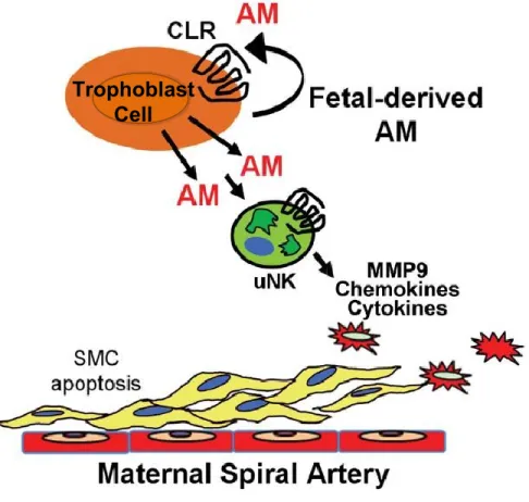
Discussion
These data are part of a larger study showing, through the use of both loss-of-function and gain-of-function genetic mouse models, that fetal AM serves as a trophoblast-derived factor that is critical for fetal placental vascularization and for the maternal vascular adaptation to pregnancy (modeled in Figure 2-3). Ultimately, the mechanisms of AM action appear to be largely mediated through its local effects on the decidual content and activation of maternal uNK cells. Therefore, the phenotypes associated with Adm-/- placentas and their reversal by Adm overexpression demonstrate that the dosage of AM provided by the fetus is a critical communication factor that has a profound impact on the innate immune milieu of the placenta.
It is worth noting that nearly all of the characteristic features of the Adm-/- placentas are similar to the pathological features which have been characterized in human preeclampsia. Although it is imperative to consider the anatomical and functional differences between placentas of humans and mice [19-21], the reduction in fetal vascular branching, failed spiral artery remodeling, and lack of spiral artery remodeling described in these studies have long been associated with preeclampsia in humans [22].
36
37 FIGURES
Figure 2-1. Genetic over-expression of fetal Adm facilitates spiral artery remodeling and drives uNK recruitment to the decidua
A. Admhi/hi placentas have appropriate spiral artery remodeling and are indistinguishable from their wildtype littermates. B,C. DBA staining reveals that Admhi/hi placentas have a statistically significant 30% increase in numbers of uNK cells in the decidua compared to
Adm+/+ littermate placentas. Data are expressed as mean +/- s.e.m.
A.
38
Figure 2-2. AM dosage alters the cytokine and chemokine milieu of the placenta
Using a mouse Chemokines and Receptors array platform, the expression of 85 chemokines, cytokines, and related proteins was evaluated in placental RNA extracts from Adm-/-, Admhi/hi
39
Figure 2-3. Model of fetal-derived AM action at the maternal-fetal interface
AM and its G protein-coupled receptor, CLR, are highly expressed in parietal trophoblast cells at the maternal-fetal interface at midgestation. The genetic dosage of fetal AM is directly proportional to the content of maternal uNK cells and their secreted chemokines, cytokines, and MMPs within the placenta. As a consequence, AM expression in fetal trophoblast cells is necessary for appropriate branching of the fetal vasculature and remodeling of maternal spiral arteries. Overexpression of AM within the fetal compartment is sufficient to promote uNK recruitment and activation in the decidua, a process that likely contributes to AM-induced activation of MMP9 and maternal spiral artery remodeling.
40
REFERENCES
1. Li M, Schwerbrock NM, Lenhart PM, Fritz-Six KL, Kadmiel M, et al. (2013) Fetal-derived adrenomedullin mediates the innate immune milieu of the placenta. J Clin Invest.
2. Lenhart PM, Caron KM (2012) Adrenomedullin and pregnancy: perspectives from animal models to humans. Trends Endocrinol Metab 23: 524-532.
3. Redman CW, Sargent IL (2010) Immunology of pre-eclampsia. Am J Reprod Immunol 63: 534-543.
4. James JL, Whitley GS, Cartwright JE (2010) Pre-eclampsia: fitting together the placental, immune and cardiovascular pieces. J Pathol 221: 363-378.
5. Moffett-King A (2002) Natural killer cells and pregnancy. Nat Rev Immunol 2: 656-663. 6. Myatt L, Webster RP (2009) Vascular biology of preeclampsia. J Thromb Haemost 7:
375-384.
7. Sankaralingam S, Arenas IA, Lalu MM, Davidge ST (2006) Preeclampsia: current understanding of the molecular basis of vascular dysfunction. Expert Rev Mol Med 8: 1-20.
8. Bulmer JN, Williams PJ, Lash GE (2010) Immune cells in the placental bed. Int J Dev Biol 54: 281-294.
9. Smith SD, Dunk CE, Aplin JD, Harris LK, Jones RL (2009) Evidence for immune cell involvement in decidual spiral arteriole remodeling in early human pregnancy. Am J Pathol 174: 1959-1971.
10. Whitley GS, Cartwright JE (2009) Trophoblast-mediated spiral artery remodelling: a role for apoptosis. J Anat 215: 21-26.
11. Lash GE, Robson SC, Bulmer JN (2010) Review: Functional role of uterine natural killer (uNK) cells in human early pregnancy decidua. Placenta 31 Suppl: S87-92.
12. Pijnenborg R, Vercruysse L, Hanssens M (2006) The uterine spiral arteries in human pregnancy: facts and controversies. Placenta 27: 939-958.
41
14. Hanna J, Goldman-Wohl D, Hamani Y, Avraham I, Greenfield C, et al. (2006) Decidual NK cells regulate key developmental processes at the human fetal-maternal interface. Nat Med 12: 1065-1074.
15. Burke SD, Barrette VF, Gravel J, Carter AL, Hatta K, et al. (2010) Uterine NK cells, spiral artery modification and the regulation of blood pressure during mouse pregnancy. Am J Reprod Immunol 63: 472-481.
16. Croy BA, van den Heuvel MJ, Borzychowski AM, Tayade C (2006) Uterine natural killer cells: a specialized differentiation regulated by ovarian hormones. Immunol Rev 214: 161-185.
17. Greenwood JD, Minhas K, di Santo JP, Makita M, Kiso Y, et al. (2000) Ultrastructural studies of implantation sites from mice deficient in uterine natural killer cells. Placenta 21: 693-702.
18. Caron KM, Smithies O (2001) Extreme hydrops fetalis and cardiovascular abnormalities in mice lacking a functional Adrenomedullin gene. Proc Natl Acad Sci U S A 98: 615-619.
19. Maltepe E, Bakardjiev AI, Fisher SJ (2010) The placenta: transcriptional, epigenetic, and physiological integration during development. J Clin Invest 120: 1016-1025.
20. Pijnenborg R, Robertson WB, Brosens I, Dixon G (1981) Review article: trophoblast invasion and the establishment of haemochorial placentation in man and laboratory animals. Placenta 2: 71-91.
21. Watson ED, Cross JC (2005) Development of structures and transport functions in the mouse placenta. Physiology (Bethesda) 20: 180-193.
22. Moffett A, Loke C (2006) Immunology of placentation in eutherian mammals. Nat Rev Immunol 6: 584-594.
23. Doods H, Arndt K, Rudolf K, Just S (2007) CGRP antagonists: unravelling the role of CGRP in migraine. Trends Pharmacol Sci 28: 580-587.
Chapter 3: Adrenomedullin Signaling Pathway Polymorphisms and Adverse Pregnancy Outcomes5
Overview
Objective: Reduced maternal plasma levels of the peptide vasodilator adrenomedullin have been associated with adverse pregnancy outcomes. We measured the extent to which genetic polymorphisms in the adrenomedullin signaling pathway are associated with birth weight, glycemic regulation, and preeclampsia risk.
Study Design: We genotyped 1353 women in the Pregnancy, Infection, and Nutrition Postpartum Study for 37 ancestry-informative markers and for single-nucleotide polymorphisms (SNPs) in adrenomedullin (ADM), complement factor H variant (CFH), and calcitonin receptor-like receptor (CALCRL). We used linear and logistic regression to model the association between genotype and birth weight, glucose loading test (GLT) results, preeclampsia, and gestational diabetes (GDM). All models were adjusted for pregravid BMI, maternal age, and probability of Yoruban ancestry. P values of <0.05 were considered statistically significant.
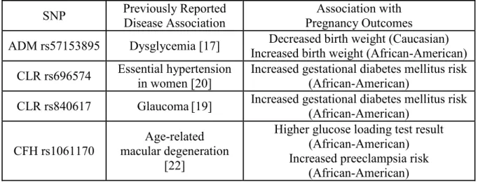
Results: Among Caucasian women, ADM rs57153895, a proxy for rs11042725, was associated with reduced birth weight z-score. Among African-American women, ADM
rs57153895 was associated with increased birth weight z-score. Two CALCRL variants were
5
43
associated with GDM risk. CFH rs1061170 was associated with higher GLT results and increased preeclampsia risk.
Conclusion: Consistent with studies of plasma adrenomedullin and adverse pregnancy outcomes, we found associations between variants in the adrenomedullin signaling pathway and birth weight, glycemic regulation, and preeclampsia.
Introduction
Adrenomedullin (ADM - human gene, Adm - mouse gene, AM - peptide), a peptide hormone vasodilator, plays critical roles in female reproductive biology. Evidence from a wide range of in vitro experiments, rat and mouse models, as well as human studies strongly supports the fact that AM is essential in the establishment and maintenance of a healthy pregnancy [1]. Throughout the course of a normal human pregnancy, AM in the maternal plasma increases, peaking at levels three- to five-fold higher than in the nonpregnant state [2-7] by the third trimester. Precise regulation of maternal AM levels may be necessary in healthy pregnancies, as complications including gestational diabetes [8], preeclampsia [7,9-13], and preterm labor [14,15] have all been associated with perturbations in AM protein levels.
44
[19] (CALCRL rs1157699, rs6759535, and rs840617), essential hypertension in women [20] (CALCRL rs696574), reduced birth weight [21] (CALCRL rs698576), age-related macular degeneration [22] (CFH rs1061170), and susceptibility to meningococcal disease [23] (CFH
rs1065489). However, despite what is known about the important role of AM in reproduction, very few of these studies have addressed the association of genetic polymorphisms in ADM and its signaling partners with adverse pregnancy outcomes. Therefore, we sought to measure the extent to which SNPs in ADM, CALCRL, and CFH are associated with birth weight, glycemic regulation in pregnancy, and preeclampsia risk in a secondary analysis of a prospective cohort study among Caucasian and African-American women in central North Carolina.
Materials and Methods
45
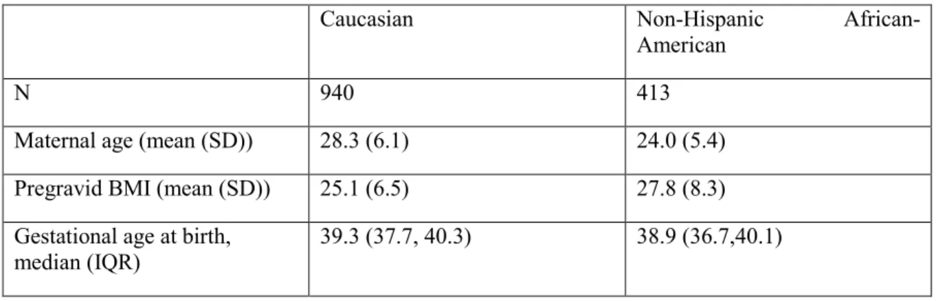
Maternal consent and DNA extracted from peripheral blood was available for 1480 pregnancies. We allowed for only one pregnancy during the study period. If data were available for multiple pregnancies (N=20), we included the pregnancy with the most complete SNP data (n = 1460). We further excluded participants with discordant self-reported race and ancestry estimates calculated from genotyped ancestry informative markers (n=5) or failed genotyping in >20% of the ancestry markers (N=64), leaving 1391 eligible participants. Finally, we excluded women who were missing data on pre-gravid BMI (n=38), leaving 1354 women available for analysis, of whom 940 were non-Hispanic Caucasian and 413 were non-Hispanic African-American.
Determination of pre-gravid BMI
Pre-gravid BMI was calculated based on self-reported pre-gravid weight and height at the first prenatal visit. Self-reported pre-gravid weights were examined for biological plausibility and imputed if deemed appropriate (<5% of weights were imputed) according to a previously-described algorithm [24]. This imputed weight was calculated using the measured weight at the first prenatal visit (if taken prior to 15 weeks) minus the recommended amount of weight to be gained in the first and second trimesters as defined by the Institute of Medicine [25].
Study covariates
46
race/ethnicity (non-Hispanic Caucasian, non-Hispanic African-American, and other) and maternal age was self-reported by the mother.
Outcome assessment
Birth weight z-score was determined using reference populations for sex and self-reported race [26]. Glucose homeostasis was evaluated using glucose loading test (GLT) screening results, which study participants underwent as part of routine clinical care at 24-29 weeks gestation. Trained abstractors ascertained gestational diabetes through prospective review of prenatal records. Participants with GLT values ≥140 mg/dL at UNC sites or ≥130 mg/dL at Wake County sites underwent a diagnostic 100g oral glucose tolerance test (OGTT). Individuals with 2 or more values above established cut points (fasting >95 mg/dL, 1 hour >180mg/dL, 2 hour > 155 mg/dL, 3 hour >140mg/dL ) were diagnosed with gestational diabetes [27].