*Departments of Genetics and Biochemistry, Comenius University in Bratislava, Faculty of Natural Sciences, Mlynska dolina, Ilkovicova 6, 842 15 Bratislava, Slovak Republic
†Department of Biology, Masaryk University, Kamenice 5/A7, Brno 625 00, Czech Republic
‡International Clinical Research Center, St. Anne’s University Hospital in Brno, Brno 60200, Czech Republic
§Lineberger Comprehensive Cancer Center, University of North Carolina at Chapel Hill, NC 27599, U.S.A. National Center for Biomolecular Research, Masaryk University, Kamenice 5/A7, Brno 625 00, Czech Republic
Synopsis
Yeast mtDNA is compacted into nucleoprotein structures called mitochondrial nucleoids (mt-nucleoids). The principal mediators of nucleoid formation are mitochondrial high-mobility group (HMG)-box containing (mtHMG) proteins. Al-though these proteins are some of the fastest evolving components of mt-nucleoids, it is not known whether the divergence of mtHMG proteins on the level of their amino acid sequences is accompanied by diversification of their biochemical properties. In the present study we performed a comparative biochemical analysis of yeast mtHMG pro-teins fromSaccharomyces cerevisiae(ScAbf2p),Yarrowia lipolytica(YlMhb1p) andCandida parapsilosis(CpGcf1p). We found that all three proteins exhibit relatively weak binding to intact dsDNA. In fact,ScAbf2p andYlMhb1p bind quantit-atively to this substrate only at very high protein to DNA ratios andCpGcf1p shows only negligible binding to dsDNA. In contrast, the proteins exhibit much higher preference for recombination intermediates such as Holliday junctions (HJ) and replication forks (RF). Therefore, we hypothesize that the roles of the yeast mtHMG proteins in maintenance and compaction of mtDNAin vivoare in large part mediated by their binding to recombination/replication intermediates. We also speculate that the distinct biochemical properties ofCpGcf1p may represent one of the prerequisites for frequent evolutionary tinkering with the form of the mitochondrial genome in the CTG-clade of hemiascomycetous yeast species.
Key words: DNA-binding protein, DNA compaction, HMG-box containing protein, Holliday junction, mitochondrial DNA (mtDNA), mitochondrial nucleoid.
Cite this article as: Bioscience Reports (2016)36, e00288, doi:10.1042/BSR20150275
INTRODUCTION
The compaction of DNA into chromosomes enables not only its accommodation into the confines of the cell, but also provides a means for spatial regulation of gene expression and protection from DNA damage. Given its importance, it is not surprising that the compaction of DNA into nucleosomes is mediated by a complex of highly conserved proteins called histones [1]. Their extremely high level of conservation is exemplified by the fact that only eight of 102 amino acids differ between the H4 histones of
. . . . Abbreviations:Cp,Candida parapsilosis; D-loop, displacement loop; ds, double-stranded; EMSA, electrophoretic-mobility shift assay; HJ, Holliday junctions; HMG, high-mobility group; mt, mitochondrial; nHJ, nicked Holliday junction; RF, replication forks;Sc,Saccharomyces cerevisiae; ss, single-stranded; TFAM, transcription factor A, mitochondrial; t-loop, telomeric loop;Yl,Yarrowia lipolytica.
1 To whom correspondence should be addressed (email [email protected]).
such evolutionary distant species as humans andSaccharomyces cerevisiae[2]. A deficiency in a histone-encoding gene, or even an imbalance in their expression is often fatal, or can accelerate aging [3,4], further underlining the importance of these proteins in mediating principal functions in DNA maintenance and gene expression [5].
compaction is in part mediated by proteins containing two DNA-binding domains known as an high-mobility group (HMG)-box [12]. The best-characterized members of this group of proteins (mitochondrial HMG-box containing proteins; mtHMG proteins) areScAbf2p [13–15] and mammalian mitochondrial transcrip-tion factor A (TFAM) [16–20]. Apparently, during the evolution of eukaryotic cells, the host genome-encoded mtHMG proteins replaced the polypeptides that served as the major nucleoid com-ponents in the originalα-proteobacterial endosymbiont [21]. Yet, although it seems that the presence of an HMG-box is a universal feature of mitochondrial compaction proteins in all eukaryotes, their overall amino acid sequences exhibit very low similarity. In fact, mtHMG proteins seem to represent one of the most di-vergent groups of mitochondrial proteins [22]. Thus, in contrast with histones, it is very difficult to identify them by simple bioin-formatic tools and most of the mtHMG proteins were identified using proteomic analyses of purified mt-nucleoids [22–25].
The dissimilarities between mtHMG proteins can be explained either by the fast evolutionary divergence of the common ancestor or by acquisition of new features. Perhaps the heterogeneity at the level of amino acid sequences in the mtHMG proteins cor-responds to different biochemical roles in mtDNA maintenance and segregation, or possibly differences in mtDNA base compos-ition and topology. To explore this question we have performed a comparative biochemical analysis of mtHMG proteins from distantly related species. For this purpose, yeast species from the subphylum Saccharomycotina represent an ideal group of or-ganisms. They exhibit a high degree of biodiversity [26], their mtDNAs differ in size, base composition and topology [27], and mtHMG proteins were identified in a number of species separated by hundreds of millions of years of evolution [15,22–24,28–30]. However, the only biochemically characterized yeast mtHMG protein is Abf2 ofS. cerevisiae[13–15,31–35]. It was shown that
ScAbf2p prefers negatively supercoiled DNA over circular or lin-ear DNA and that, in cooperation with a DNA topoisomerase, it introduces negative supercoils into a topologically relaxed, co-valently closed circular dsDNA molecule [15,32]. Its binding to mtDNA is nonrandom, which may be accomplished by the phased distribution of short stretches of poly(dA) indicating its role in genome organization and site-specific regulation of tran-scription or DNA replication [32]. Optical trapping of single DNA molecules extended by flow and visualized by fluorescence microscopy allowed determination of the binding constant of Abf2 (Kb = 2.57+−0.74×10⁷ M−1) [31] (but see also [35]),
and relative small forces (<0.6 pN) stabilizing the condensed DNA–protein interactions [31]. AFM revealed that at high con-centrations of Abf2, the DNA is compacted into relatively loosely packaged 190 nm structures [31,33] indicating that Abf2 is in-deed the bona fide mtDNA-packaging protein. This conclusion was also reached based on the results of anin organello ChIP-on-chip assay demonstrating thatScAbf2p binds to most of the mitochondrial genome with a preference for GC-rich gene se-quences [36].
Although these studies provided important information about the DNA-binding properties ofScAbf2, they also left several important questions unanswered. First, thein vitrostudies were
mostly performed on intact dsDNA substrates, whereas yeast mitochondria contain topologically different forms of DNA gen-erated as a result of various types of transactions including replic-ation, recombination and repair [37–40]. It is known that HMG-box containing proteins recognize some of these structures with high affinity [12]. However, information about the binding of yeast mtHMG proteins to DNA substrates such as Holliday junc-tions (HJ) or replication forks (RF) is lacking. Second, the com-paction of DNA is induced at relatively highScAbf2p to DNA-binding site ratios (20 to 1) [31,33], whereas in the organelle there is one molecule ofScAbf2p per 27 bp of DNA (size of the binding site) [15] (see also below). And third, basically all bio-chemical data on mtHMG proteins are derived from the studies ofScAbf2p. As indicated above, the mtHMG proteins repres-ent the fastest evolving componrepres-ent of mt-nucleoids [22] and it is currently unknown if this divergence in amino acid sequence translates into differences in biochemical properties.
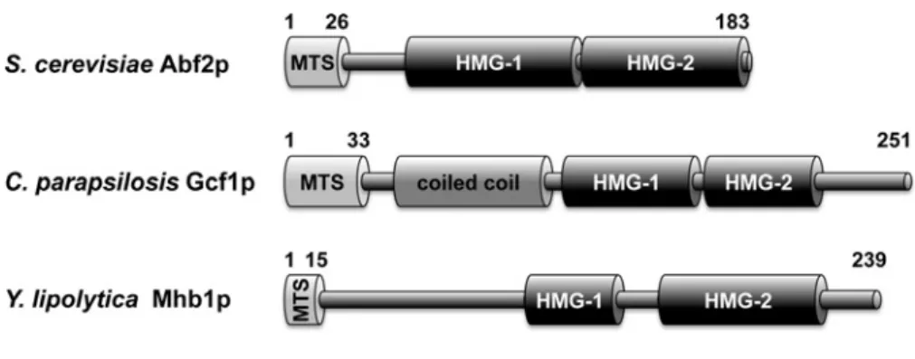
To address these questions we have performed a comparative analysis of mtHMG proteins from three distinct yeast species (Figure 1). Namely, we selectedScAbf2p fromS. cerevisiaeas the best-characterized protein thus allowing comparison of our results with those published by other authors.YlMhb1p from
Yarrowia lipolytica[23] was chosen because this species belongs to basal lineages of Saccharomycotina and phylogenetically is very distant toS. cerevisiae. Therefore, we could compare bio-chemical properties of two proteins with very divergent amino acid sequences, whose only common feature is the presence of two HMG-boxes. Finally,CpGcf1p is the mtHMG protein from
Candida parapsilosis, the yeast species with a linear mitochon-drial genome [22,41]. A detailed characterization of this protein enabled us to address the question of how biochemical proper-ties of mtHMG proteins may be associated with the evolutionary emergence of the linear mitochondrial genome forms, frequently occurring in species from the CTG-clade of Saccharomycotina encompassingC. parapsilosis.
MATERIALS AND METHODS
Microbial strains and growth conditions
Escherichia coliDH5α(F−,ϕ80dlacZΔM15,Δ(lacZYA-argF) U169, deoR, recA1, endA1, hsdR17 (rk−, mk+),λ, thi-1, gyrA96, relA1, glnV44, nupG) (Life Technologies) was used for the amp-lification of plasmid constructs.E. coliBL21 StarTM(DE3) (F−,
ompt, hsdSB, rB−mB−, gal, dcm, rne131) (Life Technologies) was used for production of recombinant proteins (ScAbf2noMP,
YlMhb1noMP,CpGcf1noMP). Bacterial cultures were grown in LB medium (1 % (w/v) bacto peptone (Difco), 0.5 % (w/v) yeast extract (Difco), 1 % (w/v) NaCl, pH 7.5) containing 100μg/ml ampicillin.
DNA manipulations
Figure 1 Yeast mtHMG proteins
Domain prediction in Abf2, Gcf1 and Mhb1 proteins is based on previous reports [15,22,23,30]. MTS – mitochondrial targeting sequence (not present in the mature protein). Note that although SMART (http://smart.embl-heidelberg.de) and InterProScan (http://www.ebi.ac.uk/interpro/) searches did not identify HMG-1 box inCpGcf1p, the corresponding region appears to be weakly conserved with HMG-1 box detected in its orthologues fromCandida lusitaniae,Candida subhashii,
Debaryomyces hanseniiandMeyerozyma guilliermondii.
Table 1 List of oligonucleotides
1, preparation of pGEX-6P-2 derived vectors.2, preparation of DNA substrates used in EMSA experiments.
Name sequence 5’→3’ Application
ScABF2noMP_F AAGGCTTCCAAGAGAACGCAGC 1
YlMHB1noMP_F AAGGAGGCTGCCACTAAGACC 1
pGEX6P2noMP_R GGGCCCCTGGAACAGAACTT 1
CpGCF1noMP_F TCAACCGCCAAAACCACTC 1
CpGCF1noMP_R TTAGATTGTGAATTTGTACTCTTGT 1
ScATP9_15_D GGAGCAGGTATTGGT 2
ScATP9_15_C ACCAATACCTGCTCC 2
ScATP9_25_D GGAGCAGGTATTGGTATTGCTATCG 2
ScATP9_25_C CGATAGCAATACCAATACCTGCTCC 2
ScATP9_50_R ACACCATTAATTAAAGCTGC 2
provided by the vendors. The oligonucleotides (Table 1) were synthesized by Microsynth. The PCRs were performed in 10– 50μl volumes using DreamTaqDNA polymerase (Life Technolo-gies) or Phusion Hot Start II High fidelity DNA polymerase (Life Technologies) and contained all four dNTPs (final concentration 200μM each), the corresponding primers (final concentra-tion 1μM), and either 100 ng of genomic DNA or mtDNA or 10 ng of plasmid DNA. The PCR fragments were purified from agarose gels using a QIAquick Gel Extraction kit (Qiagen) or Zymoclean Gel DNA recovery kit (Zymo Research).
Construction of plasmid vectors
For the expression of recombinant versions of mtHMG proteins lacking the cleavable mitochondrial import sequence (noMP) in fusion with GST a series of pGEX-6P-2 (GE Healthcare) de-rived plasmids was constructed as follows. Plasmids pGEX-6P-2-ScABF2noMP and pGEX-6P-2-YlMHB1noMP were
pre-pared by inverse PCR to eliminate the first 26 amino acids for
ScAbf2p and 14 amino acids for YlMhb1p, corresponding to cleavable mitochondrial import sequence. In the reaction, primers pGEX6P2noMP_R and ScABF2noMP_F or YlMHB1noMP_F (Table 1) were used and previously prepared plasmids pGEX-6P-2-ScABF2or pGEX-6P-2-YlMHB1[23] were used as templates. For the construction of the plasmid pGEX-6P-2-CpGCF1noMP theCpGCF1ORF lacking the first 33 amino acids, which repres-ent cleavable mitochondrial import sequence, was amplified by PCR from the genomic DNA ofC. parapsilosisstrain CBS604 using primers CpGCF1noMP_F and CpGCF1noMP_R (Table 1). The PCR product was inserted into the vector pGEX-6P-2 (GE Healthcare) linearized with SmaI. All plasmid constructs were verified by restriction enzyme mapping and DNA sequencing (Microsynth) of the inserted fragments.
Expression and purification of recombinant mtHMGp fromE. coli
Recombinant mtHMG proteins without the mitochondrial import presequence were purified from bacterial cells as described pre-viously for full lengthYlMhb1 protein [23]. The presence and purity of proteins were verified by 12 % SDS-PAGE stained with Coomassie Brilliant Blue R-250.
DNA substrates and electrophoretic-mobility shift assay (EMSA)
For electrophoretic-mobility shift assay (EMSA) experiments aimed at characterizing the length of the binding site for mtHMG proteins (Figure 2) oligonucleotides ScATP9_15_D and ScATP9_25_D (Table 1), derived from theS. cerevisiae mito-chondrial geneatp9, were radioactively labelled using T4 poly-nucleotide kinase (Life Technologies) and [γ32P]ATP. The
Table 1) in a molar ratio of 1:3. The mixtures were incub-ated at 95◦C for 5 min and cooled slowly to room temperature to allow DNA annealing. The unincorporated [γ32P]ATP was
removed from the DNA by gel filtration using Probe Quant G-50 MicroColumns (GE Healthcare). The 50 bp long DNA substrate derived from the atp9 gene was amplified by PCR from mtDNA of S. cerevisiaestrain W303-1A using primers ScATP9_15_D and ScATP9_50_R (Table 1) and terminally end-labelled using T4 polynucleotide kinase. Fluorescently end-labelled DNA substrates for EMSA were prepared as described previ-ously [42,43]. The structures of the DNA probes are schemat-ically depicted in the corresponding figures. The GC content of the probes ranged between 40 % (50 bp probe) and 53 % (15 bp probe), which is higher than the GC content in atp9 coding sequence (33%).
Indicated amounts of purified recombinant proteins
ScAbf2noMP,YlMhb1noMP orCpGcf1noMP were mixed with the individual radioactively or fluorescently labelled DNA sub-strate (3 nM) and incubated for 10 min at 30◦C in 10μl of a buffer containing 20 mM Tris/HCl pH 7.5, 1 mM EDTA/NaOH pH 8.0, 50 mM NaCl, 100μg/ml BSA. Samples were electrophoretic-ally separated in 5 or 8 % (w/v) polyacrylamide gels in 0.5× TBE buffer (45 mM Tris/borate, 1 mM EDTA/NaOH pH 8.0) at 4◦C. Note that the loading buffer contained only a final concen-tration of 5 % (v/v) glycerol, as we observed that the presence of bromophenol blue and xylene cyanol blue almost completely abolished the binding of mtHMG proteins to DNA. Radioact-ively labelled DNA substrates were visualized after exposing the gels to storage phosphor screens (Kodak) for 24–72 h using a Personal Molecular Imager FX (BioRad). Fluorescent DNA sub-strates were visualized directly using imager reader FLA-9000 Starion (Fuji) and quantified using MultiGauge V3.2 software (Fuji).
Electron microscopy
The DNA-binding reactions for EM were performed in 50μl of HEN buffer (20 mM HEPES/NaOH pH 7.5, 1 mM EDTA/NaOH pH 8.0, 50 mM NaCl) containing 2 ng/μl of the substrate DNA and 15 ng/μl of purifiedScAbf2p. Plasmid pGLGAP and pre-paration of the RF and HJ substrates were described elsewhere [44,45]. The reactions were carried out at room temperature for 15 min, followed by addition of 10μl of 1.2 % (v/v) glutaralde-hyde and incubation at room temperature for additional 6 min. To remove the unbound proteins and fixative, the samples were diluted to 50μl in HEN buffer and passed over 2 ml columns of 6 % agarose beads (ABT Inc.) equilibrated with TE buffer (10 mM Tris/HCl, pH 7.4, 0.1 mM EDTA/NaOH). Aliquots of the fractions containing the complexes were mixed with a buffer containing spermidine and adsorbed on to copper grids coated with a thin carbon film glow-charged shortly before sample ap-plication. Following adsorption of the samples for 3 min, the grids were dehydrated through a graded ethanol series and rotary shad-owcast with tungsten at 10−7torr [46]. Samples were examined
in an FEI T12 TEM equipped with a Gatan 2k×2k SC200 CCD camera.
RESULTS
Yeast mtHMG proteins differ in their affinity to dsDNA
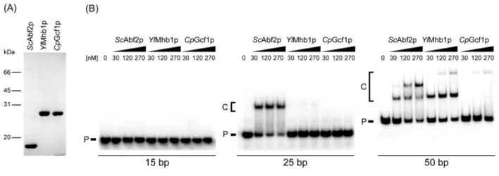
To initiate the biochemical characterization of mtHMG proteins, we expressed recombinant genes encoding the corresponding protein without the N-terminal mitochondrial targeting sequence in fusion with GST using the pGEX-6P-2 vector. To obtain native versions of the proteins, the fusion proteins purified fromE. coli
were treated withPreScissionprotease to remove the GST affinity tag (Figure 2A). The concentrations of the proteins were then adjusted to the same value and their affinity to dsDNA assessed by EMSA.
The proteins were first tested for their ability to bind dsDNA of various lengths (15–50 bp) derived from theS. cerevisiae atp9
gene (Figure 2B). Although none of the proteins was able to bind the shortest (15 bp) DNA fragment, the 25 bp DNA was almost quantitatively shifted byScAbf2p.YlMhb1p exhibited very weak binding andCpGcf1p did not bind this probe at all. The 50 bp DNA fragment was bound by all three mtHMG proteins. Both
ScAbf2p andYlMhb1p formed two DNA–protein complexes, possibly corresponding to one and two protein molecules per molecule of DNA, respectively.CpGcf1p formed only a single complex with DNA, although its mobility corresponded to the slower migrating form of DNA bound byYlMhb1p. Although the nature of this complex is unclear (see Discussion), it is evident that the affinities of the mtHMG proteins to dsDNA as well as the lengths of their corresponding DNA-binding sites differ, and that theCpGcf1p exhibits the lowest affinity towards intact dsDNA.
ScAbf2p exhibits a high preference for replication/recombination intermediates
The results presented inFigure 2B indicate that to obtain quantit-ative binding of mtHMG proteins to dsDNA, even in the case of
ScAbf2p (which exhibits the strongest binding), a relatively high protein to DNA-binding site ratio (50–100 to 1) is required. A re-latively high ratio ofScAbf2p to binding sites was also required for a complete compaction of DNA byScAbf2p as visualized by AFM [31,33]. Namely, to completely compact a linear DNA (pBR322) containing 175 binding sites, almost 4000 molecules of
ScAbf2p were needed (a ratio of 22ScAbf2p to 1 DNA-binding site) [31], and similar results were obtained by another group [33]. At such high ratios of protein to DNA, contamination of the native protein with denatured molecules or truncated species, either of which may induce a general aggregation or collapse of the DNA, becomes a significant concern.
Figure 2 Purified yeast mtHMG proteins differ in their ability to bind dsDNA of various lengths
(A) Purified mtHMG proteins were visualized by Coomassie staining of gels after their separation by 12 % SDS-PAGE. (B) The proteins (at indicated concentrations) were incubated with 3 nM of 15 bp, 25 bp or 50 bp radioactively labelled dsDNA substrates and the samples were separated in polyacrylamide gels as described in Materials and methods. P, free DNA probe; C, DNA–protein complexes.
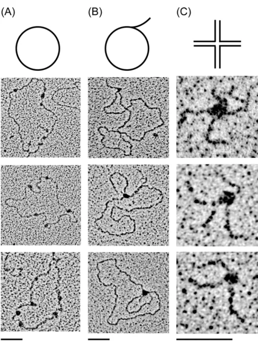
(Figure 2B; [15]), there would be 160000–320000 binding sites per cell and thus even when the highest estimate of Abf2p mo-lecules is taken into account, the stoichiometric ratio ofScAbf2p to DNA substratein vivois 1 to 1 (when considering the lower estimates, the ratio would drop dramatically). Under these con-ditions, the binding of ScAbf2p to dsDNA is 3–5-fold lower (Figure 2B) and the level of compaction is almost negligible [31,33]. Indeed, when we visualized the binding ofScAbf2p to plasmid DNA by EM at a protein to DNA-binding site ratio of 1 to 1 we observed only 5–10 protein particles per DNA molecule (Figure 3A). Therefore, we reasoned that intact dsDNA may not be the best substrate for this protein. Rather,ScAbf2 may prefer DNA forms resulting from various types of DNA transactions such as replication and recombination. This hypothesis was sup-ported by the fact that many nuclear mtHMG proteins exhibit a preference for recombination intermediates [12] and yeast mito-chondria possess a DNA recombination system involved in both DNA replication and repair [49]. We therefore tested the ability ofScAbf2p to bind RF or HJ by EM. Indeed, we found that the binding of protein to DNA occurred almost exclusively at the RF (Figure 3B) or at the junction (Figure 3C). These results promp-ted us to compare the binding of all three mtHMG proteins to various DNA substrates by EMSA.
Comparative analysis of yeast mtHMG proteins reveals differences in their binding preferences to various DNA substrates
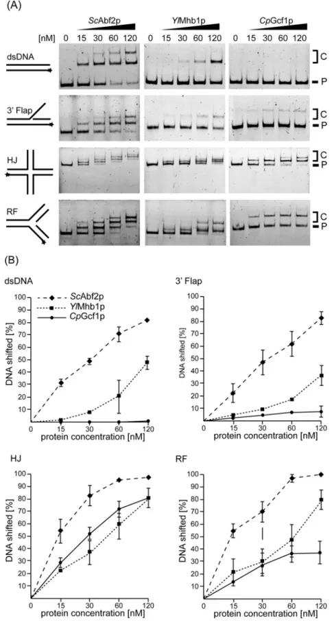
To compare the DNA-binding properties of the mtHMG proteins we tested 10 different DNA substrates, namely intact dsDNA (see alsoFigure 2B), HJ, RF, 3Flap, 5Flap, single-strand (ss), 3and 5 overhang (OH), Y-form, nicked and intact HJ (nHJ a HJ) and displacement loop (D-loop) DNAs (Figure 4). The similar-ities and differences between the proteins are best illustrated by the first four substrates (Figures 4A and4B).ScAbf2p exhib-ited the strongest binding to each of these structures with intact
dsDNA and the 3Flap substrates being the least preferred. The difference in binding between the three proteins was most evident on intact dsDNA. To shift 50 % of the probe 30 nM ofScAbf2p was sufficient, whereas 120 nM of YlMhb1p was needed, and
CpGcf1p did not shift 50 % of dsDNA even at the highest tested concentration (Figure 4B). On the other hand, RF containing DNA, and especially HJ DNAs were bound very efficiently by all three mtHMG proteins even at lower protein concentrations (Figures 4A and4B). It is of note thatCpGcf1p, which bound dsDNA and 3Flap substrates very poorly, was almost as efficient in binding to the HJ DNA asScAbf2p (Figure 4B). This demon-strates that the low affinity ofCpGcf1p to dsDNA and 3 Flap DNA is not a result of the protein being nonfunctional, but rather reflects its intrinsic biochemical preference for certain types of DNA substrates. The binding of the proteins to other DNA sub-strates, including ssDNA, 5 Flap, Y-form, 3 OH and 5 OH substrates was similar as to dsDNA, whereas the nHJ and D-loop DNAs were bound as efficiently as the HJ DNA (Figure 4C).
Intriguingly,CpGcf1p exhibits the largest difference in its abil-ity to bind recombination intermediates (HJ, nHJ, D-loop DNAs) compared with dsDNA and RF DNA. At the protein concentration of 120 nM,CpGcf1p exhibits an 8-fold higher affinity towards HJ compared with 3Flap DNA (Figure 5A). Therefore, we have performed competition experiments, where the protein is incub-ated simultaneously with both dsDNA and HJ DNA. Whereas the former is basically unrecognized, there is a relatively robust binding of the protein to the HJ substrate (Figure 5B).
DISCUSSION
Figure 3 ScAbf2 protein exhibits a binding preference for RF and HJ
15 ng/μl of purifiedScAbf2p were incubated with 2 ng/μl of pGLGAP (A) RF (B) or HJ (C) substrates for 15 min at room temperature. The samples for EM were prepared as described in Materials and methods. The bars represent 50 nm.
dsDNA; however, to achieve a quantitative binding, high protein to DNA ratios were needed (Figure 2B). WhereasYlMhb1p ex-hibited similar binding to the 50 bp long dsDNA asScAbf2p, it bound very weakly to 25 bp dsDNA. This might be caused by a longer binding site ofYlMhb1p compared with ScAbf2p. The binding ofCpGcf1p to dsDNA was very weak for all sub-strates even at the highest protein to DNA ratios (Figures 2B and5). In case of the 50 bp probe the shift by theCpGcf1p cor-responds to the supershift observed byYlMhb1p andScAbf2p suggesting that protein dimers are bound to the probe. The pres-ence of a coiled-coil domain inCpGcf1p might be responsible for the formation of oligomeric complexes, but this possibility
needs to be tested experimentally. The role of dimerization in DNA binding and compaction was studied in detail in case of the mammalian mtHMG protein (TFAM) and is still a matter of de-bate. Two crystal structures of TFAM bound to the heavy strand promoter or to a nonspecific mtDNA sequence showed evidence of TFAM dimerization [50]. On the other hand, a more recent study demonstrated that dimerization is not required for TFAM-induced compaction of mtDNA [51] underlining the importance of further investigation of interactions of mtHMG proteins with DNA.
Figure 4 EMSA of yeast mtHMG proteins with various DNA substrates reveals both similarities and differences in their DNA-binding properties
(A) The proteins (at indicated concentrations) were incubated with 3 nM of the fluorescently labelled DNA substrates (dsDNA, 3Flap, HJ and RF DNA), whose predicted structures are indicated on the left side of each panel. Stars indicate the position of the fluorescent dye. P, free probe; C, DNA–protein complexes. (B) The percentage of the shifted DNA fragments was quantified using Multi Gauge V3.2 software (Fuji). The results represent an average of at least three independent experiments. (C) Analysis of the binding of yeast mtHMG proteins to ssDNA, 3OH, 5OH, 5Flap, Y-form, nHJ and D-loop DNA was performed as inFigure 4A. P, free probe; C, DNA–protein complexes.
genomein vivo[36]. Taking into account the ratio ofScAbf2p to DNAin vivo(at most one molecule of the protein per one 27 bp binding site, but possibly much lower (see above)) our results indicate that the role of yeast mtHMG proteins in the compaction of mtDNA might not be as straightforward as suggested by pre-vious studies. This conclusion is supported by the fact that cells lackingScAbf2p [9],YlMhb1p [23] or the Gcf1p orthologue of
Candida albicans(CaGcf1p) [29], exhibit morphologically dif-ferent, yet still functional mt-nucleoids. This can be explained by the existence of distinct mtDNA-compaction factors acting in parallel with the mtHMG proteins. Some of the candidates (such as Aco1p, Ilv5p) were identified inS. cerevisiae[7,52,53]; how-ever, the means by which these proteins mediate compaction of mtDNA are far from understood.
The second general important property of the yeast mtHMG proteins we observed is their preference for DNA structures gen-erated during replication, recombination and/or repair (Figures 3
Figure 5 CpGcf1p exhibits the most dramatic difference in binding to dsDNA and 3 Flap compared with HJ and RF substrates
(A) Quantification of the binding ofCpGcf1p to four different substrates using data from Figure 4. (B)CpGcf1p was incubated in the reaction mixture containing both dsDNA and HJ DNA and the samples were separated by electrophoresis in polyacrylamide gel as described in Materials and methods. P1, free dsDNA probe; P2, free HJ probe; C, DNA–protein complexes.
structures is not clear. A recent elegant study of Kukat et al. [51] revealed that TFAM-mediated nucleoid formationin vitro
is a multistep process initiated by TFAM aggregation and cross-strand binding. It is possible that yeast mtHMG proteins also employ similar mechanisms of mtDNA compaction. However, in contrast with their mammalian counterparts, yeast mitochondria exhibit a high level of recombination DNA intermediates [37–
40]. In fact, recombination seems to be a principal mechanism of yeast mtDNA replication [39]. The high incidence of recom-bination intermediates in yeast mitochondria combined with our results demonstrating a preference of yeast mtDNA proteins for such DNA structures indicate that these structures may represent their main substrates in vivo. Indeed, theabf2mutants ofS. cerevisiae[59,60] and knockdowngcf1− strains ofC. albicans
[29] exhibit a decrease in the level of mtDNA recombination in-termediates. Conversely, overexpression of theABF2gene inS. cerevisiaeresults in an increased level of recombination inter-mediates and destabilization of mtDNA [59]. In addition, both
abf2and mhb1 mutants ofS. cerevisiae and Y. lipolytica, respectively, are more prone to mutations [23,61]. This may be the result of both deprotection of mtDNA and/or a decreased capacity of recombination-dependent DNA repair. Although all these results indicate that binding of mtHMG proteins to various DNA structures is important for mtDNA transactions in yeasts, the molecular details of their participation in these processes are still largely unknown.
The observation of Kucej et al. [36] thatin vivoAbf2p binds to most of the mitochondrial genome with a preference for GC-rich
gene sequences can also be interpreted in light of our results. First, the ChIP-on-chip assay was performed on a population of cells and could not address the distribution ofScAbf2p at the level of single mtDNA molecules. Second, GC-clusters are hot-spots of mtDNA recombination inS. cerevisiaemitochondria [62], so it is possible that the preferential binding to these regions was caused by recombination undergoing at these sites.
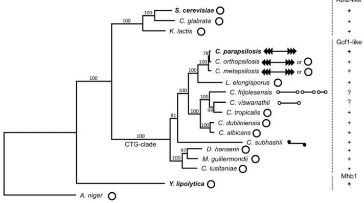
Figure 6 Phylogenetic tree illustrating the distribution of various types of mtHMG proteins and variability of the form of mitochondrial genomes in selected yeast species
The phylogeny was calculated from concatenated multiple sequence alignments of mtDNA-encoded proteins (i.e. Atp6-8-9-Cob-Cox1-2-3-Nad1-2-3-4-4L-5-6-Rps3) by the maximum likelihood algorithm and LG (Le-Gascuel) amino acids substitution model implemented in the PhyML program [80]. Bootstrap values (out of 100 replicates) are shown above the corresponding branches.Aspergillus nigerfrom the subphylum Pezizomycotina was used as an outgroup. Mitochondrial genome forms were classified as described previously [76] and are illustrated by pictograms (open circles – circular; lines with open circles at the ends – linear with terminal hairpins (i.e. type 1 linear and multipartite type 1 linear); lines with series of arrowheads – linear with array of tandem repeats (i.e. type 2 linear); line with closed circle at the ends – linear with a protein covalently bound to 5termini (i.e. type 3 linear). The species whose mtHMG proteins were investigated in the present study are shown in bold.
modifications such as phosphorylation and proteolytic cleavage [64–66].
One of the main motivations for the present study was to ad-dress the question of whether the high degree of amino acid divergence among the yeast mtHMG proteins corresponds to dif-ferences in their biochemical properties. On one hand, the pro-teins seem to be similar in their ability to complement (although to a different extent) theabf2Δmutation inS. cerevisiae[22,23]. Also, they all exhibit a preference for recombinational interme-diates compared with intact dsDNA. On the other hand, their relative affinities for various substrates differ, especially when comparingScAbf2p andYlMhb1p withCpGcf1p. In contrast to its counterparts,CpGcf1p hardly binds dsDNA, whereas its bind-ing to RF and especially HJ DNA is comparable to the other two mtHMG proteins (Figures 4A and4B). Of note is the inability ofCpGcf1p to bind to 5OH containing DNA under the condi-tions tested (Figure 4C) as this structure is present at the ends of linear mtDNA ofC. parapsilosis[41] and therefore is relatively frequentin vivo. Apparently, the ss/double-stranded (ds) junction does not seem to be a preferred site forCpGcf1p. Moreover, the 5 OH terminus of mtDNA of C. parapsilosis is covered by
the mitochondrial telomere-binding protein (mtTBP) [67–69] and it is also engaged in the formation of telomeric loops (t-loops) [70], and thus the junction would probably not be accessible for bindingin vivo.
Although, based on its biochemical properties, CpGcf1p does not seem to be present at the terminal regions of the linear mtDNA of C. parapsilosis in vivo, the dramatic dif-ferences between its ability to bind recombination intermedi-ates (HJ, nHJ, D-loop DNA) compared with dsDNA and RF DNA (Figure 5A) indicate that it is an important player em-ployed byC. parapsilosismitochondria to maintain mtDNA in general and mitochondrial telomeres in particular. When ana-lysed by 2D agarose electrophoresis, preparations of mtDNA of C. parapsilosis contain a large variety of recombination intermediates indicating that recombination plays an import-ant part in mtDNA replication [40]. Moreover, maintenance of mitochondrial telomeres composed of tandemly repeated sequences is mediated by telomeric circles (t-circles), whose formation is dependent on the recombination machinery [71–
sible that this observation can be explained by differences in the evolutionary dynamics of mitochondrial genome architec-ture in the corresponding phylogenetic branches. AlthoughS. cerevisiaeandY. lipolyticaare separated by about 500 million years [26], they belong to groups of species mostly possessing a circular-mapping mitochondrial genome, whose mode of main-tenance is likely quite similar. This would explain the similarity in biochemical properties ofScAbf2p andYlMhb1p. On the other hand,C. parapsilosisis a member of a CTG-clade of the sub-phylum Saccharomycotina exhibiting a wide repertoire of forms of mitochondrial genomes ranging from circular-mapping to lin-ear molecules with defined telomeres such as tandem repeats, hairpins, or covalently attached proteins [27,74–76]. The con-version between various forms seems to be quite frequent, often occurring in strains of the same species [77,78]. It is possible that distinct biochemical features of the Gcf1 protein described in the present study represent one of the prerequisites allowing such frequent evolutionary tinkering [79].
AUTHOR CONTRIBUTION
Jana Bakkaiova performed the experiments shown inFigures 2,4 and5. Victoria Marini prepared the DNA substrates and assisted with the experiments shown inFigures 4and5. Smaranda Willcox performed the experiments shown inFigure 3. Jozef Nosek per-formed bioinformatic analyses, preparedFigure 1and constructed the phylogenetic tree shown inFigure 6. Jack D. Griffith supervised and coordinated the electron-microscopic analysis shown in Fig-ure 3. Lumir Krejci designed and supervised the EMSA experiments
presented inFigures 4and5. Lubomir Tomaska conceived and co-ordinated the study, assisted with the experiments presented in Figures 2(B) and 3 and wrote the first draft of the manuscript. All authors reviewed the results, edited the manuscript and approved its final version.
ACKNOWLEDGEMENTS
We thank Ladislav Kovac for inspiration and continuous support, Filip Tomaska for technical assistance with the EM experiments and for help with the construction of the expression plasmid pGEX-6P-2-ScABF2noMP, Vilko Bakkai for cooperation in finalization of the EMSA experiments, and members of our laboratories for discus-sions.
FUNDING
This work was supported by the Slovak grant agencies APVV [0035-11 (to L.T.) and 14-0253 (to J.N.)], VEGA [1/03[0035-11/12 (to L.T.) and
1 Cutter, A.R. and Hayes, J.J. (2015) A brief review of nucleosome structure. FEBS Lett.589, 2914–2922 PubMed
2 Nelson, D.L. and Cox, M.M. (2005) Lehninger Principles of Biochemistry, 5th edn., W.H. Freeman
3 Das, C. and Tyler, J.K. (2013) Histone exchange and histone modifications during transcription and aging. Biochim. Biophys. Acta1819, 332–342CrossRef PubMed
4 Feser, J., Truong, D., Das, C., Carson, J.J., Kieft, J., Harkness, T. and Tyler, J.K. (2010) Elevated histone expression promotes life span extension. Mol. Cell39, 724–735CrossRef PubMed
5 Maze, I., Noh, K.M., Soshnev, A.A. and Allis, C.D. (2014) Every amino acid matters: essential contributions of histone variants to mammalian development and disease. Nat. Rev. Genet.15, 259–271CrossRef PubMed
6 Bogenhagen, D.F. (2012) Mitochondrial DNA nucleoid structure. Biochim. Biophys. Acta.1819, 914–920CrossRef PubMed
7 Chen, X.J. and Butow, R.A. (2005) The organization and inheritance of the mitochondrial genome. Nat. Rev. Genet.6, 815–825CrossRef PubMed
8 Miyakawa, I., Aoi, H., Sando, N. and Kuroiwa, T. (1984) Fluorescence microscopic studies of mitochondrial nucleoids during meiosis and sporulation in the yeast, Saccharomyces cerevisiae. J. Cell Sci.66, 21–38 PubMed
9 Miyakawa, I., Kanayama, M., Fujita, Y. and Sato, H. (2010) Morphology and protein composition of the mitochondrial nucleoids in yeast cells lacking Abf2p, a high mobility group protein. J. Gen. Appl. Microbiol.56, 455–464CrossRef PubMed
10 Kucej, M. and Butow, R.A. (2007) Evolutionary tinkering with mitochondrial nucleoids. Trends Cell Biol.17, 586–592
CrossRef PubMed
11 Kar´acsony, Z., G´acser, A., V´agv¨olgyi, C., Scazzocchio, C. and Hamari, Z. (2014) A dually located multi-HMG-box protein of Aspergillus nidulans has a crucial role in conidial and ascospore germination. Mol. Microbiol.94, 383–402CrossRef PubMed
12 Stros, M., Launholt, D. and Grasser, K.D. (2007) The HMG-box: a versatile protein domain occurring in a wide variety of DNA-binding proteins. Cell. Mol. Life Sci.64, 2590–2606CrossRef PubMed
13 Caron, F., Jacq, C. and Rouviere-Yaniv, J. (1979) Characterization of a histone-like protein extracted from yeast mitochondria. Proc. Natl. Acad. Sci. U.S.A.76, 4265–4269CrossRef PubMed
14 Certa, U., Colavito-Shepanski, M. and Grunstein, M. (1984) Yeast may not contain histone H1: the only known ‘histone H1-like’ protein in Saccharomyces cerevisiae is a mitochondrial protein. Nucleic Acids Res.12, 7975–7985CrossRef PubMed
15 Diffley, J.F. and Stillman, B. (1991) A close relative of the nuclear, chromosomal high-mobility group protein HMG1 in yeast mitochondria. Proc. Natl. Acad. Sci. U.S.A.88, 7864–7868
CrossRef PubMed
17 Fisher, R.P. and Clayton, D.A. (1988) Purification and
characterization of human mitochondrial transcription factor 1. Mol. Cell. Biol.8, 3496–3509CrossRef PubMed
18 Parisi, M.A. and Clayton, D.A. (1991) Similarity of human mitochondrial transcription factor 1 to high mobility group proteins. Science252, 965–969CrossRef PubMed
19 Gangelhoff, T.A., Mungalachetty, P.S., Nix, J.C. and Churchill, M.E. (2009) Structural analysis and DNA binding of the HMG domains of the human mitochondrial transcription factor A. Nucleic Acids Res.37, 3153–3164CrossRef PubMed
20 Ngo, H.B., Kaiser, J.T. and Chan, D.C. (2011) The mitochondrial transcription and packaging factor Tfam imposes a U-turn on mitochondrial DNA. Nat. Struct. Mol. Biol.18, 1290–1296
CrossRef PubMed
21 Wang, G. and Maier, R.J. (2015) Bacterial histone-like proteins: roles in stress resistance. Curr. Genet.61, 489–492 22 Miyakawa, I., Okamuro, A., Kinsky, S., Visacka, K., Tomaska, L.
and Nosek, J. (2009) Mitochondrial nucleoids from the yeast Candida parapsilosis: expansion of the repertoire of proteins associated with mitochondrial DNA. Microbiology-SGM155, 1558–1568CrossRef
23 Bakkaiova, J., Arata, K., Matsunobu, M., Ono, B., Aoki, T., Lajdova, D., Nebohacova, M., Nosek, J., Miyakawa, I. and Tomaska, L. (2014) The strictly aerobic yeast Yarrowia lipolytica tolerates loss of a mitochondrial DNA-packaging protein. Eukaryot. Cell13, 1143–1157CrossRef PubMed
24 Miyakawa, I. and Yawata, K. (2007) Purification of an Abf2p-like protein from mitochondrial nucleoids of yeast Pichia jadinii and its role in the packaging of mitochondrial DNA. Antonie Van Leeuwenhoek91, 197–207CrossRef PubMed
25 Sasaki, N., Kuroiwa, H., Nishitani, C., Takano, H., Higashiyama, T., Kobayashi, T., Shirai, Y., Sakai, A., Kawano, S.,
Murakami-Murofushi, K. and Kuroiwa, T. (2003) Glom is a novel mitochondrial DNA packaging protein in Physarum polycephalum and causes intense chromatin condensation without suppressing DNA functions. Mol. Biol. Cell14, 4758–4769
CrossRef PubMed
26 Dujon, B. (2006) Yeasts illustrate the molecular mechanisms of eukaryotic genome evolution. Trends Genet.22, 375–387
CrossRef PubMed
27 Nosek, J. and Tomaska, L. (2003) Mitochondrial genome diversity: evolution of the molecular architecture and replication strategy. Curr. Genet.44, 73–84CrossRef PubMed
28 Nosek, J., Tomaska, L., Bolotin-Fukuhara, M. and Miyakawa, I. (2006) Mitochondrial chromosome structure: an insight from analysis of complete yeast genomes. FEMS Yeast Res.6, 356–370CrossRef PubMed
29 Visacka, K., Gerhold, J.M., Petrovicova, J., Kinsky, S., Joers, P., Nosek, J., Sedman, J. and Tomaska, L. (2009) Novel subfamily of mitochondrial HMG box-containing proteins: functional analysis of Gcf1p from Candida albicans. Microbiology-SGM155, 1226–1240
CrossRef
30 Miyakawa, I., Sato, H., Maruyama, Y. and Nakaoka, T. (2003) Isolation of the mitochondrial nucleoids from yeast Kluyveromyces lactis and analyses of the nucleoid proteins. J. Gen. Appl. Microbiol.49, 85–93CrossRef PubMed
31 Brewer, L.R., Friddle, R., Noy, A., Baldwin, E., Martin, S.S., Corzett, M., Balhorn, R. and Baskin, R.J. (2003) Packaging of single DNA molecules by the yeast mitochondrial protein Abf2p. Biophys. J. 85, 2519–2524CrossRef PubMed
32 Diffley, J.F. and Stillman, B. (1992) DNA binding properties of an HMG1-related protein from yeast mitochondria. J. Biol. Chem.267, 3368–3374 PubMed
33 Friddle, R.W., Klare, J.E., Martin, S.S., Corzett, M., Balhorn, R., Baldwin, E.P., Baskin, R.J. and Noy, A. (2004) Mechanism of DNA compaction by yeast mitochondrial protein Abf2p. Biophys. J.86, 1632–1639CrossRef PubMed
34 Kao, L.R., Megraw, T.L. and Chae, C.B. (1993) Essential role of the HMG domain in the function of yeast mitochondrial histone HM: functional complementation of HM by the nuclear nonhistone protein NHP6A. Proc. Natl. Acad. Sci. U.S.A.90, 5598–5602
CrossRef PubMed
35 Stigter, D. (2004) Packaging of single DNA molecules by the yeast mitochondrial protein Abf2p: reinterpretation of recent single molecule experiments. Biophys. Chem.110, 171–178
CrossRef PubMed
36 Kucej, M., Kucejova, B., Subramanian, R., Chen, X.J. and Butow, R.A. (2008) Mitochondrial nucleoids undergo remodeling in response to metabolic cues. J. Cell Sci.121, 1861–1868
CrossRef PubMed
37 Bendich, A.J. (1996) Structural analysis of mitochondrial DNA molecules from fungi and plants using moving pictures and pulsed-field gel electrophoresis. J. Mol. Biol.255, 564–588
CrossRef PubMed
38 Jacobs, M.A., Payne, S.R. and Bendich, A.J. (1996) Moving pictures and pulsed-field gel electrophoresis show only linear mitochondrial DNA molecules from yeasts with linear-mapping and circular-mapping mitochondrial genomes. Curr. Genet.30, 3–11
CrossRef PubMed
39 Gerhold, J.M., Aun, A., Sedman, T., Joers, P. and Sedman, J. (2010) Strand invasion structures in the inverted repeat of Candida albicans mitochondrial DNA reveal a role for homologous recombination in replication. Mol. Cell39, 851–861
CrossRef PubMed
40 Gerhold, J.M., Sedman, T., Visacka, K., Slezakova, J., Tomaska, L., Nosek, J. and Sedman, J. (2014) Replication intermediates of the linear mitochondrial DNA of Candida parapsilosis suggest a common recombination based mechanism for yeast mitochondria. J. Biol. Chem.289, 22659–22670CrossRef PubMed
41 Nosek, J., Dinouel, N., Kovac, L. and Fukuhara, H. (1995) Linear mitochondrial DNAs from yeasts: telomeres with large tandem repetitions. Mol. Gen. Genet.247, 61–72CrossRef PubMed
42 Marini, V. and Krejci, L. (2012) Unwinding of synthetic replication and recombination substrates by Srs2. DNA Repair. (Amst)11, 789–798CrossRef PubMed
43 Matulova, P., Marini, V., Burgess, R.C., Sisakova, A., Kwon, Y., Rothstein, R., Sung, P. and Krejci, L. (2009) Cooperativity of Mus81.Mms4 with Rad54 in the resolution of recombination and replication intermediates. J. Biol. Chem.284, 7733–7745
CrossRef PubMed
44 Arat, N.O. and Griffith, J.D. (2012) Human Rap1 interacts directly with telomeric DNA and regulates TRF2 localization at the telomere. J. Biol. Chem.287, 41583–41594CrossRef PubMed
45 Subramanian, D. and Griffith, J.D. (2005) p53 Monitors replication fork regression by binding to “chickenfoot” intermediates. J. Biol. Chem.280, 42568–42572CrossRef PubMed
46 Griffith, J.D. and Christiansen, G. (1978) Electron microscope visualization of chromatin and other DNA-protein complexes. Annu. Rev. Biophys. Bioeng.7, 19–35CrossRef PubMed
47 Ghaemmaghami, S., Huh, W.K., Bower, K., Howson, R.W., Belle, A., Dephoure, N., O’Shea, E.K. and Weissman, J.S. (2003) Global analysis of protein expression in yeast. Nature425, 737–741
CrossRef PubMed
48 Chong, Y.T., Koh, J.L., Friesen, H., Duffy, K., Cox, M.J., Moses, A., Moffat, J., Boone, C. and Andrews, B.J. (2015) Yeast proteome dynamics from single cell imaging and automated analysis. Cell 161, 1413–1424CrossRef PubMed
49 Dujon, B. (1981) Mitochondrial genetics and function. In The Molecular Biology of the Yeast Saccharomyces: Life Cycle and Inheritance (Jones, E.W. and Broach, J.R., eds), pp. 505–635, Cold Spring Harbor Laboratory Press, Cold Spring Harbor, NY 50 Ngo, H.B., Lovely, G.A., Phillips, R. and Chan, D.C. (2014) Distinct
structural features of TFAM drive mitochondrial DNA packaging versus transcriptional activation. Nat. Commun.5, 3077
mitochondrial DNA stability. EMBO J.14, 3268–3276
PubMed
54 Bustin, M. (1999) Regulation of DNA-dependent activities by the functional motifs of the high-mobility-group chromosomal proteins. Mol. Cell. Biol.19, 5237–5246CrossRef PubMed
55 Ohno, T., Umeda, S., Hamasaki, N. and Kang, D. (2000) Binding of human mitochondrial transcription factor A, an HMG box protein, to a four-way DNA junction. Biochem. Biophys. Res. Commun.271, 492–498CrossRef PubMed
56 Brown, T.A., Tkachuk, A.N. and Clayton, D.A. (2015) Mitochondrial Transcription Factor A (TFAM) Binds to RNA Containing 4-Way Junctions and Mitochondrial tRNA. PLoS One10, e0142436
CrossRef PubMed
57 Ohsato, T., Muta, T., Fukuoh, A., Shinagawa, H., Hamasaki, N. and Kang, D. (1999) Binding of human mitochondrial transcription factor A, an HMG box protein, to a four-way DNA junction. Biochem. Biophys. Res. Commun.255, 1–5CrossRef PubMed
58 Thyagarajan, B., Padua, R.A. and Campbell, C. (1996) Mammalian mitochondria possess homologous DNA recombination activity. J. Biol. Chem.271, 27536–27543CrossRef PubMed
59 MacAlpine, D.M., Perlman, P.S. and Butow, R.A. (1998) The high mobility group protein Abf2p influences the level of yeast mitochondrial DNA recombination intermediates in vivo. Proc. Natl. Acad. Sci. U.S.A.95, 6739–6743
CrossRef PubMed
60 Zelenaya-Troitskaya, O., Newman, S.M., Okamoto, K., Perlman, P.S. and Butow, R.A. (1998) Functions of the high mobility group protein, Abf2p, in mitochondrial DNA segregation, recombination and copy number in Saccharomyces cerevisiae. Genetics148, 1763–1776 PubMed
61 Sia, R.A., Carrol, S., Kalifa, L., Hochmuth, C. and Sia, E.A. (2009) Loss of the mitochondrial nucleoid protein, Abf2p, destabilizes repetitive DNA in the yeast mitochondrial genome. Genetics181, 331–334CrossRef PubMed
62 Dieckmann, C.L. and Gandy, B. (1987) Preferential recombination between GC clusters in yeast mitochondrial DNA. EMBO J.6, 4197–4203 PubMed
63 Chow, C.S., Whitehead, J.P. and Lippard, S.J. (1994) HMG domain proteins induce sharp bends in cisplatin-modified DNA.
Biochemistry33, 15124–15130CrossRef PubMed
64 Cho, J.H., Lee, Y.K. and Chae, C.B. (2001) The modulation of the biological activities of mitochondrial histone Abf2p by yeast PKA and its possible role in the regulation of mitochondrial DNA content during glucose repression. Biochim. Biophys. Acta1522, 175–186CrossRef PubMed
65 Kasashima, K., Sumitani, M. and Endo, H. (2012) Maintenance of mitochondrial genome distribution by mitochondrial AAA+ protein ClpX. Exp. Cell Res.318, 2335–2343
CrossRef PubMed
Candida parapsilosis. J. Biol. Chem.272, 3049–3056
CrossRef PubMed
69 Tomaska, L., Makhov, A.M., Nosek, J., Kucejova, B. and Griffith, J.D. (2001) Electron microscopic analysis supports a dual role for the mitochondrial telomere-binding protein of Candida
parapsilosis. J. Mol. Biol.305, 61–69CrossRef PubMed
70 Tomaska, L., Makhov, A.M., Griffith, J.D. and Nosek, J. (2002) t-Loops in yeast mitochondria. Mitochondrion1, 455–459
CrossRef PubMed
71 Nosek, J., Rycovska, A., Makhov, A.M., Griffith, J.D. and Tomaska, L. (2005) Amplification of telomeric arrays via rolling-circle mechanism. J. Biol. Chem.280, 10840–10845CrossRef PubMed
72 Tomaska, L., Nosek, J., Makhov, A.M., Pastorakova, A. and Griffith, J.D. (2000) Extragenomic double-stranded DNA circles in yeast with linear mitochondrial genomes: potential involvement in telomere maintenance. Nucleic Acids Res.28, 4479–4487
CrossRef PubMed
73 Tomaska, L., Nosek, J., Kramara, J. and Griffith, J.D. (2009) Nat. Telomeric circles: universal players in telomere maintenance? Struct. Mol. Biol.16, 1010–1015CrossRef
74 Fricova, D., Valach, M., Farkas, Z., Pfeiffer, I., Kucsera, J., Tomaska, L. and Nosek, J. (2010) The mitochondrial genome of the pathogenic yeast Candida subhashii: GC-rich linear DNA with a protein covalently attached to the 5’ termini. Microbiology-SGM 156, 2153–2163CrossRef PubMed
75 Nosek, J., Tomaska, L., Fukuhara, H., Suyama, Y. and Kovac, L. (1998) Linear mitochondrial genomes: 30 years down the line. Trends Genet.14, 184–188CrossRef PubMed
76 Valach, M., Farkas, Z., Fricova, D., Kovac, J., Brejova, B., Vinar, T., Pfeiffer, I., Kucsera, J., Tomaska, L., Lang, B.F. and Nosek, J. (2011) Evolution of linear chromosomes and multipartite genomes in yeast mitochondria. Nucleic Acids Res.39, 4202–4219
CrossRef PubMed
77 Kosa, P., Valach, M., Tomaska, L., Wolfe, K.H. and Nosek, J. (2006) Complete DNA sequences of the mitochondrial genomes of the pathogenic yeasts Candida orthopsilosis and Candida metapsilosis: insight into the evolution of linear DNA genomes from mitochondrial telomere mutants. Nucleic Acids Res.34, 2472–2481CrossRef PubMed
78 Rycovska, A., Valach, M., Tomaska, L., Bolotin-Fukuhara, M. and Nosek, J. (2004) Linear versus circular mitochondrial genomes: intraspecies variability of mitochondrial genome architecture in Candida parapsilosis. Microbiology-SGM150, 1571–1580
CrossRef
79 Jacob, F. (1977) Evolution and tinkering. Science196, 1161–1166
CrossRef PubMed
80 Guindon, S. and Gascuel, O. (2003) A simple, fast, and accurate algorithm to estimate large phylogenies by maximum likelihood. Syst. Biol.52, 696–704CrossRef PubMed
Received 4 November 2015/16 November 2015; accepted 1 December 2015