STRUCTURE-FUNCTION STUDIES OF THE INTIATION RESPONSE OF HUMAN MISMATCH REPAIR PROTEINS TO DNA CONTAING A MISMATCH
Kira Caitlin Bradford
A dissertation submitted to the faculty at the University of North Carolina at Chapel Hill in partial fulfillment of the requirements for the degree of Doctor of Philosophy in the Department
of Chemistry.
Chapel Hill 2015
ABSTRACT
Kira Caitlin Bradford: Structure-Function Studies of the Initiation Response of Human Mismatch Repair Proteins to DNA Containing a Mismatch
(Under the direction of Dorothy A. Erie)
DNA mismatch repair (MMR) is the post-replicative process that recognizes and repairs misincorporated bases that occur during replication. Deficiencies in MMR are linked to greater than 80% of hereditary non-polyposis colorectal cancer. To better understand the initiation of MMR in humans, two proteins, MSH2-MSH6 (hMutS) and MLH1-PMS2 (hMutL), were
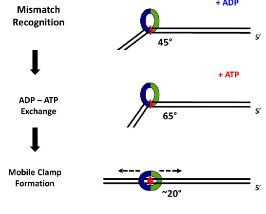
studied. hMutS first recognizes and binds to the mismatch in an ADP-dependent manner;
however, it then undergoes an ATP-dependent conformational change(s) into a mobile clamp. The ATP activated form of hMutS also recruits hMutL to the DNA. Together, these proteins
signal the downstream events of MMR that results in the repair of the misincorporated base. Currently, the conformations of the hMutS-DNA complexes in the presence of ATP are
not well characterized. hMutS in the presence of ADP has previously been crystallized in
complex with mismatched DNA. This crystal structure shows that hMutS induces a kink on the
DNA, consistent with other studies done with MutS homologs. However, no crystal structures have yet been solved of hMutS in the presence of ATP. Additionally, the conformational state
of hMutS that interacts with hMutL is not well understood, and little is known on the
In this work, I used atomic force microscopy (AFM) to examine the conformational properties of hMutS and hMutS-hMutL on DNA containing a single mismatch and in
various adenine nucleotide conditions. The data suggests that hMutS recognizes and binds to
the mismatch in the presence of ADP, and, surprisingly, remains localized to the mismatch in the presence of ATP. The data also show that ATP induces hMutS-hMutS interactions, and
multiple hMutS form a complex on the mismatch. I also characterized the hMutS-induced
DNA bend angle properties and saw unique changes in bending depending on the adenine nucleotide conditions. Additionally, I observed unique properties of the complexes of hMutS
and hMutL in the presence of ATP including: complex volumes becoming larger, DNA lengths
To Robert,
For all of his love, support, and encouragement,
To my family,
ACKNOWLEDGEMENTS
First, I’d like to thank my advisor, Dorothy Erie. I am forever grateful that she is a “gambling” woman that took a chance with me and accepted me as a graduate student in her lab.
She gave me the freedom and independence to pursue the research I found most intriguing, and made time for me once I banged my head against the brick wall a time too many. She also supported me with my endeavors outside the lab, as I try and find my path in life. She
encouraged, supported, and guided me as I navigated through my research project, and taught me how to think like a scientist. I will always be grateful that I was lucky to have the opportunity to learn from her.
I want to thank my colleagues within the Erie lab. I’d like to thank all of the Erie lab members past and present for listening to my stumbling research talks and providing constructive feedback. I especially want to thank Vanessa, Jake, Hunter, Jackie, Zimeng, and Sarah for
reading the drafts of my paper and my thesis. Thank you all for the Fridays, for the lunchroom crossword puzzles, and for the meaningful scientific discussions. Each of you helped me to grow as a scientist and forced me to learn how to be a better lab citizen. You all have helped keep me sane and made me realize time and again that there is light at the end of the tunnel.
would like to thank my committee members, especially Tom Kunkel for giving me valuable expertise.
I’d like to thank my friends who have become my new family here in NC. I am lucky to
have you all in my life. I am grateful for the time commiserating our failed experiments, encouraging each other out of graduate student guilt, and for the many fun game nights that we had. I am also grateful for our many runs, during the day, and especially at night.
I would also like to thank my husband, Robert. Thanks do not begin to adequately
describe how your strength, encouragement, and tough love have gotten me to this point. You are my rock that keeps me centered and strong. You put up with the emotional turmoil, the ups and downs of science, and the many tears of failure and despair. When I want to do something risky or off the wall, you do not hold me back but help me find ways to achieve it. You are so
wonderful and I am so happy and grateful that you are in my life.
Finally, I would like to thank my family especially Papa Jim for encouraging me to venture into science. I would also like to thank my mom, whose strength and sheer force of will are characteristics that I will always strive to achieve. My brothers, who always believe in me, even when “the thingy isn’t thingy-ing.” The love and support of my family are what helps keep
TABLE OF CONTENTS
LIST OF TABLES ... xiv
LIST OF FIGURES ... xv
LIST OF ABBREVIATIONS ... xvii
CHAPTER 1: DNA MISMATCH REPAIR AND ATOMIC FORCE MICROSCOPY: STUDYING BLOBS AND TWISTING KNOBS ... 1
Introduction ... 1
Human MMR ... 2
MutS... 3
Structure of MutS ... 3
Properties and Functions of MutS ... 7
DNA Bending by MutS ... 11
ATPase activity of MutS and the Formation of the Mobile Clamp ... 12
MutL ... 15
Structure of MutL ... 15
Properties of MutL ... 18
MutL Endonuclease Activity ... 19
The Strand Discrimination Signal ... 20
MutS and MutL Complexes ... 20
Atomic Force Microscopy ... 24
Thesis Statement ... 29
REFERENCES ... 31
CHAPTER 2: STRUCTURE-FUNCTION INVESTIGATION OF HUMAN MUTSΑ AND MUTSΑ–MUTLΑ ON DNA CONTAINING A MISMATCH ... 37
Introduction ... 37
Materials and Methods ... 40
Protein expression and purification ... 40
DNA substrate preparation ... 40
Sample preparation and deposition ... 40
Imaging ... 41
Image Analysis ... 41
Results ... 43
In the presence of ADP, one MutSα binds to the mismatch ... 43
In the presence of ATP, MutSα binds with high specificity to the mismatch ... 46
ATP induces mismatch-dependent multimerization of MutSα ... 47
MutS and MutL form large complexes on the DNA ... 49
Large MutS–MutL complexes exhibit shortened DNA lengths ... 51
MutS–MutL complexes are less specific to the mismatch ... 54
Discussion ... 57
MutL traps MutS mobile clamps and multiple MutL
are recruited ... 58
REFERENCES ... 62
CHAPTER 3: INVESTIGATION OF THE ROLE OF ATP HYDROLYSIS IN MUTSΑ MOBILE CLAMP FORMATION AND MOVEMENT: AN AFM STUDY WITH ATPS ... 65
Introduction ... 65
Materials and Methods ... 67
Protein expression and purification ... 67
DNA substrate preparation ... 67
Sample preparation and deposition ... 68
Imaging ... 68
Image Analysis ... 69
Results ... 69
In the presence of ATPγS, up to two MutSα bind the mismatch ... 69
In the presence of ATPS and ADP, one MutSα binds the mismatch ... 75
Discussion ... 77
Future Directions ... 78
REFERENCES ... 80
CHAPTER 4: EXAMINATION OF THE EFFECT OF DIFFERENT ADENINE NUCLEOTIDES ON MUTS-INDUCED DNA BENDING: AN AFM BEND ANGLE ANALYSIS STUDY ... 84
Introduction ... 84
Protein expression and purification ... 86
DNA substrate preparation ... 87
Sample preparation and deposition ... 87
Imaging ... 88
Image Analysis ... 88
Results ... 90
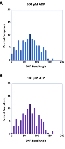
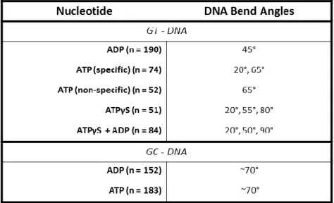
In the presence of ADP, MutS at the mismatch bends the DNA at 45 ... 90
Mismatch-bound MutS in the presence of ATP exhibits two DNA bend angles ... 91
MutS induced DNA bend angles are independent of ATP hydrolysis ... 97
Discussion ... 100
Future Directions ... 102
REFERENCES ... 104
CHAPTER 5: AFM STUDIES TO GAIN INSIGHT INTO OTHER BIOMOLECULAR PROCESSES ... 107
Introduction ... 107
REFERENCES ... 108
CHAPTER 5.1: STRUCTURE-FUNCTION INVESTIGATION OF ANGPTL4 ... 109
Introduction ... 109
Materials and Methods ... 110
Protein Purification ... 110
Results ... 111
Discussion ... 114
REFERENCES ... 116
CHAPTER 5.2: STRUCTURE-FUNCTION INVESTIGATIONS OF TRAK... 117
Introduction ... 117
Materials and Methods ... 118
TraK and DNA substrate purification ... 118
Sample preparation and deposition ... 118
Imaging ... 119
Image Analysis ... 119
Results ... 120
TraK exists as a monomer and dimer in vitro ... 120
Large complexes of TraK form on DNA ... 120
Conclusions and Future Work ... 123
REFERENCES ... 124
CHAPTER 5.3: AFM STUDIES OF THE OLIGMERIZATION STATE OF THE VACCINIA VIRUS ENDORIBONUCLEASE H5 ... 125
Introduction ... 125
Materials and Methods:... 126
Protein Purification ... 126
Atomic Force Microscopy ... 126
Conclusions and Future Work ... 128
REFERENCES ... 130
APPENDIX A: SUPPLEMENTAL FIGURES - CHAPTER 2 ... 131
Supplemental Figures ... 131
Supplemental Methods Figures ... 137
APPENDIX B: AFM DATA ANALYSIS PROTOCOL ... 140
Part 1: Flattening raw AFM images ... 140
Part 2: Volume Analysis ... 140
Part 3: DNA Contour Trace analysis ... 143
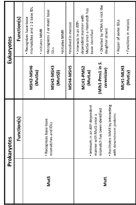
LIST OF TABLES Table 1.1 : Comparison of prokaryotic and eukaryotic MutS
and MutL homologs ... 6 Table 4.1 : Summary of MutSα-induced DNA bend angles ... 99
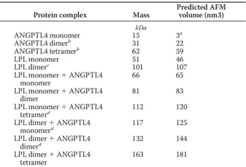
Table 5.1 : Predicted AFM volumes based on the linear dependency of AFM volume to protein molecular weight: V = 1.2 * M – 14.7, where
LIST OF FIGURES
Figure 1.1 : DNA Mismatch Repair in Humans ... 5
Figure 1.2 : Crystal Structure of Human MutS ... 10
Figure 1.3 : Structure of MutL ... 17
Figure 1.4 : Models of MutS–MutL Interactions ... 23
Figure 1.5 : AFM Experimental Methods ... 28
Figure 2.1 : AFM images and position and volume analysis of MutSα bound to GT–DNA ... 45
Figure 2.2 : AFM images and volume analysis of MutSα–MutLα complexes bound to GT–DNA ... 53
Figure 2.3 : Position analysis of MutSα–MutLα complexes bound to GT–DNA ... 56
Figure 2.4 : Model of MutSα and MutLα Interaction ... 59
Figure 3.1 : AFM images and analysis of MutSα bound to GT–DNA in the presence of ATPS ... 71
Figure 3.2 : AFM images and analysis of MutSα bound to circular GT–DNA in the presence of ATPS ... 74
Figure 3.3 : AFM images and analysis of MutSα bound to GT–DNA in the presence of ATPS and ADP ... 76
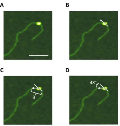
Figure 4.1 : AFM bend angle analysis ... 89
Figure 4.2 : MutSα-induced GT-DNA bend angles in the presence of various adenine nucleotides ... 93
Figure 4.3 : MutSα-induced GC-DNA bend angles ... 94
Figure 4.4 : Non-specifically bound MutSα-induced GT-DNA bend angles in the presence of ATP ... 96
Figure 5.1 : Distribution of AFM particle volumes in cubic nanometers ... 113
Figure 5.2 : AFM images and volume analysis of TraK... 121
Figure 5.3 : AFM image and position analysis of TraK on DNA... 122
Figure 5.4 : AFM images and volume analysis of H5 ... 127
Figure S2.1 : Number of complexes bound to DNA ... 131
Figure S2.2 : Position distribution of MutSα on GC–DNA in the presence of ADP vs.ATP ... 132
Figure S2.3 : Images of multimers of MutSα on GT–DNA in the presence of ATP ... 133
Figure S2.4 : Asymmetry of multimers of MutSα on GT–DNA in the presence of ATP ... 134
Figure S2.5 : Distribution of the conformational shapes of MutSα and MutSα–MutLα complexes on GT–DNA. ... 135
Figure S2.6 : AFM images of MutSα and MutSα–MutLα complexes bound to GT–DNA ... 136
Figure SM2.1 : AFM images of the non–crosslinked control experiment of MutSα bound to GT–DNA in the presence of ATP ... 137
Figure SM2.2 : Normalization of MutSα volumes ... 138
LIST OF ABBREVIATIONS
Degree
Prime
m Micron
M Micromolar
3D Three-dimensional
ABC ATP binding cassette
ADP Adenine diphosphate
AFM Atomic force microscopy
ANGPTL4 Angiopoietin-like Protein 4
ATP Adenine triphosphate
ATPS Adenine 5-(gamma-thio) triphosphate
Bp Base pair
CC Cytosine-cytosine mismatch
ddH20 Doubly deionized water
DNA Deoxyribonucleic acid
dsDNA Double stranded DNA
DTT Dithiothreitol
E. coli Escherichia coli
EDTA Ethylamine diamine tetraacetic acid
EXO1 Exonuclease I
FRET Fluorescence resonance energy transfer
HEPES 4-(2-hydroxyethyl)-1-piperazineethanesulfonic acid HNPCC Hereditary non-polyposis colorectal cancer
IDL Insertion deletion loop
IRC Initial recognition complex
kcat Catalysis rate constant
KCl Potassium chloride
Kd Dissociation constant
LPL Lipoprotein Lipase
m Meter
M Molar
mg Milligram
Mg Magnesium
MgCl2 Magnesium chloride
MgOAc Magnesium Acetate
min Minute
mL Milliliter
MLH1 Human MutL homolog 1
Mlh1 S. cerevisiae MutL homolog 1
MLH1-PMS2 Human MutL homolog 1 and postmeoitic segregation increased 2 Mlh1-Pms1 S. cerevisiae MutL homolog 1 and postmeoitic segregation increased 1
mM Millimolar
mmDNA Mismatch DNA
mol Mole
MSH2 Human MutS homolog 2
Msh2 S. cerevisiae MutS homolog 2 MSH2-MSH6 Human MutS homologs 2 and 6 Msh2-Msh6 S. cerevisiae MutS homologs 2 and 6
MSH6 Human MutS homolog 6
Msh6 S. cerevisiae MutS homolog 6
MutS() MutS and MutS
MutS Heterodimer of eukaryotic MutS homologs 2 and 6
MW Molecular weight
NaCl Sodium chloride
NaOAc Sodium acetate
NaOH Sodium hydroxide
NIDDK National Institute of Diabetes and Digestive and Kidney Diseases NIH National Insititutes of Health
nm Nanometer
nM Nanomolar
nm3 Cubic nanometer
PAGE Polyacrylamide gel electrophoresis PCNA Proliferating cell nuclear antigen PDB ID Protein database identification
PMS2 Human postmeiotic segregation increased 2
Pol Polymerase
Pol Polymerase
Pol Polymerase
RFC Replication factor C
RPA Replication protein A
s Second
S. cerevisiae Saccharomyces cerevisiae
SDS Sodium dodecyl sulfate
smFRET Single-molecule fluorescence resonance energy transfer
ssDNA Single stranded DNA
T bulge Single thymine insert
Taq Thermus aquaticus
TIRF Total internal reflection fluorescence Tris tris(hydroxymethyl)aminomethane buffer
URC Ultimate recognition complex
VLDL Very low-density lipoproteins
WT Wild type
CHAPTER 1: DNA MISMATCH REPAIR AND ATOMIC FORCE MICROSCOPY: STUDYING BLOBS AND TWISTING KNOBS Introduction
Human MMR
Mismatch repair proteins, MutS and MutL homologs, recognize and initiate the repair of misincorporated bases following DNA replication, shown in Figure 1.1. In humans, initiation of MMR occurs when MutS recognizes an error in the DNA. A base-base mismatch or a small insertion-deletion loop is recognized by MSH2-MSH6 (MutS) and large insertion deletion
loops are recognized by MSH2-MSH3 (MutS). MutS (or MutS) then binds to the mismatch
site and undergoes an ATP-dependent conformational change(s) into a mobile clamp that facilitates the interactions with one or more MutL (Kunkel and Erie 2005, Iyer, Pluciennik et
al. 2006, Hsieh and Yamane 2008, Erie and Weninger 2014). PCNA activates the latent endonuclease activity of MutL and directs the MutS–MutL complex to nick the daughter
strand either distally (preferentially, depicted in Figure 1.1 by the larger lightning bolt) or proximally up to hundreds of base pairs away (Kadyrov, Dzantiev et al. 2006, Kadyrov, Holmes et al. 2007). Subsequently, MutS interacts with and increases the processivity of EXO1. EXO1
excises the mismatch-containing DNA in a 5' to 3' direction, and RPA binding stabilizes single-stranded DNA (Genschel, Bazemore et al. 2002, Constantin, Dzantiev et al. 2005). A DNA polymerase, such as Pol for lagging strand replication, synthesizes across the gap using the
MutS
Structure of MutS
MutS in humans is a heterodimer composed of two subunits: MutS homolog 2 (MSH2)
and MutS homolog 6 (MSH6). The crystal structure of a truncated mutant of MutS with 341
amino acid (aa) residues deleted from the N-terminus of MSH6 is shown in Figure 1.2A
(Warren, Pohlhaus et al. 2007). MutS has two ATPase sites, located at the C-terminus of each
subunit (1.2A and 1.2B, red arrows pointed to the red ATP molecules) and a DNA-binding domain near the N-terminus (1.2A and 1.2C, orange arrows) (Warren, Pohlhaus et al. 2007). Yellow spheres seen in Figure 1.2A indicate magnesium ions that play an important role in ADP/ATP binding to the nucleotide binding sites (Kunkel and Erie 2005, Iyer, Pluciennik et al. 2006). In Figure 1.2C, the DNA is bent by MutS, which is consistent with other MutS homolog
crystal structures that also show a MutS-induced kink in the DNA (Lamers, Perrakis et al. 2000, Obmolova, Ban et al. 2000, Natrajan, Lamers et al. 2003). Once a mismatch has been located, the highly conserved mismatch-recognition motif (located in MSH6 of human MutS),
Phe-X-Glu, comes into play (Kunkel and Erie 2005, Hsieh and Yamane 2008, Erie and Weninger 2014). The mispaired base stacks with the aromatic ring of the phenyalanine, and the glutamate forms a hydrogen bond with the N3 of a mismatched thymine or the N7 of mismatched purines.
Replacing either of these conserved regions within MutS results in impaired mismatch
Figure 1.1 : DNA Mismatch Repair in Humans
Mismatch generation occurs when DNA polymerase, such as Pol , misincorporates the wrong
base. The heterodimer MutS initiates repair by recognizing and binding the mismatch. MutS
then undergoes an ATP–dependent conformational change(s) that allows MutS to form a
mobile clamp (black dashed arrows) and facilitates its recruitment of MutL. PCNA activates
(red double-headed arrow) the endonuclease activity of MutL to nick the daughter strand.
MutS recruits EXO1 to excise the mismatch-containing strand. RPA stabilizes single-stranded
Properties and Functions of MutS
Human MutS and its homologs (MutS()) are highly conserved proteins in prokaryotes
and eukaryotes. As shown in Table 1.1, one of the main roles of MutS homologs in all organisms is to serve as the mismatch-recognition signal (Kunkel and Erie 2005, Iyer, Pluciennik et al. 2006, Spampinato, Gomez et al. 2009, Erie and Weninger 2014). In humans, MutS is primarily
responsible for recognizing and repairing single base-base mismatches and one to two base insertions or deletions (Kunkel and Erie 2005, Iyer, Pluciennik et al. 2006, Erie and Weninger 2014). How MutS is able to scan hundreds of thousands of base pairs in search of a single
mismatch appears to be an enormous task. To accomplish this task, data suggests that MutS
binds non-specifically to the DNA and then bends it in search of a mismatch (Wang, Yang et al. 2003, Kunkel and Erie 2005, Erie and Weninger 2014). MutS has a high affinity for binding to
a mismatch; however, different mismatches, like GT and CC mismatches, are repaired with different efficiencies. One hypothesis for why MutS repairs different mismatches with different
efficiencies is that the efficiency of repair is governed by the stability of the MutS-mismatch
complex, which depends on both the type of mismatch and its sequence context (Kunkel and Erie 2015). Previous studies, however, indicate that binding of MutS to the mismatch alone is not sufficient to induce repair (Su, Lahue et al. 1988). Other studies with human MutS shows that
MutS has varying affinities for different mismatches that are dependent on the presence or
absence of ATP (Mazurek, Johnson et al. 2009). This and previous results suggests that the ATP–dependent conformational change of MutS is required for mismatch repair (Gradia,
Acharya et al. 1997).
It is widely accepted that ATP and a mismatch are required for interactions of MutS
important MutS function is to recruit one or more MutL to the mismatch site. It is unclear
whether MutS recruits MutL before or after forming a mobile clamp. In Sacchromyces
cerevisiae (S. cerevisiae), studies done with an ATPase mutant of MutS that is incapable of
forming a mobile clamp show that MutL is able to interact with MutS. This observation
suggests that MutS is able to undergo multiple ATP-induced conformational changes before
forming a mobile clamp, and that one or more of these states is able to interact with MutL
Figure 1.2 : Crystal Structure of Human MutS
(A) Front view of the human MutS heterodimer in complex with DNA (orange arrow pointing
at the orange helix) containing a single GT mismatch (PDB ID: 208B). The blue subunit is MSH2 and the green subunit is MSH6. Red stick models (indicated by red arrows) indicate ADP in the ATPase sites and yellow spheres indicate magnesium ions. (B) Zoom-in of the ATPase sites of the heterodimer of MutS. Red arrows point to the red stick models that show ADP in
the ATPase sites. (C) 90 rotation of the front-view of MutS. An orange arrow points to DNA
DNA Bending by MutS
As noted previously, the crystal structure in Figure 1.2C shows that human MutS binds
to a mismatch and bends the DNA at the GT mismatch at a ~ 45° angle (Warren, Pohlhaus et al. 2007). This result is similar to crystal structures of other MutS homologs, such as Thermus aquaticus (Taq) and E. coli MutS that were found to bend the DNA at the mismatch at a ~ 60° (Lamers, Perrakis et al. 2000, Obmolova, Ban et al. 2000, Natrajan, Lamers et al. 2003). AFM studies of E. coli and Taq MutS with homoduplex DNA and DNA containing a mismatch showed that MutS induces different bends in the DNA depending if MutS is located at the mismatch or at a homoduplex site (Wang, Yang et al. 2003, Erie and Weninger 2014). When MutS was bound to homoduplex DNA, the DNA was slightly bent; however, when MutS was incubated with DNA containing a single mismatch, two populations of DNA bend angles emerged. One population is associated with unbent DNA, the second is a bent population where MutS induced a bend angle of ~ 60°. These data suggested that MutS scanned the DNA in a slightly bent state, and Wang et. al. proposed that upon recognition of the a mismatch, MutS kinks the DNA (Initial Recognition Complex, IRC) before forming a mobile clamp and unbending the DNA (Ultimate Recognition Complex, URC). Single molecule fluorescence studies of Taq MutS further suggested that MutS is conformationally dynamic when searching the DNA for a mismatch, but upon mismatch binding, the conformations become limited (Qiu, DeRocco et al. 2012). While prokaryotic MutS exists in different dynamic states that have different DNA bend angles associated with them, it is not known if human MutS follows the
ATPase activity of MutS and the Formation of the Mobile Clamp
MutS() proteins are members of the ABC transporter ATPase superfamily, and the
ATPase activity of the human MutS subunits, MSH2 and MSH6, is essential for MMR in
humans (Kunkel and Erie 2005, Iyer, Pluciennik et al. 2006, Hsieh and Yamane 2008). The heterodimer of MutS has two composite nucleotide bindings sites. These nucleotide binding
sites are composed of six highly conserved motifs that include the Walker A and Walker B motifs from one protein subunit, and an ABC signature motif contributed by the other subunit. The loss of subunit dimerization results in the loss of ATPase activity because the ATP binding sites are “composite”, meaning that they are formed as a result of the interactions between the two subunits (Kunkel and Erie 2005, Hsieh and Yamane 2008, Spampinato, Gomez et al. 2009). Notably, the majority of the conserved residues across all homologs of MutS are within the ATPase domain (Spampinato, Gomez et al. 2009). Mutations in the conserved residues in the ATPase active sites of either MSH2 or MSH6 impair MMR activity (Schofield and Hsieh 2003).
Studies have shown that each composite ATPase site of MutS has different affinities
for nucleotide binding. Numerous biochemical studies show that MSH6 has a higher affinity for binding ATP whereas MSH2 has a higher affinity for ADP (Antony and Hingorani 2003, Bjornson and Modrich 2003, Martik, Baitinger et al. 2004, Antony, Khubchandani et al. 2006). Both subunits of MutS can bind adenine nucleotides simultaneously, and each subunit can have
different adenine nucleotides bound. Because the two subunits can bind different adenine nucleotides, this can lead to multiple liganded species of MutS (Kunkel and Erie 2005, Iyer,
Both homoduplex and heteroduplex DNA (DNA containing a mismatch) stimulate the ADP–ATP exchange activity of MutS, but only heteroduplex DNA appears to delay the
turnover of ATP. Binding heteroduplex DNA increases the lifetime of MutS bound with ATP and suggests that in the presence of heteroduplex DNA, the rate-limiting step for turnover of ATP occurs at or prior to hydrolysis of ATP (Kunkel and Erie 2005, Iyer, Pluciennik et al. 2006). MutS exhibits different ATPase activity in the presence of homoduplex or heteroduplex DNA,
suggesting that the recognition of a mismatch leads to a longer lived ATP-bound state of MutS
(Kunkel and Erie 2005, Iyer, Pluciennik et al. 2006).
Multiple studies agree that upon binding ATP, mismatch-bound MutS can form a
mobile clamp that is able to move along the DNA (Blackwell, Martik et al. 1998, Gradia, Subramanian et al. 1999, Blackwell, Bjornson et al. 2001, Acharya, Foster et al. 2003, Jeong, Cho et al. 2011, Qiu, DeRocco et al. 2012). It is important to note, however, that not all MutS()
proteins that recognize and bind to a mismatch go on to form a mobile clamp state (Qiu, DeRocco et al. 2012). In a fluorescence study with Taq MutS, results indicate that ADP-bound MutS can bind to the mismatch and then dissociate without ever transitioning into forming a mobile clamp state (Qiu, DeRocco et al. 2012). Additionally, the states of MutS() complexes
depend on the conformations of the MutS()-DNA complexes as well as the ligation state of the
MutS() ATPase sites (Tessmer, Yang et al. 2008, Qiu, DeRocco et al. 2012). There is some
debate in the field about what liganded state of MutS ATPase sites leads to the formation of a
further result in a doubly liganded ATP state of MutS (Qiu, DeRocco et al. 2012). However, these results contrast with another study that suggested that the ADP:ATP liganded state may be a “dead-end” state (Heinen, Cyr et al. 2011). This study suggested that human MutS activity
was controlled by MSH2 with Mg+2 and ADP, and found that destabilization of Mg+2 resulted in the loss of ADP with MSH6 rapidly binding ATP. MSH6-ATP promotes binding of ATP to MSH2, and the doubly liganded ATP state of MutS is thought to form the mobile clamp
(Heinen, Cyr et al. 2011).
While it appears that ATP hydrolysis is not needed for MutS to form a mobile clamp,
there is much debate on whether ATP hydrolysis is required for the movement of these MutS
mobile clamps along the DNA (Iyer, Pluciennik et al. 2006, Erie and Weninger 2014). Studies that examined the rate of dissociation of MutS from short DNA substrates with blocked or unblocked ends upon the addition of ATP or ATPS found that MutS is able to form a mobile
clamp in the absence of ATP hydrolysis (Acharya, Wilson et al. 1996, Gradia, Acharya et al. 1997, Schofield, Nayak et al. 2001). Because these studies used shorter DNA substrates, it is unclear if ATP hydrolysis is required for MutS mobile clamps to travel long distances on the DNA (Erie and Weninger 2014). Other studies found that MutS failed to form long-lived
mobile clamps in the presence of slowly hydrolyzing, ATPS or non-hydrolyzable ATP analogs,
AMPPNP (Blackwell, Martik et al. 1998). These studies were further supported with other results that found that using a mutant of MutS that was able to bind ATP, but not able to
hydolyze it also could not form a long lived mobile clamp (Iaccarino, Marra et al. 2000).
There is much debate on how the mobile clamp of MutS is formed and if the movement
forming a mobile clamp as multiple studies indicate that ATP induces more than one
conformational change in MutS homologs (Hess, Gupta et al. 2002, Qiu, DeRocco et al. 2012, Groothuizen, Winkler et al. 2015). It is also unknown if ATP hydrolysis is important for the formation of the MutS–MutL complexes (Iyer, Pluciennik et al. 2006). In spite of these
questions, it is generally accepted that MutS and MutL interactions play a key role in MMR
signaling (Kunkel and Erie 2005, Iyer, Pluciennik et al. 2006, Hsieh and Yamane 2008, Spampinato, Gomez et al. 2009).
MutL
Structure of MutL
MutL is a highly conserved protein that is a heterodimer composed of MutL homolog 1
(MLH1) and post meiotic segregation increased 2 (PMS2) in humans (see Table 1.1 for MutL homologs) (Kunkel and Erie 2005, Iyer, Pluciennik et al. 2006, Hsieh and Yamane 2008,
Spampinato, Gomez et al. 2009). Figure 1.3A shows a cartoon depiction of human MutL. Both
prokaryotic and eukaryotic MutL dimerize at the C-terminal domains, and PMS2 (Pms1 in S.
cerevisiae) contains the endonuclease site (Figure 1.3A). The N-terminal domains of both MLH1 and PMS2 contain ATPase and DNA binding activities. Long flexible linker arms link the N- and C- terminal domains together (Gueneau, Dherin et al. 2013). Figure 1.3B shows the
endonuclease site in the C-terminus of Pms1 in yeast (yellow). This figure shows that a portion of Mlh1 contributes to the endonuclease site (green). AFM studies of both human and yeast MutL in the presence of ATP shows large changes in the structure of MutL. These studies
Figure 1.3 : Structure of MutL
(A) Cartoon structure of the human MutL heterodimer. MutL is composed of two subunits
that dimerize at the C-termini. The endonuclease activity of MutL is located in the C-terminal
and C- termini together, or semi-condensed structures where either the N-terminus of PMS2 or MLH1 is brought close to the C-terminus of that subunit (Sacho, Kadyrov et al. 2008).
Properties of MutL
MutL homologs are members of the GHL ATPase family, which includes DNA Gyrase and Hsp90 (Hsieh and Yamane 2008, Erie and Weninger 2014). ATP binding and ATP
hydrolysis induces significant conformational changes in GHL proteins, which are thought to be important for the signaling function of these proteins (Geng, Sakato et al. 2012, Erie and
Weninger 2014). Like MutS homologs, MutL homologs have two asymmetric nucleotide binding sites that affect the interactions and functions of MutL homologs (Hsieh and Yamane 2008). However, in contrast to MutS homologs, MutL proteins have weak ATPase activity (Hall, Shcherbakova et al. 2002). The weak ATPase activity is indicated by a low turnover of ATP, which suggests that ATP binding rather than hydrolysis facilitates MutL interactions with other
proteins by inducing conformational changes (Kunkel and Erie 2005). AFM studies of MutL
found that adenine nucleotides induce large asymmetrical conformational changes. These conformational changes are thought to mediate the interactions of MutL with other proteins in
the MMR pathway (Sacho, Kadyrov et al. 2008).
While MutL and MutL exhibit weak DNA binding in physiological salt conditions, they
do have differing DNA binding preferences. Prokaryotic MutL preferentially binds single-stranded DNA; in contrast, MutL preferentially binds double-single-stranded DNA (Ban and Yang
1998, Hall, Shcherbakova et al. 2003). Furthermore, AFM studies that investigated the DNA-binding properties of MutL under low salt conditions revealed that MutL is able to bind
MutL is able to interact with two different strands of duplex DNA simultaneously (Hall, Wang
et al. 2001).
MutL Endonuclease Activity
The endonuclease activity in the C–terminal domain of S. cerevisiae Pms1 (PMS2 in humans) shown in Figure 1.3B is essential for repair (Deschenes, Tomer et al. 2007, Erdeniz, Nguyen et al. 2007, Kadyrov, Holmes et al. 2007, van Oers, Roa et al. 2010). In vitro studies have shown that the endonuclease activity of MutL is dependent on a mismatch, ATP, MutS,
and PCNA (Kadyrov, Dzantiev et al. 2006). Studies in S. cerevisiae with Pms1E707K, a mutant of MutL that has impaired endonuclease activity, resulted in a strong mutator phenotype
(Kadyrov, Holmes et al. 2007). Other studies using endonuclease-deficient PMS2E702K knock-in mice had increased genetic mutation rates and cancer predisposition (van Oers, Roa et al. 2010).
The location of the nick that results from the endnoculease activity of MutL remains a
perplexing question in the field. In vitro studies of MutL show that it will strand-specifically
nick the DNA in the vicinity of the mismatch on either the 5 or 3 side of the mismatch, though
preferentially distally to the mismatch (Kadyrov, Dzantiev et al. 2006, Hsieh and Yamane 2008, Pluciennik, Dzantiev et al. 2010, Pluciennik, Burdett et al. 2013). These and other studies, however, also found that MutL is capable of nicking the daughter strand up to hundreds of base
pairs away from the mismatch (Kadyrov, Dzantiev et al. 2006, Kadyrov, Holmes et al. 2007). This question becomes further complicated when taking into account that PCNA must be able to activate the endonuclease activity of MutL to strand-specifically nick the DNA when MutL is
in complex with MutS. All of these actions must be tightly and temporally coordinated in vivo
The Strand Discrimination Signal
Once MutS has recruited MutL to the mismatch site, PCNA activates MutL to nick
the DNA in a strand-specific manner. To nick the correct strand of DNA, there must be a strand discrimination signal, which in eukaryotes is hypothesized to be PCNA, because PCNA is loaded onto the DNA asymmetrically at the replication fork or at a nick by RFC (Kadyrov, Dzantiev et al. 2006, Hsieh and Yamane 2008, Pluciennik, Dzantiev et al. 2010). Previous studies utilizing a covalently closed DNA plasmid containing a single-stranded bubble that allowed RFC to load PCNA onto DNA without strand specific orientation found that repair of a mismatch in these bubble substrates is no longer strand specific because PCNA loads randomly on either strand (Pluciennik, Burdett et al. 2009, Pluciennik, Dzantiev et al. 2010). These studies suggest that the initial orientation that PCNA is loaded onto the DNA (either at the replication fork or a nicked site) is what allows PCNA to correctly discern the daughter strand for MutL to nick. To
determine if the location of a preexisting nick affected MutL activity, studies were done in both
a reconstituted system and extracts used plasmid DNA containing a nick. These studies found that if the nick is 3 to the mismatch, MutL is required for repair, but MutL is not required if
the nick is 5 to the mismatch (Kadyrov, Dzantiev et al. 2006, Kadyrov, Holmes et al. 2007).
MutS and MutL Complexes
Role of MutS–MutL Complexes
Fluorescence experiments, DNase1 footprinting, and other experiments have been conducted with MutS and MutL, and they provide some understanding as to how these proteins interact.
presence of ATP resulted in a very large DNaseI footprint, which indicates the presence of multiple proteins bound to the DNA (Selmane, Schofield et al. 2003). However, the assembly of MutS-MutL complexes for either prokaryotic or eukaryotic MMR proteins requires heteroduplex DNA substrates that are longer than 60 bp in vitro (Blackwell, Wang et al. 2001, Schofield, Nayak et al. 2001). It is unclear if longer DNA lengths is required for the assembly of MutS–
MutL complexes, and further characterization of these complexes are needed.
Despite our current knowledge on how MMR is initiated by MutS–MutL complexes,
there are many questions that remain to be addressed. MutS forms a mobile sliding clamp upon
binding ATP and a mismatch, but it is not known if this state or an alternate ATP-dependent state of MutS interacts with MutL. Additionally, we do not know what the MutS conformational
states are once it binds to the mismatch and undergoes the ATP-dependent changes, and if ATP hydrolysis has an impact on the interactions between MutS and MutL. Furthermore, there is
little known about how MutS and MutL interact, whether there are multiple proteins that
interact, or if there is a typical stoichiometric ratio of the two proteins. Finally, it remains unclear how MutL in complex with MutS interact with PCNA to potentially nick the daughter strand
either near the mismatch or up to hundreds to base pairs away from the mismatch.
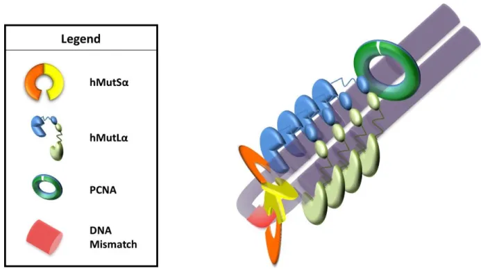
Several disparate models exist to address these questions. One model posits that MutLα joins MutSα to form MutSα–MutLα mobile clamps that diffuse along the DNA to interact PCNA
(Figure 1.4A). This model was originally suggested based on the observation that MutSα can form mobile clamps in conjunction with fluorescence data that noted movement of MutSα– MutLα complexes (Gradia, Subramanian et al. 1999, Gorman, Wang et al. 2012). However, this
Figure 1.4 : Models of MutS–MutL Interactions
(A) MutL joins MutS at the mismatch and forms a larger mobile clamp. This clamp moves
away from the mismatch to allow for a second MutS to load onto the mismatch to form
multiple MutS–MutL mobile clamps. (B) MutS at the mismatch forms a mobile clamp that
slides away from the mismatch. MutL interacts with the mobile clamp of MutS, traps MutS,
and prevents further movement. (C) MutS recruits multiple MutL to the mismatch site,
forming long MutL polymers along the DNA. (D) MutL interacts with MutS either at the
mismatch (Figure 1.4B). This complex could then facilitate DNA looping (Figure 1.4D) to interact with downstream proteins in MMR (Kunkel and Erie 2005, Iyer, Pluciennik et al. 2006). A third model suggests that MutSα induces polymerization of MutLα along the DNA (Figure 1.4C). This model (originally proposed by Paul Modrich (Modrich 1987)) is supported by in vivo
fluorescence studies in S. cerevisiae and E. coli suggesting that MMR foci contain more MutLα than MutSα proteins (Hombauer, Campbell et al. 2011, Elez, Radman et al. 2012). Each of these
models predicts distinct MutSα-MutLα complexes, though the models may not be mutually exclusive. With structural information on the MutSα-MutLα complexes, we can gain further insight into the mechanism of MMR initiation.
Atomic Force Microscopy
Atomic force microscopy (AFM) is a high resolution, single-molecule technique that can be used to gain structural insight into biomolecular processes. Gerd Binnig developed the first AFM in 1986 from scanning tunneling microscopy (Binnig, Quate et al. 1986). The advantages of using AFM are: 1) AFM results in a three-dimensional (3D) image; 2) AFM can visualize non-conductive materials such as DNA and proteins; 3) and AFM can be conducted in air and in solution (Hansma, Laney et al. 1995).
A flow chart detailing how a general AFM experiment is conducted is shown in Figure 1.5A. Samples with protein and DNA complexes or protein alone are incubated at room temperature before glutaraldehyde is added. Glutaraldehyde is a crosslinking agent that
For the purposes of this thesis, the method of tapping mode AFM will be discussed. AFM images are produced by scanning a cantilever across a sample surface, while reflecting a laser beam off the back of the cantilever (Figure 1.5B). As the cantilever progresses across the sample, small changes in sample height (z–direction) are detected as a function of changes in the position of the laser reflection in the photodiode detector. A feedback loop signals the repositioning of the piezo stage such that the position of the reflected laser beam in the detector is maintained at the same point (Figure 1.5C). Adjustments in piezo height (z) are plotted as a function of XY position on the mica surface (Last, Russell et al. 2010).
Figure 1.5 : AFM Experimental Methods
(A) Schematic overview of sample preparation. Samples are incubated before being crosslinked. Crosslinked samples are filtered through a size-exclusion column, and fractions are collected. Fractions are then deposited onto mica to be imaged. (B) The mica sample is placed onto the piezo and an oscillating tip attached to the cantilever moves across the surface in an x-y manner. A laser reflects off of the back of the tip into a photodiode. (C) The tip encounters a molecule (orange circle), and this changes the position of the reflection of the laser into the photodiode (dashed line). A feedback loop results a change in the z-movement of the piezo, and these changes are recorded to create a three-dimensional image. (D) Sample AFM image of proteins and protein-DNA complexes. This image is 2 x 2 m at a resolution of 512 x 512 pixels. Scale
Thesis Statement
This project is an investigation into the initial signaling mechanism of MMR. I used AFM to investigate and gain structural insight into how MutS and MutL interact and form
complexes on DNA containing a single Guanine-Thymine (GT–DNA) mismatch or on perfectly paired DNA (GC–DNA). I used a variety of adenine nucleotides, such as ADP, ATP, and ATPϒS as well as different incubation times to see how complexes change in various conditions. A collaboration with Peggy Hsieh’s laboratory at NIDDK (NIH, Bethesda) provided me with the DNA substrate as well as with purified human MutS and MutL proteins. Additionally, other
collaborations done with Paul Modrich’s laboratory at Duke University gave us purified human MutS. In this work, I characterized the binding positions, conformational and stoichiometric,
properties of MutS complexes on GT– and GC– linear and circular DNA in the presence of
ADP, ATP, and ATPϒS. I also examined the binding positions, stoichiometries, and conformational properties of MutS–MutL on GT–DNA. Additionally, I examined how
MutS-induced DNA bend angles change as a function of adenine nucleotide conditions.
Furthermore, I used my expertise in AFM in a collaboration with Saskia Neher’s laboratory at University of North Carolina at Chapel Hill to examine the stoichiometric properties of ANGPTL4 and LPL.
In this body of work, AFM was the primary technique used to study protein-DNA interactions, protein conformational changes, and differences in protein stoichiometries. In Chapters 2 and 3, we examine the initial signaling mechanism of MMR by using AFM to determine the stoichiometries and conformation of MutSα–DNA and MutSα–MutLα–DNA
used to measure the position of MutS and MutS–MutL complexes on the DNA relative to a
mismatch, the volumes of complexes to estimate stoichiometries of proteins within these
complexes, as well as changes in the conformation of complexes in different adenine nucleotide conditions (Chapter 2 and 3). AFM was used to measure DNA bend angles in different adenine nucleotide conditions to see how MutS induced DNA bend angles differed depending on the
REFERENCES
Acharya, S., P. L. Foster, P. Brooks and R. Fishel (2003). "The coordinated functions of the E. coli MutS and MutL proteins in mismatch repair." Mol Cell 12(1): 233-246.
Acharya, S., T. Wilson, S. Gradia, M. F. Kane, S. Guerrette, G. T. Marsischky, R. Kolodner and R. Fishel (1996). "hMSH2 forms specific mispair-binding complexes with hMSH3 and hMSH6." Proc Natl Acad Sci U S A 93(24): 13629-13634.
Antony, E. and M. M. Hingorani (2003). "Mismatch recognition-coupled stabilization of Msh2-Msh6 in an ATP-bound state at the initiation of DNA repair." Biochemistry 42(25): 7682-7693.
Antony, E., S. Khubchandani, S. Chen and M. M. Hingorani (2006). "Contribution of Msh2 and Msh6 subunits to the asymmetric ATPase and DNA mismatch binding activities of Saccharomyces cerevisiae Msh2-Msh6 mismatch repair protein." DNA Repair (Amst) 5(2): 153-162.
Ban, C. and W. Yang (1998). "Crystal structure and ATPase activity of MutL: implications for DNA repair and mutagenesis." Cell 95(4): 541-552.
Binnig, G., C. F. Quate and C. Gerber (1986). "Atomic force microscope." Phys Rev Lett 56(9): 930-933.
Bjornson, K. P. and P. Modrich (2003). "Differential and simultaneous adenosine di- and triphosphate binding by MutS." J Biol Chem 278(20): 18557-18562.
Blackwell, L. J., K. P. Bjornson, D. J. Allen and P. Modrich (2001). "Distinct MutS DNA-binding modes that are differentially modulated by ATP DNA-binding and hydrolysis." J Biol Chem 276(36): 34339-34347.
Blackwell, L. J., D. Martik, K. P. Bjornson, E. S. Bjornson and P. Modrich (1998). "Nucleotide-promoted release of hMutSalpha from heteroduplex DNA is consistent with an ATP-dependent translocation mechanism." J Biol Chem 273(48): 32055-32062.
Blackwell, L. J., S. Wang and P. Modrich (2001). "DNA chain length dependence of formation and dynamics of hMutSalpha.hMutLalpha.heteroduplex complexes." J Biol Chem 276(35): 33233-33240.
Constantin, N., L. Dzantiev, F. A. Kadyrov and P. Modrich (2005). "Human mismatch repair: reconstitution of a nick-directed bidirectional reaction." J Biol Chem 280(48): 39752-39761.
Elez, M., M. Radman and I. Matic (2012). "Stoichiometry of MutS and MutL at unrepaired mismatches in vivo suggests a mechanism of repair." Nucleic Acids Res 40(9): 3929-3938.
Erdeniz, N., M. Nguyen, S. M. Deschenes and R. M. Liskay (2007). "Mutations affecting a putative MutLalpha endonuclease motif impact multiple mismatch repair functions." DNA Repair (Amst) 6(10): 1463-1470.
Erie, D. A. and K. R. Weninger (2014). "Single molecule studies of DNA mismatch repair." DNA Repair (Amst) 20: 71-81.
Geng, H., M. Sakato, V. DeRocco, K. Yamane, C. Du, D. A. Erie, M. Hingorani and P. Hsieh (2012). "Biochemical analysis of the human mismatch repair proteins hMutSalpha MSH2(G674A)-MSH6 and MSH2-MSH6(T1219D)." J Biol Chem 287(13): 9777-9791. Genschel, J., L. R. Bazemore and P. Modrich (2002). "Human exonuclease I is required for 5'
and 3' mismatch repair." J Biol Chem 277(15): 13302-13311.
Gorman, J., F. Wang, S. Redding, A. J. Plys, T. Fazio, S. Wind, E. E. Alani and E. C. Greene (2012). "Single-molecule imaging reveals target-search mechanisms during DNA mismatch repair." Proc Natl Acad Sci U S A 109(45): E3074-3083.
Gradia, S., S. Acharya and R. Fishel (1997). "The human mismatch recognition complex hMSH2-hMSH6 functions as a novel molecular switch." Cell 91(7): 995-1005.
Gradia, S., D. Subramanian, T. Wilson, S. Acharya, A. Makhov, J. Griffith and R. Fishel (1999). "hMSH2-hMSH6 forms a hydrolysis-independent sliding clamp on mismatched DNA." Mol Cell 3(2): 255-261.
Groothuizen, F. S., I. Winkler, M. Cristovao, A. Fish, H. H. Winterwerp, A. Reumer, A. D. Marx, N. Hermans, R. A. Nicholls, G. N. Murshudov, J. H. Lebbink, P. Friedhoff and T. K. Sixma (2015). "MutS/MutL crystal structure reveals that the MutS sliding clamp loads MutL onto DNA." Elife 4.
Gueneau, E., C. Dherin, P. Legrand, C. Tellier-Lebegue, B. Gilquin, P. Bonnesoeur, F. Londino, C. Quemener, M. H. Le Du, J. A. Marquez, M. Moutiez, M. Gondry, S. Boiteux and J. B. Charbonnier (2013). "Structure of the MutLalpha C-terminal domain reveals how Mlh1 contributes to Pms1 endonuclease site." Nat Struct Mol Biol 20(4): 461-468.
Hall, M. C., P. V. Shcherbakova, J. M. Fortune, C. H. Borchers, J. M. Dial, K. B. Tomer and T. A. Kunkel (2003). "DNA binding by yeast Mlh1 and Pms1: implications for DNA mismatch repair." Nucleic Acids Res 31(8): 2025-2034.
Hall, M. C., H. Wang, D. A. Erie and T. A. Kunkel (2001). "High affinity cooperative DNA binding by the yeast Mlh1-Pms1 heterodimer." J Mol Biol 312(4): 637-647.
Hansma, H. G., D. E. Laney, M. Bezanilla, R. L. Sinsheimer and P. K. Hansma (1995). "Applications for atomic force microscopy of DNA." Biophys J 68(5): 1672-1677. Heinen, C. D., J. L. Cyr, C. Cook, N. Punja, M. Sakato, R. A. Forties, J. M. Lopez, M. M.
Hingorani and R. Fishel (2011). "Human MSH2 (hMSH2) protein controls ATP processing by hMSH2-hMSH6." J Biol Chem 286(46): 40287-40295.
Hess, M. T., R. D. Gupta and R. D. Kolodner (2002). "Dominant Saccharomyces cerevisiae msh6 mutations cause increased mispair binding and decreased dissociation from mispairs by Msh2-Msh6 in the presence of ATP." J Biol Chem 277(28): 25545-25553. Hombauer, H., C. S. Campbell, C. E. Smith, A. Desai and R. D. Kolodner (2011). "Visualization
of eukaryotic DNA mismatch repair reveals distinct recognition and repair intermediates." Cell 147(5): 1040-1053.
Hsieh, P. and K. Yamane (2008). "DNA mismatch repair: molecular mechanism, cancer, and ageing." Mech Ageing Dev 129(7-8): 391-407.
Iaccarino, I., G. Marra, P. Dufner and J. Jiricny (2000). "Mutation in the magnesium binding site of hMSH6 disables the hMutSalpha sliding clamp from translocating along DNA." J Biol Chem 275(3): 2080-2086.
Iyer, R. R., A. Pluciennik, V. Burdett and P. L. Modrich (2006). "DNA mismatch repair: functions and mechanisms." Chemical reviews 106(2): 302-323.
Jeong, C., W. K. Cho, K. M. Song, C. Cook, T. Y. Yoon, C. Ban, R. Fishel and J. B. Lee (2011). "MutS switches between two fundamentally distinct clamps during mismatch repair." Nat Struct Mol Biol 18(3): 379-385.
Jiang, Y. and P. E. Marszalek (2011). "Atomic force microscopy captures MutS tetramers initiating DNA mismatch repair." EMBO J 30(14): 2881-2893.
Kadyrov, F. A., L. Dzantiev, N. Constantin and P. Modrich (2006). "Endonucleolytic function of MutLalpha in human mismatch repair." Cell 126(2): 297-308.
Kadyrov, F. A., S. F. Holmes, M. E. Arana, O. A. Lukianova, M. O'Donnell, T. A. Kunkel and P. Modrich (2007). "Saccharomyces cerevisiae MutLalpha is a mismatch repair
endonuclease." J Biol Chem 282(51): 37181-37190.
Kaur, G., A. Masoud, N. Raihan, M. Radzi, W. Khamizar and L. S. Kam (2011). "Mismatch repair genes expression defects & association with clinicopathological characteristics in colorectal carcinoma." The Indian journal of medical research 134(2): 186-192.
Kunkel, T. A. and D. A. Erie (2015). Eukaryotic Mismatch Repair in Relation to DNA Replication.
Lamers, M. H., A. Perrakis, J. H. Enzlin, H. H. Winterwerp, N. de Wind and T. K. Sixma (2000). "The crystal structure of DNA mismatch repair protein MutS binding to a G x T
mismatch." Nature 407(6805): 711-717.
Last, J. A., P. Russell, P. F. Nealey and C. J. Murphy (2010). "The applications of atomic force microscopy to vision science." Invest Ophthalmol Vis Sci 51(12): 6083-6094.
Longley, M. J., A. J. Pierce and P. Modrich (1997). "DNA polymerase delta is required for human mismatch repair in vitro." J Biol Chem 272(16): 10917-10921.
Martik, D., C. Baitinger and P. Modrich (2004). "Differential specificities and simultaneous occupancy of human MutSalpha nucleotide binding sites." J Biol Chem 279(27): 28402-28410.
Mazur, D. J., M. L. Mendillo and R. D. Kolodner (2006). "Inhibition of Msh6 ATPase activity by mispaired DNA induces a Msh2(ATP)-Msh6(ATP) state capable of hydrolysis-independent movement along DNA." Mol Cell 22(1): 39-49.
Mazurek, A., C. N. Johnson, M. W. Germann and R. Fishel (2009). "Sequence context effect for hMSH2-hMSH6 mismatch-dependent activation." Proc Natl Acad Sci U S A 106(11): 4177-4182.
Modrich, P. (1987). "DNA mismatch correction." Annual review of biochemistry 56: 435-466. Modrich, P. (2006). "Mechanisms in eukaryotic mismatch repair." J Biol Chem 281(41):
30305-30309.
Natrajan, G., M. H. Lamers, J. H. Enzlin, H. H. Winterwerp, A. Perrakis and T. K. Sixma (2003). "Structures of Escherichia coli DNA mismatch repair enzyme MutS in complex with different mismatches: a common recognition mode for diverse substrates." Nucleic Acids Res 31(16): 4814-4821.
Obmolova, G., C. Ban, P. Hsieh and W. Yang (2000). "Crystal structures of mismatch repair protein MutS and its complex with a substrate DNA." Nature 407(6805): 703-710. Pluciennik, A., V. Burdett, C. Baitinger, R. R. Iyer, K. Shi and P. Modrich (2013). "Extrahelical
(CAG)/(CTG) triplet repeat elements support proliferating cell nuclear antigen loading and MutLalpha endonuclease activation." Proc Natl Acad Sci U S A 110(30): 12277-12282.
Pluciennik, A., V. Burdett, O. Lukianova, M. O'Donnell and P. Modrich (2009). "Involvement of the beta clamp in methyl-directed mismatch repair in vitro." J Biol Chem 284(47):
Pluciennik, A., L. Dzantiev, R. R. Iyer, N. Constantin, F. A. Kadyrov and P. Modrich (2010). "PCNA function in the activation and strand direction of MutLalpha endonuclease in mismatch repair." Proceedings of the National Academy of Sciences of the United States of America 107(37): 16066-16071.
Qiu, R., V. C. DeRocco, C. Harris, A. Sharma, M. M. Hingorani, D. A. Erie and K. R. Weninger (2012). "Large conformational changes in MutS during DNA scanning, mismatch
recognition and repair signalling." EMBO J 31(11): 2528-2540.
Ratcliff, G. C. and D. A. Erie (2001). "A novel single-molecule study to determine protein--protein association constants." Journal of the American Chemical Society 123(24): 5632-5635.
Sacho, E. J., F. A. Kadyrov, P. Modrich, T. A. Kunkel and D. A. Erie (2008). "Direct
visualization of asymmetric adenine-nucleotide-induced conformational changes in MutL alpha." Molecular cell 29(1): 112-121.
Schofield, M. J. and P. Hsieh (2003). "DNA mismatch repair: molecular mechanisms and biological function." Annu Rev Microbiol 57: 579-608.
Schofield, M. J., S. Nayak, T. H. Scott, C. Du and P. Hsieh (2001). "Interaction of Escherichia coli MutS and MutL at a DNA mismatch." J Biol Chem 276(30): 28291-28299.
Selmane, T., M. J. Schofield, S. Nayak, C. Du and P. Hsieh (2003). "Formation of a DNA mismatch repair complex mediated by ATP." J Mol Biol 334(5): 949-965.
Spampinato, C. P., R. L. Gomez, C. Galles and L. D. Lario (2009). "From bacteria to plants: a compendium of mismatch repair assays." Mutation research 682(2-3): 110-128.
Su, S. S., R. S. Lahue, K. G. Au and P. Modrich (1988). "Mispair specificity of methyl-directed DNA mismatch correction in vitro." J Biol Chem 263(14): 6829-6835.
Tessmer, I., Y. Yang, J. Zhai, C. Du, P. Hsieh, M. M. Hingorani and D. A. Erie (2008).
"Mechanism of MutS searching for DNA mismatches and signaling repair." J Biol Chem 283(52): 36646-36654.
van Oers, J. M., S. Roa, U. Werling, Y. Liu, J. Genschel, H. Hou, Jr., R. S. Sellers, P. Modrich, M. D. Scharff and W. Edelmann (2010). "PMS2 endonuclease activity has distinct biological functions and is essential for genome maintenance." Proc Natl Acad Sci U S A 107(30): 13384-13389.
Wang, H., Y. Yang, M. J. Schofield, C. Du, Y. Fridman, S. D. Lee, E. D. Larson, J. T.
Warren, J. J., T. J. Pohlhaus, A. Changela, R. R. Iyer, P. L. Modrich and L. S. Beese (2007). "Structure of the human MutSalpha DNA lesion recognition complex." Mol Cell 26(4): 579-592.
CHAPTER 2: STRUCTURE-FUNCTION INVESTIGATION OF HUMAN MUTSΑ AND MUTSΑ–MUTLΑ ON DNA CONTAINING A MISMATCH Introduction
DNA Mismatch Repair (MMR) is a highly conserved process in prokaryotes and
eukaryotes that repairs misincorporated bases and insertion-deletion loops that arise during DNA replication (Kunkel and Erie 2005, Iyer, Pluciennik et al. 2006, Hsieh and Yamane 2008). In humans, mutations in the MMR genes are linked to greater than 80% of hereditary non-polyposis colorectal cancers, which highlights the importance of understanding the mechanism of MMR (Hsieh and Yamane 2008, Martin-Lopez and Fishel 2013). In all organisms, MMR is initiated by MutS and MutL homologs. Eukaryotes have multiple MutS and MutL homologs and in humans, two of these homologs are the heterodimers MSH2-MSH6 (MutSα) and MLH1-PMS2 (MutLα). Both MutSα and MutLα contain two ATPase sites and have DNA-binding activities that are
essential for MMR (Kunkel and Erie 2005, Iyer, Pluciennik et al. 2006, Hsieh and Yamane 2008). MutSα recognizes and binds to the mispaired base and forms a complex with MutLα in the presence of ATP. MutLα subsequently preferentially nicks the daughter strand near the mismatch and initiates the MMR pathway. This nicking activity has been shown in vivo to be a fundamental step in the repair of replication errors (Deschenes, Tomer et al. 2007, Erdeniz, Nguyen et al. 2007, Kadyrov, Holmes et al. 2007, van Oers, Roa et al. 2010); however, there is controversy in the field about how the MutSα–MutLα complex accomplishes this task.
MMR is initiated by the recognition of a mismatch by MutSα and its subsequent
with this MutSα–MutLα–DNA complex. PCNA activates the latent endonuclease activity of MutLα to nick the daughter strand preferentially near the mismatch, and thus signals the
downstream events of MMR (Kadyrov, Dzantiev et al. 2006, Pluciennik, Dzantiev et al. 2010). It is well established that upon ATP-binding, MutSα forms a mobile clamp and moves away from the mismatch; however, it is unclear how MutSα and MutLα signal for MMR, and several disparate models have been proposed. One model posits that MutLα joins MutSα to form MutSα–MutLα mobile clamps that diffuse along the DNA. This model was originally suggested based on the observation that MutSα can form mobile clamps. This model, however, does not
provide an explanation for how these clamps would result in preferential nicking near the mismatch (Blackwell, Martik et al. 1998, Blackwell, Wang et al. 2001, Gorman, Wang et al. 2012). Another model proposes that MutLα traps MutSα mobile clamps near the mismatch (Schofield, Nayak et al. 2001, Groothuizen, Winkler et al. 2015, Qiu, Sakato et al. 2015). This complex could then facilitate DNA looping to interact with downstream proteins in MMR. A third model suggests that MutSα induces polymerization of MutLα along the DNA. This model
originally proposed by Modrich (Modrich 1987), is supported by recent in vivo fluorescence studies in Saccharomyces cerevisiae (S. cerevisiae) and Escherichia coli (E. coli) suggesting that MMR foci contain more MutLα than MutSα proteins (Hombauer, Campbell et al. 2011, Elez,
Radman et al. 2012). These models are not mutually exclusive, but each of these models predicts structurally distinct MutSα–MutLα complexes. With structural data we can gain insight into
which model (or models) closely represents how the initiation of MMR occurs.
between each proposed model by directly visualizing these complexes bound to DNA. The resulting images can be used to measure: 1) the position of MutS and MutS–MutL
complexes on the DNA relative to a mismatch, 2) the volumes of complexes to estimate the stoichiometries of the proteins within these complexes, and 3) the changes in the conformation of the complexes in different adenine nucleotide conditions or upon the addition of MutLα. In
addition, AFM allows for the observation of rare events that may be essential to the initiation of MMR. Using these data, we will be able to distinguish between each of these disparate models so that we can shed insight into the initial signaling mechanism of MMR.
In this study, we compare MutS–DNA complexes formed in the presence of ADP or
ATP on perfectly paired DNA or on DNA containing a single guanine-thymine (GT) mismatch. We also compare these complexes to MutS–MutL on GT mismatch DNA (GT–DNA) in the
presence of ATP. Our studies demonstrate that MutSα in the presence of ATP maintains a high
specificity for the mismatch; however, we were surprised to see the formation of MutSα multimers localized to the mismatch. We also observed that the MutSα–MutLα experiments
Materials and Methods
Protein expression and purification
Human MutS (MutS) and human MutL (MutL) were purified as previously
described (Geng, Du et al. 2011, Geng, Sakato et al. 2012) and generously provided by Dr. Peggy Hsieh (NIDDK, Bethesda, MD) and by Dr. Paul Modrich (Duke University).
DNA substrate preparation
We modified a pSCW02 plasmid to make the GT–DNA substrates that were used for AFM as done previously (Geng, Du et al. 2011, Geng, Sakato et al. 2012). To create linear GT– DNA or linear GC–DNA (using unmodified pSCW02 DNA), the plasmid was linearized using an endonuclease, Xmn1, which cut the DNA such that the mismatch was 375 bp (124 nm) from one end. The plasmid and DNA substrates were made by Dr. Chunwei Du and generously provided to us by Dr. Peggy Hsieh (NIDDK, Bethesda, MD).
Sample preparation and deposition
Freshly cleaved ruby mica discs (Spruce Pine Mica Company, Spruce Pine, NC) were placed in a desiccator next a piece of Parafilm containing 30 microliters of (3-aminopropyl) triethoxysilane (APTES) for 15 minutes to modify the mica surface to facilitate DNA deposition. For experiments with MutS alone, MutS was diluted to a concentration of 125 nM with 100
M ADP, 100 M ATP, 500 M ATP, or 1 mM ATP incubated with 1 ng/l of the DNA
substrate for 2 or 5 minutes at room temperature in imaging buffer (25 mM HEPES, pH 7.5, 100 mM NaOAc, 10 mM Mg(OAc)2, 1 mM DTT, 5% glycerol) in a total volume of 20 microliters. Experiments with both MutS and MutL used final concentrations of 125 nM MutS and 125
or 5 minutes at room temperature in imaging buffer (25 mM Hepes, pH 7.5, 100 mM NaOAc, 10 mM Mg(OAc)2, 1 mM DTT, 5% glycerol) in a total volume of 20 microliters. The protein–DNA samples were cross-linked with 0.85% glutaraldehyde for 1-5 minutes. Cross-linking conditions were optimized to minimize artifacts, and non-cross-linked control experiments with MutS
alone were conducted (see Supplemental Methods 2.1). Variable extents of cross-linking were observed for each experiment, but the relative populations of species were found to be
independent of cross-linking efficiency. Cross-linking was most important in experiments incubating MutS and MutL with the DNA substrate to observe a surface free from excess
proteins. These cross-linked samples were filtered through a 4% agarose bead gel filtration column prior to deposition to remove excess free proteins. Fractions were collected from the filtration column and deposited onto the APTES-treated mica, rinsed with water, blotted dry, and then dried under a stream of nitrogen before imaging.
Imaging
The images were captured in air with a Nanoscope IIIa (Digital Instruments, Santa Barbara, CA) microscope in tapping mode. Pointprobe Plus tapping mode silicon cantilevers (NANOSENSORS, Switzerland) with resonance frequencies from 146-236 kHz were used. The images were collected at a speed of 1.97 Hz, a size of 2 m, and at a resolution of 512 x 512
pixels.
Image Analysis
Software, Reading, PA) was used to generate statistical plots for each data set. For each data set, 20-70 images from two to three independent experiments were analyzed, compared, and pooled.
Volume analysis was performed as previously described using ImageSXM v 1.95 (Ratcliff and Erie 2001, Wang, Yang et al. 2003, Sacho, Kadyrov et al. 2008). Volumes were normalized in the MutS alone experiments, but not in the MutS–MutL experiments.
Individual data sets were normalized to the first peak (example shown in Supplemental Methods 2.2), which was consistent with the volume of 1 MutS. Raw volumes were divided by the
volume of the first peak to normalize each data set. The normalized individual data sets were then pooled.
DNA contour length and position analyses for the MutS alone experiments were done
as previously described using ImageJ64 software (Wang, Yang et al. 2003). To determine the positions of MutS binding on the DNA fragments, the distance from the center of the bound
MutS complex to each end of the DNA fragment was measured. Because the mismatch is 124
nm from end of the DNA, there will be a “short arm” and a “long arm” DNA length when MutS is bound at the mismatch (Supplemental Methods 2.3). Complexes with centers within
two standard deviations of the expected mismatch position are categorized as specific complexes. We did not end label the DNA to identify the DNA ends; thus, some nonspecific complexes will be counted as specific complexes but not vice versa.
DNA contour length and position analyses for the MutS and MutL experiments were
done using a custom MATLAB program. We observed in experiments incubating MutS and
MutL that the total DNA length appears to shorten after a 5 minute incubation time because
described previously will result in more complexes being miscounted as non-specifically bound to the DNA. The purpose of this program was to remove bias in measuring the DNA contour length when a large protein complex was present, so that we can account for the DNA length inside the protein complex more systematically. The positions of the complexes along the DNA were measured at the start of the complex, the end of the complex, and the total length of the DNA was also recorded. The DNA length missing from each DNA molecule was then calculated as the difference between the expected DNA length and the apparent length. We assumed the missing DNA length was included in the protein complex, so the missing DNA length was added to the overall protein complex length as shown in Figure 2.3B. A protein–DNA complex was considered specific if any part of the complex (including the missing DNA length) was within two standard deviations (as determined in the MutS experiments conducted with ADP) of the
mismatch.
Results
In the presence of ADP, one MutSα binds to the mismatch
We examined the properties of human MutSα and MutSα–MutLα bound to 2 kb DNA
fragments containing either perfectly paired (GC–DNA) or a single GT mismatch 375 bp (124 nm) from one end (GT–DNA) (Figure 2.1A schematic). Because we know the position of the mismatch on the DNA, we can determine whether or not the MutSα and MutSα–MutLα