Review
Mouse modelling of the MDM
2
/MDMX
−
p
53
signalling axis
Nicole R. Tackmann
1,2and Yanping Zhang
1,3,*
1 Department of Radiation Oncology, Lineberger Comprehensive Cancer Center, University of North Carolina at Chapel Hill, Chapel Hill, NC27514, USA 2 Curriculum in Genetics and Molecular Biology, School of Medicine, University of North Carolina at Chapel Hill, Chapel Hill, NC27514, USA
3 Jiangsu Center for the Collaboration and Innovation of Cancer Biotherapy, Cancer Institute, Xuzhou Medical College, Xuzhou221002, China
* Correspondence to: Yanping Zhang, Tel:+1-919-966-7713, Fax:+1-919-966-7681, E-mail: [email protected]
It is evident that p53activity is critical for tumour prevention and stress response through its transcriptional activation of genes
affecting cellular senescence, apoptosis, cellular metabolism, and DNA repair. The regulation of p53 is highly complex, and
MDM2and MDMX are thought to be critical for deciding the fate of p53, both through inhibitory binding and post-translational
modification. Many mouse models have been generated to study the regulation of p53in vivo, and they have altered our
inter-pretations of how p53is regulated by MDM2and MDMX. Although MDM2is absolutely required for p53regulation, certain
func-tions are dispensable under unstressed condifunc-tions, including the ability of MDM2to degrade p53. MDMX, on the other hand,
may only be required in select situations, like embryogenesis. These models have also clarified how cellular stress signals
mod-ify the p53-inhibiting activities of MDM2and MDMXin vivo. It is clear that more work will need to be performed to further
understand the contexts for each of these signals and the requirements of various MDM2and MDMX functions. Here, we will
discuss what we have learned from mouse modelling of MDM2and MDMX and underscore the ways in which these models
could inform future therapies.
Keywords:p53, MDM2, MDMX, E3ubiquitin ligase, cancer
Introduction
The role of p53as a tumour-suppressing transcription factor is abundantly clear, and it is well known that p53 is frequently mutated or inactivated in various cancers (Muller and Vousden, 2013). It is also apparent that p53regulation is highly complex, but two proteins are critically important for proper control of p53: MDM2and MDMX (also known as MDM4) (Wade et al.,2010). p53 transcription and translation are thought to occur ubiquitously, while MDM2 and MDMX cooperate to control both the post-translational stability and activity of p53 (Hu et al.,2007;Wade et al.,2010). MDM2is also a transcriptional target of p53(Barak et al.,1993), which contributes to a feedback loop of regulation.
MDM2, but not MDMX, harbours E3 ubiquitin ligase activity towards p53(Haupt et al.,1997;Honda et al.,1997;Kubbutat
et al.,1997; Jackson and Berberich,2000), and both proteins can directly bind to the p53transactivation domain and inhibit transcription (Chen et al., 1993; Shvarts et al.,1996). MDM2 and MDMX interact to form a heterodimer (Tanimura et al., 1999), which is thought to promote more efficient p53inhibition. Although these activities have been clearly demonstratedin vitro, the relative importance of MDM2−p53 and/or MDMX−p53 binding, MDM2–MDMX heterodimer formation, or MDM2E3 lig-ase activity towardsin vivop53activity has been incompletely understood. For instance, it was previously thought that MDM2E3ligase activity was essential for basal p53regulation, but evidence from mouse models suggests that MDM2E3 lig-ase activity is dispensable under normal conditions (Tollini et al.,2014).
The mechanisms of p53regulation are still being elucidated. Studies in mouse models have both confirmed existing hypoth-eses and often challenged widely held beliefs about how MDM2 and MDMX function together to regulate p53. This review will address howin vitroandin vivoevidences have conflicted. We will first discuss what MDM2and MDMX knockout mouse mod-els have told us about how p53is differentially regulated during embryogenesis and adulthood. Then, we will explore how knockin mouse models have clarified the mechanistic Received November23,2016. Revised December19,2016. Accepted January12,
2017.
©The Author (2017). Published by Oxford University Press on behalf ofJournal of Molecular Cell Biology, IBCB, SIBS, CAS.
cooperation of MDM2and MDMX and the upstream signals that regulate their inhibition of p53. Last, we will comment on how mouse models could inform the discovery of novel drug targets or treatment strategies to fight cancer.
Temporal and tissue-specific roles for MDM2and MDMX:
lessons from knockout mice
Mdm2knockout mice
In the following section, we will review work from whole body MDM2and MDMX knockout studies. For a more comprehensive discussion of tissue-specific deletion studies, please refer to an accompanying review by Guillermina Lozano and her colleagues (Moyer et al.,2017) in this special issue.
Earlyin vitrowork demonstrated that MDM2could bind to p53 and mask p53transactivation activity (Chen et al.,1993;Oliner et al., 1993). However, the degree of MDM2importance to p53 regulation was not fully appreciated until the creation ofMdm2 deletion alleles in the mouse (Montes de Oca Luna et al.,1995;
Jones et al.,1995). Interestingly, mice deficient for p53are viable, but tend to develop tumours (typically lymphomas) and die by 6 months of age (Donehower et al.,1992). Surprisingly, mice defi-cient for MDM2die between embryonic days4.5–6.5, with pro-nounced levels of apoptosis. This embryonic lethality caused by loss of MDM2 is rescued by concomitant loss of p53, sug-gesting that the primary function of MDM2during embryogen-esis is to inhibit undue p53 activation or accumulation. These studies also established that MDM2and p53are expressed ubi-quitously during embryonic development.
It is also apparent that although MDM2 expression is found throughout the embryo and required during embryogenesis in the presence of p53, MDM2-mediated p53regulation remains essen-tial in the adult mouse as a whole. The p53-dependent embryonic lethality caused by MDM2deficiency renders the study of MDM2 in p53regulation difficultin vivo. To address this,Christophorou et al. (2005)developed a mouse model expressing the hormone-binding domain of a modified oestrogen receptor placed at the3′ end of thep53 coding sequence, therefore generating a switch-able chimeric p53protein (p53ER hereafter) able to be rendered inactive or active by withdrawal or addition of tamoxifen or 4-hydroxytamoxifen, respectively. The p53ER protein behaves like a null allele in the absence of tamoxifen, which allows for the generation of MDM2-deficient mice and the study of MDM2 -dependent p53regulation in the adult mouse.Ringshausen et al. (2006) crossed p53ER/− mice with Mdm2+/− mice to generate
Mdm2−/−;p53ER/−mice. Then, they injected tamoxifen into these mice,
rendering p53ER able to be active. Strikingly, all Mdm2−/−;p53ER/−
mice died within 5−6 days after a single tamoxifen injection, presenting severe anaemia and bone marrow ablation, suggest-ing that p53regulation is most critical in radio-sensitive tissues. Several proliferative tissues were also severely atrophied, including small intestine and colon tissue. On the other hand, classically radio-insensitive tissues such as the heart and kidney appeared normal following tamoxifen treatment. However, in all tissues analysed, p53was more transcriptionally active, though not to a level necessarily causing extensive cell death,
suggesting that the loss of MDM2 allows for spontaneous p53 activation throughout the body (Ringshausen et al., 2006). Interestingly, onlyMdm2−/−;p53ER/−mice, but notMdm2−/−;p53ER/ ERmice, were recovered from these crosses, which suggests that
the p53ER protein may have ‘leaky’ activity.
In a similar study, Zhang et al. (2014a) used a conditional Mdm2deletion allele (Mdm2FM) (Grier et al.,2002) coupled with a whole body, tamoxifen-inducible, Cre-mediated recombination allele (CreER) to study the effects of whole body Mdm2 loss at various stages of aging, since p53 activity has been shown to decline with age (Feng et al.,2007). Similar toMdm2−/−;p53ER/−
mice, 2 to 4-month-old Mdm2FM/−;CreER mice experience p53
-mediated morbidity within a few days after tamoxifen injection. Mdm2FM/−;CreERmice also display extensive levels of apoptosis
and atrophy in the kidney and liver, radio-insensitive tissues, in addition to extensive damage to radio-sensitive tissues. Loss of Mdm2results in p53stabilization and activation in most organs, including the brain, spleen, kidney, liver, and heart. Interestingly, MDM2is still required for viability in aged mice, but p53activation and stabilization is less severe in radio-insensitive tissues.
Several other studies (Table1) have generated tissue-specific deletions ofMdm2using conditionalMdm2deletion alleles com-bined with tissue-specific Cre expression, including expression in differentiated intestinal smooth muscle cells, erythroid, and cardiac tissue (Boesten et al.,2006;Grier et al.,2006;Xiong et al., 2006; Maetens et al., 2007). Others have coupled whole body Mdm2deletions with tissue-specific reintroduction of p53(Francoz et al.,2006). Most tissues in whichMdm2has been deleted, espe-cially those that are highly proliferative, exhibit substantially increased levels of apoptosis, advocating that MDM2-mediated p53regulation is critical in nearly all tissues in the mouse.
In contrast toMdm2deletion or reduction, transgenicMdm2 overexpression in the mouse supports increased tumour devel-opment, presumably because of increased p53inhibition (Jones et al.,1998). This, in combination withMdm2deletion studies, strengthens the importance of MDM2to proper p53regulation at all stages of development.
MdmX knockout mice
Similar to MDM2, loss of MDMX in the mouse has also pro-ven to be embryonic lethal, with concomitantp53deletion res-cuing the lethality (Parant et al.,2001), suggesting that MDM2 and MDMX play non-redundant roles in the inhibition of p53 activation or stabilization. Interestingly, overexpression of an MDM2 transgene (Mdm2Tg/+) can rescue MDMX deletion
(Steinman et al.,2005), hinting that MDM2is perhaps capable of restraining undue p53activity in vivo but its efficiency is compromised by MDMX loss. From these studies, it is possible to speculate that MDMX serves to either directly enhance MDM2inhibitory functions or enhance its stability.
It also appears that MDMX is less important to p53regulation in the adult mouse than MDM2.Garcia et al. (2011) combined MdmX+/−mice with the p53ER model to generateMdmX−/−;p53ER/−
injected with tamoxifen daily live an average of 29 days. Spontaneous p53 activity was also observed in select tissues. Six hours after tamoxifen injection and in the absence of MDMX, the mRNA expression of p53cell cycle target genecdkn1a(p21) was significantly increased in almost all tissues examined. However, the mRNA expression ofpuma, a p53apoptotic target, was only significantly increased in radio-sensitive tissues. The expression of cdkn1a following p53ER restoration correlated with decreased proliferation in tissues, while the expression of puma correlated with increased apoptosis. In contrast to Mdm2−/−;p53ER/−mice, which die5–6days after a single
tam-oxifen injection (Ringshausen et al., 2006), MdmX−/−;p53ER/−
mice are remarkably tolerant to temporary p53ER restoration. After daily injections of tamoxifen for1week,MdmX−/−;p53ER/−
mice displayed significant loss of cell proliferation in the spleen, bone marrow, and thymus tissue, but following withdrawal of
tamoxifen, the mice were able to recover without long-term adverse consequences. Tissue-specific p53 restoration studies in MdmX−/− mice and tissue-specific deletions of MdmX have
further indicated that the necessity of MDMX in p53 regulation is context dependent; conversely, many conditional deletion studies support the idea that MDM2is critical in the suppres-sion of basal p53in almost all situations.
Consistent with this idea, several groups have suggested that MDMX serves to enhance MDM2-mediated p53 degradation (Badciong and Haas,2002;Gu et al.,2002;Linke et al.,2008). The relatively better survival of MdmX−/−;p53ER/− mice
com-pared to Mdm2−/−;p53ER/− mice in the presence of transient
p53ER restoration suggests that MDM2 is at least capable of restraining p53 on its own for short periods of time, but it is conceivable that efficient MDM2-mediated p53inhibition or deg-radation is required for long-term viability. Indeed, the levels of Table1Reduction ofMdm2orMdmXexpression in mice in various tissues and stages.
Tissue MDM model p53alleles Cre transgene Phenotypes and p53responses References
Whole body Mdm2puro Wild type N/A Decreased body weight, haematopoietic defects, increased apoptosis, increased p53activity
Mendrysa et al. (2003)
MdmXΔEx6
(truncation)
Wild type N/A Embryonic lethality, increased p53activity onp53ΔP/ΔPbackground Bardot et al. (2015) Central nervous
system
Mdm2−/− p53LSL/− Nestin-Cre Embryonic lethality, increased p53protein levels and activity, increased apoptosis
Francoz et al. (2006)
MdmX−/− p53LSL/− Nestin-Cre Microcephaly, growth retardation, increased p53activity and cell cycle arrest
Mdm2FM/FM Wild type Nestin-Cre Neonatal lethality, hydrancephaly, increased p53protein levels and activity, aberrant apoptosis and proliferation
Grier et al. (2002),Xiong et al. (2006)
MdmXFX/FX Wild type Nestin-Cre Neonatal lethality, porencephaly, increased p53activity, aberrant apoptosis and proliferation
Mdm2−/− p53ER/− N/A No discernable phenotypes Ringshausen et al. (2006)
MdmX−/− p53ER/− N/A Increased p53activity, increased apoptosis in subventricular zones Garcia et al. (2011) Bone Mdm2F11-12 Wild type Col3.6-Cre E19.5lethality, skeletal defects, elevated p53activity but not
protein levels, reduced proliferation
Lengner et al. (2006)
Intestine Mdm2FM/FM Wild type Villin-Cre Normal lifespan, intestinal abnormalities with eventual recovery, increased p53activity and protein levels
Valentin-Vega et al. (2008)
MdmXFX/FX Wild type Villin-Cre No major defects, increased p53-dependent apoptosis and activity in proliferating cells
Valentin-Vega et al. (2009)
Mdm2−/− p53ER/− N/A Atrophy of villi and crypts, increased apoptosis Ringshausen et al. (2006)
MdmX−/− p53ER/− N/A Increased apoptosis Garcia et al. (2011)
Mdm2FM/− Wild type CAG-Cre (Tamoxifen)
Atrophy in villi and increased apoptosis in crypts of2−4months old mice, no phenotypes in16−18months old mice
Heart Mdm2FM/− Wild type αMyhc-Cre E13.5lethality, severe defects, increased p53protein and apoptosis Grier et al. (2006)
MdmXFX/− Wild type αMyhc-Cre Normal, with some premature death at12months of age
Mdm2FM/− Wild type CAG-Cre (Tamoxifen)
Tissue fibrosis, increased p53activity and protein levels
Mdm2−/− p53ER/− N/A No discernable phenotypes Ringshausen et al. (2006)
MdmX−/− p53ER/− N/A No discernable phenotypes Garcia et al. (2011)
Endothelium Mdm2FM/FM Wild type Tie2-Cre Embryonic lethality, severe vascular defects, increased p53activity Zhang et al. (2012) Skin Mdm2F11-12 Wild type K5-Cre Progressive hair loss and decreased skin elasticity, increased p53
protein levels and activity, increased senescence
Gannon et al. (2011)
Smooth muscle Mdm2FM/FM Wild type Sm22-CreERT2 Death within12days after tamoxifen injection, increased p53
protein levels and activity, increased apoptosis
Boesten et al. (2006)
MdmXFX/FX Wild type Sm22-CreERT2 No discernable phenotypes
Red blood cells Mdm2FM/FM Wild type EpoR-GFP-Cre E13lethality, defects in erythropoiesis, increased p53activity Maetens et al. (2007)
MdmXFX/FX Wild type EpoR-GFP-Cre Death between E12.5and21days after birth, anaemia, increased p53activity
Lens epithelial cells Mdm2FM/FM Wild type Le-Cre Defects in lens development, normal birth ratios but hyperglycaemia and neonatal lethality (1week) present, increased p53levels and apoptosis, decreased cell proliferation
Zhang et al. (2014b)
p53 are increased in MdmX−/−;p53ER/− mouse embryonic
fibro-blasts (MEFs) compared to MEFs containing MDMX (Garcia et al., 2011), supporting the idea that MDMX plays some role in regulat-ing p53 stability in vivo. It is possible that in MdmX−/−;p53ER/−
mice, p53ER could continue to accumulate. Theoretically, stably elevated p53 levels could eventually mandate MDMX enhance-ment of MDM2-mediated p53inhibition, indicated by the eventual lethality of continuous tamoxifen injection in MdmX−/−;p53ER/−
mice.
It appears that splice variations of MDMX may differentially play a role in its regulation of p53. Recently, Bardot et al. (2015) modelled a conserved splice variant of MDMX, generat-ing mice with an allele ofMdmX that obligatorily skips exon6 (MdmXΔE6), preventing the expression of full-length MDMX and increasing the mRNA expression of a short allele of MdmX (MdmX-S). High expression of theMdmX-Ssplice variant is cor-related with poor survival in several cancers (Bartel et al.,2005;
Prodosmo et al.,2008;Lenos et al.,2012), and overexpression-based studies have suggested that MDMX-S may be a more potent p53inhibitor than MDMX (Rallapalli et al.,1999,2003). Although MdmX-S mRNA expression was vastly increased in MdmX+/ΔE6 mice, MDMX-S protein levels were low, suggesting that it might be quickly degraded by the proteasome. It also appears thatin vivoMDMX-S is much less efficient than MDMX at controlling p53activity.Bardot et al. (2015)propose that the upregulation of MdmX-S that is observed in cancers could instead serve to prevent the expression of full-length MDMX, and tumours containing overexpression ofMdmX-Swould likely correlate with mutated p53.
Overall, MDM2 and MDMX deletion models have suggested the following notions about MDM2- and MDMX-mediated p53 regulation: (i) MDM2 is the master regulator of p53 and is necessary to prevent p53-dependent cell death at all stages fol-lowing embryonic day 5; (ii) MDMX may serve to enhance MDM2-mediated p53 inhibition and/or degradation in a devel-opmental and tissue-specific manner.
Mechanisms of MDM2- and MDMX-mediated p53regulation:
lessons from knockin mice
Previous in vitro studies suggested that the primary mech-anism of MDM2- and MDMX-dependent p53 inhibition was mediated through direct MDM2and MDMX binding to the p53 transactivation domain, causing disruption of p53 activity. These studies also revealed that MDM2could act as an E3 ubi-quitin ligase towards p53, causing its degradation by the prote-asome (Haupt et al.,1997; Honda et al., 1997). Shortly after this discovery, it was observed that MDM2harboured autoinhi-bitory ubiquitination activity, causing its destabilization in the presence of DNA damage (Honda and Yasuda,2000; Stommel and Wahl, 2004) and allowing for further p53 stabilization. MDM2and MDMX were also found to be homologous, sharing highly similar p53-binding domains and RING domains (Shvarts et al.,1996), but unlike MDM2, MDMX does not harbour E3 ubi-quitin ligase activity (Jackson and Berberich, 2000). Some in vitro studies have suggested that through their respective
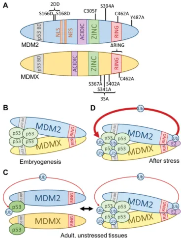
RING domains (Tanimura et al.,1999), MDMX serves to facilitate MDM2-mediated p53 ubiquitination (Linares et al., 2003). This facilitation could occur indirectly, meaning that MDMX could redirect presumable MDM2 autoinhibitory ubiquitination unto itself, or it could occur directly, meaning that MDMX could dir-ectly enhance the transfer of ubiquitin to p53. Several mouse models (Figure 1A and Table 2) have helped to clarify the mechanisms of MDM2- and MDMX-mediated p53regulation.
Figure 1 p53 regulation requirements are context dependent.
MDM2/MDMX−p53binding and MDM heterodimer formation To directly test whether or not MDM2/MDMX−p53 binding alone could restrain p53 activity in vivo,Itahana et al. (2007)
created mice carrying a mutation in the MDM2 RING domain (MDM2C462A), thus disrupting MDM2 E3 ligase activity and MDMX binding. Homozygous MDM2C462A mutation results in p53-dependent embryonic lethality before embryonic day 7.5, suggesting that MDM2/MDMX−p53interaction alone is not suffi-cient to permit embryonic development. Unpublished observations in our laboratory also suggest that MDM2−p53 or MDMX−p53 interaction may not be sufficient for p53suppression in the adult mouse. In our hands,Mdm2C462A/C462A;p53ER/−mice die within4–6 days of tamoxifen injection, which is similar to results obtained fromMdm2−/−;p53ER/−mice, suggesting that MDM2–MDMX het-erodimer formation and/or MDM2E3 ligase activity, rather than MDM−p53 transactivation domain binding, may be the primary mechanisms for MDM-mediated p53suppressionin vivo.
Studies inMdmXknockin mice also appear to corroborate that MDM2/MDMX−p53 binding is insufficient for p53 inhibition, particularly during embryogenesis.Pant et al. (2011)generated an allele carrying an in-frame deletion of the MDMX RING domain (MdmXΔRING). At the same time,Huang et al. (2011)generated an allele carrying a point mutation in the MDMX RING domain (MdmXC462A). Both of these alleles disrupt MDMX–MDM2 inter-action without altering MDM2. However, mice homozygous for eitherMdmXΔRINGorMdmXC462Aexhibit p53-dependent embryonic lethality. In the presence of MDMXΔRING, MDM2E3ligase activity appears to remain intact in MEFs (Pant et al.,2011), suggesting that MDM2-mediated ubiquitination of p53 and MDM2–p53 or MDMX–p53 binding in the absence of heterodimer formation is not sufficient to permit embryonic development. Although these two mouse models both disrupt MDMX–MDM2binding and pre-sent p53-dependent homozygous embryonic lethality, there are
several observations in apparent contradiction. First, when com-bined with thep53neoallele, which expresses ~15% of wild type p53levels, MEFs containing MDMXΔRINGappear to display greater p53 activity with no difference in p53stabilization compared to MEFs containing wild type MDMX, suggesting that MDMX does not necessarily contribute to MDM2-mediated p53 degradation. On the other hand, MdmXC462A/C462A embryos present both increased p53 abundance and activity. These observations sug-gest that the MDM2–MDMX interaction is required for efficient p53 inhibition, but may or may not be required for p53 degrad-ation during embryogenesis.
Complicating things further, Pant et al. (2011) also gener-ated a Cre-inducible MDMX RING deletion allele (MDMXflxRING) and crossed these mice with mice containing a tamoxifen-dependent Cre allele (CreER). When adultMdmXflxRING/ΔRING;CreER mice were injected with tamoxifen to generate recombined MdmXΔRING/ΔRINGmice, the mice appeared healthy. Most p53target genes, with the exception ofp21, showed little change in expres-sion, suggesting that MDM2–MDMX heterodimer formation is dis-pensable for the regulation of p53activity in adult mice. Whether p53stability is affected by this loss has not been determined and remains an interesting question.
Then, what is the contribution of MDMX to MDM2-mediated p53regulationin vivo? It is clear that the necessity of MDMX is context specific. During embryogenesis, it appears that MDM2– MDMX heterodimer formation is critical for p53suppression, but the mechanism of this inhibition is incompletely understood. Several possibilities exist, including that the MDM2–MDMX het-erodimer facilitates more efficient MDM2/MDMX−p53 binding and transcriptional inhibition than either protein alone. In support of this idea, Pant et al. (2011) observed somewhat decreased binding of MDMXΔRINGto p53R172H, which harbours a missense mutation rendering it transcriptionally inactive (Lang et al.,2004). Table2Mdm2andMdmXknockin mice.
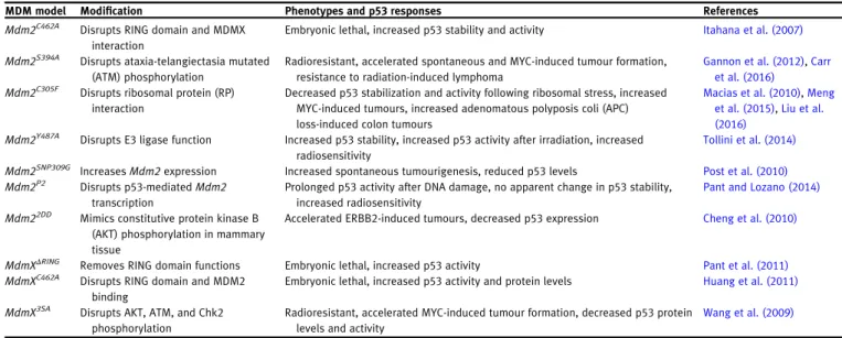
MDM model Modification Phenotypes and p53responses References
Mdm2C462A Disrupts RING domain and MDMX interaction
Embryonic lethal, increased p53stability and activity Itahana et al. (2007)
Mdm2S394A Disrupts ataxia-telangiectasia mutated (ATM) phosphorylation
Radioresistant, accelerated spontaneous and MYC-induced tumour formation, resistance to radiation-induced lymphoma
Gannon et al. (2012),Carr et al. (2016)
Mdm2C305F Disrupts ribosomal protein (RP) interaction
Decreased p53stabilization and activity following ribosomal stress, increased MYC-induced tumours, increased adenomatous polyposis coli (APC) loss-induced colon tumours
Macias et al. (2010),Meng et al. (2015),Liu et al. (2016)
Mdm2Y487A Disrupts E3ligase function Increased p53stability, increased p53activity after irradiation, increased radiosensitivity
Tollini et al. (2014)
Mdm2SNP309G IncreasesMdm2expression Increased spontaneous tumourigenesis, reduced p53levels Post et al. (2010)
Mdm2P2 Disrupts p53-mediatedMdm2
transcription
Prolonged p53activity after DNA damage, no apparent change in p53stability, increased radiosensitivity
Pant and Lozano (2014)
Mdm22DD Mimics constitutive protein kinase B (AKT) phosphorylation in mammary tissue
Accelerated ERBB2-induced tumours, decreased p53expression Cheng et al. (2010)
MdmXΔRING Removes RING domain functions Embryonic lethal, increased p53activity Pant et al. (2011)
MdmXC462A Disrupts RING domain and MDM2 binding
Embryonic lethal, increased p53activity and protein levels Huang et al. (2011)
MdmX3SA Disrupts AKT, ATM, and Chk2 phosphorylation
Radioresistant, accelerated MYC-induced tumour formation, decreased p53protein levels and activity
We have also noticed that MDM2/MDMX−p53binding is impaired in Mdm2−/−;p53ER/− and MdmX−/−;p53ER/− MEFs, respectively,
compared top53ER/−MEFs (our unpublished data). On the other
hand, it appears that MDM2–MDMX binding is dispensable to p53inhibition in the adult mouse (Pant et al.,2011).
MDM2E3ligase activity
The recently developed MDM2Y487Amouse model (Tollini et al., 2014) has provided insight into both basal and stress-dependent p53 regulation by MDM2 and MDMX. As an extension of the MDM2C462A model, in which both MDM2 E3 ligase activity and MDM2–MDMX interaction are disrupted, the MDM2Y487Amutation disrupts MDM2 E3 ubiquitin ligase activity while maintaining MDM2–MDMX interaction. Surprisingly, unlike MDM2C462A, MDMXΔRING, or MDMXC462Amice, MDM2Y487A mice survive into adulthood, with little phenotypic difference from wild type mice under normal, unstressed conditions. This clearly indicates that MDM2E3ligase activity is not essential for regulating p53 dur-ing embryonic development. No degradation of p53is observed in MEFs, and although MDMX levels are also increased, p53 activity is greater than in wild type. This perhaps suggests that either MDM2–p53 or MDMX–p53 binding is not sufficient for complete p53 activity suppression, or that without E3 ligase-mediated degradation by MDM2, increased levels of p53 are also spontaneously more active.
Although Mdm2Y487A/Y487A mice appear normal under un-stressed conditions, these mice are highly sensitive to even sub-lethal doses of ionizing radiation (IR), dying within ~20days after exposure due to p53-dependent haematopoietic failure, indicat-ing that the MDM2E3ligase activity is necessary for p53 degrad-ation and suppression during DNA damaging conditions.
MDM2 has been shown to inhibit p53 acetylation by p300 (Kobet et al.,2000;Ito et al.,2001;Jin et al.,2002).Tollini et al. (2014) also compared the total and acetylated p53 levels of Mdm2C462A/C462A;p53ER/−andMdm2Y487A/Y487A;p53ER/−MEFs and found that although total p53 levels were equivalent between the two, p53 acetylation levels were much greater in Mdm2C462A/C462A;p53ER/− MEFs. In addition, p53–p300 binding was increased in the absence of the MDM2–MDMX heterodimer, possibly indicating that the MDM2–MDMX heterodimer is more efficient than MDM2alone in inhibiting p53acetylation by p300, suggesting another mechanism through which the heterodimer could inhibit p53activityin vivo.
MDM2–MDMX heterodimerization appears to be particularly important for suppressing chronic, basal levels of p53 activa-tion, such as what might occur during embryogenesis, while MDM2E3ligase activity is dispensable under these conditions. However, under stressed conditions where p53is acutely acti-vated, such as DNA damaging conditions, the MDM2–MDMX heterodimer appears to be insufficient for restraining p53 in the adult mouse. These conditions appear to require the fur-ther degradation of p53, mandating use of MDM2 E3 ligase activity.
p53–Mdm2feedback
In addition to regulating p53stability and activity,Mdm2is a p53target gene (Barak et al.,1993). This feedback loop of regu-lation is thought to be important for returning p53 to basal levels and activity following a p53-activating insult. To directly address the importance of the p53–MDM2feedback loop to p53 regulation in vivo, Pant and Lozano (2014) generated the Mdm2P2allele, in which point mutations were introduced to two p53-binding sites within theMdm2promoter region. p53 stabil-ization in response to several stresses occurred in a similar manner to wild typeMdm2mice, but p53activity persisted long-er in Mdm2P2 mice and MEFs, suggesting that basal levels of MDM2 are sufficient for p53 regulation in unstressed cells, but the p53–MDM2feedback loop is required for restraining stress-induced p53 in vivo. In addition, the heterozygous deletion of MdmXappeared to enhance p53stability inMdm2P2/P2;MdmX+/−
MEFs, suggesting that MDMX may enhance the degradation of stress-induced p53.
Disrupting upstream p53signalling through MDM2and MDMX mutation
DNA damage.Knockin mouse models have allowed us to appre-ciate the complex interactions of MDM2and MDMX in p53 regu-lation, but they have also been used by several groups to determine the contributions of various upstream signals to p53 activation (Table 2). Activation of p53requires transient inhib-ition of the activities of MDM2and/or MDMX, which is thought to be mediated through upstream signalling factors. For instance, in vitro studies have shown that in response to DNA damage, ATM phosphorylates MDM2, inhibiting MDM2E3ligase activity and RING domain-dependent oligomer formation (Cheng et al.,2009,2011). To test the importance of MDM2 phosphoryl-ation at serine394(serine395in human),Gannon et al. (2012)
generated the MDM2S394Amouse, replacing serine394with an alanine and disrupting MDM2 phosphorylation in vivo. Basal p53 levels and activity were unchanged in these mice. In response to lethal doses of IR, MDM2S394A mice experience reduced p53 stabilization and activation, translating to increased survival compared to wild type mice, indicating that MDM2serine394phosphorylation is an important event preced-ing the propagation of p53stabilization and activation following IR-mediated DNA damage. Conversely,Gannon et al. (2012)also generated mice containing a substitution of serine 394with a phosphomimetic aspartate residue (MDM2S394D). Basal p53 levels and activity were unchanged compared to wild type mice, indicating that phosphorylation at serine 394 is not sufficient for p53 stabilization. Following IR, however, p53 stabilization and activation was greater and persisted longer in MDM2S394D mice, suggesting perhaps that the serine 394phosphorylation mark is responsible for maintaining activation of p53or is typic-ally removed shortly after p53activation.
MDMX degradation following IR treatment (see next section). ATM phosphorylates MDMX serine 403(402in mouse) (Pereg et al.,2005), while Chk2can phosphorylate serine342and ser-ine 367(341and 367in mouse) (Chen et al.,2005; Okamoto et al.,2005;LeBron et al., 2006;Pereg et al.,2006). To study the importance of MDMX phosphorylation to p53activation fol-lowing DNA damage, Wang et al. (2009) generated MDMX3SA
mice, in which serine341, serine367, and serine402of MDMX are replaced with alanine residues. Upon loss of MDMX phos-phorylation capability, MDMX3SA appears to be stabilized at
basal levels. Following IR treatment, MDMX3SA remains stable
compared to MDMX, and p53protein levels and transcriptional activity appear to be lower in MDMX3SA MEFs and thymuses. In
addition, MDMX3SA mice are resistant to lethal IR treatment and
sensitive to MYC-induced lymphomagenesis. These results are in congruence with the reduced basal and DNA damage-induced p53 activity observed in MDMX3SAmice, suggesting that MDMX
phos-phorylation and subsequent degradation is important for proper p53activation. These results also suggest that basal MDMX phos-phorylation could be required for basal levels of p53activity.
Oncogene activation.It is known that p53responds to a variety of stresses in order to perform various survival or pro-apoptotic functions, but the upstream signals of p53activation are still being elucidated. MDM2S394Amice are somewhat
sus-ceptible to tumour formation, indicating that ATM-mediated MDM2 phosphorylation is likely important for allowing proper p53activation in response to endogenous, cancer-causing DNA damage events. Carr et al. (2016)analysed this propensity for tumourigenesis in mice harbouring the MDM2S394Amutation by
performing a regimen of repeated low doses of IR and by cross-ing Mdm2S394A mice with Eµ-Myc mice. MDM2S394A mice are
resistant to IR-induced lymphomagenesis yet are highly suscep-tible to c-MYC-induced tumourigenesis, suggesting that the role of MDM2phosphorylation in p53regulation and tumour preven-tion is highly context- and stress type-specific.
Although oncogenes can invoke p53 stabilization by inducing DNA damage, it has been recently appreciated that the acceler-ated growth of cancer cells can also invoke p53stabilization. For instance, accelerated cell growth mandates the increased pro-duction of ribosomes. The c-MYC oncogene is a master regulator of ribosomal biogenesis and directly upregulates the transcrip-tion of many RPs (Van Riggelen et al.,2010). Several RPs have been foundin vitroto bind to the central zinc finger domain of MDM2 and prevent p53inhibition (Zhang et al., 2003; Dai and Lu, 2004; Dai et al., 2004; Chen et al., 2007; Zhang and Lu, 2009). In order to directly test the role of RP–MDM2interaction towards p53 activation in vivo,Macias et al. (2010) generated the MDM2C305Fmouse. The MDM2C305Fmutation resides in the
region of RPL11and RPL5binding and thus prevents their inter-action with MDM2. Mice with the MDM2C305Fmutation display
normal responses to DNA damage, but are highly susceptible to c-MYC overexpression- and adenomatous polyposis coli loss-induced lymphomagenesis and colorectal tumourigenesis, respectively (Macias et al.,2010;Meng et al., 2015; Liu et al.,
2016). On the other hand, MDM2C305F mice are surprisingly
resistant to HRASG12V-mediated melanomagenesis (Meng et al.,
2016), possibly due to increased MDM2–RPL23 interaction mediated by the MDM2C305Fmutation.
Nutrient availability. It has become increasingly apparent that MDM2 and MDMX also serve to regulate p53 in response to nutrient availability. This is not surprising, as it is clear that p53 itself regulates cellular energy homoeostasis (Vousden and Ryan,2009;Zhang et al.,2010). It has also been reported that changes in nutrient abundance can drastically alter riboso-mal biogenesis (Boulon et al.,2010). Altered ribosomal biogen-esis induces the binding of RPs to MDM2(Zhang and Lu,2009), which inhibits MDM2-mediated p53degradation, activating p53 -induced (or dependent) metabolic alterations. Consistently, the MDM2C305Fmouse, with its impaired RP–MDM2 binding, is defi-cient in p53-mediated fatty acid oxidation in response to fasting (Liu et al., 2014) and p53-mediated fat storage in response to sustained high-fat diet feeding (unpublished data). Although fasting or high-fat diet treatments appear to activate p53 sig-nalling through the RP–MDM2 interaction, p53 activation in response to glucose deprivation appears to be MDMX-dependent (He et al., 2014).He et al. (2014) showed that glucose depriv-ation enhances 5´ adenosine monophosphate-activated protein kinase (AMPK) phosphorylation of MDMX at serine 342 (serine 341 in mouse). They suggest that MDMX S342phosphorylation reduces its activity against p53by enhancing MDMX interaction with 14-3-3, which allows p53to become stabilized. Using the MDMX3SAmouse, this study suggested that loss of MDMX phos-phorylation is correlated with reduced p53stability and activity in response to AMPK induction.
MDM2and MDMX stability regulation in vivo
Previous hypotheses proposed that MDM2E3 ligase activity was important not only for p53regulation, but also for stability of MDM2 and MDMX. In fact, in vitro mutations in the MDM2 RING domain result in increased stability of overexpressed MDM2 protein (Honda and Yasuda, 2000). However, mouse models have opposed these observations. The MDM2C462Aand
MDM2Y487A mouse models have specifically challenged the
notion that MDM2autoubiquitination occursin vivo.
The MDM2C462Aand MDM2Y487Amutations disrupt MDM2E3
ligase activityin vivo, yet the half-life and ubiquitination levels of MDM2 do not change in these mice (Itahana et al., 2007;
Tollini et al.,2014), suggesting that in the live mouse, MDM2 stability is mediated by the activity of other E3 ligases. However, some discrepancies exist between these models and other knockin mice. For example, the MDM2C462Amutation
dis-rupts MDM2–MDMX interaction but does not affect MDM2 deg-radation (Itahana et al., 2007). Yet, the MDMXC462A mutation
also disrupts MDM2–MDMX interaction without directly altering MDM2, and in the absence of MDM2–MDMX binding, Huang et al. (2011)observed that MDM2ubiquitination was disrupted. However, since the MDM2C462A mutation and the MDMXC462A
is thought to be important for the structure of these proteins (Poyurovsky et al., 2007), it is possible that these mutations alter the functions of the proteins beyond simple loss of RING domain function. In addition, mice containing MDMXΔRING, which
also does not interact with MDM2, display no difference in MDM2 half-life compared to mice containing wild type MDMX (Pant et al.,2011). Resolving these conflicting observations is important to understand MDM2stabilityin vivo. The MDM2Y487A
mouse has provided some clarification of this problem, because the MDM2Y487Amutation does not occur in the RING domain of
MDM2, and would not likely alter MDM2 structure greatly. Lacking MDM2E3ligase activity and maintaining MDM2–MDMX binding, MDM2Y487A is degraded equally quickly compared to
MDM2(Tollini et al.,2014). Two independent E3ligase activity-disrupting mutations of MDM2have shown that MDM2E3ligase activity is not required for basal MDM2 degradation in vivo (Itahana et al.,2007;Tollini et al.,2014), although whether or not MDM2–MDMX interaction is required for MDM2 ubiquitina-tion is still unclear in the present mouse models.In vitroMDMX overexpression has been shown to stabilize MDM2, and this sta-bilization is dependent on the RING domain of each protein (Tanimura et al.,1999;Linares et al.,2003). Conversely, knock-down of MDMX has resulted in reduced MDM2expression (Gu et al., 2002). Because of this, it was previously proposed that MDMX could redirect MDM2E3ligase activity from MDM2unto itself and stabilize MDM2, but if MDM2autoubiquitination does not truly occurin vivo, this may not be the case.
In vivomodels have advocated that MDM2does in fact con-trol MDMX stability. For example, MDM2Y487A mice lacking
MDM2 E3 ligase activity have increased protein levels of MDMX (Tollini et al.,2014), which is in line within vitrostudies suggesting that MDM2 E3 ligase activity acts to ubiquitinate MDMX (Kawai et al.,2003; Pan and Chen,2003). In addition, MDMXΔRING, which does not have the ability to interact with
MDM2, is not degraded compared to MDMX in untreated or IR-treated MEFs (Pant et al.,2011).
MDM2and MDMX degradation following IR has been observed by many groups in cell culture (Kawai et al.,2003;Stommel and Wahl, 2004). This regulation has been recapitulated in several mouse models. For example, MDM2S394A, which cannot be
phos-phorylated by ATM, appears to be resistant to IR-induced deg-radation (Carr et al.,2016). MDMX3SA is also more stable than
MDMX in multiple tissues and is resistant to DNA damage-induced degradation (Wang et al.,2009). This indicates both that MDMX may have some level of constitutive phosphorylation that is important for its normal degradation in vivo and that DNA damage-induced phosphorylation is necessary for proper regula-tion of MDMX stability. Following IR treatment, MDMX3SA also
interacts with MDM2 similarly to MDMX, which suggests that DNA damage-induced phosphorylation does not hinder MDM2– MDMX interaction.
Using mouse models to inform future therapies
p53 mutation occurs in ~50% of human cancers, and p53 is often functionally inactivated in tumours harbouring wild type
p53, due to aberrantly expressed MDM2or MDMX (Tovar et al., 2006). A cancer-associated human single nucleotide polymorph-ism (SNP) (Bond et al.,2004) in the second promoter ofMdm2 contributing to increased Mdm2 transcription was recently modelled in the mouse (Post et al., 2010). Mice containing a T to G human SNP (SNP309) were susceptible to decreased p53 function and increased tumourigenesis. This model suggests that even naturally occurring MDM2 ‘overexpression’ (as opposed to transgenic overexpression) does in fact contribute to p53functional inactivation.
A growing number of studies have suggested targeting mutant p53or restoring wild type p53as cancer treatment strat-egy (Burgess et al., 2016; Soragni et al., 2016). Many drugs specifically targeting MDM2–p53 interaction, MDMX–p53 inter-action, or MDM2-mediated ubiquitination of p53 have been developed (Vassilev et al., 2004; Wade et al., 2013; Burgess et al., 2016). For example, MDMX loss in c-MYC-driven tumours extends survival after p53ER restoration (Garcia et al.,2011). In addition, CreER-mediated p53neo restoration in transplanted MDM2-overexpressing tumours also appears to extend survival in mice (Li et al.,2014). However, so far these treatment strat-egies have enjoyed limited efficacy in the clinic.
In vivo studies have also suggested that other approaches could be taken to restore p53function in human cancers har-bouring wild type p53, such as inhibiting MDM2–MDMX binding or inhibiting MDM2 E3 ligase activity. The inhibition of MDM2 E3 ligase activity may be especially attractive as a treatment strategy, because the MDM2Y487Amouse model shows that
gen-etic ablation of MDM2E3ligase activity is tolerated by the adult mouse as well as the developing embryo (Tollini et al., 2014), which suggests that this strategy could avoid toxicity issues. In response to p53-activating stimuli, cells containing MDM2Y487A
demonstrate increased p53 stability and activity. Observations from our laboratory support this strategy in principle, as homozy-gous MDM2Y487Amutation appears to allow for prolonged survival
in response to c-MYC-induced tumourigenesis (our unpublished data). Although several inhibitors of MDM2E3ligase activity have been identified and shown to stabilize p53 (Yang et al., 2005;
Herman et al.,2011;Roxburgh et al.,2012), their activity and spe-cificity may not yet be sufficient for human use. To our knowl-edge, MDM2E3ligase inhibitors have not been tested in humans, but several other small-molecule MDM2antagonists are currently in Phase I trials (Burgess et al.,2016).
Mouse models have also suggested that complete restoration of p53function in the presence of radiation should be used with caution, as abundant p53 activity is especially toxic to radio-sensitive tissues (Ringshausen et al.,2006; Tollini et al.,2014;
Zhang et al.,2014a). It is possible that tissue-targeted therapies will need to be used in combination with any p53-reactivating therapies to avoid this problem.
Concluding remarks
MDMX proteins are master p53regulators. However, several questions remain. Although several in vitro studies suggest that MDMX may facilitate MDM2-mediated p53degradation, we still do not have a clear understanding of whether this occurs in vivo. We still do not completely understand how MDM2is degraded. In addition, MDM2and MDMX appear to have differing activities in p53transcriptional inhibition, but we do not understand how or why this may occur. Although many questions remain, the tools presented in this review are indicative of the importance ofin vivomodelling and point to a bright future of continued research in the MDM2/MDMX– p53field.
Acknowledgements
The authors would like to thank Hui Tian, Jing Yang, and Derek Franklin (Department of Radiation Oncology, University of North Carolina at Chapel Hill) for their helpful discussions of this manuscript. The authors apologize if they failed to cite any relevant articles.
Funding
This review was supported by grants from the National Institutes of Health (CA127770, CA 100302, and CA167637), the Natural Science Foundation of China (NSFC) to Y.Z., and the National Institute of General Medical Sciences (5T32GM007092) to N.R.T.
Conflict of interest: none declared.
References
Badciong, J.C., and Haas, A.L. (2002). MdmX is a RING finger ubiquitin ligase capable of synergistically enhancing Mdm2ubiquitination. J. Biol. Chem.
277,49668–49675.
Barak, Y., Juven, T., Haffner, R., et al. (1993). mdm2expression is induced by wild type p53activity. EMBO J.12,461.
Bardot, B., Bouarich-Bourimi, R., Leemput, J., et al. (2015). Mice engineered for an obligatory Mdm4exon skipping express higher levels of the Mdm4 -S isoform but exhibit increased p53activity. Oncogene34,2943–2948. Bartel, F., Schulz, J., Bo¨hnke, A., et al. (2005). Significance of HDMX-S (or
MDM4) mRNA splice variant overexpression and HDMX gene amplification on primary soft tissue sarcoma prognosis. Int. J. Cancer117,469–475. Boesten, L., Zadelaar, S., De Clercq, S., et al. (2006). Mdm2, but not Mdm4,
protects terminally differentiated smooth muscle cells from p53-mediated caspase-3-independent cell death. Cell Death Differ.13,2089–2098. Bond, G.L., Hu, W., Bond, E.E., et al. (2004). A single nucleotide
polymorph-ism in theMDM2promoter attenuates the p53tumor suppressor pathway and accelerates tumor formation in humans. Cell119,591–602.
Boulon, S., Westman, B.J., Hutten, S., et al. (2010). The nucleolus under stress. Mol. Cell40,216–227.
Burgess, A., Chia, K.M., Haupt, S., et al. (2016). Clinical overview of MDM2/ X-targeted therapies. Front. Oncol.6,7.
Carr, M.I., Roderick, J.E., Gannon, H.S., et al. (2016). Mdm2phosphorylation regulates its stability and has contrasting effects on oncogene and radiation-induced tumorigenesis. Cell Rep.16,2618–2629.
Chen, D., Zhang, Z., Li, M., et al. (2007). Ribosomal protein S7as a novel modulator of p53–MDM2 interaction: binding to MDM2, stabilization of p53protein, and activation of p53function. Oncogene26,5029–5037. Chen, J., Marechal, V., and Levine, A.J. (1993). Mapping of the p53and
mdm-2interaction domains. Mol. Cell. Biol.13,4107–4114.
Chen, L., Gilkes, D.M., Pan, Y., et al. (2005). ATM and Chk2-dependent phos-phorylation of MDMX contribute to p53 activation after DNA damage. EMBO J.24,3411–3422.
Cheng, Q., Chen, L., Li, Z., et al. (2009). ATM activates p53by regulating MDM2oligomerization and E3processivity. EMBO J.28,3857–3867. Cheng, Q., Cross, B., Li, B., et al. (2011). Regulation of MDM2E3ligase
activ-ity by phosphorylation after DNA damage. Mol. Cell. Biol.31,4951–4963. Cheng, X., Xia, W., Yang, J.-Y., et al. (2010). Activation of murine double
minute2by Akt in mammary epithelium delays mammary involution and accelerates mammary tumorigenesis. Cancer Res.70,7684–7689. Christophorou, M.A., Martin-Zanca, D., Soucek, L., et al. (2005). Temporal
dissection of p53function in vitro and in vivo. Nat. Genet.37,718–726. Dai, M.-S., and Lu, H. (2004). Inhibition of MDM2-mediated p53ubiquitination
and degradation by ribosomal protein L5. J. Biol. Chem.279,44475–44482. Dai, M.-S., Zeng, S.X., Jin, Y., et al. (2004). Ribosomal protein L23activates p53by inhibiting MDM2function in response to ribosomal perturbation but not to translation inhibition. Mol. Cell. Biol.24,7654–7668.
Donehower, L.A., Harvey, M., Slagle, B.L., et al. (1992). Mice deficient for p53 are developmentally normal but susceptible to spontaneous. Nature356,19. Feng, Z., Hu, W., Teresky, A.K., et al. (2007). Declining p53function in the aging process: a possible mechanism for the increased tumor incidence in older populations. Proc. Natl Acad. Sci. USA104,16633–16638.
Francoz, S., Froment, P., Bogaerts, S., et al. (2006). Mdm4and Mdm2 cooper-ate to inhibit p53activity in proliferating and quiescent cells in vivo. Proc. Natl Acad. Sci. USA103,3232–3237.
Gannon, H.S., Donehower, L.A., Lyle, S., et al. (2011). Mdm2–p53signaling regulates epidermal stem cell senescence and premature aging pheno-types in mouse skin. Dev. Biol.353,1–9.
Gannon, H.S., Woda, B.A., and Jones, S.N. (2012). ATM phosphorylation of Mdm2Ser394regulates the amplitude and duration of the DNA damage response in mice. Cancer Cell21,668–679.
Garcia, D., Warr, M.R., Martins, C.P., et al. (2011). Validation of MdmX as a thera-peutic target for reactivating p53in tumors. Genes Dev.25,1746–1757. Grier, J.D., Xiong, S., Elizondo-Fraire, A.C., et al. (2006). Tissue-specific
differ-ences of p53inhibition by Mdm2and Mdm4. Mol. Cell. Biol.26,192–198. Grier, J.D., Yan, W., and Lozano, G. (2002). Conditional allele of mdm2which
encodes a p53inhibitor. Genesis32,145–147.
Gu, J., Kawai, H., Nie, L., et al. (2002). Mutual dependence of MDM2and MDMX in their functional inactivation of p53. J. Biol. Chem. 277, 19251–19254.
Haupt, Y., Maya, R., Kazaz, A., et al. (1997). Mdm2promotes therapid deg-radation of p53. Nature387,296–299.
He, G., Zhang, Y.W., Lee, J.H., et al. (2014). AMP-activated protein kinase induces p53by phosphorylating MDMX and inhibiting its activity. Mol. Cell. Biol.34,148–157.
Herman, A.G., Hayano, M., Poyurovsky, M.V., et al. (2011). Discovery of Mdm2-MdmX E3ligase inhibitors using a cell-based ubiquitination assay. Cancer Discov.1,312–325.
Honda, R., Tanaka, H., and Yasuda, H. (1997). Oncoprotein MDM2is a ubi-quitin ligase E3for tumor suppressor p53. FEBS Lett.420,25–27. Honda, R., and Yasuda, H. (2000). Activity of MDM2, a ubiquitin ligase,
toward p53or itself is dependent on the RING finger domain of the ligase. Oncogene19,1473–1476.
Hu, B., Gilkes, D.M., and Chen, J. (2007). Efficient p53activation and apop-tosis by simultaneous disruption of binding to MDM2and MDMX. Cancer Res.67,8810–8817.
Huang, L., Yan, Z., Liao, X., et al. (2011). The p53inhibitors MDM2/MDMX complex is required for control of p53activity in vivo. Proc. Natl Acad. Sci. USA108,12001–12006.
Itahana, K., Mao, H., Jin, A., et al. (2007). Targeted inactivation of Mdm2 RING finger E3ubiquitin ligase activity in the mouse reveals mechanistic insights into p53regulation. Cancer Cell12,355–366.
Jackson, M.W., and Berberich, S.J. (2000). MdmX protects p53from Mdm2 -mediated degradation. Mol. Cell. Biol.20,1001–1007.
Jin, Y., Zeng, S.X., Dai, M.-S., et al. (2002). MDM2inhibits PCAF (p300 /CREB-binding protein-associated factor)-mediated p53acetylation. J. Biol. Chem.
277,30838–30843.
Jones, S.N., Hancock, A.R., Vogel, H., et al. (1998). Overexpression of Mdm2 in mice reveals a p53-independent role for Mdm2in tumorigenesis. Proc. Natl Acad. Sci. USA95,15608–15612.
Jones, S.N., Roe, A.E., Donehower, L.A., et al. (1995). Rescue of embryonic lethality in Mdm2-deficient mice by absence of p53. Nature378,206–208. Kawai, H., Wiederschain, D., Kitao, H., et al. (2003). DNA
damage-induced MDMX degradation is mediated by MDM2. J. Biol. Chem.278, 45946–45953.
Kobet, E., Zeng, X., Zhu, Y., et al. (2000). MDM2inhibits p300-mediated p53 acetylation and activation by forming a ternary complex with the two pro-teins. Proc. Natl Acad. Sci. USA97,12547–12552.
Kubbutat, M.H.G., Jones, S.N., and Vousden, K.H. (1997). Regulation of p53 stability by Mdm2. Nature387,299–303.
Lang, G.A., Iwakuma, T., Suh, Y.-A., et al. (2004). Gain of function of a p53 hot spot mutation in a mouse model of Li-Fraumeni syndrome. Cell119, 861–872.
LeBron, C., Chen, L., Gilkes, D.M., et al. (2006). Regulation of MDMX nuclear import and degradation by Chk2and14-3-3. EMBO J.25,1196–1206. Lengner, C.J., Steinman, H.A., Gagnon, J., et al. (2006). Osteoblast
differenti-ation and skeletal development are regulated by Mdm2–p53signaling. J. Cell Biol.172,909–921.
Lenos, K., Grawenda, A.M., Lodder, K., et al. (2012). Alternate splicing of the p53inhibitor HDMX offers a superior prognostic biomarker than p53 muta-tion in human cancer. Cancer Res.72,4074–4084.
Li, Q., Zhang, Y., El-Naggar, A.K., et al. (2014). Therapeutic efficacy of p53 res-toration in Mdm2-overexpressing tumors. Mol. Cancer Res.12,901–911. Linares, L.K., Hengstermann, A., Ciechanover, A., et al. (2003). HdmX
stimu-lates Hdm2-mediated ubiquitination and degradation of p53. Proc. Natl Acad. Sci. USA100,12009–12014.
Linke, K., Mace, P., Smith, C., et al. (2008). Structure of the MDM2/MDMX RING domain heterodimer reveals dimerization is required for their ubiqui-tylation in trans. Cell Death Differ.15,841–848.
Liu, S., Tackmann, N., Yang, J., et al. (2016). Disruption of the RP-MDM2-p53 pathway accelerates APC loss-induced colorectal tumorigenesis. Oncogene, doi:10.1038/onc.2016.301.
Liu, Y., He, Y., Jin, A., et al. (2014). Ribosomal protein–Mdm2–p53pathway coordinates nutrient stress with lipid metabolism by regulating MCD and promoting fatty acid oxidation. Proc. Natl Acad. Sci. USA 111, E2414–E2422.
Macias, E., Jin, A., Deisenroth, C., et al. (2010). An ARF-independent c-MYC-activated tumor suppression pathway mediated by ribosomal protein-Mdm2Interaction. Cancer Cell18,231–243.
Maetens, M., Doumont, G., De Clercq, S., et al. (2007). Distinct roles of Mdm2and Mdm4in red cell production. Blood109,2630–2633.
Mendrysa, S.M., McElwee, M.K., Michalowski, J., et al. (2003). mdm2is crit-ical for inhibition of p53during lymphopoiesis and the response to ioniz-ing irradiation. Mol. Cell. Biol.23,462–472.
Meng, X., Carlson, N., Dong, J., et al. (2015). Oncogenic c-Myc-induced lym-phomagenesis is inhibited non-redundantly by the p19Arf–Mdm2–p53and RP–Mdm2–p53pathways. Oncogene34,5709–5717.
Meng, X., Tackmann, N.R., Liu, S., et al. (2016). RPL23 links oncogenic RAS signaling to p53-mediated tumor suppression. Cancer Res. 76, 5030–5039.
Montes de Oca Luna, R., Wagner, D.S., and Lozano, G. (1995). Rescue of early embryonic lethality in mdm2-deficient mice by deletion of p53. Nature378,203–206.
Moyer, S.M., Larsson, C.A., and Lozano, G. (2017). Mdm proteins: critical regulators of embryogenesis and homoeostasis. J. Mol. Cell Biol. 9, 16–25.
Muller, P.A., and Vousden, K.H. (2013). p53mutations in cancer. Nat. Cell Biol.15,2–8.
Okamoto, K., Kashima, K., Pereg, Y., et al. (2005). DNA damage-induced phosphorylation of MdmX at serine367activates p53by targeting MdmX for Mdm2-dependent degradation. Mol. Cell. Biol.25,9608–9620. Oliner, J.D., Pietenpol, J.A., Thiagalingam, S., et al. (1993). Oncoprotein
MDM2conceals the activation domain of tumour suppressor p53. Nature
362,857–860.
Pan, Y., and Chen, J. (2003). MDM2promotes ubiquitination and degradation of MDMX. Mol. Cell. Biol.23,5113–5121.
Pant, V., and Lozano, G. (2014). Dissecting the p53-Mdm2feedback loop in vivo: uncoupling the role in p53stability and activity. Oncotarget5,1149–1156. Pant, V., Xiong, S., Iwakuma, T., et al. (2011). Heterodimerization of Mdm2
and Mdm4is critical for regulating p53activity during embryogenesis but dispensable for p53and Mdm2stability. Proc. Natl Acad. Sci. USA108, 11995–12000.
Parant, J., Chavez-Reyes, A., Little, N.A., et al. (2001). Rescue of embryonic lethality in Mdm4-null mice by loss of Trp53suggests a nonoverlapping pathway with MDM2to regulate p53. Nat. Genet.29,92–95.
Pereg, Y., Lam, S., Teunisse, A., et al. (2006). Differential roles of ATM-and Chk2-mediated phosphorylations of Hdmx in response to DNA damage. Mol. Cell. Biol.26,6819–6831.
Pereg, Y., Shkedy, D., de Graaf, P., et al. (2005). Phosphorylation of Hdmx mediates its Hdm2-and ATM-dependent degradation in response to DNA damage. Proc. Natl Acad. Sci. USA102,5056–5061.
Post, S.M., Quinta´s-Cardama, A., Pant, V., et al. (2010). A high-frequency regulatory polymorphism in the p53pathway accelerates tumor develop-ment. Cancer Cell18,220–230.
Poyurovsky, M.V., Priest, C., Kentsis, A., et al. (2007). The Mdm2 RING domain C-terminus is required for supramolecular assembly and ubiquitin ligase activity. EMBO J.26,90–101.
Prodosmo, A., Giglio, S., Moretti, S., et al. (2008). Analysis of human MDM4 variants in papillary thyroid carcinomas reveals new potential markers of cancer properties. J. Mol. Med.86,585–596.
Rallapalli, R., Strachan, G., Cho, B., et al. (1999). A novel MDMX transcript expressed in a variety of transformed cell lines encodes a truncated protein with potent p53 repressive activity. J. Biol. Chem. 274, 8299–8308.
Rallapalli, R., Strachan, G., Tuan, R.S., et al. (2003). Identification of a domain within MDMX-S that is responsible for its high affinity interaction with p53and high-level expression in mammalian cells. J. Cell. Biochem.
89,563–575.
Ringshausen, I., O’Shea, C.C., Finch, A.J., et al. (2006). Mdm2is critically and continuously required to suppress lethal p53activity in vivo. Cancer Cell
10,501–514.
Roxburgh, P., Hock, A.K., Dickens, M.P., et al. (2012). Small molecules that bind the Mdm2RING stabilize and activate p53. Carcinogenesis33,791–798. Shvarts, A., Steegenga, W., Riteco, N., et al. (1996). MDMX: a novel p53
-binding protein with some functional properties of MDM2. EMBO J.15, 5349.
Soragni, A., Janzen, D.M., Johnson, L.M., et al. (2016). A designed inhibitor of p53aggregation rescues p53 tumor suppression in ovarian carcinomas. Cancer Cell29,90–103.
Steinman, H.A., Hoover, K.M., Keeler, M.L., et al. (2005). Rescue of Mdm4 -deficient mice by Mdm2reveals functional overlap of Mdm2and Mdm4in development. Oncogene24,7935–7940.
Stommel, J.M., and Wahl, G.M. (2004). Accelerated MDM2auto-degradation induced by DNA-damage kinases is required for p53activation. EMBO J.
23,1547–1556.
Tanimura, S., Ohtsuka, S., Mitsui, K., et al. (1999). MDM2interacts with MDMX through their RING finger domains. FEBS Lett.447,5–9.
Tovar, C., Rosinski, J., Filipovic, Z., et al. (2006). Small-molecule MDM2 antagonists reveal aberrant p53signaling in cancer: implications for ther-apy. Proc. Natl Acad. Sci. USA103,1888–1893.
Valentin-Vega, Y., Okano, H., and Lozano, G. (2008). The intestinal epithe-lium compensates for p53-mediated cell death and guarantees organismal survival. Cell Death Differ.15,1772–1781.
Valentin-Vega, Y.A., Box, N., Terzian, T., et al. (2009). Mdm4loss in the intes-tinal epithelium leads to compartmentalized cell death but no tissue abnormalities. Differentiation77,442–449.
Van Riggelen, J., Yetil, A., and Felsher, D.W. (2010). MYC as a regulator of ribosome biogenesis and protein synthesis. Nat. Rev. Cancer 10, 301–309.
Vassilev, L.T., Vu, B.T., Graves, B., et al. (2004). In vivo activation of the p53 pathway by small-molecule antagonists of MDM2. Science303,844–848. Vousden, K.H., and Ryan, K.M. (2009). p53and metabolism. Nat. Rev. Cancer
9,691–700.
Wade, M., Li, Y.-C., and Wahl, G.M. (2013). MDM2, MDMX and p53in onco-genesis and cancer therapy. Nat. Rev. Cancer13,83–96.
Wade, M., Wang, Y.V., and Wahl, G.M. (2010). The p53orchestra: Mdm2and Mdmx set the tone. Trends Cell Biol.20,299–309.
Wang, Y.V., Leblanc, M., Wade, M., et al. (2009). Increased radioresistance and accelerated B cell lymphomas in mice with Mdmx mutations that
prevent modifications by DNA-damage-activated kinases. Cancer Cell16, 33–43.
Xiong, S., Van Pelt, C.S., Elizondo-Fraire, A.C., et al. (2006). Synergistic roles of Mdm2and Mdm4for p53inhibition in central nervous system develop-ment. Proc. Natl Acad. Sci. USA103,3226–3231.
Yang, Y., Ludwig, R.L., Jensen, J.P., et al. (2005). Small molecule inhibitors of HDM2ubiquitin ligase activity stabilize and activate p53in cells. Cancer Cell7,547–559.
Zhang, Q., He, X., Chen, L., et al. (2012). Synergistic regulation of p53by Mdm2and Mdm4is critical in cardiac endocardial cushion morphogenesis during heart development. J. Pathol.228,416–428.
Zhang, X.-D., Qin, Z.-H., and Wang, J. (2010). The role of p53in cell metabol-ism. Acta Pharmacol. Sin.31,1208–1212.
Zhang, Y., and Lu, H. (2009). Signaling to p53: ribosomal proteins find their way. Cancer Cell16,369–377.
Zhang, Y., Wolf, G.W., Bhat, K., et al. (2003). Ribosomal protein L11 nega-tively regulates oncoprotein MDM2and mediates a p53-dependent riboso-mal-stress checkpoint pathway. Mol. Cell. Biol.23,8902–8912.
Zhang, Y., Xiong, S., Li, Q., et al. (2014a). Tissue-specific and age-dependent effects of global Mdm2loss. J. Pathol.233,380–391.