THE INFLUENCE OF MOVEMENT PROFILE ON THE FEMALE ATHLETE’S BIOMECHANICAL RESILIENCE & TRAINING LOAD RESPONSE TO CONTROLLED
EXERCISE EXPOSURE
Barnett S. Frank
A dissertation submitted to the faculty of The University of North Carolina at Chapel Hill in partial fulfillment of the requirements for the degree of Doctor of Philosophy in Interdisciplinary
Human Movement Science
Chapel Hill 2016
ABSTRACT
Barnett S. Frank: The Influence Of Movement Profile On The Female Athlete’s Biomechanical Resilience & Training Load Response To Controlled Exercise Exposure
(Under the Direction of Darin A. Padua)
Background: “Stiff” landing biomechanics and excessive frontal plane knee motion, such as limited trunk, hip, and knee flexion and medial knee displacement have been identified as risk factors or movement patterns associated with lower extremity musculoskeletal injury and elevated joint loads. Additionally, high training load exposure has similarly be linked to
musculoskeletal injury in the physically active population. There is a significant volume of evidence supporting high training loads and high-load biomechanics to independently influence injury risk. However there is a lack evidence describing the influence of an individual’s baseline movement quality profile on their systemic and musculoskeletal tissue stress experienced
secondary to high training load exposure. An individual’s global resilience to high training loads may be influenced by the mechanical demands of their inherent movement profile during
physical activity and sport participation.
Aim: Investigate the influence of an individual’s inherent baseline movement profile on their biomechanical, systemic stress, and musculoskeletal system stress response to an acute bout of high training load exposure.
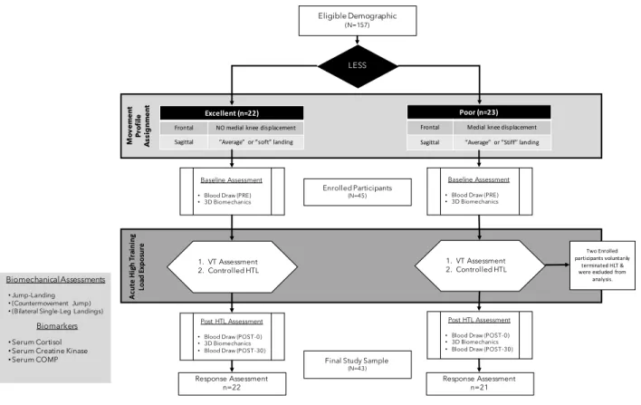
Methods: 43 physically active, healthy, college-aged females were enrolled in this study and were assigned to a poor high-load or excellent low-load movement profile group
biomechanics and blood samples were collected prior to and following a metabolically controlled acute high training load exercise protocol (HTL). Changes in biomechanics and circulating biomarkers of global systemic stress (cortisol), and musculoskeletal system tissue stress (sCOMP & CK-MM) were compared between poor and excellent movement profiles to better understand the influence of movement profile on the body’s response to the demands of HTL.
Results: The poor group was observed to experience greater degradation of
neuromuscular control strategies that effectively and efficiently dissipate mechanical stresses experienced during high-intensity exercise. Furthermore, we observed movement profile to influence systemic stress hormone levels (cortisol). A poor movement profile was associated with an elevated stress level in contrast to their excellent movement counter parts. Furthermore, it seems the excellent movement profile is linked to greater deployment of dynamic muscle tissue to efficiently dissipate the high mechanical stresses experienced during HTL activities, as the excellent movement profile was associated with greater circulation of CK-MM following acute HTL exposure.
TABLE OF CONTENTS
LIST OF TABLES ... vii
LIST OF FIGURES ... viii
LIST OF ABBREVIATIONS & SYMBOLS ... ix
CHAPTER 1 ... 1
INTRODUCTION ...1
OPERATIONAL DEFINITIONS ... 8
LIMITATIONS & ASSUMPTIONS ... 13
DELIMITATIONS ... 14
INDEPENDENT VARIABLE ... 15
DEPENDENT VARIABLES ... 15
RESEARCH QUESTIONS ... 16
HYPOTHESES ... 18
REFERENCES ... 20
CHAPTER 2 ... ..26
SECTION ONE: Epidemiology of Sport-Related Musculoskeletal Injury ... 27
Lower Extremity Musculoskeletal Injury in Sport & Physical Activity ... 27
Anterior Cruciate Ligament Injury is Responsible for High-Severity Knee Injury ... 31
The Landscape of ACL Injury in Sport ... 33
SECTION TWO: Biomechanical Mechanisms and Risk Factors for
Sport-Related Noncontact ACL Injury ... 47
Sagittal Plane Knee, Hip, and Trunk Biomechanics Associated with Noncontact ACL Injury ... 49
Frontal Plane Knee, Hip, and Trunk Biomechanics Associated with Noncontact ACL Injury ... 64
ACL Injury Biomechanics Summary and their Proposed Influence on Sport-Related Noncontact ACL Injury in a High-Risk Athlete Population ... 78
SECTION THREE: The Proposed Interaction Between Movement Profile and Total-Body Physiological Response to High-Intensity Exercise-Induced Fatigue ... 88
Exercise-Induced Fatigue is an Interactive Process that Influences Muscle Function ... 88
The Influence of Exercise-Induced Fatigue on Neuromuscular Function and Control of Human Movement ... 92
Evidence of an Interaction Between Biomechanics and Exercise-Induced Physiological Demand on the Human Body ... 101
Biomarker Assessment for Training Stress & Fatigue Monitoring in Athletes ... 112
REFERENCES ... 134
CHAPTER 3 ... 163
RATIONALE ... 163
POPULATION ... 165
Subjects ... 165
Power Analysis ... 169
Initial LESS Screening ... 171
LESS Instrumentation & Setup ... 171
LESS Screening Procedure ... 172
TESTING SESSION ... 173
DATA COLLECTION ... 176
Procedures ... 182
DATA PROCESSING & REDUCTION ... 187
Blood Sample Processing & Long-Term Serum Sample Storage Preparation ... 187
Marker Identification & Processing ... 188
Kinetic Calculations ... 189
Data Reduction ... 189
DATA ANALYSIS ... 191
Biomarker Analysis ... 191
Biomechanical Analysis ... 192
REFERENCES ... 195
CHAPTER 4 ... 201
PARTICIPANT DEMOGRAPHICS ... 201
Acute High Training Load Exposure Protocol ... 202
AIM #1 – Evaluate the effects of movement quality on MSK tissue stress biomarkers at rest and in response to an acute HTL. ... 204
AIM #2 – Investigate the influence of movement quality on systemic stress biomarkers at rest and in response to an HTL. ... 207
AIM #3 – Determine if movement quality moderates biomechanical responses to an acute HTL. ... 207
REFERENCES ... 215
CHAPTER 5: MANUSCRIPT #1 ... 216
Overview ... 216
INTRODUCTION ... 218
METHODS ... 220
Participants ... 220
Participant Preparation ... 222
Three-Dimensional Motion Analysis ... 223
Pre-HTL Jump-Landing Assessment ... 223
Ventilatory Threshold Assessment ... 224
Controlled High-Intensity Exercise Exposure (HTL) ... 225
Post-HTL Jump-Landing Assessment ... 226
Data Reduction & Analysis ... 227
RESULTS ... 230
Participants Demographic ... 230
Controlled High-Intensity Exercise Exposure Metabolics & Perceived Intensity ... 230
Biomechanics ... 230
DISCUSSION ... 231
NEW KEY FINDINGS ... 239
HOW MIGHT IT IMPACT CLINICAL PRACTICE IN THE FUTURE? ... 239
REFERENCES ... 249
CHAPTER 6: MANUSCRIPT #2 ... 258
Overview ... 258
INTRODUCTION ... 260
METHODS ... 262
Participants ... 262
Pretest Guidelines ... 263
Participant Preparation ... 264
Ventilatory Threshold Assessment ... 265
Biochemical Analysis ... 268
Statistical Analysis ... 268
RESULTS ... 270
Circulating Systemic & Musculoskeletal Tissue Stress Biomarkers ... 270
DISCUSSION ... 272
Cortisol ... 273
Creatine Kinase (CK-MM) ... 275
Cartilage Oligomeric Matrix Protein (COMP) ... 279
REFERENCES ... 288
APPENDIX 1: 17-Item LESS Operational Definitions ...297
APPENDIX 2: Pre-Test Guidelines ...298
LIST OF TABLES
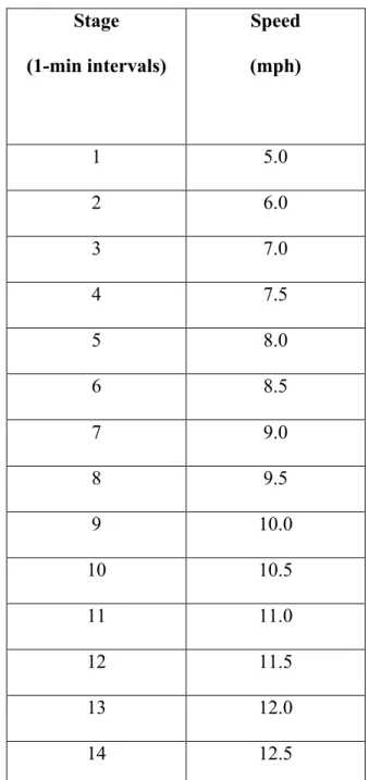
1. Table 3.1: Speed-Only Graded Submaximal Aerobic Power Assessment Protocol ...180 2. Table 4.1: Group Demographic & Fitness Level
Descriptive Statistics: Group Means & (SDs) ...201 3. Table 4.2: Controlled acute high training load
exposure stage metabolic & intensity perception data ...203 4. Table 4.3: Group-by-time Raw (ng/ml), natural logarithm-transformed,
& %∆ serum biomarker concentrations per and post-acute HTL ...206 5. Table 4.4: Summary of pre, post and biomechanical variable change
response relative to acute HTL exposure ...209 6. Table 5.1: Group Demographic Descriptive Statistics ...240 7. Table 5.2: Controlled Acute High Training Load
LIST OF FIGURES
Figure 1.1: Conceptual Model of the Influence of Movement Profile on
High Training Load Responses ... 6
Figure 1.2: Proposed Study Methodology ... 8
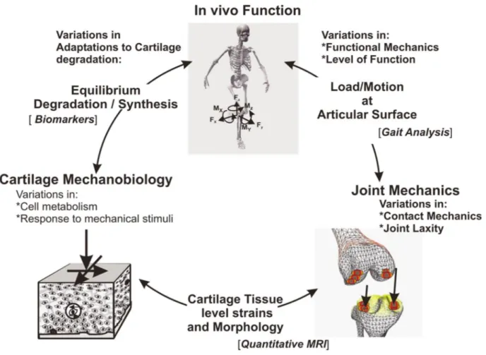
Figure 2.1: Andriacchi et al’s. 2004 theoretical framework explaining the relationship between in vivo function, non-physiological joint biomechanics, joint loading, and articular cartilage mechanical and biological responses ... 103
Figure 3.1: Movement Profile LESS Inclusion Criteria ... 167
Figure 3.2: Study Overview Diagram ... 169
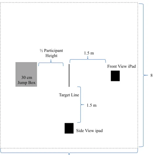
Figure 3.3: Overhead view of the jump-landing LESS testing set-up ... 172
Figure 3.4: Detailed Testing Session Overview ... 175
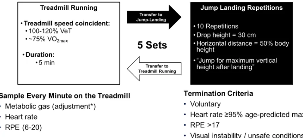
Figure 3.5: High Training Load Exposure Exercise Protocol ... 185
Figure 4.1: Acute HLT protocol speed, perceptual measures (RPE) and associated metabolic responses (Heart Rate, Oxygen Uptake, RER) ... 203
Figure 4.2: Log [C] of values and acute HTL response behavior of CK-MM (a), COMP (b), and cortisol (c) ... 206
Figure 4.3: Sagittal plane trunk, hip, and knee motion group ensemble curves and associated 95% confidence interval waveforms over the stance phase of the jump-landing task ... 210
Figure 4.4: Frontal plane hip and knee motion group ensemble curves and associated 95% confidence interval waveforms over the stance phase of the jump-landing task ... 211
Figure 4.5: Sagittal plane net internal hip and knee moment group ensemble curves and associated 95% confidence interval waveforms over the stance phase of the jump-landing task ... 212
Figure 4.6: Frontal plane net internal hip and knee moment group ensemble curves and associated 95% confidence interval waveforms over the stance phase of the jump-landing task ... 213 Figure 4.7: Vertical ground reaction force (VGRF) and
Figure 5.1: Study Overview & Biomechanical Adaptations to
acute High Training Load Exposure ... 242 Figure 5.2: Biomechanics Methodology Protocol ... 243 Figure 5.3: Pre-HTL, Post-HTL, & Change Responses for Sagittal
Plane Trunk, Hip, & Knee Kinematics ... 244 Figure 5.4: Pre-HTL, Post-HTL, & Change Responses for
Frontal Plane Hip & Knee Kinematics ... 245 Figure 5.5: Pre-HTL, Post-HTL, & Change Responses for
Sagittal Plane Hip & Knee Moments ... 246 Figure 5.6: Pre-HTL, Post-HTL, & Change Responses for
Frontal Plane Hip & Knee Moments ... 247 Figure 5.7: Pre-HTL, Post-HTL, & Change Responses for
Vertical Ground Reaction and Anterior Tibial Shear Forces ... 248 Figure 6.1: Group Demographic Descriptive Statistics ... 282 Figure 6.2: Controlled Acute High Training Load Exercise Exposure Stage Metabolic &
Intensity Perception Data ... 283 Figure 6.3: Group-by-time Raw (ng/ml), Natural Logarithm-transformed,
& %∆ serum biomarker concentrations pre and post acute HTL ... 284 Figure 6.4: Raw (ng/ml), Natural Logarithm-Transformed,
LIST OF ABBREVIATIONS & SYMBOLS
ACL: Anterior Cruciate Ligament
C1,2C: Carboxy-Terminus of 3/4 Peptide From Cleavage of Type-I & Type-II Collagen C2C: Neoepitope From Cleavage Of Type-II Collagen
CK: Creatine Kinase
COMP: Cartilage Oligomeric Matrix Protein CPII: Type-II Procollagen Carboxy-Propeptide CTx-II: C-Telopeptide Of Type-II Collagen ELISA: Enzyme Linked Immuosorbent Assay HIE: High-Intensity Exercise
HTL: High Training Load IL-6: Interleukin-6
LESS: Landing Error Scoring System MKD: Medial Knee Displacement MSK: Musculoskeletal
OA: Osteoarthritis
RPE: Rating of Perceived Exertion
sCOMP: Serum Cartilage Oligomeric Matrix Protein sCORT: Serum Cortisol
SOVO2submax: Speed-Only Graded Submaximal Aerobic Capacity Assessment
VeT: Ventilatory Threshold
CHAPTER 1 INTRODUCTION
Musculoskeletal (MSK) injuries during sport and physical activity are common (Conn, Annest, and Gilchrist 2003), costly (Woolf and Pfleger 2003; Jacobs 2008), and have long-term health consequences (Lohmander et al. 2007; Maffulli et al. 2010), representing a substantial socioeconomic burden (Cumps et al. 2008; Shephard 2003). Injury severity is the primary determinant of an injury’s cost to society (van Mechelen 1997; van Mechelen, Hlobil, and Kemper 1992). Individuals who sustain a high-severity sport-related MSK injury such as an anterior cruciate ligament (ACL) rupture experience sizeable direct and indirect medical costs, acute and long-term decreases in productivity that result in a reduction in human capital, and decreases in quality of life (van Mechelen 1997; van Mechelen, Hlobil, and Kemper 1992; Cumps et al. 2008). Thus, there is a considerable need to understand underlying factors that may contribute to an increased risk of experiencing a high-severity MSK during sport and physical activity to reduce the socioeconomic burden of MSK injury while maximizing the health benefits of sport and physical activity participation.
Interestingly, over 50% to 70% of sport-related ACL injuries are reported to be the result of a noncontact mechanism of injury (Agel, Arendt, and Bershadsky 2005; Boden et al. 2000; Mihata, Beutler, and Boden 2006; Mountcastle et al. 2007). Noncontact mechanisms causing sport-related ACL rupture are described as “forces applied to the knee at the time of injury that result from an athlete’s own movement that did not involve contact with another athlete of object” (Marshall, Padua, and McGrath, n.d.). Thus, an athlete’s self-imposed motion is responsible for injury.
Recent studies suggest that high training load (HTL) exposure similar to the physical demands of sport participation elicits changes in lower extremity biomechanics associated with noncontact ACL injury (Quammen et al. 2012; Santamaria and Webster 2010; Webster et al. 2012; SCHMITZ et al. 2014). Current evidence links fatigue (Galambos et al. 2005) and high training loads (Gabbett 2004; Gabbett and Jenkins 2011) to increased injury rates. Furthermore, the physiological effects of sport participation can result in high levels of markers of total body / systemic stress (Rietjens et al. 2005; Thorpe and Sunderland 2012), muscle damage (Rietjens et al. 2005; Thorpe and Sunderland 2012), and joint loading (Andriacchi et al. 2004; Dominguese and Seegmiller 2012; Santamaria and Webster 2010) that may explain an individual’s
predisposition to injury.
(Andriacchi et al. 2004). Furthermore, when individuals adopt non-sagittal plane energy absorption strategies they may recruit frontal and transverse plane dynamic stabilizing
musculature to maintain safe hip and knee joint control, reducing energy absorption efficiency, resulting in a potentially higher total body physiological demand. Therefore, the volume of total body stress, muscle damage, and joint stress experienced by those with poor biomechanics may be greater after HTL exposure. However, the influence of poor biomechanics on total body stress, muscle damage, and joint load during HTL exposure is currently unknown. Developing an understanding of the influence of poor lower extremity biomechanics on systemic stress and MSK system tissue damage is important, because poor biomechanics are modifiable, and can be improved during sport and physical activity participation (Distefano et al. 2010; Distefano et al. 2011; Padua et al. 2011; Dempsey et al. 2009). Therefore, clinicians may be able to decrease systemic and MSK tissue stress during HTL scenarios by means of improving biomechanics through corrective exercise interventions, reducing an individual’s susceptibility to injury during sport or physical activity participation.
Clinical movement screenings such as The Landing Error Scoring System (LESS) can validly and reliably discriminate between individuals with poor and excellent movement profiles (APPENDIX 1) (Padua et al. 2009). Biomechanics associated with a poor movement profile and the readily identifiable medial knee displacement (MKD) movement pattern are linked to
numerous lower extremity injuries, including ACL injury (Shultz et al. 2010), patellofemoral pain syndrome (Mizuno et al. 2001; Elias et al. 2004), medial collateral ligament injury (Hull et al. 1996), lower-leg stress fracture (Cameron, Peck, and Owens 2014), as well as the progression of knee osteoarthritis (OA) (Sharma et al. 2001; Brouwer et al. 2007). Mounting evidence
can significantly improve lower extremity biomechanics to limit MKD and other non-sagittal plane loading patterns that characterize a poor movement profile during functional tasks (Bell et al. 2013; Zebis et al. 2008; Barendrecht et al. 2011); improving lower extremity neuromuscular control and subsequent movement efficiency during athletic participation. Although corrective exercise and lower extremity injury prevention programs are consistently reported to effectively reduce knee injury incidence during sport participation (Sadoghi, Keudell, and Vavken 2012; Taylor et al. 2013), the underlying mechanisms responsible for injury risk reduction remain elusive.
The influence of an individual’s movement profile on their mechanical tissue loading and physiological response to sport and physical activity participation is not currently described. Previous research has focused on the influence of lower extremity neuromuscular control on future risk of lower extremity injury. Abnormal knee and hip biomechanics such as increased knee abduction moment (Farrokhi et al. 2013; 2002; 2013) and hip internal rotation (2007) are associated with increased joint loading at the tibiofemoral and patellofemoral joint in
pathological populations (Farrokhi et al. 2013). However, the combined influence of an
individual’s pre-existing / baseline movement profile and HTL exposure on MSK tissue loading and systemic physiological stress has not been evaluated.
systemic physiological response to exercise manifest in circulating markers of total body hormonal stress response, muscle tissue damage, and subjective perceived exertion (Knicker et al. 2011; Purvis, Gonsalves, and Deuster 2010; Thorpe and Sunderland 2012). Individuals with deficits in lower extremity neuromuscular control such as a poor movement profile and MKD may experience greater tissue loading and total body stress responses driving a potential
Figure 1.1 - Conceptual model depicting the potential influence of and individual’s baseline movement profile on their systemic and musculoskeletal system tissue stress in response to high-intensity exercise exposure.
Currently, the strongest link between training stress and MSK injury is a subjective measure of training load; calculated by multiplying an athlete’s session rate of perceived exertion (RPE) by the duration of the activity (Gabbett and Jenkins 2011). Exposure to higher training loads has been linked to increases in the hormone cortisol, an endocrine system marker of systemic stress (Gomes et al. 2013). Similarly, elevated levels of creatine kinase (CK), a marker of skeletal muscle damage has been observed during periods of higher intensity training or competition in athletes (Coutts et al. 2007; Uchida et al. 2009). Evidence implicates subjective assessment of training load to be a sufficient measure of objectively assessed markers of systemic physiological and MSK system tissue stress imposed on the body during sport participation, as elevated levels of cortisol and CK are both linked to higher levels of training
Mo ve m en t P ro fil e Poor
“stiff” or “average” saggital plane (+MKD)
Total Body Physiological Stress Musculoskeletal System Stress
Total-Body Biomechanics Functional Joint Loading
Physical Function Foundation High-Intensity Athletic Activity Exposure • Cortisol • IL-6
• Creatine Kinase
• IL-6
• Biochemical markers of cartilage metabolism
• Trunk
• Hip Joint
• Knee Joint
Excellent “soft” or “average” sagittal plane
lack of information regarding the underlying physiological link between increased training load and injury risk.
A majority of research has focused on the effects of lower extremity corrective exercise / injury prevention programs on biomechanics and injury rates in the active population, however there is a lack of understanding regarding how an individual’s movement profile affects the athlete’s systemic stress response, consequent changes in biomechanics associated with injury, and MSK system tissue damage. An investigation regarding the effects of aberrant lower
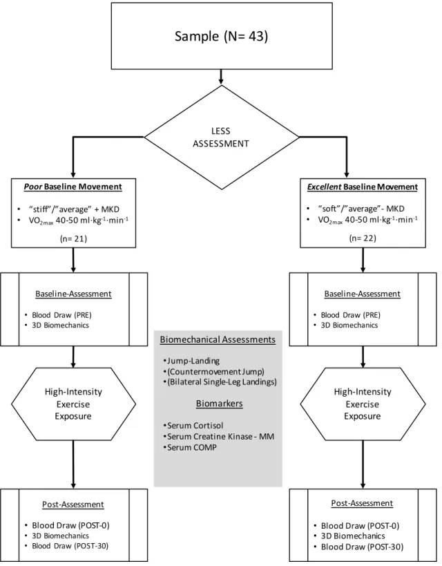
extremity biomechanics on consequential physiological stress markers and biological markers of MSK system tissue damage and metabolism is warranted (figure 1.2). The overall aim of this study was to evaluate the effect of an individual’s baseline movement profile on MSK system tissue damage, systemic stress and biomechanical response to HTL exposure simulating the physical demands of field and court sport. A total of 43 female court and field and court sport athletes with poor (n= 21) and excellent (n= 22) movement profiles was recruited for
Figure 1.2 – Overview of the P study methodology to evaluate the effects of an individual’s baseline movement profile on their musculoskeletal tissue damage and systemic stress response to high-intensity exercise exposure.
Sample (N= 43)
LESS ASSESSMENT
PoorBaseline Movement
• “stiff”/”average” + MKD
• VO2max40-50 ml·kg-1·min-1
(n= 21) High-Intensity Exercise Exposure High-Intensity Exercise Exposure Baseline-Assessment
• Blood Draw (PRE)
• 3D Biomechanics
Baseline-Assessment
• Blood Draw (PRE)
• 3D Biomechanics
Post-Assessment
• Blood Draw (POST-0)
• 3D Biomechanics
• Blood Draw (POST-30)
Biomechanical Assessments
•Jump-Landing
•(Countermovement Jump)
•(Bilateral Single-Leg Landings)
Biomarkers
•Serum Cortisol
•Serum Creatine Kinase - MM
•Serum COMP
Post-Assessment
• Blood Draw (POST-0)
• 3D Biomechanics
• Blood Draw (POST-30)
Excellent Baseline Movement
• “soft”/”average”- MKD
• VO2max40-50 ml·kg-1·min-1
Operational Definitions
1. High Training Load (HTL) Protocol: An exercise protocol lasting approximately 28 minutes comprised of 6 sets of a 5-minute interval of treadmill running at a speed coincident with 115 – 120% of a participant’s ventilatory threshold (VeT) and 10
repetition jump-landing interval. This protocol has been identified to induce elevations in measures of systemic stress and global fatigue responses associated with the high
physical demands of field and court sport participation.
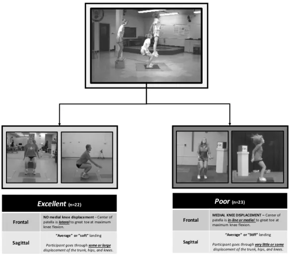
2. Poor Baseline Movement Profile: An “average” or “stiff” landing characterized as “very little, if any trunk, hip, and knee displacement.” with medial knee displacement
(APPENDIX 1).
3. Excellent Baseline Movement Profile: An “average” or “soft” landing characterized as “large displacement of the trunk, hips, and knees” with medial knee displacement (APPENDIX 1).
4. Medial Knee Displacement (MKD): Visually observed frontal plane medial displacement of the center of the patella relative to the first ray during the loading phase of a jump-landing.
5. Submaximal Aerobic Capacity (VO2max): The maximal volume of oxygen (ml) consumed
per unit (kg) body mass per unit time (minute) measured via ventilatory gas exchange during a speed only graded exercise test (ml•kg-1•min-1).
6. Ventilatory Threshold (VeT): Quantified using the V-slope method (Albouaini et al. 2007), an exercise intensity (treadmill speed) representative of the point at which
7. Serum Cartilage Oligomeric Matrix Protein (sCOMP) Concentration: Venous blood sample serum concentration (ng·dl-1) assessed using an enzyme-linked immunosorbent assay (ELISA) reflective of cartilage matrix disruption / degradation.
8. Serum Cortisol Concentration: Venous blood sample serum concentration (ng·dl-1) assessed using an ELISA reflective of systemic stress level.
9. Serum Creatine Kinase MM-Isoform (CK-MM) Concentration: Venous blood sample serum concentration (ng·dl-1) assessed using an ELISA representative of exercise-induced muscle damage.
10.Jump-Landing Task: A functional movement task imposing physical demands similar to landing from a jump during sport activity as in “rebounding” during basketball.
Participants jump down from a 30 cm high “jump box” to a target line placed ½ the participant’s height anterior to the “jump box” and immediately jumping upward for maximal height.
11.Sagittal Plane Knee Angle: Local coordinate system angulation of the shank segment rigid body relative to the thigh segment rigid body about the knee joint’s medio-lateral axis ((+) Flexion / (-) Extension).
12.Frontal Plane Knee Angle: Global coordinate system angulation of the shank segment rigid body and the thigh segment rigid body axes ((+) Varus / (-) Valgus).
14.Frontal Plane Knee Moment: Net internal soft-tissue force acting about the knee joint’s anterior-posterior axis formed by the moment arms of shank segment and thigh segment rigid bodies ((+) Varus / (-) Valgus).
15.Proximal Anterior Tibial Shear Force: The net linear force applied in the anterior direction at the tibiofemoral joint causing anterior translation of the shank rigid body in the reference frame of the x-axis of the femur translated to the distal end of the femur rigid body.
16.Sagittal Plane Hip Angle: Local coordinate system angulation of the thigh segment rigid body relative to the pelvis segment rigid body about the hip joint’s medio-lateral axis ((+) Flexion / (-) Extension).
17.Frontal Plane Hip Angle: Local coordinate system angulation of the thigh segment rigid body relative to the pelvis segment rigid body about the hip joint’s antero-posterior axis ((+) Adduction / (-) Abduction).
18.Sagittal Plane Hip Moment: Net internal soft-tissue force acting about the hip joint’s medio-lateral axis formed by the moment arms of thigh segment and pelvis segment rigid bodies ((+) Flexion / (-) Extension).
19.Frontal Plane Hip Moment: Net internal soft-tissue force acting about the hip joint’s antero-posterior axis formed by the moment arms of thigh segment and pelvis segment rigid bodies ((+) Adduction / (-) Abduction).
21.Frontal Plane Trunk Angle: Local coordinate system angulation of the thorax segment rigid body relative to the pelvis segment rigid body about the L5-S1 joint interspace’s anterior-posterior axis ((+) Rightward / (-) Leftward).
22.Vertical Ground Reaction Force: The vertical components of the ground reaction force vectors of the right and left force platforms equal in magnitude and opposite in direction to the force imparted by participants when they are in contact with the ground atop the right and left force platforms normalized to the participant’s mass.
23.Initial Ground Contact: The first time point during each jump-landing trial when the right or left force platform registers a vertical ground reaction force >10N.
24.Toe-Off: The first time point during each jump-landing trial when the right or left force platform registers a vertical ground reaction force <10N after initial ground contact. 25.Stance Phase: The period of time between initial ground contact and toe-off, representing
the period of time in which the participant’s right or left foot is in contact with the right or left force platform during the jump-landing task.
26.Biomechanical Response Change Score and Confidence Interval Waveforms: All biomechanical data will be analyzed as continuous normalized waveforms during the stance phase of the jump-landing (Kuenze et al. 2014). Interpolated kinematic and kinetic data will be normalized to 201 data points (knots) over the stance phases of the middle 3 jump-landing task trials using a cubic spline function. Each knot will be calculated as the mean value of the respective derived knots from each of the 3 middle jump-landing tasks (eq. 1) (trial 2, trial 3, trial 4).
To calculate changes in biomechanical variables from baseline to post-exercise, the difference between the respective individual baseline and post-HIE knot values (knotbi &
knotfi) was calculated to form a 201 knot waveform reflecting the change in the
biomechanical variable of interest (eq. 2).
!"#$∆%…'() = !"#$2% − !"#$4%
(eq. 2)
Average change score ensemble means and 95% confidence interval waveforms will be calculated for sagittal and frontal plane trunk, hip, and knee joint angles. Change score waveforms will be calculated for sagittal and frontal plane internal net hip and knee joint moments. Furthermore, change score waveforms will also be calculated for proximal anterior tibial shear force and vertical ground reaction force variables.
LIMITATIONS & ASSUMPTIONS
1. Biomechanical calculations from the motion analysis system and biomechanical software are reliable and valid.
2. The principal investigator is an “expert” LESS rater, and thus accurately and reliably is able to identify poor and excellent baseline movement profiles of study candidates. 3. The HIE protocol represents a generalizable simulation of the physical demands of field
and court sports.
4. Participants honestly report their rate of perceived exertion during the VeT assessment and the HTL exercise exposure.
6. The ELISA kits are reliable within <10% inter and intra-assay coefficients of variation. 7. Circulating serum concentrations of sCOMP accurately and reliably reflect articular
cartilage metabolism.
8. Circulating serum concentration of cortisol accurately and reliably reflects systemic stress level.
9. Circulating serum CK-MM accurately and reliably reflects levels of exercise-induced skeletal muscle damage.
10.The MSK system tissue damage, systemic stress, and biomechanical responses of college-aged club field and court sport athletes to HTL is generalizable to the high-risk female athlete population at high-risk for sport-related noncontact ACL injury.
DELIMITATIONS
1. 43 female participants (21 poor & 22 excellent) will be recruited from the local university population.
2. All participants were between the ages of 18 – 24 years of age.
3. All participants were healthy with no history of upper or lower extremity joint surgery, spine surgery, or neurological or metabolic disorders.
4. All participants were injury-free at the time of testing, and had no history of lower or upper extremity MSK injury that limited their participation from sport or exercise for more than 3 days.
6. All participants had an estimated maximal aerobic capacity between 40 – 50 ml•kg-1•min1.
7. Participants demonstrate MKD and an “average” or “stiff” landing OR participants demonstrate a “soft” or “average” landing without MKD.
8. Segment kinematic data was collected from the trunk, thigh, shank, and foot using a 10-camera optoelectric motion capture system.
9. Bilateral ground reaction force data was collected using two conductive in-ground mounted force platforms.
10.All serum biomarker concentrations were measured using ELISA and spectrophotometry.
INDEPENDENT VARIABLE 1. Baseline movement profile
a. Excellent vs. Poor
DEPENDENT VARIABLES
1. Baseline and Post-HIE Exposure Serum Biomarker Concentrations: a. sCOMP
b. Cortisol c. CK-MM
2. Change Score and 95% Confidence Interval Ensemble Waveforms for the Following Biomechanical Variables Normalized to 202 Data Points Over The Stance Phase of the Jump-Landing Task:
b. Frontal plane knee thigh-shank segment angle c. Sagittal plane hip joint angle
d. Frontal plane hip joint angle e. Sagittal plane trunk angle f. Frontal plane trunk angle
g. Net internal sagittal plane knee joint moment h. Net internal frontal plane knee joint moment i. Net internal sagittal plane hip joint moment j. Net internal frontal plane hip joint moment k. Proximal anterior tibial shear force
l. Vertical ground reaction force
RESEARCH QUESTIONS
1. What are the effects of an individual’s baseline movement profile on changes in
circulating biomarkers of MSK system tissue damage and mechanical stress in response to HTL?
a. Compare the magnitude and direction of changes from pre to post-HTL in serum sCOMP concentration between participants with poor and excellent baseline movement profiles.
2. What are the effects of an individual’s baseline movement profile on changes in circulating biomarkers of systemic stress and peripheral fatigue in response to HIE?
a. Compare the magnitude and direction of changes from pre to post-HIE in serum cortisol concentration between participants with poor and excellent baseline movement profiles.
3. What are the effects of an individual’s baseline movement profile on changes in
biomechanics associated with sport-related noncontact ACL injury in response to HIE? a. Compare the ensemble change scores and associated 95% confidence interval
waveforms for sagittal and frontal plane trunk, hip, and knee kinematics during the stance phase of the jump-landing task between individuals with poor and excellent baseline movement profiles.
b. Compare the ensemble change scores and associated 95 confidence interval waveforms for sagittal and frontal plane hip and knee moments during the stance phase of the jump-landing task between individuals with poor and excellent baseline movement profiles.
c. Compare the ensemble change score and associated 95confidence interval waveforms for proximal anterior tibial shear force during the stance phase of the jump-landing task between individuals with poor and excellent baseline
movement profiles.
d. Compare the ensemble change score and associated 95% confidence interval waveforms for the vertical ground reaction force during the stance phase of the jump-landing task between individuals with poor and excellent baseline
HYPOTHESES
1. Individuals with poor and excellent baseline movement profiles will experience different magnitudes of MSK system tissue stress in response to HTL exposure such that:
a. The poor group will experience greater elevations in markers of cartilage degradation, with the poor group experiencing greater elevations in sCOMP relative to baseline following HTL exposure compared to the excellent group. b. The poor group will exhibit greater elevations in exercise-induced muscle
damage, with the poor group experiencing greater elevations in CK-MM relative to baseline following HTL exposure compared to the excellent group.
2. Individuals with poor and excellent baseline movement profiles will experience greater magnitudes of systemic stress in response to HTL exposure characterized by:
a. The poor group will experience greater elevations in serum cortisol compared to the excellent group.
3. Individuals with poor baseline movement profiles will exhibit a greater tendency and magnitude in changes toward biomechanics associated with sport-related noncontact ACL injury in response to HTL exposure compared to individuals with excellent baseline movement profiles such that:
demonstrate greater changes toward forward and lateral trunk flexion motion compared to the excellent group during the stance phase of the jump-landing task. b. The poor group will exhibit greater decreases in sagittal plane hip extension
moment with concomitant increases in sagittal plane knee extension moment compared to the excellent group. Additionally, the poor group will exhibit greater increases in internal hip adduction and knee varus moment during the stance phase of the jump-landing task compared to the excellent group.
c. The poor group will experience greater increases in proximal anterior tibial shear force during the stance phase of the jump-landing compared to the excellent group.
REFERENCES
Agel, Julie, Elizabeth A Arendt, and Boris Bershadsky. 2005. “Anterior Cruciate Ligament Injury in National Collegiate Athletic Association Basketball and Soccer: a 13-Year Review..” American Journal of Sports Medicine 33 (4): 524–30. doi:10.1177/0363546504269937. Albouaini, Khaled, Mohaned Egred, Albert Alahmar, and David Justin Wright. 2007.
“Cardiopulmonary Exercise Testing and Its Application..” Postgraduate Medical Journal 83 (985): 675–82. doi:10.1136/hrt.2007.121558.
Andriacchi, Thomas P, Anne Mündermann, R Lane Smith, Eugene J Alexander, Chris O Dyrby, and SEUNGBUM KOO. 2004. “A Framework for the in Vivo Pathomechanics of Osteoarthritis at the Knee..” Annals of Biomedical Engineering 32 (3): 447–57.
doi:10.1023/b:abme.0000017541.82498.37.
Barendrecht, Maarten, Harry C A Lezeman, Jacques Duysens, and Bouwien C M
Smits-Engelsman. 2011. “Neuromuscular Training Improves Knee Kinematics, in Particular in Valgus Aligned Adolescent Team Handball Players of Both Sexes..” Journal of Strength and
Conditioning Research / National Strength & Conditioning Association 25 (3): 575–84. doi:10.1519/JSC.0b013e3182023bc7.
Bell, David R, D Craig Oates, Micheal A Clark, and Darin A Padua. 2013. “Two- and 3-Dimensional Knee Valgus Are Reduced After an Exercise Intervention in Young Adults with Demonstrable Valgus During Squatting..” Journal of Athletic Training 48 (4): 442–49. doi:10.4085/1062-6050-48.3.16.
Boden, B P, G. S. Dean, J A Feagin, and W E Garrett. 2000. “Mechanisms of Anterior Cruciate Ligament Injury..” Orthopedics 23 (6): 573–78.
Boocock, M, P McNair, F Cicuttini, A Stuart, and T Sinclair. 2009. “The Short-Term Effects of Running on the Deformation of Knee Articular Cartilage and Its Relationship to Biomechanical Loads at the Knee..” Osteoarthritis and Cartilage / OARS, Osteoarthritis Research Society 17 (7): 883–90. doi:10.1016/j.joca.2008.12.010.
Brouwer, G M, A W van Tol, A P Bergink, J N Belo, R M D Bernsen, M Reijman, H A P Pols, and S M A Bierma-Zeinstra. 2007. “Association Between Valgus and Varus Alignment and the Development and Progression of Radiographic Osteoarthritis of the Knee..” Arthritis &
Rheumatism 56 (4): 1204–11. doi:10.1002/art.22515.
Cameron, K L, K Y Peck, and B D Owens. 2014. “Landing Error Scoring System (LESS) Items Are Associated with the Incidence Rate of Lower Extremity Stress Fracture.” Journal of Sports …. doi:10.1177/2325967114S00080.
Muscular Strength, Power, and Endurance Measures During Deliberate Overreaching and Tapering in Rugby League Players..” International Journal of Sports Medicine 28 (2): 116–24. doi:10.1055/s-2006-924145.
Cumps, E, E Verhagen, L Annemans, and R Meeusen. 2008. “Injury Rate and Socioeconomic Costs Resulting From Sports Injuries in Flanders: Data Derived From Sports Insurance Statistics 2003..” British Journal of Sports Medicine 42 (9): 767–72. doi:10.1136/bjsm.2007.037937. Dallinga, Joan M, Anne Benjaminse, and Koen A P M Lemmink. 2012. “Which Screening Tools Can Predict Injury to the Lower Extremities in Team Sports?: a Systematic Review..” Sports Medicine 42 (9): 791–815. doi:10.2165/11632730-000000000-00000.
Dempsey, A R, D G Lloyd, B C Elliott, J R Steele, and B J Munro. 2009. “Changing Sidestep Cutting Technique Reduces Knee Valgus Loading.” The American Journal of Sports Medicine 37 (11): 2194–2200. doi:10.1177/0363546509334373.
Distefano, Lindsay J, Darin A Padua, J Troy Blackburn, William E Garrett, Kevin M
Guskiewicz, and Stephen W Marshall. 2010. “Integrated Injury Prevention Program Improves Balance and Vertical Jump Height in Children..” Journal of Strength and Conditioning Research / National Strength & Conditioning Association 24 (2): 332–42.
doi:10.1519/JSC.0b013e3181cc2225.
Distefano, Lindsay J, J Troy Blackburn, Stephen W Marshall, Kevin M Guskiewicz, William E Garrett, and Darin A Padua. 2011. “Effects of an Age-Specific Anterior Cruciate Ligament Injury Prevention Program on Lower Extremity Biomechanics in Children..” The American Journal of Sports Medicine 39 (5): 949–57. doi:10.1177/0363546510392015.
Dominguese, D J, and J Seegmiller. 2012. “Alterations in Peak Ground-Reaction Force During 60-Cm Drop Landings Caused by a Single Session of Repeated Wingate Anaerobic Tests..” Journal of Sport ….
Elias, J J, S M Mattessich, M Kumagai, Y Mizuno, A J Cosgarea, and E Y Chao. 2004. “In Vitro Characterization of the Relationship Between the Q-Angle and the Lateral Component of the Quadriceps Force..” Proceedings of the Institution of Mechanical Engineers. Part H, Journal of Engineering in Medicine 218 (1): 63–67.
Farrokhi, Shawn, Carrie A Voycheck, Scott Tashman, and G Kelley Fitzgerald. 2013. “A Biomechanical Perspective on Physical Therapy Management of Knee Osteoarthritis..” Journal of Orthopaedic and Sports Physical Therapy 43 (9): 600–619. doi:10.2519/jospt.2013.4121. Gabbett, Tim J. 2004. “Influence of Training and Match Intensity on Injuries in Rugby League..” Journal of Sports Sciences 22 (5): 409–17. doi:10.1080/02640410310001641638.
Gabbett, Tim J, and David G Jenkins. 2011. “Relationship Between Training Load and Injury in Professional Rugby League Players..” Journal of Science and Medicine in Sport / Sports
Predictors of Injury Among Elite Athletes..” British Journal of Sports Medicine 39 (6): 351–4– discussion351–4. doi:10.1136/bjsm.2005.018440.
Gomes, R V, A Moreira, L Lodo, K Nosaka, A J Coutts, and M S Aoki. 2013. “Monitoring Training Loads, Stress, Immune-Endocrine Responses and Performance in Tennis Players..” Biology of Sport / Institute of Sport 30 (3): 173–80. doi:10.5604/20831862.1059169.
Hull, M L, G S Berns, H Varma, and H A Patterson. 1996. “Strain in the Medial Collateral Ligament of the Human Knee Under Single and Combined Loads..” Journal of Biomechanics 29 (2): 199–206.
Jacobs, Joshua. 2008. Burden of Musculoskeletal Diseases in the United States: Prevalence, Societal and Economic Cost. 1st ed. Rosemont, IL: American Academy of Orthopaedic Surgeons.
Knicker, Axel J, Ian Renshaw, Anthony R H Oldham, and Simeon P Cairns. 2011. “Interactive Processes Link the Multiple Symptoms of Fatigue in Sport Competition..” Sports Medicine (Auckland, N.Z.) 41 (4): 307–28. doi:10.2165/11586070-000000000-00000.
Krosshaug, Tron, Atsuo Nakamae, Barry P Boden, Lars Engebretsen, Gerald Smith, James R Slauterbeck, Timothy E Hewett, and Roald Bahr. 2007. “Mechanisms of Anterior Cruciate Ligament Injury in Basketball: Video Analysis of 39 Cases..” American Journal of Sports Medicine 35 (3): 359–67. doi:10.1177/0363546506293899.
Kuenze, Christopher, Jay Hertel, ARTHUR WELTMAN, DAVID R DIDUCH, Susan Saliba, and Joseph M Hart. 2014. “Jogging Biomechanics After Exercise in Individuals with ACL-Reconstructed Knees.” Medicine & Science in Sports & Exercise 46 (6): 1067–76.
doi:10.1249/MSS.0000000000000217.
Lohmander, L Stefan, P Martin Englund, Ludvig L Dahl, and Ewa M Roos. 2007. “The Long-Term Consequence of Anterior Cruciate Ligament and Meniscus Injuries: Osteoarthritis..” The American Journal of Sports Medicine 35 (10): 1756–69. doi:10.1177/0363546507307396. Maffulli, N, U G Longo, N Gougoulias, M Loppini, and V Denaro. 2010. “Long-Term Health Outcomes of Youth Sports Injuries..” British Journal of Sports Medicine 44 (1): 21–25. doi:10.1136/bjsm.2009.069526.
Marshall, Stephen W, Darin A Padua, and Melanie McGrath. n.d. “Incidence of ACL Injury.” In Understanding and Preventing Noncontact ACL Injury, edited by Stephen W Marshall, Darin A Padua, and Melanie McGrath. Champaign, IL.
Mihata, Leanne C S, Anthony I Beutler, and Barry P Boden. 2006. “Comparing the Incidence of Anterior Cruciate Ligament Injury in Collegiate Lacrosse, Soccer, and Basketball Players: Implications for Anterior Cruciate Ligament Mechanism and Prevention..” American Journal of Sports Medicine 34 (6): 899–904. doi:10.1177/0363546505285582.
Orthopaedic Research 19 (5): 834–40. doi:10.1016/S0736-0266(01)00008-0.
Mountcastle, Sally B, Matthew Posner, John F Kragh, and Dean C Taylor. 2007. “Gender Differences in Anterior Cruciate Ligament Injury Vary with Activity: Epidemiology of Anterior Cruciate Ligament Injuries in a Young, Athletic Population..” The American Journal of Sports Medicine 35 (10): 1635–42. doi:10.1177/0363546507302917.
Naredo, E, C Acebes, I Moller, F Canillas, J J de Agustin, E de Miguel, E Filippucci, et al. 2009. “Ultrasound Validity in the Measurement of Knee Cartilage Thickness..” Annals of the
Rheumatic Diseases 68 (8): 1322–27. doi:10.1136/ard.2008.090738.
Niehoff, A, M Müller, L Brüggemann, T Savage, F Zaucke, F Eckstein, U Müller-Lung, and G-P Brüggemann. 2011. “Deformational Behaviour of Knee Cartilage and Changes in Serum
Cartilage Oligomeric Matrix Protein (COMP) After Running and Drop Landing..” Osteoarthritis and Cartilage / OARS, Osteoarthritis Research Society 19 (8): 1003–10.
doi:10.1016/j.joca.2011.04.012.
Padua, Darin A, Lindsay J Distefano, Michael DiStefano, Anthony I Beutler, and Stephen W Marshall. 2011. “Duration of Training Affects Retention of Movement Pattern Changes Following an ACL Injury Prevention Program.” Medicine & Science in Sports & Exercise 43 (Suppl 1): 14–15. doi:10.1249/01.MSS.0000402711.36978.66.
Padua, Darin A, Stephen W Marshall, Michelle C Boling, Charles A Thigpen, William E Garrett, and Anthony I Beutler. 2009. “The Landing Error Scoring System (LESS) Is a Valid and
Reliable Clinical Assessment Tool of Jump-Landing Biomechanics: the JUMP-ACL Study..” The American Journal of Sports Medicine 37 (10): 1996–2002. doi:10.1177/0363546509343200. Purvis, Dianna, Stephen Gonsalves, and Patricia A Deuster. 2010. “Physiological and
Psychological Fatigue in Extreme Conditions: Overtraining and Elite Athletes..” PM & R : the Journal of Injury, Function, and Rehabilitation 2 (5): 442–50. doi:10.1016/j.pmrj.2010.03.025. Quammen, David, Nelson Cortes, Bonnie L Van Lunen, Shawn Lucci, Stacie I Ringleb, and James Onate. 2012. “Two Different Fatigue Protocols and Lower Extremity Motion Patterns During a Stop-Jump Task..” Journal of Athletic Training 47 (1): 32–41.
Rietjens, G J W M, H Kuipers, J J Adam, W H M Saris, E van Breda, D van Hamont, and H A Keizer. 2005. “Physiological, Biochemical and Psychological Markers of Strenuous Training-Induced Fatigue..” International Journal of Sports Medicine 26 (1): 16–26. doi:10.1055/s-2004-817914.
Sadoghi, Patrick, Arvind von Keudell, and Patrick Vavken. 2012. “Effectiveness of Anterior Cruciate Ligament Injury Prevention Training Programs..” The Journal of Bone and Joint Surgery 94 (9): 769–76. doi:10.2106/JBJS.K.00467.
SCHMITZ, RANDY J, John C Cone, Amanda J Tritsch, Michele L Pye, Melissa M
Montgomery, Robert A Henson, and Sandra J Shultz. 2014. “Changes in Drop-Jump Landing Biomechanics During Prolonged Intermittent Exercise..” Sports Health 6 (2): 128–35.
doi:10.1177/1941738113503286.
Sharma, L, J Song, D T Felson, S Cahue, E Shamiyeh, and D D Dunlop. 2001. “The Role of Knee Alignment in Disease Progression and Functional Decline in Knee Osteoarthritis..” JAMA : the Journal of the American Medical Association 286 (2): 188–95.
Shephard, R J. 2003. “Can We Afford to Exercise, Given Current Injury Rates?.” Injury Prevention 9 (2): 99–100. doi:10.1136/ip.9.2.99.
Shimokochi, Yohei, and Sandra J Shultz. 2008. “Mechanisms of Noncontact Anterior Cruciate Ligament Injury..” Journal of Athletic Training 43 (4): 396–408. doi:10.4085/1062-6050-43.4.396.
Shultz, Sandra J, RANDY J SCHMITZ, ANH-DUNG NGUYEN, Ajit M Chaudhari, Darin A Padua, Scott G McLean, and Susan M Sigward. 2010. “ACL Research Retreat v: an Update on ACL Injury Risk and Prevention, March 25-27, 2010, Greensboro, NC..” In, 45:499–508. doi:10.4085/1062-6050-45.5.499.
Shultz, Sandra J, RANDY J SCHMITZ, Anne Benjaminse, Ajit M Chaudhari, Malcolm Collins, and Darin A Padua. 2012. “ACL Research Retreat VI: an Update on ACL Injury Risk and Prevention..” In, 47:591–603. doi:10.4085/1062-6050-47.5.13.
Taylor, Jeffrey B, Justin P Waxman, Scott J Richter, and Sandra J Shultz. 2013. “Evaluation of the Effectiveness of Anterior Cruciate Ligament Injury Prevention Programme Training
Components: a Systematic Review and Meta-Analysis..” British Journal of Sports Medicine, August. doi:10.1136/bjsports-2013-092358.
Thorpe, Robin, and Caroline Sunderland. 2012. “Muscle Damage, Endocrine, and Immune Marker Response to a Soccer Match..” Journal of Strength and Conditioning Research / National Strength & Conditioning Association 26 (10): 2783–90.
doi:10.1519/JSC.0b013e318241e174.
Uchida, Marco C, Blair T Crewther, Carlos Ugrinowitsch, Reury Frank P Bacurau, Anselmo S Moriscot, and Marcelo S Aoki. 2009. “Hormonal Responses to Different Resistance Exercise Schemes of Similar Total Volume..” Journal of Strength and Conditioning Research / National Strength & Conditioning Association 23 (7): 2003–8. doi:10.1519/JSC.0b013e3181b73bf7. van Mechelen, W. 1997. “The Severity of Sports Injuries..” Sports Medicine 24 (3): 176–80. van Mechelen, W, H Hlobil, and H C Kemper. 1992. “Incidence, Severity, Aetiology and Prevention of Sports Injuries. a Review of Concepts..” Sports Medicine 14 (2): 82–99.
Woolf, Anthony D, and Bruce Pfleger. 2003. “Burden of Major Musculoskeletal Conditions..” Bulletin of the World Health Organization 81 (9): 646–56.
Zebis, Mette K, Jesper Bencke, Lars L Andersen, Simon Døssing, Tine Alkjaer, S Peter
CHAPTER 2
Physical Activity Participation Must be Promoted to Improve Population Health
Non-communicable disease represents 65% of all-cause mortality and 44% of premature deaths per year (World Health Organization, 2010). Physical inactivity was responsible for 9% of the world’s premature mortality in 2008, and has been linked to the leading causes of death classified as non-communicable diseases (Lee et al. 2012). Physical inactivity is directly
attributable to 6% of coronary heart disease, 7% of type II diabetes, and 10% of colon and breast cancer (Lee et al. 2012). Non-communicable disease represents a significant socioeconomic burden on the world’s population (Pratt et al. 2014), thus interventions and behaviors that limit the prevalence of non-communicable disease should be promoted (Hallal et al. 2012; Garber et al. 2011). Regular exercise participation significantly reduces an individual’s risk of non-communicable disease (Garber et al. 2011; Lee et al. 2012). High-level evidence implicates exercise significantly reduces an individual’s risk of developing coronary heart disease, breast and colon cancer, and type II diabetes by 20-40% (United States Department of Health & Human Services, 2008, (Burns and Murray 2014). Physical activity participation is perhaps the single-most effective health behavior to reduce non-communicable disease risk (Lee et al. 2012). Thus improving and maintaining physical activity participation in the population is a significant priority to improve world health.
activity participation has an overall unintentional injury incidence of 25.9 injuries per 1,000 persons, with musculoskeletal injury amassing to over 50% of all injuries sustained (Conn, Annest, and Gilchrist 2003). Two hundred and seventeen million Americans engaged in regular physical activity in 2012, representing 76% of the United States population aged six years or greater (Council 2012). Considering the unintentional injury incidence associated with physical activity, the total number of musculoskeletal injuries associated with physical activity is large and may create a significant burden on the healthcare system.
Musculoskeletal (MSK) injury represents an economic burden surmounting to 9% of The United States gross domestic product (Jacobs 2008). Thus, as a population we are presented with a dilemma; physical inactivity has adverse health consequences (Lee et al. 2012; Trost, Blair, and Khan 2014) but participation in sport increases one’s risk for sustaining MSK injury (Conn, Annest, and Gilchrist 2003). Understanding the epidemiology of sport and physical activity related injury helps to direct interventions aimed at reducing an active individual’s risk for sustaining MSK injury during sport and physical activity participation. Enabling safe sport and physical activity participation permits the population to participate in an effective protective health behavior while reducing the negative consequences of sustaining a MSK injury.
SECTION ONE: Epidemiology of Sport-Related Musculoskeletal Injury
Lower Extremity Musculoskeletal Injury in Sport & Physical Activity
injuries (31.5%) represent a majority of injury diagnoses compared to other MSK conditions, rivaled only by the frequency of fractures (22%) (Conn, Annest, and Gilchrist 2003). Extremity injuries account for over 70% of the sprains and strains sustained by athletes and the physically active population (Conn, Annest, and Gilchrist 2003). While upper extremity injuries are common in sport and physical activity participation, lower extremity MSK diagnoses consistently amount to a majority of reported sport-related injuries in multiple populations, ranging from the recreationally active (Conn, Annest, and Gilchrist 2003) through youth (Clausen et al. 2014; Conn, Annest, and Gilchrist 2003; Fernandez, Yard, and Comstock 2007; Ruedl et al. 2012), collegiate (Hootman, Dick, and Agel 2007), and professional athlete
populations (Walden, Hagglund, and Ekstrand 2005; Hägglund et al. 2013).
Specifically, knee and lower-leg conditions represent a majority of the sport-related injuries across all levels of athletic participation (Fernandez, Yard, and Comstock 2007; Hootman, Dick, and Agel 2007; Cumps et al. 2008; Hägglund, Waldén, and Ekstrand 2005; Walden, Hagglund, and Ekstrand 2005). In a secondary analysis of the High School Sport-Related Injury Surveillance System Study data, Fernandez et al. observed ankle (40.3%) and knee (25.3%) injuries to account for the majority of all sport-related injuries, of which 50% of injuries were diagnosed as ligamentous injury (Fernandez, Yard, and Comstock 2007). While it is clear ankle injuries occur with a higher frequency compared to knee injuries, the most
Similar trends in MSK injuries requiring surgical intervention are observed at the extreme levels of sport participation. Amateur youth (Stracciolini et al. 2014; Gottschalk and Andrish 2011; D. Caine, Purcell, and Maffulli 2014) and elite professional (Waldén et al. 2011; Hägglund, Waldén, and Ekstrand 2005; Walden, Hagglund, and Ekstrand 2005; Hawkins and Fuller 1999) athletes have high incidences of knee injury requiring surgery. Furthermore, lower extremity MSK injury is a significant burden within the military (Hauret et al. 2010), a
population exposed to high-intensity physical training and combat activity comparable to the physical demands of sport (Teyhen et al. 2014). A study by Hauret et al. in 2010 concluded that lower extremity injury accounted for 39% of all MSK injuries in the military, with injuries to the knee and lower-leg representing 22.4% of all injuries; more than double all upper extremity injuries combined (14.1% of total injuries) (Hauret et al. 2010). It is clear lower extremity MSK injury, specifically injuries to the knee, lower leg, and ankle occur at substantially higher rates compared to MSK injuries at other body locations in the physically active population. While MSK injuries to the lower-leg and ankle commonly have a higher incidence than knee injury, knee injury is responsible for a majority of surgeries associated with sport-related injury, contributing to a significant socioeconomic cost that reflects the surging burden of severe MSK injury and disease in the current population (Jacobs 2008; van Mechelen 1997; Turkiewicz et al. 2014).
Sport-Related Knee Injury is a Severe Injury
injury; Direct Costs, Indirect Costs, and Social Costs (van Mechelen 1997; H. G. Tolpin, Vinger, and Tolpin 1981). Direct Costs represent the costs of medical treatment such as diagnostic expenses, physician / clinician and admissions fees, pharmacological treatments, material
products (i.e. orthopaedic braces, orthotics, home-care equipment, & assistive mobility devices), and assistive labor (H. G. Tolpin, Vinger, and Tolpin 1981; van Mechelen 1997). Indirect Costs are expenditures incurred due to elevated levels of morbidity that are linked to disability,
preventing individuals from executing their professional or career objectives effectively and efficiently, thus decreasing an individual’s level of productivity, resulting in lost or decreased income and a depreciated human capital for society (Cumps et al. 2008; H. G. Tolpin, Vinger, and Tolpin 1981; van Mechelen 1997; Knowles et al. 2007). Social Costs are implicated to be less quantifiable compared to direct and indirect costs that are based on quantifiable monetary and time-loss measures. However, Social Costs represent the impact of injury on an individual’s quality of life, ranging from indices that aim to assess levels of physical function, pain, general physical health, and mental health (H. G. Tolpin, Vinger, and Tolpin 1981; van Mechelen 1997). Together, the above costs represent the socioeconomic burden of an injury that affects both the injury victim and society as a whole, driving a flux in resource demand from the healthcare system on the broader economy in order to effectively manage and treat a sport-related injury acutely and over time.
Ekstrand 2005). The knee has been observed to account for over 81% of complete ligament sprains in athletes (Darrow et al. 2009). Complete ligamentous ruptures commonly require high-cost surgical intervention for treatment and long-duration rehabilitation (greater than 6 months), thus resulting in substantial time loss from sport and working time loss, further contributing to the higher severity of knee injury compared to other sport-related MSK conditions (D. Caine, Purcell, and Maffulli 2014; de Loës, Dahlstedt, and Thomée 2000; Cumps et al. 2008).
The direct link between high-severity sport-related knee injury and high-cost surgical intervention is evident. Knee injury represents 53.9% of all severe injuries requiring surgery in the high school athlete population, with 41.9% of all diagnoses requiring surgery involving ligament sprains (Darrow et al. 2009). Furthermore, in a study of Swiss youth athletes, severe knee injuries account for only 10% and 13% of all sport-related injuries in males and females. Yet, sport-related knee injury contributes to the highest cost-per-hour injury in sport
participation, amounting to 27% and 33% of total sport-related injury expenditures (de Loës, Dahlstedt, and Thomée 2000). In a Flemish population-based study, non-specific knee injury (including meniscal and articular cartilage involvement) was second only to anterior cruciate ligament (ACL) injury direct costs (Cumps et al. 2008). ACL injury direct costs more than double the direct medical costs of any other costly sport-related injuries (Cumps et al. 2008).
Anterior Cruciate Ligament Injury is Responsible for High-Severity Knee Injury
The body of sport injury epidemiology research recognizes the knee to be the most common body location to sustain a severe injury during sport participation spanning all levels of sport competition (Conn, Annest, and Gilchrist 2003; Darrow et al. 2009; Gottschalk and
van Mechelen and Tolpin et al. As a diagnosis involving rupture of one of the primary passive stabilizers of the knee joint, the nature of the injury implicates a relatively complex surgical repair with a high direct cost ($11,500 (Gottlob et al. 1999) – $12,713 (Mather et al. 2013) per reconstruction) in attempt to restore physiological function of the joint.
ACL injuries also carry a high indirect cost as a result of decreased human capital. A single anterior cruciate ligament reconstruction (ACLR) represents a $38,121 mean lifetime cost to society, while conservative management of a ACL rupture has been reported to surmount to $88,538 mean lifetime cost to society (Mather et al. 2013). A long-duration recovery to pre-injury physical functioning following a ACL rupture results in longer sport time and work time lost compared to other MSK conditions such as less a severe ligamentous injury to the ankle (Hagglund, Walden, and Ekstrand 2006; Walden, Hagglund, and Ekstrand 2005; Conn, Annest, and Gilchrist 2003; Fernandez, Yard, and Comstock 2007).
sport-related injuries implicates sport-sport-related ACL injury should be a target for preventative interventions within the population (van Mechelen 1997).
The Landscape of ACL Injury in Sport
In order to have the largest public health impact, an intervention must target both a population and context that contributes to the highest socioeconomic burden regarding a health condition, disease, or injury condition (Fixsen et al. 2005; Hanson et al. 2014). Identifying populations with high incidence of ACL injury will help aim interventions to promote a public health impact. ACL injury incidence has been studied on a basis of sport participation, sex, age, previous injury history, and level of participation. While sport-related ACL injury is not 100% avoidable, previous findings implicate more than 50% to 70% of ACL injuries are a result of a noncontact mechanism (Agel, Arendt, and Bershadsky 2005; Boden et al. 2000; Mihata, Beutler, and Boden 2006; Mountcastle et al. 2007). A noncontact mechanism is described as “forces applied to the knee at the time of injury that result from an athlete’s own movement that did not involve contact with another athlete of object.” (Marshall, Padua, and McGrath, n.d.) Thus, an athlete’s self-imposed motion is responsible for injury, suggesting improvement of an athlete’s control of his or her own motion can decrease likelihood of ACL injury.
to more effectively decrease the burden of ACL injury on society. The remainder of this literature review aims to identify a target population at high risk for ACL injury that results in substantial socioeconomic impact. Furthermore, this review will identify a potential context for heightened ACL injury risk, proposing the interaction between the high-risk population and a high-load training environment that may further explain underlying mechanisms of ACL injury. The Influence of Sport Participation on ACL Injury Incidence – Targeting High-Risk Sports
denominator of athlete exposure. Common practices are to report athlete exposures as athletes per unit time (i.e. player-hours) or per unit of training or competition session (i.e. team practice or match play). While there is considerable inconsistency in incidence calculations, it is clear that sports involving rapid changes in direction and landing from a jump exhibit the highest
incidences of ACL injury (Boden et al. 2000; Myklebust et al. 1998; Myklebust, Skjølberg, and Bahr 2013; Olsen et al. 2004).
College-aged female soccer, basketball, team handball, rugby, and lacrosse athletes represent a population at high risk for sustaining a noncontact ACL injury.
ACL Injury Incidence as a Function of Sex, Sport, and Level of Play or Age
The sex disparity in injury incidence is perhaps one of the most highlighted
epidemiological features of sport-related ACL injury research (Prodromos et al. 2007). Initial reports and subsequent reviews implicated an overall higher incidence of ACL injury in the female athlete when compared to their male counterparts (Arendt and Dick 1995; Myklebust et al. 1997; Myklebust and Bahr 2005; Messina, Farney, and DeLee 1999; Shea et al. 2004). The notion that females were between 2 (Myklebust et al. 1997) and 4 times (Arendt and Dick 1995) greater risk for sustaining a ACL injury was generalized across sports and levels of play / age groups (Renstrom et al. 2008; Renström 2013). However, these initial epidemiological studies focused on a sex disparity in ACL injury incidence between three specific sports; soccer (Arendt and Dick 1995; Shea et al. 2004), basketball (Arendt and Dick 1995; Messina, Farney, and DeLee 1999), and team handball (Myklebust et al. 1997).
The rationale for comparing ACL injury incidence between males and females in these three sports was sound, as soccer, basketball, and team handball represent sports with high levels of participation and substantially higher ACL injury rates over other team sports (Agel, Arendt, and Bershadsky 2005; Hootman, Dick, and Agel 2007; Swenson et al. 2013; Joseph et al. 2013; Cumps et al. 2008). In collegiate-level soccer, females were identified to have over 3 times the risk of males for sustaining a noncontact ACL injury (Arendt and Dick 1995). A higher
increased risk of ACL injury compared to males, with 90% of ACL injuries sustained by a noncontact mechanism (Myklebust et al. 1997). The greatest sex disparity in ACL injury incidence was initially observed in collegiate and high school basketball. Female college basketball athletes were observed to be at 4 times greater risk for sustaining an ACL injury compared to males (Arendt and Dick 1995). As in soccer, the trend toward higher injury rates in females was reflected in younger athletes; female high school basketball athletes exhibited 3.79 times greater risk of sustaining a ACL injury compared to males (Messina, Farney, and DeLee 1999). While these earlier sport-related ACL injury epidemiology studies provided significant insight to a sex disparity in ACL injury incidence within specific sports, these initial studies did not consider the broader context of sport participation, and did not include sports such as rugby, hockey, lacrosse, men’s football, volleyball, field hockey, wrestling, gymnastics, baseball, and softball. Thus the results from earlier sport-related injury epidemiology studies should not be interpreted such that all female athletes are at an overall higher risk for suffering an ACL injury across all sports and levels of participation.
2013), suggesting little difference between high school male ACL injury rates inclusive of 20 high school sports from 100 nationally representative schools in The United States.
While the overall ACL injury incidence between sexes is similar at the youth and high school athlete levels, a recent cohort study of college and high school-aged athletes by Beynnon et al. observed females to have a higher overall first-time noncontact ACL injury incidence compared to male athletes competing in the same sports at both the collegiate and high school levels (Beynnon et al. 2014). Beynnon et al’s. methodology was the first to simultaneously assess the effects of sex, sport, and level of play (high school & college) on first-time noncontact ACL injury incidence using a Poisson regression analysis. Interestingly, Beynnon et al. did not identify any significant interactions between the three demographic predictors for first-time noncontact ACL injury. Beynnon et al. observed their Poisson regression model to predict similar incidences of injury for male and female sports at both the college and high school level. The results of Beynnon et al’s. study implicate the observed overall adjusted 2.10-fold increased risk of first time noncontact ACL injury for females over their male counterparts is independent of sport and level of play (Beynnon et al. 2014). Additionally, Beynnon et al. observed a similar adjusted relative risk of 2.38 for first-time noncontact ACL injury in male and female college-level athletes compared to high-school athletes participating in the same sport. Furthermore, the relative risk for first-time noncontact ACL injury in Beynnon et al’s cohort was higher for soccer (1.77) and rugby (2.23) athletes over lacrosse players independent of sex and level of play
College-aged females are at greater risk for sustaining ACL injury compared to high school-aged female athletes (Beynnon et al. 2014; Prodromos et al. 2007). Female college-aged athletes are at substantially higher risk of ACL injury compared to their male counterparts participating in the same sport (Beynnon et al. 2014; Prodromos et al. 2007). As previously discussed, sex and level of competition are not the only factors to consider when identifying a population at high risk for sustaining a noncontact ACL injury. Rugby, team handball,
basketball, and soccer represent four sex-comparable team sports that require rapid changes in direction, cutting / pivoting, and landings with significant disparity in ACL injury rates between college-aged males and females. In descending order the female-to-male ratios of injury
The Influence of Workload, Time-of-Season, Phase-of-Play, and Training & Competition
Activity on Injury Incidence in Sport
Epidemiological evidence supports a link between higher magnitude cumulative and acute training loads and injury incidence in sport participation (Finch, Williamson, and O'Brien 2011). However the underlying mechanisms that influence a higher incidence of injury
secondary to high training load exposure are not well understood. Previous literature has
described sport injury incidence in the context of workloads and rest breaks (training load), time-of-season, phase-of-play, and activity session type (training versus competition) effects (Finch, Williamson, and O'Brien 2011).
Higher Magnitude Training Loads Throughout a Sport Season are Associated with an Increased Injury Incidence
Elevations in training loads throughout a season represent periods of higher-intensity work and activity congestion in which physical activity demands are higher relative to athlete conditioning / fitness and recovery capacity such as pre-season training when conditioning and fitness development are commonly a training goal (Gabbett 2000; Gabbett and Jenkins 2011; Gabbett and Domrow 2007). Similarly, training loads may increase during post-season play when competition congestion can occur in combination with higher work intensities due to increased levels of play (Gabbett 2000; Gabbett and Jenkins 2011; Gabbett and Domrow 2007). During periods of high training load exposure, the acute and cumulative influence of fatigue is hypothesized to drive observed increases in athletic injury incidence