RESEARCH ARTICLE
Comparison of different methods to assess alveolar cleft defects in
cone beam CT images
1
Gabriella Lopes de Rezende Barbosa,
2Jeyhan S Wood,
3Luiz A Pimenta,
1Solange Maria de Almeida
and
4Donald A Tyndall
1Department of Oral Diagnosis, Division of Oral Radiology, Piracicaba Dental School, University of Campinas, Piracicaba, S~
ao Paulo, Brazil;2Division of Plastic and Reconstructive Surgery, School of Medicine, University of North Carolina at Chapel Hill,
Chapel Hill, NC, USA;3Department of Dental Ecology, School of Dentistry, University of North Carolina at Chapel Hill, Chapel
Hill, NC, USA;4Department of Diagnostic Sciences, School of Dentistry, University of North Carolina at Chapel Hill, Chapel
Hill, NC, USA
Objectives: This study aimed to evaluate the accuracy of three different methods for assessing the volume of cleft defects in CBCT images. The influence of field of view (FOV) and voxel sizes was also assessed.
Methods: Using three radio-opaque plastic skulls, unilateral defects were created to mimic alveolar clefts and were filled with wax following the contralateral side contours. They were scanned in a CBCT unit using four different acquisition protocols, varying FOV and voxel sizes. Using three different methods, the defect/wax volume was evaluated on the images by defining: (1) the width, height and facial-palatal length of the defect in maximum intensity projection; (2) the areas of the defect on axial slices; and (3) the threshold and segmentation of the region of interest. The values obtained from each method using different acquisition protocols were compared with the real volume of the wax (gold standard) using ANOVA and Tukey’s test.
Results: Methods 2 and 3 did not differ from the gold standard (p.0.05). Conversely, Method 1 presented statistically significant overestimated values (p,0.01). No differences were found among the different FOV and voxel sizes (p.0.05).
Conclusions: CBCT volumes proved reliable for the volumetric assessment of alveolar cleft defects, when using Methods 2 and 3 regardless of FOV and voxel sizes. It may be possible to improve surgical planning and outcomes by knowing the exact volume of grafting material needed prior to the surgical intervention.
Dentomaxillofacial Radiology(2016)45, 20150332.doi: 10.1259/dmfr.20150332
Cite this article as: de Rezende Barbosa GL, Wood JS, Pimenta LA, Maria de Almeida S, Tyndall DA. Comparison of different methods to assess alveolar cleft defects in cone beam CT images.Dentomaxillofac Radiol2016;45: 20150332.
Keywords: orofacial cleft; CBCT; alveolar bone grafting
Introduction
Orofacial clefts (OFC) are congenital malformations characterized by incomplete formation of structures in the nasal and oral cavities, affecting approximately 1 child in 600 live births with considerable ethnic and geographical variations.1 OFC can affect the lip,
alveolar process, and hard and soft palates. It may also vary in size, as well as occur unilaterally or bilaterally. The alveolar involvement affects 75% of the patients with clef lip.2This osseous defect of the alveolar process of the maxilla requires a particular osseous resolution that plays a special role in the OFC management. Al-veolar bone grafting (ABG) is the method used to add bone for correction of these defects in order to restore function and form of the arch. ABG aims to stabilize Correspondence to: Ms Gabriella Lopes de Rezende Barbosa. E-mail:
Received 14 October 2015; revised 1 December 2015; accepted 7 December 2015
the maxillary segments,3provide bony support for ad-jacent teeth, close oronasal fistulae and improve support for the alar base.1
As OFC vary in size, extension and severity, patient-specific evaluation must be done during the treatment of patients with cleft. In ABG stages, the individualized pre-operative planning plays an important role in the procedure. The evaluation of the shape and measure-ment of the size of the bone defect is useful for an ac-curate pre-operative planning, which would result in a more predictable procedure, allowing a more precise assessment of the volume of grafting material needed and a successful ABG. This predictability may also re-sult in decreased morbidity, reduction of total hospital stay and overall reduced cost.4
Initially, conventional two-dimensional (2D) radio-graphs were the available method for cleft assessment before surgery. As a result, most investigations relied on linear measurements and subjective evaluations of pano-ramic, occlusal and periapical radiographs.5–8However, the shifting from 2D to three-dimensional (3D) approach with the incorporation of CT images in the treatment of patients with clefts allowed the visualization of the defects in all three planes without superposition of structures.
Currently, CBCT is becoming increasingly more popular in dentistry and craniofacial care. This imaging modality has advantages as lower cost and lower radi-ation dose for the patients when compared with multi-slice CT (MSCT). The image quality of CBCT scans and its task-specific diagnostic ability can be influenced by several variables such as the scanning unit and dif-ferent acquisition parameters, such as the field of view (FOV), tube voltage, tube amperage and voxel size.9,10 Moreover, an accurate quantitative assessment of the dimensions of the defect is possible. And, for this task, few methods have been described in the literature.
Quereshy et al,11 in 2012, proposed a volumetric es-timation of the defect using anatomic landmarks: the cleft width, height and facial-palatal length. The authors indicated this process as an accurate alternative to quantitatively assess the cleft volume. Feichtinger et al,12 2007, also suggested a methodology for volu-metric appraisal of the clefts. In this method, the areas of the defects were determined in every axial slice that comprised the cleft and posteriorly applied to a pro-posed formula for volume calculation. The latter one has been used in studies of patients with cleft.4,13
The above-mentioned methods use linear measure-ments and area calculation: one-dimensional and 2D attributes, respectively. In this sense, we hypothesized
that a 3D appraisal of the entire defect would be more accurate to determine its dimensions. Since segmenta-tion is increasingly present in dental applicasegmenta-tions, it is now possible to isolate structures in the maxillofacial region even using CBCT images. Hereof, the segmen-tation of the cleft and calculation of its volume would be possible. It is worth mentioning that there is a paucity of literature regarding the influence of the acquisition parameters in this 3D evaluation. In this sense, the aim of the study was to evaluate the accuracy of three dif-ferent methods for assessing the volume of cleft defects in CBCT images. In addition, the influence of FOV and voxel sizes was also assessed.
Methods and materials
Three radio-opaque plastic skulls (3B Scientific, Ham-burg, Germany) were used for the study. In order to simulate an alveolar cleft, unilateral defects varying in size and shape were created, by a plastic surgeon, on the left side of the skulls using a RemB reciprocating saw (Stryker Corporation, Kalamazoo, MI) and a thin ex-tended blade (ref 5100-337-233, Stryker Corporation) for surgical bone removal and reshaping. The clefts were filled with utility wax following the contralateral side contours.
The skulls were then scanned in a CS9300 CBCT unit (Carestream Dental LLC, Atlanta, GA) operating at 85 kVp and 4 mA. Four different acquisition protocols (Table 1) were used, varying the FOV and voxel sizes: (1) 17311 cm FOV, 0.25 mm voxel; (2) 17311 cm FOV, 0.5 mm voxel; (3) 17313.5 cm FOV, 0.3 mm voxel; and (4) 17313.5 cm FOV, 0.5 mm voxel.
The 12 resultant CBCT volumes (3 skulls34 proto-cols) were saved in DICOM files. The assessments were performed in a secluded room with dim light by an oral and maxillofacial radiologist with experience in tomo-graphic appraisal and cleft management. Three different methods were used to evaluate the volume of the cleft/ wax in the images:
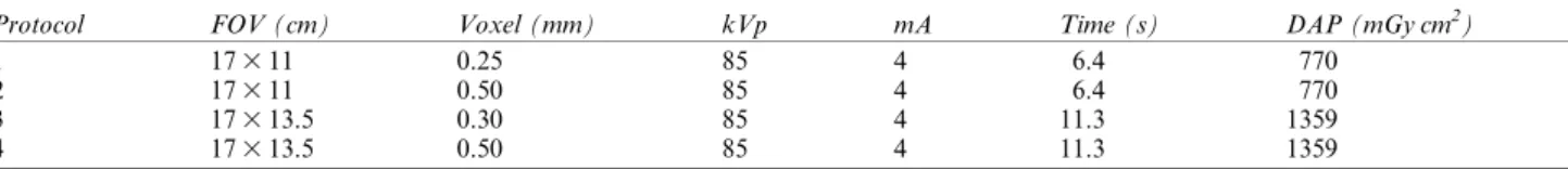
(1) The first one, proposed by Quereshy et al, 2012, was performed using inVivo software (Anatomage, San Jose, CA). Initially, the 3D reconstruction in maximum intensity projection was created. Using landmarks, linear measurements corresponding to the cleft width, height and facial-palatal length were collected. These values were used to calculate the estimated volume of the defect (Figure 1).
Table 1 Imaging protocols used in the research
Protocol FOV (cm) Voxel (mm) kVp mA Time (s) DAP (mGy cm2)
1 17311 0.25 85 4 6.4 770
2 17311 0.50 85 4 6.4 770
3 17313.5 0.30 85 4 11.3 1359
4 17313.5 0.50 85 4 11.3 1359
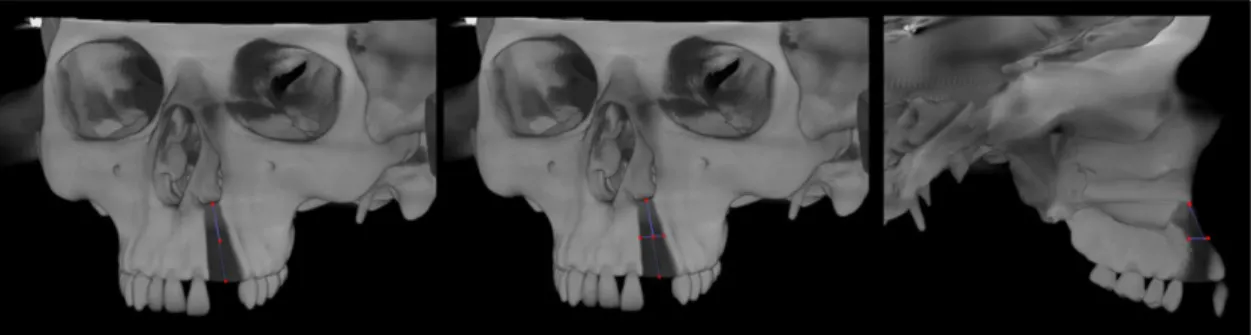
(2) The second method, first described by Feichtinger et al, 2007, was also executed using inVivo software (Anatomage). In this technique, the defects were outlined on each axial slice and the area was automatically given. After determination of the area in every slice that comprised the cleft, the volume was calculated using the following formula: Volume5[A1
3S]1[A23S]1 …1[An3S] (A, area; S, thickness of the slice; andn, number of slices) (Figure 2). (3) The third method consisted of a 3D evaluation using
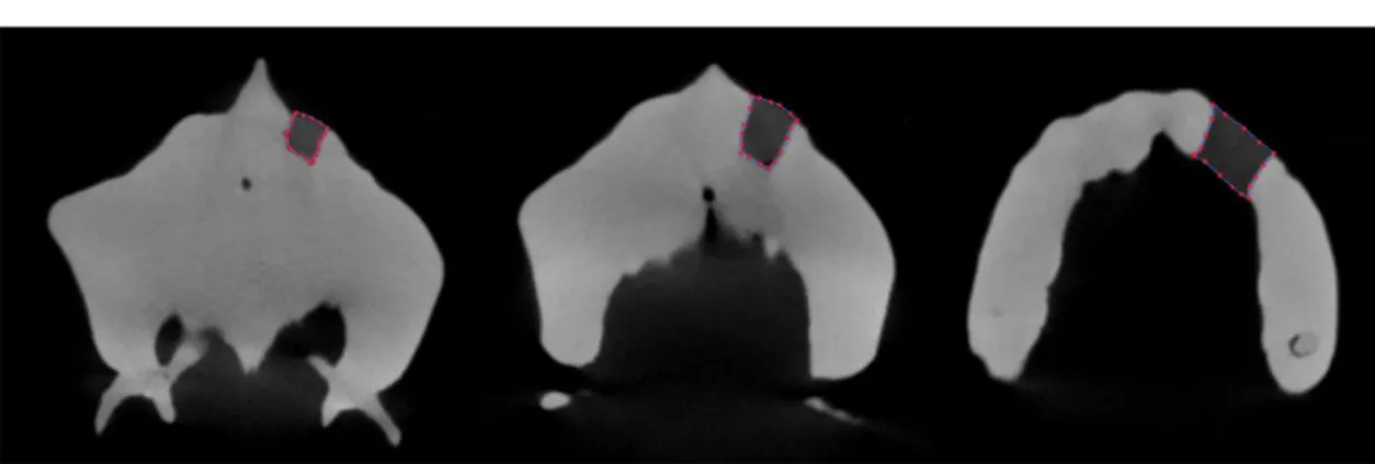
Mimics® software (v. 16.0, Materialise Medical, Leuven, Belgium). For this, the threshold compris-ing the region of interest (cleft/wax) was defined. The volumetric region of interest was cropped to comprise only the cleft for posterior segmentation. After threshold selection, 3D editing was used to obtain refined surfaces of the segmentation, resulting in a volumetric region of interest that was rendered into a shaded surface mesh, and the segmented volume was calculated (Figure 3).
In order to compare the methods, the calculation of the real volume of the wax was selected as the gold standard of the study. For this assessment, the skull was immersed in warm water and the entire wax was care-fully removed using a dental floss. Next, a graduated cylinder was filled with water up to a reference line. The entire wax model was then submerged into the cylinder and the new volume occupied by the water was delimited. The real volume was measured using Archi-medes’ principle of water displacement. This analysis was performed twice for each wax model by one well-trained evaluator using a digital caliper. After a month of interval, the evaluations of all CBCT images were repeated to evaluate intra-observer reliability.
After data collection, the values obtained from each method using different acquisition protocols were com-pared with the real volume of the wax using two-way ANOVA and Tukey’s test, using BioEstat for Windows (v. 5.0; BioEstat, Bel´em, PA, Brazil). For intra-observer analysis, Pearson’s correlation coefficient was performed using SigmaStat® for Windows (v. 3.5; Systat Software Inc., Erkrath, Germany). The level of significance was set at p,0.05. The values for agreement evaluation were interpreted as poor (0.00–0.20), fair (0.21–0.40),
moderate (0.41–0.60), substantial (0.61–0.80) or almost perfect (0.811).
Results
The final sample of the study consisted of 12 CBCT volumes of 3 skulls with simulated unilateral alveolar defects scanned under 4 different protocols each, vary-ing the FOV and voxel sizes.
Intra-observer values indicated almost perfect agree-ment for all methods (Method 1 : 0.98, Method 2 : 0.99 and Method 3 : 0.98).
The overall values of the volumetric measurements for each skull and method are summarized in Table 2. In relation to the comparison among the methods for assessment of the cleft volume, Methods 2 and 3 did not differ from the gold standard (p.0.05). Conversely, Method 1 presented statistically significant over-estimated values (p,0.01).
In relation to the influence of different acquisition parameters, no differences were found among the se-lected protocols. The variation of FOV and voxel sizes did not influence the reliability of the methods (p.0.05).
Discussion
The increased utilization of faster CT examinations of lower cost allowed the increase in referrals for CBCT examinations by dental practitioners, including during pre-operative and treatment planning of the patients with cleft.14,15 In addition, these examinations enable reduc-tions up to 12.3-fold in the effective dose to which the patients are exposed when compared with MSCT, justi-fying the indication of CBCT examinations in cases of patients with cleft, according to SEDENTEXCT guide-lines.16,17In the present study, the two different adopted FOV resulted in variation in exposure doses. The smallest FOV had a dose–area product of 770 mGy cm2,while the largest one presented an estimation of 1359 mGy cm2 (Table 1). It is true that these values are estimated dose values provided by the manufacturer without weighting based on tissues. Even so, such values can provide an
overall idea for the health practitioners and improve ex-amination indication and selection of parameters.
Based on our results, the variation of FOV did not influence the methods’performance (p.0.05). For this reason, a protocol of lower dose exposure should be selected in accordance with SEDENTEXCT guidelines. Also, in several CBCT units, such as the one used in the present study, when a larger FOV is used, the scanning time increases and more likely the patient will move during image acquisition, leading to artefact movements and image degradation. In our study, only the two ex-tended FOVs available in the CBCT unit were evalu-ated. They are the indicated volume for scanning patients with clefts in the Craniofacial Center where the study was conducted, as well as in many other centers. It allowed the clinical and practical application of our results, being the smallest volume size compatible with the situation and most indicated for scanning the 173 11 cm FOV.
In relation to the voxel size, its influence on image resolution and diagnostic ability of different diseases has been the object of study of several reports.9In the evaluated CBCT unit, two options of voxel size per FOV are provided by the acquisition software. When using the 17311 cm FOV, 0.25 and 0.5 mm voxel sizes can be selected; and with the 17313.5 cm FOV, 0.3 and 0.5 mm voxel sizes can be selected. In our study, we tested all the available possibilities, and as observed for
FOV, the voxel size did not influence the performance of all three methods.
It is well known that CBCT linear measurements are accurate and do not show difference in relation to an-atomical truths,18,19 even when acquisition parameters are altered in a range of acceptable image quality.20,21 These reports of the literature corroborate our findings that the imaging parameters did not influence the results when the methods that use linear measurements were performed (Methods 1 and 2). However, as little has been studied regarding the influence of acquisition parameters in the accuracy of 3D models for volumetric assessment, the present study aimed to evaluate the scanning possibilities in order to provide a better in-dication of these examinations for this purpose. The results observed disagree with the findings of da Silveira et al,222014, who detected that protocols with different voxel sizes in CBCT significantly changed volumetric measurements. However, these authors evaluated such measures in simulated internal root resorptions, lesions of much smaller dimensions than the defects assessed in our study. It is possible, or even probable, that voxel sizes do have an influence on accuracy in small defects. Furthermore, the lack of influence observed in Method 3 can be the foundation for further research for prac-titioners of craniofacial teams that aim to incorporate the segmentation in the treatment of patients with cleft. Moreover, no reports in the available literature
Figure 2 Method 2: measurement of the area of the cleft slice by slice on axial view.
regarding the influence of acquisition protocols in the assessment of cleft volumes were found.
Regarding the comparison of methods for volume assessment of clefts, Method 1 presented overestimated values, not being suitable for this purpose. This result disagrees with the reports of the study that suggested the method.11 However, differently from our study, this previous report did not have a gold standard with known dimension for comparison. An overestimation of the necessary amount of bone for ABG would lead to unnecessary bone removal from the donor site, in-creasing morbidity.1
Conversely, Methods 2 and 3 proved reliable in our study. This finding is in agreement with previous studies.4,12,13Nonetheless, Feichtinger et al, 2007, and Choi et al, 2012, used images from patients who un-derwent ABG, and did not have a gold standard for comparison of the obtained volume and proper evalu-ation of the method itself. Even with the reports of Albuquerque et al, 2011, regarding the reliability of these methods using the same gold standard as our study, it was important to evaluate this methodology using CBCT images, since the technology is becoming increasingly more present in the treatment of clefts. Along with Albuquerque et al, 2011, the cited studies evaluated the method in MSCT images, which vary significantly in quality and resolution when compared with CBCT, which could lead to different results in different imaging modalities.
With reference to Method 3, no reports were found using the exact same method and software. However, studies that evaluate cleft volumes by 3D methods also found values that did not differ from a gold stan-dard.23,24 The edition of boundaries and volume of in-terest in previous reports was done slice by slice in the study of Amirlak et al, 2013, and applying an algorithm in the study of Kasaven et al, 2013. These studies were similar to ours in that they used simulated defects in skulls but differed from our study in the methodology
and software selected for volume assessment. The use of skulls for this type of research is a double-edged sword: the possibility of having a gold standard with accurate known dimensions at one side; on the other, the po-tential shortcoming of reproducing the true clinical sit-uation. In studies similar to the present one, the evaluation of a method’s reliability by a gold standard is mandatory, which makes the use of models such as skulls necessary and is also a limitation of the study at the same time.
Currently, the validation of assessments based on segmentation and 3D models of CBCT examinations is essential considering that such images were easily pro-duced only by MSCT. The production and evaluation of these virtual models is a big advance for all types of treatment. In addition, Hamada et al,25 2005, stated that these images would be especially advantageous for pre-operative imaging of the morphology of residual alveolar clefts.
Conclusion
CBCT volumes proved reliable for the volumetric as-sessment of alveolar cleft defects, when using methods of area determination in axial reconstructions and seg-mentation with 3D rendering of the volume, regardless of FOV and voxel sizes, in the evaluated methodology. It may be possible to improve surgical results by knowing the exact volume of grafting material needed prior to the surgery itself.
Acknowledgments
We are grateful to CAPES Foundation (Brazil) for the scholarship support and to the University of North Carolina at Chapel Hill (NC, USA) for the technical support to the first author while she was a visiting postgraduate fellow.
References
1. Cobourne MT.Cleft lip and palate: epidemiology, aetiology, and treatment (vol 16). Front oral biol. Basel: Karger; 2012. 2. Bell WH, Proffit WR, White RP. Residual alveolar and palatal
cleft. In: Bell WH, Proffit WR, White RP, eds.Surgical correction of dentofacial deformities. Philadelphia, PA: WB Saunders; 1980. pp. 1329–67.
3. Turvey TA, Vig K, Moriarty J, Hoke J. Delayed bone grafting in the cleft maxilla and palate: a retrospective multidisciplinary
analysis.Am J Orthod1984;86: 244–56. doi:10.1016/0002-9416 (84)90376-2
4. Choi HS, Choi HG, Kim SH, Park HJ, Shin DH, Jo DI, et al. Influence of the alveolar cleft type on preoperative estimation using 3D CT assessment for alveolar cleft.Arch Plast Surg2012;
39: 477–82. doi:10.5999/aps.2012.39.5.477
5. Aurouze C, Moller KT, Bevis RR, Rehm K, Rudney J. The presurgical status of the alveolar cleft and success of secondary
Table 2 Average volumes of the simulated defects according to the methods (mm3)
Gold standard SD Method 1 SD Method 2 SD Method 3 SD
Skull 1 1384.40 A 0.10 1410.47 B 58.03 1355.57 A 37.39 1406.58 A 56.82
Skull 2 1582.80 A 0.08 2540.19 B 125.22 1471.84 A 37.89 1501.71 A 51.57
Skull 3 760.70 A 0.02 1117.70 B 46.75 680.25 A 14.61 681.33 A 28.66
Average volume 1242.6 A 428.99 1689.45 B 751.16 1169.22 A 427.43 1196.54 A 448.71
SD, standard deviation.
bone grafting. Cleft Palate Craniofac J 2000; 37: 179–84. doi:
10.1597/1545-1569(2000)036,0179:TPSOTA.2.3.CO;2
6. Bergland O, Semb G, Abyholm FE. Elimination of the residual alveolar cleft by secondary bone grafting and subsequent ortho-dontic treatment.Cleft Palate J1986;23: 175–205.
7. Helms JA, Speidel TM, Denis KL. Effect of timing on long-term clinical success of alveolar cleft bone grafts.Am J Orthod Den-tofacial Orthop 1987; 92: 232–40. doi: 10.1016/0889-5406(87) 90417-3
8. Long RE Jr, Spangler BE, Yow M. Cleft width and secondary alveolar bone graft success. Cleft Palate Craniofac J1995; 32: 420–7. doi: 10.1597/1545-1569(1995)032,0420:CWASAB.2.3. CO;2
9. Spin-Neto R, Gotfredsen E, Wenzel A. Impact of voxel size variation on CBCT-based diagnostic outcome in dentistry: a sys-tematic review.J Digit Imaging2013;26: 813–20. doi:10.1007/ s10278-012-9562-7
10. Kamburoglu K, Murat S, Kolsuz E, Kurt H, Y ¨uksel S, Paksoy C.˘ Comparative assessment of subjective image quality of crosssec-tional cone-beam computed tomography scans.J Oral Sci2011;
53: 501–8. doi:10.2334/josnusd.53.501
11. Quereshy FA, Barnum G, Demko C, Horan M, Palomo JM, Baur DA, et al. Use of cone beam computed tomography to volumetrically assess alveolar cleft defects–preliminary results.J Oral Maxillofac Surg 2012; 70: 188–91. doi: 10.1016/j. joms.2011.01.027
12. Feichtinger M, Mossb ¨ock R, K¨archer H. Assessment of bone resorption after secondary alveolar bone grafting using three-dimensional computed tomography: a three-year study. Cleft Palate Craniofac J2007;44: 142–8. doi:10.1597/06-047.1
13. Albuquerque MA, Gaia BF, Cavalcanti MG. Oral cleft volu-metric assessment by 3D multislice computed tomographic images.Int J Oral Maxillofac Surg2011;40: 1280–8. doi:10.1016/ j.ijom.2011.05.015
14. Mozzo P, Procacci C, Tacconi A, Martini PT, Andreis IA. A new volumetric CT machine for dental imaging based on the cone-beam technique: preliminary results.Eur Radiol1998;8: 1558–64. doi:10.1007/s003300050586
15. Scarfe WC, Farman AG, Sukovic P. Clinical applications of cone-beam computed tomography in dental practice.J Can Dent Assoc
2006;72: 75–80.
16. Ludlow JB, Ivanovic M. Comparative dosimetry of dental CBCT devices and 64-slice CT for oral and maxillofacial radiology.Oral
Surg Oral Med Oral Pathol Oral Radiol Endod2008;106: 106–14. doi:10.1016/j.tripleo.2008.03.018
17. SEDENTEXCT guidelines on CBCT for Dental and Maxillofa-cial Radiology. Radiation protection no. 172. 2012. Available from: http://www.sedentexct.eu/content/guidelines-cbct-dental-and-maxillofacial-radiology
18. Ganguly R, Ruprecht A, Vincent S, Hellstein J, Timmons S, Qian F. Accuracy of linear measurement in the Galileos cone beam computed tomography under simulated clinical conditions. Den-tomaxillofac Radiol 2011; 40: 299–305. doi: 10.1259/dmfr/ 72117593
19. Stratemann SA, Huang JC, Maki K, Miller AJ, Hatcher DC. Comparison of cone beam computed tomography imaging with physical measures.Dentomaxillofac Radiol2008;37: 80–93. doi:
10.1259/dmfr/31349994
20. Torres MG, Campos PS, Segundo NP, Navarro M, Cruso´e-Rebello I. Accuracy of linear measurements in cone beam com-puted tomography with different voxel sizes.Implant Dent2012;
21: 150–5. doi:10.1097/ID.0b013e31824bf93c
21. Vasconcelos TV, Neves FS, Queiroz de Freitas D, Campos PS, Watanabe PC. Influence of the milliamperage settings on cone beam computed tomography imaging for implant planning.Int J Oral Maxillofac Implants 2014; 29: 1364–8. doi: 10.11607/ jomi.3524
22. da Silveira PF, Vizzotto MB, Liedke GS, da Silveira HL, Montagner F, da Silveira HE. Detection of vertical root fractures by conven-tional radiographic examination and cone beam computed tomog-raphy—an in vitro analysis.Dent Traumatol 2013;29: 41–6. doi:
10.1111/j.1600-9657.2012.01126.x
23. Kasaven CP, Ivekovic S, McIntyre GT, Gillgrass T, Thomson DA, Menhinick A, et al. Validation of the volumetric measure-ment of a simulated maxillary alveolar bone defect using cone-beam computed tomography.Cleft Palate Craniofac J2013;50: e115–120. doi:10.1597/12-161
24. Amirlak B, Tang CJ, Becker D, Palomo JM, Gosain AK. Volu-metric analysis of simulated alveolar cleft defects and bone grafts using cone beam computed tomography. Plast Reconstr Surg
2013;131: 854–9. doi:10.1097/PRS.0b013e3182818e4f