Comparative therapeutic ef
fi
cacy of remdesivir and
combination lopinavir, ritonavir, and interferon beta
against MERS-CoV
Timothy P. Sheahan
1,5
*, Amy C. Sims
1,5
, Sarah R. Leist
1
, Alexandra Schäfer
1
, John Won
1
, Ariane J. Brown
1
,
Stephanie A. Montgomery
2
, Alison Hogg
3
, Darius Babusis
3
, Michael O. Clarke
3
, Jamie E. Spahn
3
,
Laura Bauer
3
, Scott Sellers
3
, Danielle Porter
3
, Joy Y. Feng
3
, Tomas Cihlar
3
, Robert Jordan
3
,
Mark R. Denison
4
& Ralph S. Baric
1
*
Middle East respiratory syndrome coronavirus (MERS-CoV) is the causative agent of a
severe respiratory disease associated with more than 2468 human infections and over 851
deaths in 27 countries since 2012. There are no approved treatments for MERS-CoV infection
although a combination of lopinavir, ritonavir and interferon beta (LPV/RTV-IFNb) is
cur-rently being evaluated in humans in the Kingdom of Saudi Arabia. Here, we show that
remdesivir (RDV) and IFNb have superior antiviral activity to LPV and RTV in vitro. In mice,
both prophylactic and therapeutic RDV improve pulmonary function and reduce lung viral
loads and severe lung pathology. In contrast, prophylactic LPV/RTV-IFNb slightly reduces
viral loads without impacting other disease parameters. Therapeutic LPV/RTV-IFNb
improves pulmonary function but does not reduce virus replication or severe lung pathology.
Thus, we provide in vivo evidence of the potential for RDV to treat MERS-CoV infections.
https://doi.org/10.1038/s41467-019-13940-6
1Department of Epidemiology, University of North Carolina at Chapel Hill, Chapel Hill, NC, USA.2Department of Pathology & Laboratory Medicine,
T
he coronavirus (CoV) family has a propensity for
emer-gence into new hosts often causing novel severe disease. In
2012, Middle East respiratory syndrome coronavirus
(MERS-CoV), was discovered as the causative agent of a severe
respiratory syndrome in the Kingdom of Saudi Arabia (KSA), has
since caused at least 2468 cases and 851 deaths globally
1.
MERS-CoV is endemic in camels, the zoonotic reservoir host, with
evidence of infection going back at least 30 years
2. Camels in the
Middle East and perhaps in East Africa continue to seed human
infections which may require hospitalization especially in aged
individuals with preexisting comorbidities
1,3,4. Similar to severe
acute respiratory syndrome CoV (SARS-CoV), MERS-CoV has
spread to over 27 countries via air travel of infected people
5. In
2014, a single imported case caused an outbreak of 186 cases in
South Korea, while a more recent case imported from the Middle
East was contained as a result of rapid implementation of public
health measures
6. MERS-CoV continues to cause human
infec-tions globally and thus is listed as a priority pathogen with
pandemic potential by World Health Organization (WHO) and
the Coalition for Epidemic Preparedness Innovations (CEPI).
Presently, there are no approved treatments for MERS-CoV or
any other human CoV.
Emerging viral diseases typically have very few if any effective
treatment options. As such, treatments designed and approved
for other diseases are administered to patients with emerging viral
syndromes empirically based on limited clinical or laboratory
data. Multiple U.S. Food and Drug Administration (FDA)
approved therapies have been evaluated for antiviral activity
against MERS-CoV in vitro including lopinavir (LPV), ritonavir
(RTV), and interferon beta (IFNb). LPV is a human
immuno-de
fi
ciency virus 1 (HIV-1) protease inhibitor that is usually
combined with RTV to increase LPV half-life through the
inhi-bition of cytochrome P450
7. Although the antiviral activity of
LPV against MERS-CoV has been reported in Vero cells
(con-centration causing a 50% reduction in replication (EC
50)
=
8 µM),
other studies report complete inactivity similar to that of RTV
8,9.
In contrast, studies evaluating the antiviral activity of type I and
type II interferons have reported IFNb as the most potent
interferon (EC
501.37
–
17 IU/mL) in reducing MERS-CoV
repli-cation in vitro
8,10. The only in vivo study assessing the
ther-apeutic ef
fi
cacy of LPV/RTV or IFNb against MERS-CoV
published thus far was performed in common marmosets where
modest improvements in clinical outcomes were noted
11. In
human MERS-CoV patients, two published case reports describe
con
fl
icting results on the use of a combination of LPV/RTV,
pegylated interferon, and ribavirin with one of two patients
surviving
12,13. To this end, a randomized control trial (MIRACLE
Trial) aimed at conclusively determining if LPV/RTV-IFNb
improves clinical outcomes in MERS-CoV patients was initiated
in 2016 and has thus far enrolled 76 patients in KSA
14,15.
Remdesivir (RDV, GS-5734) is a broad-spectrum antiviral
nucleotide prodrug with potent in vitro antiviral activity against a
diverse panel of RNA viruses such as Ebola virus (EBOV),
Marburg, MERS-CoV, SARS-CoV, respiratory syncytial virus
(RSV), Nipah virus (NiV), and Hendra virus
16–18. The
mechan-ism of RDV
’
s anti-MERS-CoV activity is likely through
pre-mature termination of viral RNA transcription as shown in
biochemical assays using recombinant EBOV, NiV, and RSV
polymerases
18–20. In primary human lung epithelial cell cultures,
RDV is potently antiviral against circulating contemporary
human CoVs, SARS-CoV (EC
50=
0.07 µM), MERS-CoV (EC
50=
0.07 µM), and related zoonotic bat CoVs
17,21. We recently
reported that therapeutic RDV improves disease outcomes and
reduces viral loads in SARS-CoV-infected mice
17. Since similar
studies had not been performed with MERS-CoV, we generated a
transgenic mouse with a humanized MERS-CoV receptor
(dipeptidyl peptidase 4,
hDPP4
) and deleted for carboxylesterase
1c (
Ces1c
) to improve the pharmacokinetics of nucleotide
pro-drugs such that it better approximates the drug exposure pro
fi
le
in humans
22. Here, we show that RDV provides superior antiviral
activity against MERS-CoV in vitro and in vivo as compared with
LPV/RTV-IFNb. In addition, RDV was the only therapeutic
treatment to signi
fi
cantly reduce pulmonary pathology. Thus, we
provide in vivo evidence of the potential for RDV to treat
MERS-CoV infections.
Results
RDV and IFNb have superior antiviral activity to LPV and
RTV
. We utilized a recombinant MERS-CoV engineered to
express a reporter nanoluciferase (MERS-nLUC) for our in vitro
antiviral activity assays. To ensure that our reporter virus behaved
similarly to WT MERS-CoV, we
fi
rst demonstrated that
MERS-nLUC and wild-type (WT) MERS-CoV EMC 2012 strain
repli-cate to similar levels in the absence of drug treatment and are
similarly susceptible to the antiviral activity of RDV (WT EC
50=
0.12 µM; MERS-nLUC EC
50=
0.09 µM) in the human lung
epi-thelial cell line, Calu-3 (Supplementary Fig. 1). These data are in
agreement with those for WT MERS Jordan strain (EC
50=
0.3 µM) reported by Warren et al.
18. Thus, future reporter virus
data should be representative of WT MERS-CoV.
We then performed parallel antiviral assays in Calu-3 cells with
MERS-nLUC comparing LPV, RTV, IFNb, and RDV (Fig.
1;
Supplementary Fig. 2)
17. Similar to our previous reports, RDV
showed potent inhibition of MERS-CoV replication with a EC
50of 0.09 µM, no observable cytotoxicity up to 10
μM and a
selectivity index (SI
=
EC
50/CC
50) > 100
17. (Fig.
1a). In contrast,
the respective EC
50values generated for LPV and RTV were 11.6
and 24.9 µM with CC
50values >50 µM (Fig.
1b, c). Thus, the SI
for LPV and RTV was > 4.3 and > 2, respectively. Combination
LPV and RTV (LPV:RTV, 4.6:1 molar ratio) is currently under
evaluation in the MIRACLE trial
14. The antiviral activity of LPV/
RTV (EC
50=
8.5 µM) was similar to LPV alone (EC
50=
11.6 µM,
P
=
0.43, Wilcoxon matched-pairs signed rank test), suggesting
the effect was largely driven by LPV (Fig.
1d). We found potent
inhibition of MERS-CoV with IFNb (EC
50=
175 international
units (IU)/mL) (Fig.
1e), CC
50values >2800 IU/mL and an SI >
16. Together, these data demonstrate that RDV and IFNb have
superior in vitro antiviral activity compared with LPV and RTV,
and that RTV does not signi
fi
cantly enhance the antiviral activity
of LPV in vitro.
IFNb activity is not improved when combined with LPV/RTV
.
Cell culture medium and human plasma have different
con-centrations and types of proteins, which directly affects the levels
of biologically available free drug unbound to protein
compli-cating the comparison of drug levels the respective systems
23. To
address this issue, we utilized comparative equilibrium dialysis
(CED) to determine the differences in free unbound drug between
human plasma and cell culture medium revealing that the
max-imal plasma concentration (
C
max) in human plasma (15 µM) had
the same amount of free unbound LPV as 5 µM LPV in 10% FBS
containing cell culture medium. Thus, we combined LPV (5 µM)
and RTV (1.09 µM) at a
fi
xed molar ratio of 4.6:1 in combination
with increasing concentrations of IFNb. The antiviral activity of
the LPV/RTV-IFNb combination (EC
50=
160 IU/mL) was
indistinguishable from that of IFNb alone (EC
50=
175 IU/mL)
A MERS-CoV mouse model for the testing of nucleotide
pro-drugs
. Unlike humans, mice have high levels of a serum esterase
(carboxylesterase 1c,
Ces1c
) that drastically reduces the stability of
RDV in mice requiring ef
fi
cacy studies be performed in
Ces1c
−/−mice to better approximate the pharmacokinetics and drug
exposure pro
fi
le in humans
17. MERS-CoV infection of standard
laboratory mice is prevented due to differences in human and
mouse dipeptidyl peptidase 4 (DPP4), the entry receptor for
MERS-CoV. To enable testing of RDV in mice, we bred
Ces1c
−/−mice with mice harboring a modi
fi
ed
DPP4
humanized via
CRISPR/Cas9 at residues 288 and 330 (
hDPP4
). The resultant
Ces1c
−/−hDPP4
mice had indistinguishable virus replication and
pathogenesis from
hDPP4
mice when infected with MERS-CoV
(Supplementary Fig. 3)
22,24.
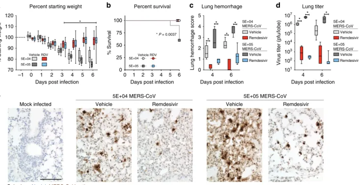
Prophylactic RDV diminishes MERS-CoV replication and
disease
. Using the new
Ces1c
−/−hDPP4
mouse model, we sought
to determine if prophylactic RDV could ameliorate MERS-CoV
disease. As shown in Fig.
2a, prophylactic RDV (25 mg/kg, BID)
administered 1 day prior to infection signi
fi
cantly diminished
MERS-CoV-induced weight loss in mice infected with 5E
+
04
(
P
< 0.0001, two-way ANOVA with Tukey
’
s multiple comparison
test) or 5E
+
05 plaque-forming units (pfu) (
P
< 0.0001, two-way
ANOVA with Tukey
’
s multiple comparison test) as compared
with similarly infected vehicle-treated animals. Moreover, RDV
administered prior to infection also prevented mortality (
P
=
0.0037, Mantel
–
Cox test) in those administered a lethal dose (i.e.,
5E
+
05 pfu) (Fig.
2b). In contrast to vehicle-treated animals, lung
hemorrhage was signi
fi
cantly reduced (
P
< 0.0001, two-way
ANOVA with Tukey
’
s multiple comparison test) with RDV
prophylaxis (Fig.
2c)
22,25,26. Importantly, RDV prophylaxis
sig-ni
fi
cantly reduced virus lung titers > 3 logs on both 4 (5E
+
04
P
=
0.0240, 5E
+
04
P
=
0.0001, two-way ANOVA with Sidek
’
s
multiple comparison test) and 6 days post infection (dpi) (5E
+
05
P
=
0.0001, two-way ANOVA with Sidek
’
s multiple
compar-ison test) (Fig.
2d), which was corroborated by viral antigen
labeling in lung tissue sections (Fig.
2e).
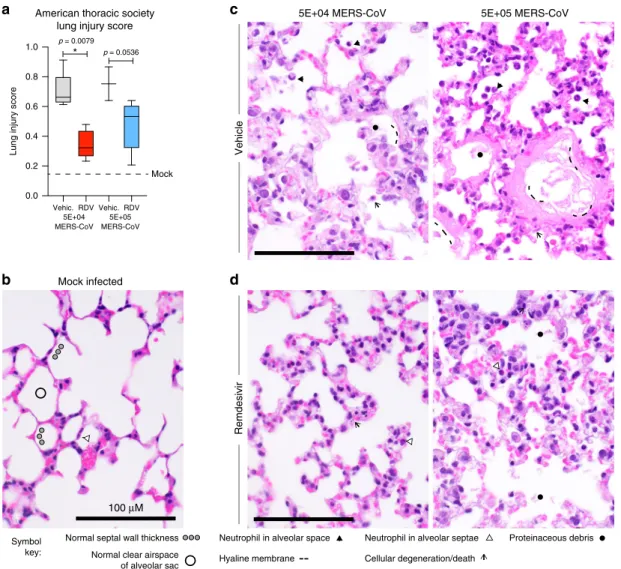
RDV prophylaxis reduces features of ALI
. The American
Thoracic Society (ATS) issued a consensus document with
de
fi
ned parameters and tools to more accurately translate small
animal models of acute lung injury (ALI) to the human
condi-tion
26. Using the ATS Lung Injury Scoring System designed to
quantitate histopathological features of ALI (Fig.
3a), we blindly
scored
fi
elds of hematoxylin and eosin stained lung tissue sections
from the mice in Fig.
2
for the following features: neutrophils in
the alveolar and interstitial space, hyaline membranes,
protei-naceous debris
fi
lling the air spaces, and alveolar septal
thicken-ing
26. As compared with control mice, the ATS lung injury scores
were signi
fi
cantly reduced in RDV-treated mice (
P
=
0.0079,
Mann
–
Whitney test) infected with 5E
+
04 pfu MERS-CoV and
approached statistical signi
fi
cance (
P
=
0.0536, Mann
–
Whitney
test) in those infected with the higher virus dose (5E
+
05 pfu)
(Fig.
3a). In Fig.
3, we also show examples of the quantitated
pathological features. In normal healthy mock-infected control
mice (Fig.
3b), the alveolar air spaces are free of debris and
in
fl
ammatory cells, the walls (i.e., septae) of the alveolar sac are
thin which facilitates ef
fi
cient gas exchange and rare neutrophils
in circulation are seen in the capillaries within alveolar septae, but
not in the air spaces. In contrast, vehicle-treated
MERS-CoV-infected animals in both viral dosage groups (Fig.
3c) had
mul-tiple histologic features of ALI including notable immune cell
in
fi
ltration into the alveolar septae and resultant septal wall
thickening, scattered degenerating and dying cells, proteinaceous
debris in the air spaces resulting from capillary leakage some of
which is organized into hyaline membranes and neutrophils in
alveolar septae as well as in the air spaces. Interestingly, the early
hyaline membranes (i.e., thin pink material lining alveoli) were
noted in vehicle-treated animals infected with the lower dose of
MERS-CoV (Fig.
3c, left), while more developed hyaline
mem-branes (i.e., thick pink material lining alveoli) were observed in
vehicle-treated animals infected with the higher dose of virus
(Fig.
3c, right). Importantly, the air spaces of RDV-treated
ani-mals infected with the lower dose of MERS-CoV (Fig.
3d, left)
predominantly lacked cellular debris, immune cells, and hyaline
membranes although alveolar septal thickening due to increased
a
b
c
d
e
f
0.1 1 10 100
–30 0 30 60 90 120 –30 0 30 60 90 120
Lopinavir [µM] Ritonavir [µM]
% Inhibition
EC50 11.6 µM
EC50 175 IU/ml EC50 160 IU/ml % Cytoxicity Lopinavir
Interferon beta Lopinavir:Ritonavir + Interferon beta Remdesivir
0.1 1 10 100
–30 0 30 60 90 120 –30 0 30 60 90 120 % Inhibition % Cytoxicity
0.1 1 10 100
–30 0 30 60 90 120 –30 0 30 60 90 120 % Inhibition % Cytoxicity Lopinavir:Ritonavir Ritonavir EC50 24.9 µM
Lopinavir:Ritonavir (4.6:1) [µM]
EC50 8.5 µM
10 100 1000
–30 0 30 60 90 120 –30 0 30 60 90 120
Interferon beta [IU/mL] 6 µM Lopinavir:Ritonavir + Interferon beta [IU/mL]
% Inhibition
% Cytoxicity
10 100 1000
–30 0 30 60 90 120 –30 0 30 60 90 120 % Inhibition % Cytoxicity
0.01 0.1 1 10
–30 0 30 60 90 120 –30 0 30 60 90 120
Remdesivir [µM]
% Inhibition
% Cytoxicity
EC 50 0.09 µM
Fig. 1 RDV and IFNb have superior antiviral activity to LPV and RTV.Graphs depict mean % inhibition of MERS-CoV replication (leftY-axis) and %
cytotoxicity (rightY-axis) of antivirals. Calu-3 cells were infected in sextuplicate with MERS-CoV nanoluciferase (nLUC) at a multiplicity of infection (MOI)
of 0.08 in the presence of a dose response of drug for 48 h, after which replication was measured through quantitation of MERS-CoV–expressed nLUC.
immune cells was observed. RDV-treated animals infected with
the higher dose of MERS-CoV (Fig.
3d, right) similarly lacked
debris and in
fl
ammatory in
fi
ltrates in the air spaces, but exhibited
increased alveolar septal thickening. Thus, our blind pathological
evaluation of lung tissue sections using the ATS Lung Injury
Scoring System demonstrates that prophylactic RDV diminished
the pathological features of ALI in MERS-CoV-infected mice
26.
The minimal effect of prophylactic LPV/RTV-IFNb on
MERS-CoV
. Prophylactic studies provide a best case scenario to evaluate
in vivo antiviral activity since time is given for the metabolization
and accumulation of antiviral agents within cells targeted by virus
prior to infection. To determine if prophylactic LPV/RTV-IFNb
improved outcomes following MERS-CoV infection, we
fi
rst
con
fi
rmed that subcutaneous human equivalent doses (H.E.D.) of
IFNb exerted a biological effect in mice. Administration of a 1 × ,
2.5x, or 25x H.E.D. of mouse IFNb rapidly induced
dose-dependent expression of interferon stimulated gene (ISG),
Mx1
,
in peripheral blood mononuclear cells (PBMCs) in
Ces1c
−/−mice
(Supplementary Fig. 4a). Importantly, the kinetics of
Mx1
gene
expression in target organ of MERS-CoV, the lung, was similar in
both
Ces1c
−/−and
Ces1c
−/−hDPP4
mice (Supplementary
Fig. 4b). Similarly, a single dose of IFNb signi
fi
cantly induced
sustained expression (
P
< 0.05, two-way ANOVA with Sidek
’
s
multiple comparison test) of interferon gamma-induced protein
10 (IP-10, CXCL-10) in the serum of both strains of mice
(Supplementary Fig. 4c). Thus, we observed an expected
ISG response in the blood and lung tissue of both
Ces1
−/−and
Ces1
−/−hDPP4
mice following IFNb treatment.
We then evaluated if LPV/RTV-IFNb prophylaxis could
improve outcomes in
Ces1
−/−hDPP4
mice infected with 5E
+
04 pfu MERS-CoV. We compared vehicle (oral: propylene glycol
and ethanol, subcutaneous: PBS) to three different treatment
scenarios, including LPV/RTV-IFNb high (25x H.E.D. IFNb),
LPV/RTV-IFNb low (1x H.E.D. IFNb), or IFNb-high alone (25x H.
E.D. IFNb) (Fig.
4). Prophylactic RDV and vehicle were included
as controls. Since ISG expression peaks 2
–
4 h after IFNb
administration (Supplementary Fig. 4), we initiated IFNb dosing
2 h prior to MERS-CoV infection to maximize the potential
antiviral effect. Similar to our previous studies, RDV (25 mg/kg,
BID) or vehicle were administered subcutaneously every 12 h to
obtain exposures in mice similar to that observed in humans
17.
The dosing levels and frequencies of LPV/RTV (oral once daily)
and IFNb (subcutaneously every other day) were chosen to mirror
those in the MIRACLE trial
27. Unlike RDV-treated mice (Fig.
4a),
vehicle, LPV/RTV-IFNb, or IFNb alone did not prevent weight loss
(Fig.
4b). In fact, animals administered IFNb alone lost
signi
fi
cantly more weight than vehicle (
P
=
0.01), LPV/RTV-IFNb
high (
P
=
0.0001) and low (
P
=
0.006, all two-way ANOVA with
Tukey
’
s multiple comparison test) groups (Fig.
4b). On 6 dpi, RDV
treatment reduced virus lung titers the most (>3 log reduction,
vehicle median
=
1.4E
+
05 pfu/lobe, RDV median
=
50 pfu/lobe)
(Fig.
4a), while those in LPV/RTV-IFNb low treated were modestly
reduced (~4-fold, median
=
5.5E
+
03 pfu/lobe) as compared with
its vehicle (median
=
2.3E
+
04 pfu/lobe) (
P
=
0.04, two-way
a
b
d
e
c
Mock infected
101 102 103 104 105 106 107
Virus titer (pfu/lobe)
Vehicle Remdesivir Vehicle Remdesivir
5E+04 MERS-CoV
Nuclei MERS-CoV antigen
Color key
5E+05 MERS-CoV
–1 0 1 2 3 4 5 6
70 80 90 100 110 120
Days post infection
% Starting weight
Percent starting weight
*
5E+04 5E+05
Vehicle RDV
0 1 2 3 4 5 6 0
25 50 75 100
Days post infection
% Survival
Percent survival
5E+04 5E+05
Vehicle RDV
4 6
0 1 2 3 4 5
Days post infection
4 6
Days post infection
Lung hemorrhage score
Lung hemorrhage
* *
*
* *
* *
Vehicle Remdesivir Vehicle Remdesivir 5E+04 MERS-CoV
5E+05 MERS-CoV Vehicle
Remdesivir Vehicle Remdesivir 5E+04 MERS-CoV
5E+05 MERS-CoV
Lung titer
* P = 0.0037
Fig. 2 Prophylactic RDV reduces MERS-CoV replication and disease. aPercent starting weight of 9–12-week-old male and femaleCes1c−/−hDPP4mice
prophylactically administered subcutaneous vehicle or remdesivir (RDV, 25 mg/kg) BID the day prior to infection with either 5E+04 (vehiclen=14, RDV
n=14) or 5E+05 (vehiclen=14, RDVn=15) plaque-forming units (pfu) MERS M35C4. Asterisks indicate statistically significant differences (P< 0.05)
as determined by two-way ANOVA and Tukey’s multiple comparison test.bPercent survival of each cohort and survival analysis by Mantel–Cox test (P<
0.05,Nper group noted ina).cLung hemorrhage scored on a scale of 0–4, where 0 is a normal pink healthy lung and 4 is a completely dark red lung. On 4
dpi,N=4/group, and on 6 dpi the remaining animals are plotted. Asterisks indicate statistically significant differences (P< 0.05) as determined by
two-way ANOVA and Tukey’s multiple comparison test.dMERS-CoV lung titer on 4 (N=4) and 6 dpi (all remaining animals). Asterisks indicate statistically
significant differences (P< 0.05) as determined by two-way ANOVA and Sidek’s multiple comparison test. Fora,c,d, the boxes encompass the 25th to
75th percentile, the line is at the median, while the whiskers represent the range.eHematoxylin (nuclei, blue) and immunostaining for MERS-CoV antigen
(brown) in lung tissue sections from 4 dpi. All photos were taken with the same magnification. The black bar indicates 100µM scale. Images from
ANOVA with Sidek
’
s multiple comparison test) (Fig.
4b). Upon
measuring pulmonary function by whole-body plethysmography
(WBP), we found only RDV prophylaxis improved pulmonary
function (Fig.
4a, b) (
P
=
0.002 to 0.0001, two-way ANOVA with
Sidek
’
s multiple comparison test). Interestingly, animals receiving
IFNb-alone had signi
fi
cantly worse pulmonary function than
companion groups at later times post infection (4
–
5 dpi,
P
=
<
0.05, two-way ANOVA with Tukey
’
s multiple comparison test). To
determine if initiation time or dose level would affect outcomes in
the IFNb only group, we performed a similar study but
administered IFNb at a lower dose (i.e., 1x H.E.D.) 24 h prior to
infection (Supplementary Fig. 5). Alteration of IFNb dose or time
of administration did not improve outcomes from those in Fig.
4,
and the modest improvement in virus titer seen with
LPV/RTV-IFNb noted above was not observed. Taken together, prophylactic
LPV/RTV-IFNb caused modest reductions in lung viral load in
one of two studies, but had minimal impact on other disease
metrics while IFNb alone did not impact virus replication and
exacerbated disease.
Therapeutic RDV reduces MERS-CoV replication and
pathol-ogy
. To more stringently assess the therapeutic potential of RDV
and LPV/RTV-IFNb and to better model the human scenario
where MERS-CoV patients most likely would initiate treatment
after infection, we performed a series of therapeutic ef
fi
cacy
studies in mice. We initiated the following treatments in
Ces1
−/−hDPP4
mice infected with 5E
+
04 pfu MERS-CoV on 1 dpi:
RDV or vehicle, LPV/RTV-IFNb low (1× human equivalent),
LPV/RTV-IFNb high (25× human equivalent) or their vehicles.
Dose route, amount, and frequency were similar to the
prophy-lactic studies above. Only therapeutic RDV substantially reduced
body weight loss (
P
=
0.019 to < 0.0001, two-way ANOVA with
Tukey
’
s multiple comparison test) (Fig.
5a, b) and lung
hemor-rhage on 6 dpi (Fig.
5c) (
P
< 0.0001, one-way ANOVA with
Kruskal
–
Wallis test). Similarly, only RDV treatment signi
fi
cantly
reduced virus lung titers on 6 dpi (vehicle median 7.8E
+
05 pfu/
lobe, RDV median 125 pfu/lobe,
P
=
0.0001, one-way ANOVA
with Kruskal
–
Wallis test) (Fig.
5d), which we corroborated with
viral antigen labeling in lung tissue sections (Fig.
5e). Similar
Mock infected5E+04 MERS-CoV 5E+05 MERS-CoV
100 µM American thoracic society
lung injury score
Vehicle
Remdesivir
Vehic. RDV 5E+04 MERS-CoV
Vehic. RDV 5E+05 MERS-CoV p = 0.0079
* p = 0.0536
0.0 0.2 0.4 0.6 0.8 1.0
Lung injury score
Mock
Symbol key:
Normal septal wall thickness Neutrophil in alveolar space Neutrophil in alveolar septae
Normal clear airspace
of alveolar sac Hyaline membrane Cellular degeneration/death
Proteinaceous debris
a
b
c
d
Fig. 3 Remdesivir prophylaxis reduces features of acute lung injury.The histological features of acute lung injury were blindly scored using the American Thoracic Society Lung Injury Scoring system creating an aggregate score for the following phenotypes: neutrophils in the alveolar and interstitial space,
hyaline membranes, proteinaceous debrisfilling the air spaces, and alveolar septal thickening. Three randomly chosen high power (×60)fields of diseased
lung were assessed per mouse. Representative images are shown for mock infected as well as those administered prophylactic vehicle or RDV and infected
with either 5E+04 or 5E+05 pfu mouse adapted MERS-CoV. The numbers of mice scored per group: Vehicle 5E+04 pfu MERS-CoVN=5, Vehicle
5E+05 pfu MERS-CoVN=3, RDV 5E+04 pfu MERS-CoVN=5, RDV 5E+05 pfu MERS-CoVN=5. Symbols identifying example features of disease
are indicated in thefigure. All images were taken at the same magnification. The black bar indicates 100µm scale. For the graph, the boxes encompass the
therapeutic studies (Supplementary Fig. 6) were performed but
with a lethal dose of MERS-CoV (5E
+
05 pfu) where no
treat-ment improved survival (Suppletreat-mentary Fig. 6a, d) or lung
hemorrhage (Supplementary Fig. 6b, e), but therapeutic RDV
signi
fi
cantly reduced lung viral load on 6 dpi (Supplementary
Fig. S6c) (
P
=
0.03, Mann
–
Whitney test). Thus, therapeutic LPV/
RTV-IFNb failed to improve weight loss, lung hemorrhage, and
virus lung titer after 5E
+
04 pfu MERS-CoV and did not
improve survival following a lethal dose of MERS-CoV
(Supple-mentary Fig. 6d
–
f). In contrast, therapeutic RDV diminished
weight loss, lung hemorrhage, and virus replication during an
ongoing MERS-CoV infection, but the degree of clinical bene
fi
t is
dependent on viral dose and time of treatment initiation.
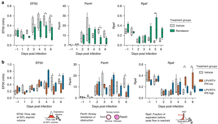
Therapeutic RDV and LPV/RTV-IFNb improve pulmonary
function
. In the therapeutic antiviral ef
fi
cacy studies described in
Fig.
5, we used WBP to assess pulmonary function (Fig.
6). In
contrast to vehicle-treated animals, RDV-treated animals had
reduced
fl
ow rate at 50% of the expired volume (EF50) (
P
=
0.01,
two-way ANOVA with Sidek
’
s multiple comparison test), and
PenH (
P
=
0.04, two-way ANOVA with Sidek
’
s multiple
com-parison test), a surrogate measure of airway
resistance/obstruc-tion (Fig.
6a)
28. Similarly, Rpef (the fraction of expiration before
peak expiratory
fl
ow is reached), an indicator of
bronchocon-striction, returned to baseline in RDV-treated animals by 5 dpi
(
P
=
0.002, two-way ANOVA with Sidek
’
s multiple comparison
test) yet remained suppressed in vehicle-treated animals.
Unlike the prophylactic study in Fig.
4b, therapeutic LPV
+
RTV-IFNb low improved pulmonary function as compared with
vehicle with signi
fi
cantly reduced EF50 and PenH (
P
< 0.05,
two-way ANOVA with Tukey
’
s multiple comparison test) and
base-line levels of Rpef by 5 dpi (
P
< 0.05, two-way ANOVA with
Tukey
’
s multiple comparison test) (Fig.
6b). While therapeutic
LPV/RTV-IFNb failed to reduce weight loss, lung hemorrhage,
and virus titer, this regimen appears to provide improvements in
pulmonary function similar to therapeutic RDV.
Therapeutic RDV but not LPV/RTV-IFNb diminishes signs of
ALI
. We then quantitated the lung pathology in therapeutically
treated animals (Fig.
7). In lung tissue sections from the 6 dpi, we
blindly evaluated and scored features of ALI using three different
and complementary approaches. In Fig.
7a, we show reduced ALI
with RDV treatment. Although sporadic apoptotic cells and
alveolar septal wall thickening (~2
–
4-fold over mock) driven by
immune cell in
fi
ltration were observed in the lungs of mice
treated with therapeutic RDV, the air spaces remained
pre-dominantly free of cellular debris and in
fl
ammatory cells similar
to mock-infected animals (Fig.
7a). In contrast, multiple
patho-logical features typical of ALI were noted in both vehicle groups
(RDV and LPV/RTV vehicles) as well as both LPV/RTV-IFNb
low and high, including altered alveolar architecture due to
pneumocyte degeneration and death, numerous in
fl
ammatory
a
b
0 1 2 3 4 5 6
60 70 80 90 100 110
Days post infection
% Starting weight
Percent starting weight *
2 6
Day post infection
Day post infection
PFU/Lobe
Virus lung titer
Virus lung titer *
*
–1 1 2 3 4 5 6
0 5 10 15 20
Days post infection
PenH
Pulmonary function
Pulmonary function
Vehicle
Remdesivir *
101 102 103 104 105 106 107 108
101 102 103 104 105 106 107 108
Treatment groups
0 1 2 3 4 5 6
60 70 80 90 100 110
Percent starting weight
Days post infection
% Starting weight
* * * *
2 6
PFU/Lobe
* *
–1 1 2 3 4 5 6
0 10 20 30 40
Days post infection
PenH
Vehicle
LPV/RTV-IFNb low
LPV/RTV-IFNb high
IFNb only *
* *
*
Treatment groups
Fig. 4 Prophylactic LPV/RTV+IFNb does not improve disease outcomes. aPercent starting weight (Left) of 12–14-week-old femaleCes1c−/−hDPP4
mice infected with 5E+04 pfu MERS M35C4 and treated BID with either vehicle (n=9) or remdesivir (RDV, 25 mg/kg,n=9) subcutaneously beginning
−1 dpi. Asterisks indicate statistically significant differences (P< 0.05) as determined by two-way ANOVA and Tukey’s multiple comparison test. (Middle)
MERS-CoV lung titer on 2 (N=3) and 6 dpi (all remaining animals). Asterisks indicate statistically significant differences (P< 0.05) as determined by
Mann–Whitney test. (Right) WBP was used to assess pulmonary function in mice. PenH is a surrogate measure of airway resistance or
bronchoconstriction. Asterisks indicate statistical differences by two-way ANOVA with Sidek’s multiple comparison test.bPercent starting weight (left),
virus lung titer (middle), and pulmonary function metric PenH (right) of cohorts of mice similar in age and sex and infected similarly with MERS-CoV as in
bbut treated with vehicle (n=9), LPV/RTV+IFNb low (1× human equivalent) (n=9), LPV/RTV+IFNb high (25× human equivalent) (n=9), or IFNb
high only (n=9). Oral vehicle or lopinavir/ritonavir (160/40 mg/kg) were administered orally once daily beginning the−1 dpi. IFNb treatment was
initiated 2 h prior to infection and every other day thereafter. To control for dosing effects, vehicle-treated mice received both LPV/RTV vehicle and subcutaneous PBS to mirror IFNb injections. Likewise, IFNb only group received oral vehicle to mirror that seen in orally dosed groups. Similar statistical
tests performed onawere performed onb. For the box and whisker plots, the boxes encompass the 25th to 75th percentile, the line is at the median, while
cells in the septae and in alveolar air spaces, neutrophils in the air
spaces, and proteinaceous debris in the air spaces organizing into
hyaline membranes (Fig.
7a). Using the ATS ALI scoring tool
described in Fig.
3, we found that only RDV therapy signi
fi
cantly
(
P
=
0.005,
one-way
ANOVA
with
Kruskal
–
Wallis
test)
decreased lung injury scores (Fig.
7b). With a complementary
histological tool, we then quantitated features of diffuse alveolar
damage (DAD), the pathological hallmark of ALI
26. We found
that only therapeutic RDV reduced DAD scores (
P
=
0.04,
one-way ANOVA with Kruskal
–
Wallis test) (Fig.
7c)
29. Since degree
of cell death appeared to correlate with protective ef
fi
cacy, we
quantitated levels of cleaved caspase-3 in lung tissue sections by
antibody labeling. Caspase-3, a widely accepted marker of
apoptosis, is a regulatory enzyme whose cleavage and activation
drive programmed cell death
30. Using the De
fi
niens software
suite, we obtained unbiased quantitative data showing that only
RDV treatment signi
fi
cantly reduced levels of cleaved caspase-3
antigen (
P
=
0.0109, one-way ANOVA with Kruskal
–
Wallis test)
(Fig.
7d). Thus, using three complementary and blinded
approaches, we obtained similar data showing that only
ther-apeutic RDV reduced histologic features of ALI.
Discussion
Emerging viral diseases have caused signi
fi
cant global pandemics
(e.g., HIV, 1918 in
fl
uenza, smallpox), epidemics (e.g.,
SARS-CoV), and devastating outbreaks (e.g., EBOV, MERS-CoV). For
CoV, metagenomic studies in wild animals have revealed a great
diversity of viruses and hosts, and have even identi
fi
ed viruses
similar to current and past epidemic strains in bats
31,32. Thus,
broad-spectrum therapies effective against known epidemic and
zoonotic strains likely to seed future emergence, have the
potential to diminish epidemic disease today and diminish future
outbreaks. Currently, there are no FDA-approved treatments for
any human CoV infection. Upon emergence of SARS- and
MERS-CoV, patients were administered off-label antivirals (e.g.,
ribavirin, LPV, RTV) and immunomodulators (e.g.,
corticoster-oids, interferon alpha-2a/2b, IFNb) as single agents or in
com-bination in an attempt to ameliorate severe disease outcomes with
very limited success
33. Without randomized controlled trials,
determining ef
fi
cacy is dif
fi
cult due to patient and treatment
variability as well as a lack of appropriate matching controls.
Although recent meta-analysis and modeling has suggested that
interferon treatment does not improve clinical outcomes in
MERS-CoV patients
4, the MIRACLE trial in the KSA is aimed at
conclusively determining if a
fi
xed dose combination of LPV/
RTV-IFNb is effective at treating MERS-CoV infections
14. Here,
we show that RDV provides superior antiviral activity against
MERS-CoV in vitro and in vivo as compared with
LPV/RTV-IFNb. In addition, RDV was the only treatment to signi
fi
cantly
reduce pulmonary pathology. Thus, we provide in vivo evidence
of the potential for RDV to treat MERS-CoV infections. Ef
fi
cacy
a
b
d
e
Vehicle-Remdesivir RemdesivirNuclei MERS-CoV antigen
Color key
Lung hemorrhage Virus lung titer
0 1 2 3 4 5 6
60 70 80 90 100 110 120
Days post infection
% Starting weight
Percent starting weight
Vehicle
Remdesivir
*
n.s. n.s.
101 102 103 104 105 106 107 108 Percent starting weight
c
Vehicle-LPV/RTV LPV/RTV-IFN low LPV/RTV-IFN high
0 1 2 3 4 5 6
60 70 80 90 100 110
Days post infection
% Starting weight
Vehicle
LPV/RTV-IFN low
LPV/RTV-IFN high
Vehicle-LPV/RTVLPV/RTV-IFN lowLPV/RTV-IFN high Vehicle-RDV
RDV
Vehicle-LPV/RTVLPV/RTV-IFN lowLPV/RTV-IFN high Vehicle-RDV
RDV
–1 0 1 2 3 4 5
Treatment group Treatment group
Lung hemorrhage score
PFU/Lobe
* *
n.s.
Fig. 5 Therapeutic RDV reduces replication and pathology.Percent starting weight of 10–12-week-old femaleCes1c−/−hDPP4mice infected with 5E+04
pfu MERS M35C4 and treated withasubcutaneous vehicle for RDV (N=13) or remdesivir (RDV, 25 mg/kg,N=14) BID beginning 1 dpi orbvehicle for
LPV/RTV-IFNb (N=15), LPV/RTV-IFNb low (N=16) or LPV/RTV-IFNb high (N=16) beginning 1 dpi. Oral vehicle or lopinavir/ritonavir (160/40 mg/kg)
was administered orally once daily. IFNb low (1x human equivalent dose of 1.6 MIU/kg) and high (25x human equivalent dose of 40 MIU/kg) or PBS
vehicle were administered via subcutaneous injection every other day. Asterisks indicate statistical differences by two-way ANOVA with Tukey’s multiple
comparison test.cLung hemorrhage 6 dpi for all animals ina,bscored on a scale of 0–4, where 0 is a normal pink healthy lung and 4 is a diffusely
discolored dark red lung.dMERS-CoV lung titer 6 dpi in mice as described ina,b. Asterisks indicate statistical significance (Ngroup described inaandb,
P< 0.05) by one-way ANOVA with Kruskal–Wallis test for (c,d). Data fora–dare compiled from two independent experiments. For the box and whisker
plots, the boxes encompass the 25th to 75th percentile, the line is at the median, while the whiskers represent the range.eRepresentative
testing of antiviral regimens in humans like those in the
MIRA-CLE trial are essential to progress antiviral development and
prioritize therapies most likely to improve clinical outcomes in
MERS-CoV patients.
Differences in protein identity and concentration complicate
the translation of in vitro antiviral activity to in vivo therapeutic
ef
fi
cacy. Although the EC
50for LPV against MERS-CoV (EC
50=
11 µM) falls within the maximum (
C
max=
15 µM) and minimum
(
C
min=
9.5 µM) levels observed in human plasma, the
biologi-cally available fraction unbound to protein in these respective
systems should be compared to more accurately translate in vitro
activity to potential in vivo ef
fi
cacy
9. For example, the trough
levels (
C
min) of total (5.8 µM) and unbound protein free LPV
(0.057 µM) in women with HIV differ by 100-fold, but LPV is still
highly active and effective since the EC
50(0.010
–
0.027 µM) falls
below the unbound protein free
C
min7,34. Together, these data
argue that levels of free biologically active LPV achieved in
humans are well below those that exert robust antiviral effects on
MERS-CoV replication in cell culture systems. Thus, the
micro-molar EC
50observed for LPV against MERS-CoV coupled with
protein binding and insuf
fi
cient levels of free LPV in plasma is
likely responsible for the modest antiviral effect with
LPV/RTV-IFNb prophylaxis and minimal impact on disease with
ther-apeutic administration in our mouse model. In contrast, human
equivalent doses of RDV are demonstrably ef
fi
cacious in mice
infected with SARS- and MERS-CoV and in nonhuman primates
infected with EBOV thus demonstrating a more suitable PK/PD
relationship
17.
Interferons are useful in treating multiple viral infections
35,36.
Since IFNb was shown to be the most potent against MERS-CoV
when comparing the antiviral activity of multiple type I and type
II interferons in Vero cells, IFNb was selected to use in the
MIRACLE trial
8,10. Mirroring the MIRACLE trial, we delivered
IFNb subcutaneously every other day, which failed to reduce
MERS-CoV viral loads and appeared to exacerbate disease in
mice. As we sought to understand this result, we found little
experimental congruency among reports detailing the
prophy-lactic or therapeutic ef
fi
cacy of type I interferons in animal
models of MERS-CoV
11,37,38. Falzarano et al. demonstrated that
IFN-alpha-2a coupled with ribavirin initiated 8 h post infection
improved outcomes in rhesus macaques and reduced viral copy
number in lung tissue, but the treatment had no effect on
infectious virus titers in bronchoalveolar lavage
fl
uid
37. In mice
where
hDPP4
is delivered to lung tissue by adenoviral
transduc-tion, intranasal IFNb given before or after MERS-CoV infection
reduced lung titers although the peak lung titers in this model are
approximately two orders of magnitude lower than the current
transgenic models, and thus may be more easily treated
38–40. The
utility of the common marmoset as a model of MERS-CoV
pathogenesis is controversial with one study detailing severe
respiratory disease yet another reporting similarly mild disease
among mock and MERS-CoV-infected animals
41,42. In
marmo-sets, Chan et al. explored the therapeutic potential of LPV/RTV or
IFNb, but the small numbers of animals used per group, lack of
time-matched viral load samples, and unexpected early mortality
in the LPV group made the resultant data dif
fi
cult to interpret
11.
Nevertheless, the studies noted above demonstrate that type I
interferon can exert an antiviral effect on MERS-CoV in vivo
when given subcutaneously (IFN alpha, rhesus macaque) and
intranasally (IFNb, adenovirus
hDPP4
model)
37–39. Our inability
to reduce MERS-CoV titer or improve outcomes with IFN as
described above may be due to inherent differences in the animal
b
–1 1 2 3 4 5 6
0.0 0.2 0.4 0.6 0.8 1.0
EF50 (ml/s)
EF50
* *
–1 1 2 3 4 5 6
0 10 20 30
Days post infection
Days post infection Days post infection
Pen
H
PenH
*
–1 1 2 3 4 5 6
0.0 0.2 0.4 0.6
Rpe
f
Rpef
Vehicle * *
LPV/RTV-IFN low
LPV/RTV-IFN high
Flow rate at 50% volume
Expiratory volume
Normal Infected Airway lumen
PenH PenH
Expiratory flow
PEF Rpef EF50: Flow rate
at 50% expired volume
PenH: Airway resistance or obstruction
Rpef: Fraction of expiration before peak flow is reached
a
EF50 PenH RpefDays post infection Days post infection Days post infection –1 1 2 3 4 5 6
0.0 0.2 0.4 0.6 0.8
EF50 (ml/s)
* *
–1 1 2 3 4 5 6 0
10 20 30 40
Pen
H
*
–1 1 2 3 4 5 6 0.0
0.2 0.4 0.6 0.8
Rpe
f
Vehicle
Remdesivir *
Treatment groups
Treatment groups
Fig. 6 Therapeutic RDV and LPV/RTV-IFNb improve pulmonary function.Whole-body plethysmography (WBP) was used to assess pulmonary function
in mice. Representative WBP data for one of the two studies depicted in Fig.5are shown. All groups wereN=4 per day. EF50 is theflow rate at 50%
expired volume. PenH is a surrogate measure of airway resistance. Rpef is the fraction of expiration before peak expiratoryflow is reached. Altered EF50,
PenH, and Rpef indicate bronchoconstriction or airway obstruction. Asterisks indicate statistical differences by two-way ANOVA with Sidek’s multiple
comparison test inaand two-way ANOVA with Tukey’s multiple comparison test inb. The boxes encompass the 25th to 75th percentile, the line is at the
a
b
c
d
100 µM
Mock Infected Remdesivir Vehicle-Remdesivir
Vehicle-LPV/RTV-IFN LPV/RTV-IFN low LPV/RTV-IFN high
American thoracic society lung injury score
0.0 0.2 0.4 0.6 0.8 1.0
0 1 2 3 4 5
Lung injury score
DAD score
Diffuse alveolar damage score
Vehicle LPV/RTV IFN
LPV/RTV IFN low
LPV/RTV IFN high
RDV Vehicle
RDV Vehicle
LPV/RTV IFN
LPV/RTV IFN low
LPV/RTV IFN high
RDV Vehicle
RDV
n.s. n.s. * p = 0.005
* p = 0.04
Vehicle
LPV/RTV-IFN LPV/RTV-IFN LowLPV/RTV-IFN High
RDV Vehicle RDV 0
2 4 6 8
Sum % cleaved caspase-3 +
Cleaved caspase-3 staining
Mock
Mock
* p = 0.0109 n.s.
Symbol key:
Normal septal wall thickness Neutrophil in alveolar space Neutrophil in alveolar septae
Normal clear airspace of alveolar sac
Hyaline membrane Cellular degeneration/death
Proteinaceous debris
Fig. 7 Therapeutic RDV but not LPV/RTV-IFNb diminishes signs of ALI. aRepresentative images of the histological features of acute lung injury 6 dpi
comparing a mock-infected mouse to the therapeutic treatment groups described in Figs.5and6. Symbols identifying example features of disease are
indicated in thefigure.bAmerican Thoracic Society Lung Injury Score derived as described in Fig.3. The numbers of animals per group quantitated: vehicle
RDVN=7, RDVN=7, vehicle LPV/RTV-IFNbN=9, LPV/RTV-IFNb lowN=7, LPV/RTV-IFNb highN=8.cDiffuse alveolar damage score quantitating
the degree of cellular sloughing, necrosis, and breakdown of barrier epithelium and vascular leakage. For bothbandc, scores were blindly assessed in three
random high power (×60)fields of diseased lung tissue sections.dQuantitation of cleaved caspase-3 antigen staining in lung tissue sections from studies
described in Figs.5–7. Cleaved caspase-3 is a marker of cell death. The numbers of animals per group quantitated for all groups wasN=5/group. For the
box and whisker plots, the boxes encompass the 25th to 75th percentile, the line is at the median, while the whiskers represent the range. Forb–d, asterisks
models, delivery route, differences in IFN subtype and/or active
viral antagonism of innate immunity. Since recent studies have
demonstrated type III IFN to be most effective in ameliorating
in
fl
uenza pathogenesis in mice, comparative studies investigating
the potency of different IFN subtypes should be pursued with
MERS-CoV
43–45.
Acute lung injury (ALI) in humans is well de
fi
ned by a set of
clinical parameters (i.e., acute onset, diffuse bilateral in
fi
ltrates on
X-ray, ratio of partial pressure of arterial oxygen to inspired
oxygen < 300, no evidence of elevated pulmonary arterial
pres-sure, etc.), which can be measured in mice but require specialized
procedures, equipment, and training not readily available to most
researchers
26,46. Moreover, animal models of ALI typically fail to
recapitulate all pathologic features observed in humans likely due
to differences in underlying anatomy, physiology, immunology,
genetics, and complex comorbidities typically associated with
hospitalized ALI patients (e.g., diabetes, kidney and liver disease,
etc.)
26. For example, mice have a distinctly different lobar
com-position with less branching in the conducting airway than
humans, a resting respiratory rate far exceeding that of humans
(250
–
300 bpm in mice, 12
–
16 bpm in humans), and very different
amounts of neutrophils in circulation (10
–
25% in mice, 50
–
70%
in humans). To address these issues, the American Thoracic
Society (ATS) has created tools to simplify the translation of
mouse models of ALI to the human condition including those to
quantitate the hallmarks of ALI in tissue sections
26. We employed
a complementary quantitative histologic assessment tool for
dif-fuse alveolar damage (DAD), the pathological hallmark of ALI in
humans
26,29. With the ATS ALI and DAD histologic assessment
tools described above, along with quantitation of cell death via
cleaved caspase-3 antigen labeling, we show that only RDV
therapy reduced ALI when initiated 1 dpi. Therefore, the evidence
from several complementary histologic approaches demonstrate
that RDV provides superior protection from ALI as compared
with LPV/RTV-IFNb.
Remdesivir (RDV, GS-5734) is a broad-spectrum antiviral with
potent in vitro ef
fi
cacy against multiple genetically unrelated
RNA viruses
16–18. RDV has demonstrated in vivo ef
fi
cacy against
EBOV in nonhuman primates which has led to its inclusion in
clinical studies evaluating the effects of RDV treatment in acute
Ebola virus disease (EVD) as well as in EVD survivors with
prolonged viral shedding
47,48. As with our SARS-CoV studies
with RDV, here we provide similar evidence for MERS-CoV with
diminished weight loss, improved pulmonary function, and
reduced virus replication with both prophylactic and therapeutic
RDV
17. The kinetics of SARS- or MERS-CoV replication and
disease in mice is substantially accelerated as compared with that
in humans. Virus replication in the lung peaks at 2 dpi in the
lungs of mice infected with SARS-CoV or MERS-CoV and
infection progresses to mortality or recovery by 7
–
10 dpi,
depending on virus dose
17,24. Therefore, the therapeutic window
in which to treat mice prior to the peak of virus replication is only
1
–
2 days. In contrast, MERS-CoV replication in the human
respiratory tract peaks at 7
–
10 days after the onset of symptoms
and the disease course resolves or progresses to death within
~21 days
49,50. Thus, the window for therapeutic administration
after the onset of symptoms but prior to the peak of virus
replication is very different in humans and experimentally
infected mice. With therapeutic RDV, we observed decreased
MERS-CoV pathogenesis and signi
fi
cant reductions in viral titers,
yet therapeutic treatment did not completely abrogate disease. In
addition, with high-titer virus inoculum, RDV was unable to
prevent mortality and loss of pulmonary function although it did
signi
fi
cantly reduce viral loads. These results are similar to those
obtained for SARS-CoV, where therapeutic treatment initiated
after peak virus titer and lung damage failed to improve outcomes
yet signi
fi
cantly reduced viral titer
17. Similar observations were
recently reported where neutralizing monoclonal antibodies failed
to reduce severe lung disease pathology or clinical disease when
administered 1 day after MERS-CoV infection in marmosets
51.
Since disease resulting from both SARS- and MERS-CoV
infec-tion is driven by both virus and host immune response factors,
depending on the stage of the disease progression, early initiation
of antiviral therapy, and/or holistic combination therapies will
likely be needed to diminish virus replication, immunopathology,
and/or promote repair and restoration of pulmonary
home-ostasis. Ongoing and future studies are aimed at determining if
MERS-CoV is capable of generating resistance to RDV in vitro
and whether the mutational spectra are similar to those obtained
by MHV and SARS-CoV which shift EC
50values only 3
–
5-fold
52.
In summary, we provide in vivo evidence of the potential utility
of RDV in treating MERS-CoV patients. Overall, our work
sug-gests that RDV may improve disease outcomes in CoV-infected
patients, serve to protect health care workers in areas with
endemic MERS-CoV and prove valuable in preventing future
epidemics in the event of novel CoV emergence in the future.
Methods
Study design. The primary goal of this study was to compare the prophylactic and
therapeutic efficacy of RDV with the combination of LPV/RTV and IFNb. First, we
assessed antiviral efficacy and cytoxicity in the Calu-3 human lung cell line as
compared with the appropriate vehicle control. Experimental conditions in vitro were performed in sextuplicate unless otherwise stated, and antiviral assays were
repeated four times per drug. Second, we evaluated the in vivo efficacy of
pro-phylactic RDV as compared to vehicle with two different doses of MERS-CoV in a new transgenic mouse model of MERS-CoV pathogenesis with improved phar-macokinetics for nucleotide prodrugs. We performed two additional prophylactic studies comparing RDV and vehicle to LPV/RTV-IFNb, IFNb-alone, and their
vehicles. Third, we assessed the therapeutic efficacy of the above treatment
regi-mens in a mouse model of MERS-CoV pathogenesis, but did not include an IFNb, only arm and these studies were performed twice. All lung histological assessments were performed in a blinded manner. In addition, we performed a single
ther-apeutic efficacy study with a lethal dose of MERS-CoV. Our in vivo efficacy studies
were designed to mirror the ongoing MIRACLE human clinical trial which is evaluating combination LPV/RTV-IFNb. Our studies were intended to generate the data required to justify further testing in nonhuman primates and collectively inform future human clinical trials. Mice were age- and sex-matched and randomly assigned into groups before infection and treatment. Exclusion criteria for in vivo studies were as follows: If a given mouse unexpectedly did not lose weight after
infection and their virus lung titers were more than 2 log10lower than the mean of
the group, this indicated that infection was inefficient, and all data related to that
mouse were censored.
Animal care and ethics statement. Efficacy studies were performed in animal biosafety level 3 facilities at UNC Chapel Hill. All works were conducted under protocols approved by the Institutional Animal Care and Use Committee at UNC Chapel Hill (IACUC protocol #16-284) according to guidelines set by the Asso-ciation for the Assessment and Accreditation of Laboratory Animal Care and the U.S. Department of Agriculture.
Virus. For in vitro studies, a MERS-CoV reporter virus expressing nanoluciferase
was employed (MERS-nLUC)17. MERS-nLUC stocks were derived from a
mole-cular clone through electroporation of Vero 81 cells (ATCC CCL-81) and isolation
of virus through harvesting of culture supernatants53. Briefly, DNA fragments A–F
encoding MERS-nLUC cDNA genome were ligated to create full-length cDNA, which was then used as a template for in vitro transcription. Full-length genomic RNA was then electroporated into Vero-81 cells yielding recombinant virus stock. The resultant stock was passaged twice in Vero 81 to generate a working stock
(1.6E+07 pfu/mL) for our studies. Wild-type MERS-CoV for comparative
anti-viral efficacy studies was derived from our EMC 2012 infectious clone as described
above to obtain a working stock with a titer of 3E+07 pfu/mL53. For in vivo
studies, we utilized mouse adapted MERS-CoV passage 35 clone 4 (MERS M35C4)24.
MERS M35C4 has 12 amino acid coding changes as well as a single-nucleotide
change in the 5ʹUTR and a large deletion in ORF4b/ORF5. This virus is a clonal
isolate generated through serial passage of MERS-CoV in mice. After 35 passages in
mice, virus was plaque purified and clone 4 was expanded two times on Vero
CCL81 cells to obtain our working stock. The working stock (1.1E+08 pfu/mL)