THE RHOA GUANINE NUCLEOTIDE EXCHANGE FACTOR, LARG, MEDIATES ICAM-1-DEPENDENT MECHANOTRANSDUCTION IN ENDOTHELIAL CELLS TO
STIMULATE TRANSENDOTHELIAL MIGRATION
Elizabeth Chase Lessey-Morillon
A dissertation submitted to the faculty of the University of North Carolina at Chapel Hill in partial fulfillment of the requirements for the degree of Doctor of Philosophy in the Department of Cell and Molecular Physiology (Cell and Developmental Biology).
Chapel Hill 2014
© 2014
ABSTRACT
Elizabeth Chase Lessey-Morillon: The RhoA guanine nucleotide exchange factor, LARG, mediates ICAM-1-dependent mechanotransduction in endothelial cells to
stimulate transendothelial migration (Under the direction of Keith Burridge)
RhoA-mediated cytoskeletal rearrangements in endothelial cells (ECs) play an active role in leukocyte transendothelial cell migration (TEM), a normal
physiological process in which leukocytes cross the endothelium to enter the underlying tissue. While much has been learned about RhoA signaling pathways downstream from ICAM-1 in ECs, little is known about the consequences of the tractional forces that leukocytes generate on ECs as they migrate over the surface before TEM. We have found that after applying mechanical forces to ICAM-1 clusters, there is an increase in cellular stiffening and enhanced RhoA signaling compared to ICAM-1 clustering alone. We have identified that the Rho GEF
DEDICATION
This thesis is dedicated to my parents, Bruce and Barbara Lessey, my husband Maurice Morillon, and to all who have supported me through my life and graduate
ACKNOWLEDGEMENTS
First, I need to thank Keith Burridge for allowing me to join his lab and in doing so, providing me with a truly wonderful graduate school experience. The current and former Burridge Lab members make the Burridge Lab an ideal place for research to occur. A huge thank you goes out to my two postdoc mentors Tom Sampson and Christophe Guilley. During my graduate career, both have supported me greatly. Beth Benson has also been a great mentor with career advice as well as with presentations and papers. I would like to thank Erika Wittchen for her guidance both in and outside of the lab. Lisa Sharek is an amazing lab manager; the Burridge Lab could not function without her. Without her support, none of this would be possible. My time in the Burridge Lab would have been very different and much harder without my fellow graduate students, Caitlin Tolbert and Amir Aghajanian.
I could not have done this project without the aid of Richard Superfine’s Laboratory in the Physics Department. I am grateful to Dr. Superfine for permitting me to use his laboratory. Luke Osborne and Tim O’Brien assisted me greatly with the physics side of my project. I’ve received fantastic support from the IVB training program and the former Cell and Developmental Department and current
TABLE OF CONTENTS
LIST OF TABLES ... ix
LIST OF FIGURES ... x
ABBREVIATIONS ... xi
Chapter 1: BACKGROUND AND SIGNIFICANCE ... 1
Leukocyte Transendothelial cell migration ... 1
The Rho Family of Small GTPase-binding proteins ... 3
ICAM-1 Signaling ... 6
Paracellular migration ... 7
Transcellular migration ... 9
Summary ... 10
Chapter 2: FROM MECHANICAL FORCE TO RHOA ACTIVATION ... 11
Introduction ... 12
The Rho pathway ... 14
GEFs and GAPs ... 17
Other Rho proteins ... 21
Experimentally manipulating force ... 22
Rigid substrata, stress fibers and focal adhesions ... 29
Cancer ... 37
Future directions ... 39
Chapter 3: THE RHOA GUANINE NUCLEOTIDE EXCHANGE FACTOR, LARG, MEDIATES ICAM-1-DEPENDENT MECHANOTRANSDUCTION IN ENDOTHELIAL CELLS TO STIMULATE TRANSENDOTHELIAL MIGRATION ... 41
Introduction ... 41
Results ... 43
Mechanical force on ICAM-1 increases cellular stiffness around ICAM-1 clusters. ... 43
RhoA is activated by mechanical force on ICAM-1 ... 46
Knockdown of LARG expression inhibits RhoA activation downstream of ICAM-1 clustering ... 50
Endothelial LARG contributes to leukocyte TEM ... 53
Discussion ... 53
Methods and materials ... 58
Chapter 4: CONCLUSIONS AND FUTURE DIRECTIONS ... 66
Summary ... 66
Substratum Stiffness ... 67
Other Rho Family GTPase ... 69
LARG Signaling ... 70
LARG as a Mechanosensor ... 71
Knockout mice ... 71
Role in Cell Spreading ... 74
Conclusions ... 75
LIST OF TABLES
Table 1: RhoA GEFs and GAPs which associate with the
LIST OF FIGURES
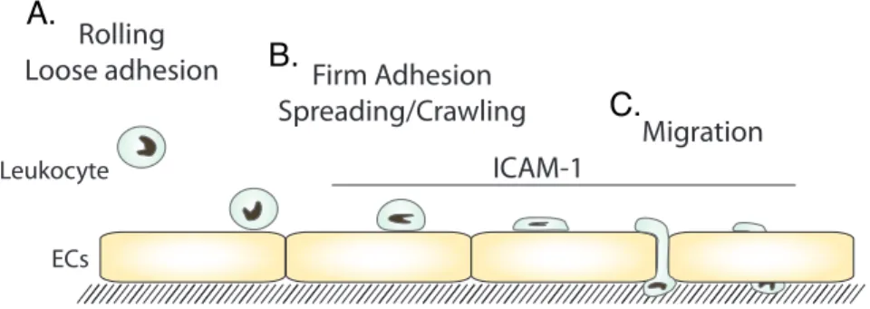
Figure 1: Steps in Leukocyte TEM ... 2
Figure 2: RhoA cycle ... 3
Figure 3: A diagram of ICAM-1 ... 7
Figure 4: RhoA effector signaling ... 14
Figure 5: Mechanical force in cell biology ... 16
Figure 6: RhoA GEFs and GAPs regulated by force ... 19
Figure 7: Mechanical force on ICAM-1 increases cellular stiffening ... 45
Figure 8: Mechanical force on ICAM-1 increases RhoA activity and MLC phosphorylation ... 47
Figure 9: LARG is activated downstream of ICAM-1 clustering alone and enhanced with mechanical force ... 49
10: Confirmation of LARG knockdown ... 51
Figure 11: LARG mediates EC response to mechanical force on ICAM-1 and affects neutrophil crawling and TEM ... 52
Figure 12:The Pathway downstream from mechanical force on ICAM-1 ... 67
Figure 13: Mechanical force on ICAM-1 does not alter other Rho family GTPases ... 70
ABBREVIATIONS
DH dbl homology
EC endothelial cell ECM extracellular matrix
GAP guanine nucleotide exchange factors GEF guanine nucleotide exchange factor ICAM-1 inter-cellular adhesion molecule-1 HUVEC human umbilical cord endothelial cell HMVEC human microvascular endothelial cells
LARG leukemia-associated Rho guanine nucleotide exchange factor LFA-1 leukocyte function-associated molecule-1
Mac-1 macrophage-1 antigen
mDia mammalian homolog of Drosophila diaphanous MHC major histocompatibility complex
MLC myosin light chain
MSC mesenchymal stem cells PH pleckstrin homology
pMLC phosphorylated myosin light chain Rho Ras homology proteins
ROCK Rho kinase
Chapter 1: BACKGROUND AND SIGNIFICANCE
Leukocyte Transendothelial cell migration
Endothelial cells (ECs) make up the lining of blood vessels and provide a protective barrier to the underlying tissue. Dynamic regulation of the ECs and cell-cell junctions is required to allow leukocyte diapedesis. During inflammation or infection, chemoattractant signaling cues the leukocytes to exit the blood stream (Figure 1). Pro-inflammatory signaling increases expression of adhesion receptors, including E-selectin, ICAM-1, and VCAM-1, on the luminal surface of the EC (1-4). These adhesion molecules bind to receptors on the leukocyte. For example,
E-selectin binds to sialylated glycoproteins on leukocytes (5), whereas ICAM-1 binds to β2 integrins such as leukocyte function-associated molecule-1 (LFA-1)(αLβ2,
CD11a/CD18) (6, 7), and macrophage-1 antigen (Mac-1) (αMβ2CD11b/CD18) (8, 9),
and VCAM-1 binds to β1 integrins such as very late antigen-4 (VLA-4) (α4β1,
CD49a/CD29) (10).
inflammation (11). While E-selectin binds the leukocyte to slow it down from the circulation, E-selectin also links to the endothelial cytoskeleton to alter EC signaling (12).
Figure 1: Steps in Leukocyte TEM
(A) The leukocyte loosely adheres to the endothelium. (B) Then, the leukocyte spreads and crawls. (C) Lastly, the leukocyte crosses the endothelium at an EC junction, paracellular migration, or though an EC,
transcellular migration.
Next, the leukocyte binds to other adhesion receptors, including ICAM-1, then spreads and crawls on the endothelial surface (Figure 1B). ICAM-1 engagement initiates many signaling pathways within the EC to assist in leukocyte TEM
(discussed later in this chapter) (13-21). The crawling leukocyte extends protrusions to probe the surface of the endothelium (20). Transmigration can occur by two routes, transcellular and paracellular diapedesis (Figure 1C) (22-24). Paracellular migration is the most well studied route of diapedesis and occurs by the leukocyte migrating through the junction between two ECs. For a leukocyte to be able to cross at an endothelial cell-cell junctions the junctional proteins must disengage (25-27). Leukocyte bindings to adhesion receptors on the EC surface induces the junctions to weaken (28). The alternative route is transcellular diapedesis, where a leukocyte migrates through a single EC. Like in paracellular migration, leukocyte engagement of EC receptors initiates the process. A combination of actin protrusions and vesicle
B. A.
C.
Rolling
Loose adhesion Firm Adhesion Spreading/Crawling
Migration
ECs
trafficking create pore in the ECs allowing the leukocyte to pass through (18, 29, 30). Both routes depend on endothelial changes in the cytoskeleton, which are controlled by Rho GTPases (14, 15, 30, 31),
The Rho Family of Small GTPase-binding proteins
Rho GTPases are a subfamily of the Ras superfamily of GTPases, which act as molecular switches. By cycling through an on/off cycle, they are able to regulate the cytoskeleton and cell contractility. There are 22 members of the Rho family in mammals (32). The most well studied family members are RhoA, Rac1 and CDC42. RhoA promotes stress fibers and actomyosin contractility (33, 34). Rac1 stimulates actin polymerization and induces the branched actin network that makes up the lamellipodia at the leading edge of a cell (33). Cdc42 also stimulates actin
polymerization and regulates protrusions known as filopodia (35, 36). In addition, these GTPases regulate many other activities (37).
Figure 2: RhoA cycle
Like most G proteins, RhoA cycles between an inactive GDP-‐bound form and an active GTP-‐bound form. Activation is mediated by GEFS that catalyze exchange of GDP for GTP. GAPs inactivate RhoA by stimulating intrinsic GTPase activity. GDI sequesters inactive GDP-‐bound RhoA in the cytoplasm.
Rho GTPases are inactivated by GTPase-activating proteins (GAPs) that enhance the intrinsic GTPase activity by stimulating hydrolysis of GTP to GDP. It is striking that there are more GEFs or GAPs than Rho family members. This most likely reflects that many signaling pathways can converge on individual Rho proteins and that different GEFs and GAPs function within these different pathways.
Some redundancy between GEFs appears to exist and is indicated by the modest or negligible phenotype induced in mice where a single GEF has been knocked out. PDZ-RhoGEF knockout mice or LARG knockout mice have no obvious phenotype, but the double knockout is lethal suggesting compensation between the GEFs in the single knockout mice (38). Also, LARG deficient mice have a lower than expected birth rate which might be a result of GEF compensation in the surviving mice (38). A close examination of Vav deficient mice show that compensation between GEFs, even closely related isoforms, does not always occur. Only Vav1, not Vav2 or Vav3, regulates thymic selection based the phenotype of the single isoform knockout animals (39-42). When Vav1 and Vav3 are knocked out, there is further impairment of thymic selection, suggesting that Vav3 is compensating for Vav1 (39, 40). However, Vav2 is unable to do so as the double Vav1 and Vav2 knockout mice have the same thymic selection defect as the single Vav1 knockout mice (43). While single GEF knockout animals frequently lack a strong phenotype, the Trio knockout mice are not viable from a neuronal and muscle defect (44).
DH domain is able to bind inactive Rho and induce dissociation of GDP, temporally leaving Rho in a nucleotide-free state. However, due the high ratio of GTP to GDP within the cell, GTP quickly binds, thus transferring Rho into its active state. The neighboring pleckstrin homology (PH) domain can associate with phosphoinositides causing plasma membrane localization as well as assisting with GTPase binding. The DH-PH domain is also responsible for the specificity of GEFs for Rho GTPases. A single DH-PH domain can exchange nucleotide for a specific GTPases or can act on multiple GTPases. For example, Leukemia-associated Rho guanine nucleotide exchange factor (LARG) can only activate RhoA (45) while Vav shows less
specificity and can activate RhoA, Rac, Cdc42 and RhoG (46). Some GEFs like, Trio, have multiple DH-PH domains each with different Rho family specificities (47).
GAP proteins are not as well characterized as GEFs but are just as important in understanding Rho GTPase function. The first identified GAP was Rho GAP (48). Subsequent family members have been identified by the presence of the GAP domain. The GAP domain binds to active GTP-bound Rho proteins and promotes hydrolysis of GTP to GDP, thus cycling Rho GTPases into their inactive form. Like GEFs, GAPs have specificities for Rho family members. This allows for tight regulation of the Rho GTPase family member.
inactivating MLC phosphatase, which then causes increased actomyosin contractility (49-52). In some cells such as fibroblasts, the activation of myosin stimulates the formation of stress fibers (53). ROCK can also signal through LIM kinase to promote actin stress fiber formation. ROCK can activate LIM kinase, which then
phosphorylates cofilin (54). Cofilin functions as an actin-severing protein and is inactivated after phosphorylation. Formins, including mammalian homolog of Drosophila diaphanous (mDia), are another class of Rho effectors that function to promote stress fiber formation by nucleating actin polymerization to create F-actin (55).
ICAM-1 Signaling
One of the most-well studied adhesion receptors in Rho GTPases signaling during leukocyte TEM is ICAM-1. ICAM-1 has 5 extracellular IgG like domains and a small 22 aa cytoplasmic tail (Figure 3). β2 integrins, such as LFA-1 or MAC-1, bind to specific IgG domains to induce ICAM-1 clustering. ICAM-1 is then translocated into the lipid insoluble regions (56). This clustering is then able to bring together the intracellular domains initiating downstream signaling cascades (57, 58).
Figure 3: A diagram of ICAM-‐1
One central consequence of ICAM-1 clustering is rearrangement of the EC cytoskeleton, which assists in
leukocyte TEM. This is predominantly due to increases in RhoA signaling and changes in the actin cytoskeleton (14-16, 59, 60). After ICAM-1 clustering, F-actin and actin binding proteins, including ezrin, moesin, radixin, and α-actinin, associate with the ICAM-1 complex to induce cytoskeletal changes (21, 31, 61-64).
FAK, paxillin, p130Cas, ezrin, and cortactin are phosphorylated in response to ICAM-1 crosslinking (64, 65). Src phosphorylation also occurs and is responsible for cortactin phosphorylation (64). Interestingly, Src also becomes phosphorylated after E-selectin clustering (64). Leukocyte-induced ICAM-1 clustering activates RhoA to assist in migration across the EC monolayer (14-16, 59, 60). Inhibiting RhoA signaling in ECs greatly attenuates leukocyte adhesion, spreading, and migration (14, 15, 59, 60). RhoA signaling leads to activation of the effector Rho-associated protein kinase (ROCK) (50). Rock dependent enhanced EC actomyosin contractility results in weakened cell-cell junctions allowing the formation of gaps through which leukocytes can migrate across the EC monolayer (49, 50).
Paracellular migration
Paracellular diapedesis is a well-studied route for leukocyte TEM and requires ECs to alter their cell-cell junctions to allow leukocytes to cross. Normally, adherens
Extracellular
TM
Intracellular
D1
D2
D3
D4
integrity of the blood vessel. Pro-inflammatory signaling can disrupt cell-cell junctions to increase permeability (23). Also, leukocyte binding to the EC induces a signaling cascade to weaken the cell-cell junctions to assist in diapedesis.
Adherens junctions are central regulators of leukocyte TEM. One of the more well studied cell adhesion molecules found in adherens junctions is vascular
endothelial-cadherin (VE-cadherin). VE-cadherin regulates the strength of EC junctions and is critical in leukocyte TEM (28). Significantly, VE-cadherin is a
downstream target of RhoA-dependent contractility. The extracellular domain of VE-cadherin creates a homophilic interaction with a VE-VE-cadherin on a neighboring cell, and this interaction is calcium dependent (66). Blocking VE-cadherin increases leukocyte TEM (25, 26). Conversely, Mice expressing a mutant VE-cadherin-α -catenin fusion protein, which prevents the disassociation of VE-cadherin at cell-cell junctions, leads to decreased permeability and leukocyte TEM (27).
The dissociation and loss of VE-cadherin from adherens junctions during leukocyte TEM triggers cell-cell junctions to weaken allowing the leukocyte to cross, then VE-cadherin returns shortly after to reseal the junction (67, 68).
Phosphorylation of VE-cadherin assists in the disruption of cell junctions (28, 69-71). Specifically, ICAM-1 crosslinking induces downstream signaling to phosphorylation of VE-cadherin by activating Src and pyk2 (28).
Endothelial junctions also contain tight junction proteins that regulate
restricted apically, whereas in ECs tight junctions extend throughout the junction interface and intermingle with the adherens junction proteins (72). The role of tight junction proteins in leukocyte TEM has not been as extensively studied as the role of adherens junction proteins. However, phosphorylation of the tight junctional proteins occludin and claudin-5 are reported during leukocyte TEM, and depend on RhoA and ROCK signaling (73). Highly regulated control of junctional proteins is required for efficient leukocyte TEM.
Transcellular migration
Transcellular diapedesis is a less anticipated route for a leukocyte to cross the endothelium. Instead of crossing that the endothelial junctions, the leukocyte passes through a single EC. Transcellular diapedesis initially was observed in electron micrographs of leukocyte TEM occurring in in vivo models of inflammation (74, 75). This route is less well characterized than paracellular TEM but does involve some of the same adhesion receptors, like ICAM-1 (17, 18). After leukocyte
adhesion, caveolin-1 co-localizes with the transcellular pore (18). ICAM-1 rich
microvilli-like structures extend from the EC around the leukocyte before it ultimately transmigrates (17, 18, 20, 28, 30).
It is unclear why transcellular or paracellular diapedesis becomes the chosen route for a leukocyte. However, it does appear that the EC type might play a role. Whereas in HUVECs there is very a small number of transcellular diapedesis events, less than 10%, it is as much as 30% in microvascular ECs (20). The route picked might also be dependent on the type of stimulus to induce inflammation. Under pro-inflammatory conditions, like treatment with VEGF, histamine, LPS or IL-1β,
paracellular diapedesis occurs more frequently (27). While paracellular diapedesis frequently occurs at a higher rate, this does not rule out an important role for transcellular diapedesis.
Summary
Regardless of the transcellular or paracellular path the leukocyte takes, the endothelium is an active player in the process in part due to the role of Rho Family GTPases. The endothelial cytoskeleton is responding to endothelial receptors
engaging with the leukocyte to assist in diapedsis. TEM illustrates the important role for Rho family GTPases in this example of cell-cell interactions, but TEM is also critical in the normal inflammatory response and in inflammatory diseases.
Chapter 2: FROM MECHANICAL FORCE TO RHOA ACTIVATION1
Throughout their lives all cells constantly experience and respond to various mechanical forces. These frequently originate externally but can also arise internally as a result of the contractile actin cytoskeleton. Mechanical forces trigger multiple signaling pathways. Several converge and result in the activation of the GTPase RhoA. In this review we focus on the pathways by which mechanical force leads to RhoA regulation, especially when force is transmitted via cell adhesion molecules that mediate either cell-matrix or cell-cell interactions. We discuss both the upstream signaling events that lead to activation of RhoA, as well as the downstream
consequences of this pathway. These include not only cytoskeletal reorganization and, in a positive feedback loop, increased myosin-generated contraction, but also profound effects on gene expression and differentiation.
1
This chapter appeared as a review article in Biochemistry. Reproduced with
Introduction
All cells are exposed to mechanical forces and to a greater or lesser degree responds to these forces. In the vertebrate body, cells experience different types of force according to their tissue location. For example, ECs lining blood vessels, as well as epithelial cells lining certain ducts or cavities, experience mechanical force from the passage of fluid over the cell surface. Cells in the skeletal system (bone and cartilage) but also many other cells are exposed to compression. Throughout most tissues, cells experience varying degrees of tension, which can arise from external forces or from within the cell as a result of actomyosin contractility. It is important to note, however, that the very high tensional forces experienced by some tissues, such as tendons and ligaments, are usually transmitted by extracellular matrix (ECM) components such as collagen fibers and the cells within these tissues are shielded from the tension by the ECM (76). Some forces on cells may be cyclical as experienced by cells in contact with the blood circulation, or as a result of
rhythmic activities such as breathing or walking, whereas other cells experience sustained force for varying periods of time.
tractional forces on the underlying substratum (81) and to be able to harness these forces to orient collagen fibers (82). Application of mechanical tension to migrating cells in culture using a microneedle inhibited extension perpendicular to the axis of tension but allowed or even promoted extension that was parallel with the force (83).
Although research in the field of mechanotransduction has been active for many years, much of it was focused on systems, tissues and cells that are very overtly affected by mechanical stimuli, such as vascular ECs and vascular smooth muscle exposed to flow and/ or stretch, or osteoblasts that experience compressive forces. However, during the past decade there has been an explosion of interest in the more universal responses of cells to mechanical forces and progress is occurring rapidly. Whether the forces are applied exogenously on cells or are generated
endogenously, they are usually transmitted to the ECM or to neighboring cells via cell adhesion molecules. Consequently, considerable interest has been directed at understanding the signaling pathways that are initiated in response to mechanical forces that are applied to adhesion molecules (84). Multiple signaling pathways have been identified, including tyrosine kinases, ion channels and GTPases (85). One of the pathways that appears to be involved in many cells responding to mechanical force involves activation of Rho family GTPases, particularly RhoA. In this review we will focus primarily on the signaling pathways that lead to activation of RhoA in
response to mechanical force and we will discuss the consequences of this pathway. The reader is directed to recent comprehensive reviews for information about
The Rho pathway
In contrast to most plant cells that have rigid cell walls, the mechanical properties of animal cells are critically dependent on their cytoskeletons, consisting of microtubules, actin microfilaments, various types of intermediate filaments and also septins (92). All of these filament systems may contribute to the mechanical properties of animal cells, although with respect to how cells respond to exogenously applied forces most attention has been directed toward the actin cytoskeleton. When actin filaments are highly crosslinked they can give rise to a relatively rigid cell
cortex. However, this can be rapidly remodeled to allow cell protrusion and changes in cell shape. The polymerization of actin filaments drives many types of cell
extension. In conjunction with myosin, actin filaments can generate contractile forces, exerting traction on the surrounding matrix or on other cells and contributing to major changes in cell morphology. The interaction of myosin with actin not only contributes to the response of cells to
exogenously applied forces but is responsible for generating endogenous forces within cells.
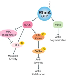
Figure 4: RhoA effector signaling
Activated RhoA interacts with effector proteins, which lead to actomyosin contractility and actin stabilization. ROCK signals by MLC phosphorylation to increase myosin II activity and LIM kinase to increase the level of actin stabilization. mDia nucleates actin polymerization.
The Rho family of GTPases are key regulators of the actin cytoskeleton. The mammalian genome encodes approximately
the most studied and each has distinct effects on the actin cytoskeleton (33, 34). In the context of mechanotransduction, most effort has been directed at determining the role of RhoA, which is the focus of this review. In large part, this reflects the fact that RhoA regulates the activity of myosin II and consequently is responsible for much of the intracellular tension and force that is generated within cells (93). RhoA cycles between an inactive GDP state and an active GTP state (Figure 2). Three classes of proteins regulate this cycle: GEFs, GAPs and guanine nucleotide-dissociation inhibitors (GDIs) (94). GEFs activate Rho proteins by catalyzing the exchange of GDP for GTP (32) and GAPs stimulate the intrinsic GTPase activity, leading to the return to the inactive state (95). The inactive pool of RhoA is
maintained in the cytosol by association with GDI (96) and it is in the active GTP-bound conformation that RhoA interacts with its effectors and performs its functions (Figure 2). With respect to regulating the activity of myosin II, the critical effector is ROCK, which exists in two isoforms, ROCK1 and ROCK2. Both isoforms promote myosin II activity by elevating the phosphorylation of the regulatory MLC. This occurs both directly by phosphorylation of the regulatory MLC (51) and indirectly by
phosphorylation and consequent inhibition of the MLC phosphatase (52). The phosphorylation of the MLC promotes assembly of myosin II into bipolar filaments and enhances the ATPase activity of myosin II. Together these effects increase the contractile force generated by myosin II on actin filaments. ROCK also
phosphorylates and activates another kinase, LIM kinase, which in turn
cofilin’s actin severing activity, this increases the stability of actin filaments. Active RhoA also promotes actin filament polymerization. This occurs by RhoA binding different effector, mDia1, which is member of the formin family of actin nucleating factors (55) (Figure 4).
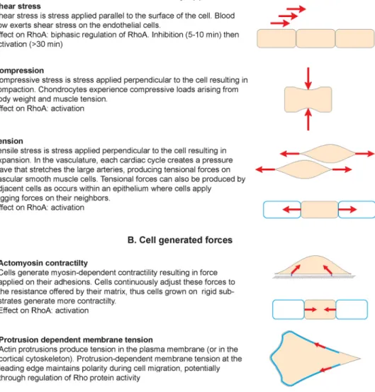
Figure 5: Mechanical force in cell biology
Diagram summarizing the different types of force that cells can experience. These can be externally applied (A) or generated by the cell itself and its own cytoskeleton (B). The effect on RhoA activity is indicated for each example. Force is a vector with magnitude and direction that causes an object with mass to change its velocity (units of newtons). Stress is force per unit of area (units of pascals).
generated by the cell itself with its cytoskeleton (97, 98) (Figure 5). Despite the apparent differences between these two signals, applied forces and cell-generated forces share some similarities in their transduction modalities and seem to regulate the same molecular mechanisms (89, 98). In both cases, cell surface adhesions, cytoskeleton and membrane tension cooperate to transmit forces which eventually affect the conformation of “mechanosensors” and trigger the mechanoresponse (85, 89). Interestingly, numerous GEFs and GAPs are known to associate with
cytoskeletal and cell adhesion components, suggesting that mechanical forces can directly affect the activity or the localization of RhoA regulators.
GEFs and GAPs
Some GEFs specific for RhoA have been found to associate with the
cytoskeleton and adhesions (Table 1). Integrin-based adhesions constitute a major site of mechanotransduction (99) and experience very diverse types of forces. For example, they are subjected to tensional forces when the ECM is stretched or when cells are grown on rigid substrates and generate more myosin-dependent
GEFs Localization References
p115 (ArhGEF1) Cell-ECM adhesion (101, 102) GEF-H1(ArhGEF2) Cell-ECM adhesion
Cell-Cell adhesion Microtubule
(101, 102) (103, 104) (105) LARG (ArhGEF12) Cell-ECM adhesion (101, 102) Vav Cell-ECM adhesion (106) p190RhoGEF Cell-ECM adhesion
mirotubule
(107) p114RhoGEF (ArhGEF18) Cell-Cell adhesion (107) Trio Intermediate filaments (108) PDZRhoGEF (ArhGEF11) Cell-ECM adhesion (109)
GAPs
p190RhoGAP Cell-ECM adhesion (110) DLC1 Cell-ECM adhesion
Cell-Cell adhesion
(111) (112) Myo-IXA Cell-Cell adhesion (113)
Table 1: RhoA GEFs and GAPs which associate with the cytoskeleton or adhesions
transactivation. Depletion of vav2, as well as EGFR inhibition, prevented stretch-induced RhoA activation (100). Applying tensional forces on fibronectin coated beads bound to fibroblasts, our group recently showed that force on integrins activates RhoA through two GEFs, GEF-H1 and LARG (101) (Figure 6B).
colleagues observed that microtubule stability, and not the Ras/ERK pathway, regulates GEF-H1 activity (115). This apparent discrepancy could be due to the difference of cell types that were used in these studies. Indeed, working with
fibroblasts another group observed that RhoA activation in response to stretch was not affected by taxol-induced microtubule stabilization (116), whereas in ECs RhoA activation in response to stretch requires GEF-H1 and is prevented by taxol (117).
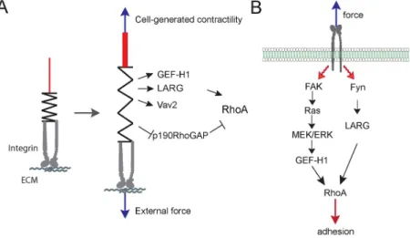
Figure 6: RhoA GEFs and GAPs regulated by force
(A) Schematic diagram showing the GEFs and GAPs whose activities are regulated by external force or cell-‐ generated tension on cell–ECM adhesions. (B) Diagram showing the signaling pathways that regulate GEF-‐ H1 and LARG activity in response to force on integrin (31).
Cell-cell adhesions are also subjected to tensional forces which are generated by neighboring cells or by the cell’s own contractile machinery (Figure 5). It is now clear that tugging forces play an important role in intercellular junction maturation and growth (see below) (118-120). Interestingly, some RhoA GEFs have been found to localize at intercellular adhesions. GEF-H1 associates with cingulin at tight
induces junction maturation (118), it would be interesting to determine if p114RhoGEF activity is regulated by tugging forces.
Mirroring the GEFs, some RhoA GAPs localize at adhesions (Table 1) and play a role during the mechanoresponse. DLC1 (111, 122-124) and p190RhoGAP (125-128) associate with Cell-ECM adhesion components. Shear stress regulates
Other Rho proteins
to depress Rac1 activity (60). Blocking myosin activity, either directly with blebbistatin or indirectly by inhibiting ROCK activity, decreased ArhGAP22 activity(136). The Rac GEF βPIX binds to Myosin II and actomyosin contractility induces βPIX dissociation from cell substrate adhesions (114). This contributes to Rac1 inhibition during adhesion maturation. It seems likely that all of these
mechanisms may synergize to confine Rac1 activity and membrane protrusion to the leading edge of migrating cells and away from regions of high mechanical tension and RhoA/ROCK activity. As a consequence of the competitive binding to RhoGDI, increasing the binding affinity of one Rho protein leads to the release and
degradation and/or activation of other Rho proteins (137). Interestingly, actomyosin contractility induces GDI dissociation from cell-matrix adhesions (114). However, the mechanism of GDI recruitment to adhesions is not known.
Experimentally manipulating force
adhesions less that 1 μm2 that generated high levels of stress that did not correlate with the size of the adhesion (147). These latter adhesions most likely relate to the adhesions studied by Beningo et al. at the leading edge of migrating cells (146).
Manipulations of the ECM and flexible substrata can be used to mimic the tensional forces cells experience in the body. Simply plating cells on more rigid rather than on more compliant substrata increases the tension generated by cells on their underlying matrix due to increased RhoA activity (see discussion below) (148). Various devices have been developed that allow investigators to stretch cells by stretching the substratum to which the cells are adhering. The development of culture dishes with a flexible base that can be stretched by applying a vacuum facilitated subjecting cells to periods of cyclic stretch (149, 150). The period of the stretch as well as the degree of stretch imposed on cells can be readily varied and a large literature now exists describing many signaling pathways that become
activated in response to cyclic stretch. Tension has also been applied to individual cellsusing glass rods or needles (83, 151). With these it is often more difficult to know the precise force that is being applied to cells, although the amount of force required to bend a needle by a certain angle can be determined experimentally.
Stretching or deforming a cell via a flexible substratum or by a glass rod or needle simultaneously affects many properties, including cell shape, the
cytoskeleton, as well as a cell’s adhesion to the matrix and/or its neighbors. In order to examine the effects of tension on specific adhesion molecules different
magnetic tweezers to exert forces on beads that are attached to cells via specific ligands or antibodies. Wang and colleagues used ferromagnetic beads that were attached to cells via integrin ligands (152). The beads were magnetized in one direction and then a second magnetic field was applied at 90° inducing the beads to twist and exert a shear force. This allowed them to show that there was a stiffening response as force was applied and that this depended on the cytoskeleton (152). Sheetz’s group used optical tweezers to manipulate beads similarly coated with integrin ligands or antibodies(153). They used the optical tweezers to restrain individual beads against the force exerted by the cell. It was observed that cells sensed the restraining force and strengthened the cytoskeletal linkages to oppose this. One advantage of optical tweezers is that beads can be individually
manipulated with great precision, allowing them to be placed at different points on a cell’s surface and to be moved in different directions. Optical tweezers can generate forces up to about 500 pN, but in the higher range of forces heat generated by the laser can be detrimental and limit the use of this approach. Whereas an advantage is the ease of examining single cell responses, optical tweezers are not suitable for bulk biochemical analyses of signaling pathways.
can also be examined. The magnitude of the force generated by magnetic tweezers can be easily varied by altering the magnetic field and bead size, resulting in forces ranging from 1 pN to 100 nN (154). This wide range of force that can be generated is a potential advantage of the technique. However, the application of force is
unidirectional and the position of the beads relative to the cell surface is essentially random, reflecting where the beads have dropped. However, a significant advantage of using magnetic beads to generate force on cells is that tension can be applied to all the cells in a dish provided that sufficient beads are added and a permanent magnet is used (156). This facilitates biochemical analysis of signaling pathways induced by sustained force (101, 157). The forces generated on cells using magnetic beads and permanent magnets have been discussed in detail elsewhere (156, 158). As an example, studying the application of collagen- coated 3 μm magnetic beads to fibroblasts growing in a 60 mm dish and using a permanent ceramic magnet 2 cm above the dish, Zhao et al. calculated that they exerted 480 pN per cell or 0.65 pN/μm2 (157).
A large body of work has examined the effects of flow and shear force particularly on ECs. Because of their location lining blood vessels, these are
exposed and respond to blood flow throughout their existence. Hemodynamic forces vary over a wide range within the vasculature, but most work has focused on the arterial system because the high flow within arteries is critical not only to their normal physiology but also is a major factor in the pathological development of
full range experienced by arterial ECs in vivo (159, 162).
Not only can exogenously applied force be experimentally modulated, but the endogenous forces generated by actomyosin contractility within cells can also be controlled by the investigator. This can be achieved by directly affecting myosin activity or by modifying upstream signaling pathways. Myosin ATPase activity can be inhibited by the drug blebbistatin, which has become a valuable tool for cell
biologists interested in decreasing endogenous tension (170). The major limitation using this drug is that it is photo-sensitive and therefore cannot easily be used with live cell imaging. Alternatively, the expression of myosin II isoforms (usually myosin IIA or IIB) can be knocked down using siRNA techniques. Given the key role of RhoA and ROCK in regulating myosin activity in cells, contractility is often manipulated by inhibiting the RhoA/ROCK signaling pathway. Direct inhibition of RhoA is achieved using treatment with the Botulinum exotransferase C3 which ADP-ribosylates RhoA. Several ROCK inhibitors have been developed, but the most frequently used
The phosphorylation state of the regulatory MLC can also be mimicked by expression of mutant MLCs in which one or both of the critical phosphorylatable residues (threonine18 and serine19) are mutated to aspartic acid. These generate constitutively active forms and have been used in several studies (see for example (173)). The difficulty with these mutants is that the dynamic nature of regulation by phosphorylation is blocked because the myosin molecules are locked into a single activated state.
Rigid substrata, stress fibers and focal adhesions
On substrata of different compliance, cells exhibit strikingly different
behaviors. Compared with when they are cultured on more rigid surfaces, on more compliant substrates fibroblasts are less able to develop stress fibers and focal adhesions but migrate more rapidly (174). Culturing cells on substrates of different compliance can also have profound effects on gene expression (175, 176). The behavior of cells on relatively soft substrata relates to the general observation that in tissue culture many cells develop stress fibers and focal adhesions, although the same cells within their host tissues rarely develop these structures (177). What is it about tissue culture and rigid substrata that promote the formation of these
structures that often dominate a cell’s cytoskeletal appearance? In tissue culture, frequently one factor is the presence of agents in serum such as LPA and S1P that activate RhoA (34). These derive from platelet secretions during blood clot
response. However, even in the absence of serum and these factors, many
fibroblasts develop stress fibers and focal adhesions when plated on rigid substrata coated with matrix proteins. Conversely, even in the presence of serum, cells
adhering to soft substrata are unable to assemble these structures (174). The rigidity of the substratum is a second factor contributing to the development of focal
adhesions and stress fibers. On rigid substrata cells such as fibroblasts generate strong tractional forces to the matrix components adsorbed to the surface of the culture dish or cover glass. The resulting isometric tension was suggested many years ago as a factor in the development of these structures (97). Subsequent work has shown that culturing cells on rigid surfaces elevates RhoA activity (178, 179). The importance of tension in the development of these structures is supported by a large body of evidence, including numerous experiments showing that inhibiting the RhoA/ROCK pathway or myosin activity blocks the development of stress fibers and focal adhesions, and leads to the disassembly of these structures if they have
already formed (34, 93, 180-182). The development of stress fibers and focal adhesions on rigid substrata is the quintessential example of endogenously
generated tension affecting the organization of the cytoskeleton and cell behavior. Synergy between endogenously generated tension and tension applied exogenously promoting the assembly of these structures was elegantly demonstrated by Riveline and coworkers who showed that applying tension on cells adhering to rigid substrata promoted the growth of focal adhesions (151).
adhesions and stress fibers, a major contribution to the activation of RhoA derives from integrin engagement with the ECM. This is a complex biphasic response, in which integrin-mediated adhesion initially depresses and then elevates RhoA activity (126, 183). The RhoA GEFs, p115/Lsc, LARG and p190RhoGEF were all shown to be activated upon adhesion to fibronectin (102, 107). Both the engagement of integrins and the mechanical tension exerted on these adhesion molecules leads to the activation of RhoA (101, 102, 107).
ECM Compliance and gene expression
differentiated phenotype can be understood in the context of RhoA/ROCK signaling. Numerous studies have led to the conclusion that the level of RhoA activity affects differentiation and gene expression (88, 186, 187). For example, Sordella and coworkers studying the phenotype of the p190-B RhoGAP null mouse discovered that mice lacking this major negative regulator of RhoA activity, not only had elevated RhoA activity, but were defective in adipogenesis and had enhanced myogenesis. They concluded that there was a Rho-dependent switch that regulated stem cells to differentiate in a myoblast direction under conditions of high RhoA activity but to differentiate into adipocytes under low RhoA activity (188). This work was extended by others. For example, McBeath and colleagues using human
there was high RhoA activity but low ROCK activity and that the level of myosin light chain phosphorylation was similarly low. Additionally, they found that inhibiting
endogenous cell tension either by disrupting the cytoskeleton with cytochalasin or by blocking myosin with blebbistatin also inhibited ROCK activity, although in the case of cytochalasin treatment this decrease in ROCK activity occurred in the presence of high RhoA activity (182). Their results suggested a positive feedback mechanism by which mechanical tension is needed to maintain high ROCK activity. In terms of the uncoupling between ROCK and RhoA activities, tyrosine phosphorylation of ROCK2 was shown to inhibit its activation by RhoA (190). This tyrosine phosphorylation was found to occur in response to adhesion and likely allows RhoA signaling to activate mDia but not ROCK, such that actin polymerization and cell spreading are promoted but contraction is inhibited. The high activity of RhoA that has been detected at the leading edge of migrating cells (191) has been difficult to explain in terms of models where RhoA drives contractility but can be easily accommodated in models where there is a regulatory bifurcation downstream from RhoA such that ROCK is inhibited while mDia is activated. However, in the case of cell rounding leading to ROCK inhibition, we suspect that this may involve other pathways because cell rounding is usually associated with decreased levels of tyrosine phosphorylation for many proteins (192).
gene expression, whereas on stiffer substrata mimicking muscle the same cells were myogenic, and on the stiffest matrices resembling collagenous bone the cells were osteogenic (176). Significantly, it was found that blocking myosin II activity inhibited the effects of matrix compliance on the resulting phenotype providing further support for the importance of myosin and tension in the sensing of matrix rigidity. Exploring how transcription may be regulated by matrix rigidity, cytoskeletal tension and RhoA activity, Piccolo’s group examined the transcriptional profiles of several cell types on substrates of differing compliance and identified the YAP/TAZ transcriptional
regulators as key factors in controlling the enhanced expression of specific genes on more rigid substrates (193). Specifically, the distribution of these factors in the
nucleus or in the cytoplasm was found to be determined by rigid versus soft matrices, respectively. Inhibiting RhoA, ROCK or myosin II activity was found to keep YAP and TAZ in the cytoplasm, whereas active RhoA drove them into the nucleus and induced the expression of genes associated with rigid matrices. It will be interesting in the future to learn how this is accomplished, but together with many of the studies mentioned above this work establishes a pathway by which a cell responds to the rigidity or compliance of its environment and alters its pattern of gene expression accordingly.
Tension at Cell-Cell junctions
contractility with opening of endothelial junctions and increased permeability (194, 195). However, other work has indicated a role for RhoA in junction assembly(121, 196-199). Not only is RhoA activity required for junction assembly, but several studies have shown that myosin-induced tension downstream from RhoA can promote junction assembly (118, 200-202). These results appear at first sight to be contradictory. We suspect that under conditions where tension is associated with junctional disruption other factors must contribute to weakening the junctions.
Support for this idea comes, for example, from studies of HIV-induced encephalitis in which there is increased monocyte passage across the blood/brain barrier and disruption of endothelial tight junctions. The weakening of tight junctions was related to ROCK-mediated phosphorylation of two tight junction proteins, occludin and claudin-5 (203). It seems likely that many agents that increase permeability and open cell-cell junctions simultaneously increase tension while weakening the adhesive strength of the junctional CAMs. On the other hand in situations where RhoA and increased contractility enhance junction assembly, we assume that the signals must segregate such that the junctional CAMs maintain their adhesive strength or increase it so that increased tension does not break the adhesions and open gaps between the cells. With respect to the strengthening of junctions in response to mechanical force, it was discovered that α-catenin changes its
on E-cadherin leads to a stiffening response that is dependent on vinculin (120). Together these studies support a model in which RhoA-mediated tension on cell-cell junctions can have opposite effects depending on the adhesive strength of the junctional CAMs and their associated protein complexes.
Cadherin engagement has been found to either decrease (204, 205) or increase RhoA activity (206-208). Differences in these results may reflect in part the different signaling pathways initiated downstream from different cadherins. However, some of the differences may be due to the presence or absence of force on the cadherins. Working with ECs and VE-cadherin, Nelson and colleagues observed that sustained adhesion via VE- cadherin resulted in a peak of RhoA activity 6 hours following VE-cadherin engagement and they provided evidence that this was
dependent on tension being transmitted to the sites of cell-cell adhesion. In contrast, the depression in RhoA activity upon E-cadherin engagement was rapid (204). Consistent with the idea that mechanical force on the cadherin may switch the signaling pathway from depressing RhoA activity to elevating it, we have found that while simple engagement of E-cadherin leads to decreased RhoA activity, applying force to the cadherins elevates RhoA activity (Marjoram and Guilluy, unpublished results). It will be interesting to identify the GEFs that become activated in response to tension on E-cadherin.
response of ECs and vascular smooth muscle cells to mechanical force (209). Subjecting both cell types to stretching stimulated proliferation but ECs required cell-cell adhesion and engagement of VE-cadherin for proliferation to occur, whereas smooth muscle cells responded to stretch by proliferation in the absence of cell-cell contact. Interestingly, the authors found that stretching ECs activated Rac1 and this was required for proliferation. However, upon stretching smooth muscle cells RhoA activation was needed for proliferation (209). This result is in contrast to the absence of RhoA activation found by Schwartz’ group when smooth muscle cells were
stretched, but differing conditions probably account for the apparent discrepancy (210).
Cancer
Many epithelial cell types adopt a more normal morphology and phenotype when grown in relatively soft 3D matrices and this is lost when the same cells are cultured on rigid two dimensional surfaces (211). Working with breast epithelial cells in culture, it was found that changing the compliance of the ECM alone promoted a more malignant cancer phenotype (179). Cells grown on a stiff matrix exhibited larger colony size, increased ERK activity, elevated RhoA activity, more focal
adhesions, and greater tractional force applied to the ECM compared to cells grown on a soft ECM. Blocking ROCK activity caused the cells on the stiff ECM to behave more like cells grown on a compliant ECM. Elevated RhoA-dependent signaling disrupted the normal epithelial morphology of the breast epithelial cells, which in soft matrices grow as spheroids with a cell polarity mimicking that found in the normal gland. On more rigid substrata the cells lost their polarized organization and the cell aggregates failed to develop lumens. These changes are reminiscent of the changes associated with malignancy (179, 212). Elevated rigidity has been shown to have protumorigenic effects in other cell types as well. For example, expression of activated forms of ROCK2 in skin resulted in increased stiffening associated with increased collagen deposition (213). This was associated with nuclear accumulation of β-catenin, transcriptional activation and hyperproliferation. Interestingly, when human skin squamous cell carcinomas were examined, the majority were found to have elevated ROCK expression and activity (213).
aligned collagen fibrils and this is promoted by increased mechanical tension within the tumor (215). When tumor cells move in tissues either they migrate as cell collectives where a group of cells migrate together while maintaining their cell-cell contacts or they migrate as individual cells (216). In the latter situation they have been found to migrate in two distinct ways, which have been described as
mesenchymal versus amoeboid or rounded (217, 218). These two types of migration appear to be interchangeable and the mesenchymal form can be driven to become the amoeboid type by inhibiting proteases involved in degrading the ECM or by elevating RhoA and ROCK activity. Conversely, the mesenchymal mode of migration is promoted by high Rac1 but low RhoA activity (136, 216).
Future directions
The discovery that mechanical forces exerted exogenously on cells or generated endogenously within them leads to Rho protein activation and signaling has many implications. This pathway is important in development, preferentially driving stem cell differentiation along one lineage versus another. With the
Chapter 3: THE RHOA GUANINE NUCLEOTIDE EXCHANGE FACTOR, LARG, MEDIATES ICAM-1-DEPENDENT MECHANOTRANSDUCTION IN ENDOTHELIAL
CELLS TO STIMULATE TRANSENDOTHELIAL MIGRATION2
Introduction
Leukocyte extravasation is a tightly controlled process that involves signaling in both the leukocyte and EC. Neutrophils are early responders to sites of infection. Pro-inflammatory signals prompt them to exit post-capillary venules and infiltrate tissues to ingest microbes or foreign bodies, destroying them with proteolytic
enzymes and/or the release of reactive oxygen species. In response to inflammatory signals, several adhesion molecules become expressed or increased on the EC surface including ICAM-1. Leukocyte TEM starts with leukocyte rolling, mediated by leukocyte binding to selectins on the surface of ECs (219). β2 integrins on the
leukocyte then bind to ICAM-1 (13-21). The strong adhesion resulting from ICAM-1
2 The citation is as follows: Lessey-Morillon, E. C., Osborne, O., Monaghan-Benson,
E., Guilluy. G., O’Brien, T. E., Superfine. R., Burridge, K., (2014) The RhoA guanine nucleotide exchange factor, LARG, mediates ICAM-1-dependent
mechanotransduction in endothelial cells to stimulate transendothelial migration. Journal of Immunology.
engagement and clustering allows leukocytes to spread and crawl on the surface of the endothelium. Finally, leukocytes cross the EC monolayer, either passing through the junctions or through the ECs themselves (20, 30, 220) to enter the underlying tissue. Without ICAM-1, leukocyte spreading, crawling and TEM are impaired (57, 59).
Engagement and clustering of ICAM-1 by leukocytes induces multiple signaling pathways within ECs (22) that promote passage of the leukocytes across the endothelium. After ICAM-1 clustering, F-actin and actin binding
Cell migration requires the cell to exert tractional forces on the underlying
substratum. The amount of traction force generated by migrating leukocytes has been estimated to be between 5 and 50 pN (225-227). It is unclear if EC signaling is altered in response to the tractional force applied by leukocytes to adhesion
molecules expressed on the EC luminal surface. At the outset of this work, we were interested in determining whether the tractional forces exerted on ICAM-1 as
leukocytes migrate affect RhoA signaling, and secondly, we were interested in identifying GEFs that activate RhoA downstream of ICAM-1. Here we identify LARG, also known as ARHGEF12, as the critical RhoA GEF activating RhoA downstream of ICAM-1, show that it is activated by SFK-dependent tyrosine phosphorylation, and demonstrate that applying mechanical force on ICAM-1 clusters equivalent to the forces generated by migrating neutrophils enhances this signaling pathway. We provide evidence that this activation of RhoA not only promotes neutrophil TEM but stiffens the endothelial surface thereby enhancing the migration of neutrophils over it.
Results
Mechanical force on ICAM-1 increases cellular stiffness around ICAM-1 clusters.
We first sought to determine if mechanical force on ICAM-1 induces a cellular response. We used beads coated with aICAM-1 mAb as a model to mimic leukocyte-induced ICAM-1 clustering (30). The beads were also magnetic, allowing us to apply force on the ICAM-1 clusters. To assess cellular stiffness, we measured
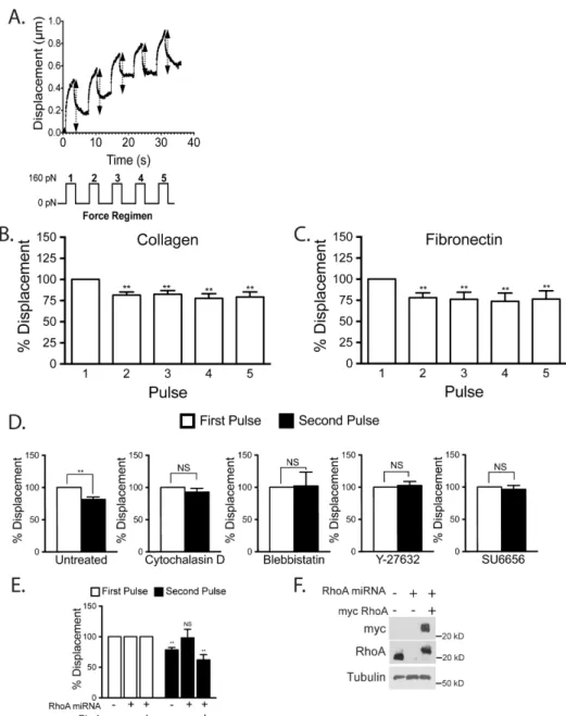
pulses of 160 pN force on ICAM-1 and tracked the bead location during each pull (Figure 7A). There was no statistically significant difference in the initial average displacements of the beads on cells grown on collagen (0.5 µm) or fibronectin (0.4 µm). We observed that after the first pulse of force subsequent pulses did not
displace the beads as much, indicating cellular stiffening (Figure 7B). This stiffening response occurred whether the ECs had been cultured on a fibronectin or collagen ECM, revealing that the response was not affected by the integrins through which the ECs were adhering to the matrix (Figure 7B,C). Since there was little change in bead displacement between the second pulse and subsequent pulses, for most experiments we have compared the bead displacement generated by the first and second pulse.
To explore the basis for the force-induced stiffening, we examined the effects of agents that perturb the cytoskeleton. The average initial bead displacement for control cells, and cells treated with blebbistatin, cytochalasin D, Y-27632 and SU6656 were 0.4, 0.6, 0.8, 0.4, and 0.6 µm, respectively. The stiffening response was blocked by disrupting the actin cytoskeleton with cytochalasin D or by inhibiting myosin activity with blebbistatin (Figure 7D). To inhibit the RhoA/ROCK pathway we used the ROCK inhibitor Y-27632 (Figure 7D) and used adenoviral delivery ofmiRNA to knockdown RhoA expression (Figure 7E, F). We found that knockdown of RhoA as well as inhibition of ROCK both inhibited the force-induced stiffening response. The SFK inhibitor, SU6656, also was able to prevent any change in bead
Figure 7: Mechanical force on ICAM-‐1 increases cellular stiffening
Magnetic beads coated with ICAM-‐1 mAb were added to a monolayer of TNF-‐treated HMVECs. Magnetic tweezers were used to apply pulses of force to individual beads and bead movement recorded with high-‐ speed video. (A) Typical displacement of a bead bound to ICAM-‐1. Arrows denote displacement distance (Top). A diagram of the 160 pN force regimen used (3s of force with 5s recovery for 5 pulses) (Lower). Percentage bead displacement in response to sequential pulses of force for ECs plated on collagen (B) or fibronectin (C). For D-‐F, the ECs were plated on collagen. (D) Bead displacements on HMVECs treated with specified inhibitors for 30 min followed by 2 pulses of force. (E) Bead displacement on HMVECs and HMVECs treated with miRNA to inhibit RhoA expression with or without rescue with myc-‐RhoA. (F) Western blotting confirms RhoA knockdown and myc-‐RhoA re-‐expression. (B-‐E) Quantification of bead displacement with each pulse normalized to the first pulse. Asterisks shows p-‐value of statistical significance compared to the control (*, p≤0.05; **, p≤0.01). The means ± SEM of ≥9 independent bead pulls are shown.
ECs respond to mechanical force on ICAM-1 and the observed stiffening response is dependent on the actin cytoskeleton, myosin activity, RhoA signaling and SFK
activity.
RhoA is activated by mechanical force on ICAM-1
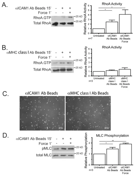
After we had determined that the cellular stiffening was dependent on RhoA expression and actomyosin contractility we next wanted to examine RhoA activity levels. A considerable body of work has revealed the importance of RhoA within ECs in facilitating the passage of leukocytes across the endothelium (15, 16, 31, 57). To cluster ICAM-1, we incubated cells for 15 min with magnetic beads coated with aICAM-1 mAb, and then applied force with a permanent magnet placed above the cell culture dish for 1 min to provide ~10 pN of force (Figure 8A). Consistent with previous findings (14, 15, 31), ICAM-1 clustering increased RhoA activity over untreated cells (Figure 8A). RhoA activity was further increased within 1 min of mechanical force on the ICAM-1 bead clusters (Figure 8A). To evaluate if the
observed activation of RhoA was specific to ICAM-1, we clustered and applied force on MHC class I. Neither clustering, nor force application on MHC class I significantly affected RhoA activity (Figure 8B), confirming that the activation of RhoA is not a universal response to tension on the cell surface. Both MHC class I beads and the ICAM-1 mAb coated beads were able to bind to the EC monolayer as seen by phase contrast microscopy (Figure 8C). MLC phosphorylation is frequently elevated
Figure 8: Mechanical force on ICAM-‐1 increases RhoA activity and MLC phosphorylation
ICAM-1 signaling activates LARG
While a downstream role for RhoA activity after ICAM-1 engagement has long been established, the GEF mediating activation of RhoA has not been determined. Using the binding of GEFs to nucleotide-free mutant RhoAG17A as an indicator of GEF activation (229), we tested several candidate GEFs including LARG,
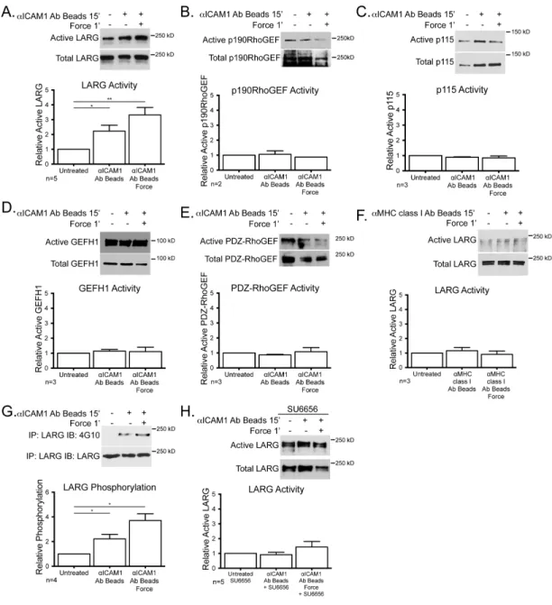
p190RhoGEF p115RhoGEF, G-H1 and PDZ-RhoGEF, but only observed activation of LARG in response to ICAM-1 clustering (Figure 9A-E). There was an additional increase in LARG activity when force was applied to the clustered ICAM-1 (Figure 9A). Neither clustering MHC class I, nor applying tension on this receptor affected LARG activity (Figure 9F). ICAM-1 clustering induced LARG tyrosine
phosphorylation and application of force on ICAM-1 further elevated this
Figure 9: LARG is activated downstream of ICAM-‐1 clustering alone and enhanced with mechanical force
TNF-‐treated HUVECs were treated with mAb-‐coated beads. (A-‐F) GEF activity was determined by affinity purification via GST-‐RhoAG17A and detected by immunoblotting for the specified GEF, LARG (A and F),
p190RhoGEF (B), p115 (C), GEF-‐H1 (D), PDZ-‐RhoGEF (E). (G) LARG was immunoprecipitated and immunoblotted for phosphotyrosine and LARG. (H) Active LARG was detected by sedimentation with GST-‐ RhoAG17A in the presence of SU6656. For all experiments, a representative blot of ≥2 independent
Knockdown of LARG expression inhibits RhoA activation downstream of ICAM-1 clustering
10: Confirmation of LARG knockdown
HMVECs were treated with control or LARG shRNA lenti-‐virus for 48 h and selected for with 2.5 ng/ml puromycin for 24 h. (A) EC lysates were immunoblotted with the indicated pAb. (B) Western blotting shows that ICAM-‐1 expression before and after TNF-‐treatment is not affected by LARG knockdown. (C) Electrical impedance was used to measure monolayer integrity for HMVECs plated at high density for 72 h. No significant difference was found in impedance values after control or LARG knockdown. n=3 independent experiments preformed in triplicate wells.
To determine if LARG knockdown affected the cellular stiffness at ICAM-1 clusters, we used magnetic tweezers as in Figure 7. We measured the stiffness of the cells with a single pulse of force on ICAM-1 (Figure 11C). The stiffness
measured was 50 Pa in control cells compared to 37 Pa in LARG knockdown cells. While there was a reproducible trend of ECs becoming softer after LARG
knockdown, this difference was not statistically significant. However, after LARG knockdown there was a loss of the adaptive stiffening at ICAM-1 clusters in
Figure 11: LARG mediates EC response to mechanical force on ICAM-‐1 and affects neutrophil crawling and TEM
To determine whether ICAM-1-induced stiffening might contribute to increased leukocyte migration over the EC surface, we disrupted this pathway by knocking down LARG expression. Neutrophils were plated on a TNF-treated EC monolayers and live cell imaging was used to calculate the average velocity of neutrophil migration. For neutrophils crawling on control knockdown ECs, the
average velocity was 3 µm/min, whereas after LARG knockdown in ECs the average velocity decreased to 2.5 µm/min (Figure 11E). Given that leukocytes migrate more rapidly over stiffer surfaces, these results are consistent with LARG-dependent stiffening of ECs induced by neutrophil traction enhancing neutrophil migration over the EC surface.
Endothelial LARG contributes to leukocyte TEM
To determine whether endothelial LARG contributes to neutrophil TEM, we counted and compared the number of neutrophils crossing a control shRNA EC monolayer with the number crossing a monolayer in which LARG expression had been decreased by shRNA. The percentage of leukocytes crossing the EC
monolayer after LARG knockdown was decreased by ~35% compared with the control EC monolayer (Figure 11F). These results show that LARG activity in ECs promotes both neutrophil migration over the endothelial surface as well as neutrophil TEM.
Discussion
occur, leukocytes must first adhere to the endothelium and this is mediated by receptors on both the leukocyte and ECs. ICAM-1 is a key endothelial receptor which functions as a ligand for β2 integrins on the surface of leukocytes, promoting
leukocyte spreading and migration (6). However, ICAM-1 is more than an adhesive ligand, its engagement and clustering by the leukocyte generates many signals in ECs that promote TEM (22). It is widely considered that increased RhoA activity downstream from ICAM-1 clustering (16, 31) contributes to leukocyte TEM both by weakening the junctions and increasing tension on them to open them (15, 22, 23, 59, 60, 230). At the outset of this work, we were interested in identifying the GEF(s) responsible for RhoA activation downstream of ICAM-1, and secondly, we were interested in determining whether the tractional forces exerted on ICAM-1 as leukocytes migrate affect RhoA signaling. Here we identify LARG as the critical RhoA GEF activating RhoA downstream of ICAM-1, show that it is activated by SFK-dependent tyrosine phosphorylation, and demonstrate that applying mechanical force on ICAM-1 clusters equivalent to the forces generated by migrating neutrophils enhances this signaling pathway. This is the first report of RhoA activation
Our first goal in this work was to identify the GEF(s) downstream from ICAM-1 responsible for activating RhoA. Several RhoA GEFs have been identified in
not sufficient to restore RhoA activity downstream of ICAM-1, however it may well be that p115RhoGEF activity compensates for other signaling pathways in the LARG knockdown cells. Our results strongly suggest a pathway in which clustering of ICAM-1 activates SFKs that phosphorylate and activate LARG. Our results also indicate that mechanical tension on ICAM-1 clusters enhances this pathway leading to higher levels of LARG tyrosine phosphorylation, increased activation and elevated levels of GTP-loaded RhoA. These findings point to LARG as the major regulator of RhoA activity downstream of ICAM-1 signaling.
Previous studies examining endothelial compliance have obtained conflicting results in response to leukocyte adhesion. Initially, using magnetic twisting cytometry to pull on integrins, it was found that clustering of ICAM-1 or adhesion of neutrophils to ECs induced a stiffening response (233, 234). This is of great interest as it is the region of the cell the leukocyte would be in contact with and sensing. In contrast, subsequent work by the same group using atomic force microscopy found transient and localized softening of the endothelial surface in a zone around where neutrophils adhered but an increased stiffening of adjacent cells (235). These differences likely result in part from the different techniques used to measure stiffness, but they may also reflect slight differences in culture conditions with the former favoring