CELL AND MATRIX DYNAMICS DURING BRANCHING MORPHOGENESIS
Jill Shizuko Harunaga
A dissertation submitted to the faculty of the University of North Carolina at Chapel Hill in partial fulfillment of the requirements for the degree of Doctor of Philosophy in the
Department of Biology.
Chapel Hill 2014
Approved by:
iii ABSTRACT
JILL S. HARUNAGA. Cell and Matrix Dynamics during Branching Morphogenesis (Under the direction of Kenneth M. Yamada)
During embryonic development when tissues are particularly plastic, cells within a tissue can often interact with their surrounding extracellular matrix in a reciprocal manner: the cells remodel the matrix, but the matrix can induce signaling and changes in cell behavior, which in turn can affect matrix remodeling to sculpt tissue architecture. A specialized type of extracellular matrix is the basement membrane, which underlies or encapsulates epithelial tissues. We have used the embryonic mouse salivary gland as a model to study cell-basement membrane interactions during branching morphogenesis.
We first focused on whether the cells in contact with the basement membrane (termed outer bud cells) behave differently from cells that remain in contact only with other epithelial cells (inner bud cells). Using a transgenic mouse expressing a photoconvertible fluorescent probe to optically highlight small populations of cells within developing salivary glands, we tracked their migration. The outer cells migrated much more rapidly than the inner cells and each cell population required different proteins for their migration. Therefore, there are two distinct populations of epithelial cells that utilize two different modes of migration in the salivary gland.
iv
v
ACKNOWLEDGEMENTS
I would like to thank my family and my husband for their unconditional support and encouragement throughout my pursuit of science. I strive to make you proud. To my advisor and the members of my committee, thank you for challenging me to be a better scientist, I have learned so much from you. To the members of the Yamada laboratory, past and present, I am so grateful for your enthusiasm and scientific thoughtfulness. You all make coming to work an enjoyable and inspiring experience. To my wonderful science teachers and mentors at Kamehameha High School, the Cancer Research Center of Hawaii, and the University of Southern California that started me on this path, I couldn’t have done it without you. And thank you to everyone at the National Institutes
vi
TABLE OF CONTENTS
LIST OF FIGURES ... x
LIST OF ABBREVIATIONS ... xii
CHAPTER 1: Dynamic cell-matrix interactions sculpt morphogenesis ... 1
The basement membrane: a specialized extracellular matrix ... 1
The embryonic salivary gland as a model for tissue-matrix interactions ... 3
Branching morphogenesis requires basement membrane proteins ... 4
Actomyosin contractility in branching morphogenesis... 7
Cell motility during branching morphogenesis ... 10
Basement membrane remodeling in development ... 11
Sculpting and stabilizing tissue ... 12
Inducing proliferation and dynamic cell motility ... 16
Inducing cell invasion ... 18
CHAPTER 2: Epithelial cell dynamics in branching morphogenesis ... 23
Preface ... 23
Introduction ... 24
vii
Epithelial cell migration and morphology are region-specific
in the salivary gland... 25
Distinct populations of epithelial cells use different modes of migration ... 27
Discussion ... 29
CHAPTER 3: Basement membrane remodeling and dynamics allows epithelial expansion during branching morphogenesis ... 41
Preface ... 41
Introduction ... 42
Results ... 43
The basement membrane surrounding several embryonic branched organs is perforated ... 43
The micro-perforations span the entire basement membrane ... 44
Basement membrane micro-perforation occurs while the tissue is rapidly expanding ... 45
Extensive perforation of the basement membrane increases its distensibility ... 46
Both protease activity and myosin II contractility are required for perforation formation and maintenance ... 47
Discussion ... 49
CHAPTER 4: Rearward translocation of the basement membrane helps to shape branching of the developing salivary gland ... 62
Preface ... 62
Introduction ... 63
viii
The epithelium translocates the basement membrane towards the duct,
where it accumulates ... 64
Basement membrane translocation requires actomyosin contractility and cell-matrix adhesion ... 66
Discussion ... 68
CHAPTER 5: Conclusions and perspective ... 78
MATERIALS AND METHODS ... 85
Dissection and ex vivo organ culture ... 85
Inhibitors ... 86
Epithelial rudiment dissection ... 86
Immunostaining ... 87
Perforation area analysis ... 87
Antibody conjugation ... 88
KikGR photoconversion and live imaging ... 88
Basement membrane and GFP live imaging ... 89
Basement membrane photobleaching ... 90
Bud expansion analysis ... 91
Basement membrane intensity analysis ... 91
Basement membrane distensibility analysis ... 92
EdU proliferation analysis ... 93
ix
x
LIST OF FIGURES
CHAPTER 1:
Figure 1.1. Branching of the mouse salivary gland ex vivo. ... 22
CHAPTER 2:
Figure 2.1. Region-specific florescent highlighting in KikGR
salivary gland epithelial cells. ... 33 Figure 2.2. The outer bud cells were highly migratory compared to the inner bud and duct cells. ... 34 Figure 2.3. Epithelial cells in different regions of the salivary gland exhibit different velocities. ... 35 Figure 2.4. The outer bud cells are highly pleomorphic. ... 36 Figure 2.5. Changes in the velocities of regional cell migration with the addition of various inhibitors. ... 37 Figure 2.6. Salivary gland morphological changes with various inhibitors. ... 39 Figure 2.7. The developing salivary gland is easily adaptable. ... 40
CHAPTER 3:
Figure 3.1. Micro-perforated basement membranes are present in
multiple embryonic organs. ... 54 Figure 3.2. Micro-perforations penetrate through the entire
basement membrane. ... 55 Figure 3.3. Basement membrane holes are associated with specific developmental stages. ... 56 Figure 3.4. Characterization of basement membrane micro-perforations. ... 57 Figure 3.5. Micro-perforations increase basement membrane distensibility. ... 58 Figure 3.6. Formation and maintenance of basement membrane micro-perforations require protease activity. ... 59 Figure 3.7. Salivary gland bud outgrowth decreases as basement membrane
xi
Figure 3.8. Both protease and myosin II activity are required for the micro-perforations. ... 61
CHAPTER 4:
Figure 4.1. The basement membrane translocates towards the duct where it accumulates. ... 73 Figure 4.2. The epithelium alone can pull the basement membrane rearward. ... 75 Figure 4.3. The epithelial cells are not moving in concert towards the duct and simply towing the basement membrane rearward. ... 76 Figure 4.4. Inhibition of cell-matrix adhesion, actomyosin contractility, and protease activity inhibit basement membrane translocation. ... 77
CHAPTER 5:
xii
LIST OF ABBREVIATIONS
ADAM A disintegrin and metalloprotease
BB-94 Batimastat
Btbd7 BTB (POZ) domain containing 7
BSA Bovine serum albumin
DMSO Dimethyl sulfoxide
E# Embryonic day #, eg. E12 means embryonic day 12, 12 days after observation of a vaginal plug
EGF Epidermal growth factor
EMT Epithelial-to-mesenchyme transition
FAK Focal adhesion kinase
FGF or FGFR Fibroblast growth factor or Fibroblast growth factor receptor
FRAP Florescence recovery after photobleaching
GAG Glycosaminoglycan
GFP Green fluorescent protein
HB-EGF Heparin binding epidermal growth factor
KikGR Kikume green-red, a photoconvertible fluorescent probe
MMP or MT-MMP Matrix metalloprotease or Membrane-tethered matrix metalloprotease
ROCK Rho associated protein kinase
1
CHAPTER 1
Dynamic cell-matrix interactions sculpt morphogenesis
The basement membrane: a specialized extracellular matrix
2
3
The embryonic salivary gland as a model for tissue-matrix interactions
Branching morphogenesis is an essential developmental process by which several mammalian organs such as the lung, kidney, mammary and salivary glands gain epithelial surface area for secretion or adsorption (Harunaga et al., 2011; Kim and Nelson, 2012; Patel et al., 2006). The process begins with a single epithelial bud surrounded by basement membrane and dense mesenchyme. The epithelium expands and divides through repetitive rounds of branching to form a highly arborized structure, which allows for maximal surface area confined within a small space. As the epithelial tissue expands and branches, it remains encapsulated by a basement membrane (Rozario and DeSimone, 2010; Timpl and Dziadek, 1986). Because the basement membrane provides such important cues, and because the epithelium expands rapidly during development, the basement membrane must also be rapidly remodeled to accommodate the expanding tissue in order to continue to surround the epithelium.
4
beginning at E12.5 when the single epithelial bud becomes divided by clefts; the clefts then elongate to delineate ducts; this branching is followed by lumen formation and differentiation beginning at E15 (Patel et al., 2006). Salivary glands cultured ex vivo are ideal for fluorescence live imaging because they flatten more than the other branched organs while still maintaining their three-dimensional shape, allowing for crisper images with less background fluorescence. Our ex vivo approach allows us to study how cell-mediated remodeling of a native basement membrane affects tissue architecture and reciprocally, how basement membrane affects cell dynamics in a whole organ.
Branching morphogenesis requires basement membrane proteins
5
suggests that collagen, which is absent from the “morphologically active” end buds, is a stabilizing component in the cleft and duct regions (Grobstein and Cohen, 1965; Kallman and Grobstein, 1965). In fact a collagenase inhibitor can stimulate cleft formation and increases collagen-like fibrils on the surface of the salivary gland epithelium early in development (Nakanishi et al., 1986). Conversely, inhibition of collagen synthesis, but not inhibition of collagen crosslinking, inhibited branching in both the salivary gland and lung (Spooner and Faubion, 1980).
The importance of glycosaminoglycans (GAGs) such as chondroitin sulfates or heparan sulfate, and their extracellular matrix proteoglycans including perlecan, in branching has also been demonstrated in the embryonic salivary gland. Complete removal of the GAGs results in a severe inhibition of salivary gland branching morphogenesis (Thompson and Spooner, 1982). The other main components of the basement membrane, laminin and fibronectin, are also required for branching morphogenesis (Rebustini et al., 2007; Sakai et al., 2003). Laminin is necessary for development: treatment of embryonic lungs or salivary glands with laminin function-blocking antibody decreased branching (Kadoya et al., 1995; Schuger et al., 1990). Mice deficient for an isoform of laminin, Lama5, exhibit a significant delay in branching of the salivary gland, and once branching does commence, the epithelium appears disorganized and lumen formation is disrupted (Rebustini et al., 2007). Fibronectin is required for branching of the embryonic salivary gland (Sakai et al., 2003), but excess fibronectin and its associated receptor α5β1 integrin inhibit branching (Joo and Yamada, 2014).
6
regulators are important as well. Proteases from the metalloprotease family are the major players in extracellular matrix remodeling, including matrix metalloproteases (MMPs) and a disintegrin and metalloprotease with thrombospondin motifs (ADAMs) (Lu et al., 2011). This very large family of proteases cleaves a wide range of matrix proteins; they also act on protease precursors and growth factors, activating them through cleavage. Their activity is often redundant, making it difficult to pinpoint specific functions (Lu et al., 2011). The Mmp-14 knock-out mouse displays decreased branching of the salivary gland and significantly reduced collagen metabolism (Holmbeck et al., 1999; Oblander et al., 2005). Adam17-null embryos exhibit decreased terminal bud numbers and increased extracellular matrix accumulation in their salivary glands compared to wild-type controls (Melnick and Jaskoll, 2000). Finally, MMP-2 and MMP-3 activities have been shown to mediate side and primary branching, respectively, in murine mammary gland organoid cultures (Wiseman et al., 2003).
7
Actomyosin contractility in branching morphogenesis
8
Branching is inhibited in embryonic mouse lungs with ROCK inhibitor treatment without affecting proliferation rates, resulting in a decreased number of enlarged end buds that fail to form clefts (Moore et al., 2005). Conversely, treatment with cytotoxic necrotizing factor-1 to increase Rho activity and contractility has a biphasic effect on lung branching, increasing the number of end buds at low doses and decreasing the bud number at high concentrations (Moore et al., 2005). Pharmacologically modulating the contractility of the lung also increases the thickness of the basement membrane, which is typically thinner at the tips of the end buds (Moore et al., 2005). How tissue contractility changes the thickness of the basement membrane is unknown, but it is postulated that protease activity is tension-mediated (Moore et al., 2005). Increased cellular tension could decrease protease activity, as it has been shown that increased tension from cell spreading and integrin binding eventually decreases MMP2 activity by decreasing expression of MT1-MMP in endothelial cells (Moore et al., 2005; Yan et al., 2000). Myosin II also regulates cell shape and branch formation in the lung (Plosa et al., 2012). Treatment with blebbistatin results in a decrease in the organization of the epithelial cells in the stalk region, which is analogous to the duct region in the salivary gland, and an increase in cell size (Plosa et al., 2012). Whether the change in cell size and organization is related to the changes in basement membrane thickness observed previously is unknown.
9
al., 2009). Localized fibronectin assembly also induces cell proliferation to expand the bud outward as the cleft progresses inward (Daley et al., 2009). In addition to mediating assembly of fibronectin in the basement membrane, ROCK regulates basement membrane deposition and polarization of the peripheral epithelial cells in a myosin II-independent manner (Daley et al., 2012). Treatment with Y27632 but not blebbistatin induces ectopic deposition of basement membrane components within the epithelial bud instead of just at the epithelial-mesenchyme interface; this disrupts polarization of the outer bud epithelial cells, which usually are columnar-shaped and well-organized along the basement membrane (Daley et al., 2012; Onodera et al., 2010).
10
mammary glands the end buds appear to collectively invade outward (Daley et al., 2009; Ewald et al., 2008; Ewald et al., 2012; Onodera et al., 2010).
Cell motility during branching morphogenesis
Branching is a complex, dynamic process that requires extensive remodeling of tissue, and the epithelial cells display surprisingly high levels of cell motility. These cell movements during branching morphogenesis have been suggested to contribute to the plasticity of tissues during the rapid architectural rearrangements of early organ formation. Confocal timelapse microscopy of GFP-adenovirus infected salivary glands revealed extensive random, individual cell motility of the epithelial cells during development (Larsen et al., 2006). The epithelial cells of the developing mouse mammary gland and Drosophila melanogaster (Drosophila) trachea also display a high degree of cell motility (Ewald et al., 2008; Ewald et al., 2012; Metzger and Krasnow, 1999). The epithelial cells of the developing kidney also move, though significantly less than observed in the salivary gland (Shakya et al., 2005). Higher-power imaging showed that salivary gland epithelial cells migrate as single cells in random patterns (Larsen et al., 2006), but epithelial cells from the mammary gland migrate both individually and as collective groups (Ewald et al., 2008; Ewald et al., 2012). Preliminary observations suggested that the salivary gland outer bud and cleft epithelial cells that are in contact with the basement membrane are especially motile.
11
adherens, tight, and desmosome-based junctions of very early epithelia are lost when oral epithelial cells undergo branching morphogenesis to form buds (Hieda et al., 1996; Kadoya and Yamashina, 1993). In post-natal branching mammary glands, adherens and tight junctions also appear to be lost during branching morphogenesis, although desmosomes remain (Ewald et al., 2012). Because the cells are so motile, the tissue is likely extremely plastic throughout development, and perhaps the basement membrane surrounding the remodeling tissue is also highly dynamic.
Basement membrane remodeling in development
12 Sculpting and stabilizing tissue
Historically, the basement membrane was typically thought to be predominantly structural, providing support to the epithelial tissue it surrounds (Rozario and DeSimone, 2010). While more recently the basement membrane has been shown to be a dynamic structure with several other functions in addition to structural support, how tissues organize and remodel their basement membranes to dictate form and function remains unclear. Several cell types can orient and slide extracellular matrix fibers, which may help to guide and stabilize tissue architecture.
13
results in rapid rounding of the egg, suggesting it is the basement membrane and not the cells themselves that maintains the elongated structure of the tissue (Haigo and Bilder, 2011). Tissue rotation induces basement membrane polarization, and the basal actin filaments in the epithelial cells become planar polarized along the same axis as the basement membrane (Gutzeit et al., 1991). Cell migration of the underlying follicle cells is also required for the polarization of the basement membrane (Lewellyn et al., 2013). However, the typical Wnt-mediated planar cell polarity signaling pathways are not required; instead the egg chamber rotation seems sufficient to orient both the basement membrane and the basal actin filaments (Bilder and Haigo, 2012; Viktorinova et al., 2009). Directed deposition of collagen IV by the rotating planar polarized epithelial cells also contributes to the polarization of basement membrane structure (Lerner et al., 2013), which indicates there are multiple facets of regulation (Horne-Badovinac, 2014).
Murine mammary gland acini assembled from single cells also rotate when they are imbedded in basement membrane extract (Tanner et al., 2012; Wang et al., 2013). The rotation is accompanied by planar polarized actin on the basal surface of the epithelial cells and an oriented laminin matrix similar to that observed in the Drosophila
14
In addition to the recent demonstrations that whole tissue movements can physically orient basement membrane, it had been previously observed that the basement membrane varies in thickness in different locations surrounding the epithelium of the branching lung, mammary and salivary glands (Bernfield and Banerjee, 1982; Fata et al., 2004; Mammoto and Ingber, 2010; Mollard and Dziadek, 1998; Moore et al., 2005; Silberstein and Daniel, 1982). In the mammary gland, the basement membrane at the tips of the expanding terminal end buds can be 10 times thinner than the basement membrane along the forming ducts (Fata et al., 2004). Salivary glands treated with collagenase exhibit regression of the clefts and decreased branching (Banerjee et al., 1977; Grobstein and Cohen, 1965). Such clefts divide an epithelial bud into two buds, eventually progressing to a duct during branching morphogenesis of the salivary gland. This regression suggests that the thicker basement membrane found in clefts and ducts could be supporting and stabilizing these structures. Ingber suggests that the varying thickness and thus stiffness of basement membrane exposes the epithelium to varying local force anisotropies, permitting directed tissue expansion only in areas where the basement membrane is thinnest (Mammoto and Ingber, 2010; Moore et al., 2005).
15
collagen III and creating a physical wedge to promote cleft progression (Nakanishi et al., 1988). Perhaps localized assembly and translocation of the basement membrane could create the differences in basement membrane thickness and force anisotropies during branching morphogenesis. Additionally, there could be differences other than thickness between the tip and duct regions in branching organs, such as the amount of crosslinking and composition of the basement membrane that contribute to the architecture of the tissue. For example, the thinner basement membrane at the tips of mammary end buds contains more hyaluronic acid than the thicker basement membrane around the ducts, which likely affects local signaling (Hinck and Silberstein, 2005; Silberstein and Daniel, 1982).
16
metalloproteases or MMPs have been shown to be required branching (Fata et al., 2004; Lelongt et al., 1997; Oblander et al., 2005).
Inducing proliferation and dynamic cell motility
In addition to removing a physical impediment to branching, proteolysis can release bioactive extracellular matrix fragments or growth factors bound in the basement membrane that increase proliferation, motility, and overall branching of the organ (Ortega and Werb, 2002; Patel et al., 2007; Rebustini et al., 2009; Sternlicht et al., 2006). In a feed-forward pathway, MT2-MMP (membrane-type 2 matrix metalloprotease or MMP15) degrades collagen IV in the basement membrane surrounding branching salivary glands, releasing bioactive NC1 domains, which signal through β1 integrin and
17
A basement membrane-associated protein, fibronectin, can signal directly through integrin α5β1 in the clefts of the embryonic mouse salivary gland and alter cell motility
and adhesion. Localized assembly and accumulation of fibronectin rapidly induces Btbd7 [BTB (POZ) domain containing 7] at the base of the clefts, which then up-regulates the transcription factor Snail2 while down regulating E-cadherin (Onodera et al., 2010; Sakai et al., 2003). This is one of the first examples of an extracellular matrix protein directly affecting transcription in the nucleus. The down-regulation of E-cadherin results in decreased cell adhesion at the base of the clefts, which cooperates with Snail2 to induce a more motile population of cells, allowing for dynamic cell rearrangements and progression of the cleft through the epithelium (Onodera et al., 2010). As basement membrane proteins can induce cell migration, they can also reduce migration. During
Xenopus laevis gastrulation, assembly of fibrillar fibronectin is required for blastocoel roof thinning and epiboly but actually slows down convergent extension (Rozario et al., 2009). This suggests that different cell types can respond differently to the same basement membrane protein at different times, adding additional layers of regulation to cell-matrix interactions.
18
Chaudhry, 1973). These contacts are transient and occur at the tips of the epithelial end buds (Cutler and Chaudhry, 1973). The tips of the rapidly expanding end buds are considered areas of high morphogenic potential, exhibiting increased cell proliferation compared to areas adjacent to the clefts in the developing lung (Mollard and Dziadek, 1998). Breaks in the basement membrane also occur in adult organs such as the intestine and lymph vessels (Komuro, 1985; Pflicke and Sixt, 2009; Trier et al., 1990). However, how these breaks in the basement membrane form and how epithelial-mesenchyme interactions may induce differentiation or branching remains unknown.
Inducing cell invasion
Cells also need to physically traverse the basement membrane during development to establish new tissue or to make connections between existing ones (Kelley et al., 2014). This process is especially difficult because the basement membrane usually functions as a barrier to migration, compartmentalizing different cell types. And instead of simply modifying the intact basement membrane as we have described in the previous sections, invading cells and tissue must actually breach a very tough, dense sheet of extracellular matrix. While this process occurs normally in many developmental processes, it can become mis-regulated in pathological conditions, such as cancer metastasis (Fata et al., 2004; Lu et al., 2011; Rowe and Weiss, 2008).
19
integrins and several actin regulatory proteins (Hagedorn et al., 2009). Once the basement membrane is breached, the invasive protrusions are consolidated into one large invadopodium by the netrin receptor, DCC (Hagedorn et al., 2013). This large protrusion physically pushes aside basement membrane locally to widen the breach point for invasion, which demonstrates that mechanical displacement by the cell is an efficient mechanism for removing the basement membrane (Hagedorn et al., 2013). Consistent with this idea, once the anchor cell has crossed the basement membrane, the pore continues to widen further by physical sliding of the basement membrane. Movement and division of the underlying cells could exert traction forces via the C. elegans integrin homolog, INA-1/PAT-3 to slide the basement membrane (Ihara et al., 2011). Protease activity has not yet been shown to be directly involved in this system (Kelley et al., 2014), but may be needed in the initial breach of the basement membrane since the matrix metalloprotease, MT1-MMP, is found at the tips of invadopodia in mammalian cancer cells and is required in invasion and degradation of the extracellular matrix (Artym et al., 2006). General protease activity could also potentially partially degrade the entire basement membrane, making it more pliable and allowing the surrounding cells to displace and slide it laterally more easily.
20
there are needed. Interestingly, the basement membrane in lymph vessels is discontinuous, and dendritic cells can enter and exit without the assistance of proteases or integrin mediated adhesion (Pflicke and Sixt, 2009). These discontinuities may be a result of localized decreased deposition of basement membrane, persisting for at least 24 hours and capable of dilating as a cell pushes through (Pflicke and Sixt, 2009).
Another system that has been recently described utilizes mechanical force to breach a weakened basement membrane in mouse embryogenesis. The developing embryo is spatially restricted by the uterus after implantation, inducing elongation and increased mechanical stress at the distal tip of the embryo (Hiramatsu et al., 2013). Decreased collagen IV deposition, but not protease activity, at the distal tip of the embryo thins the basement membrane in combination with this increased pressure, contributes to breaching of the basement membrane (Hiramatsu et al., 2013). The early epiblast cells migrate through these gaps in the basement membrane to establish the distal visceral endoderm (Hiramatsu et al., 2013). The pressure of the whole tissue on the basement membrane and decreased deposition contribute significantly to the rupture of the basement membrane, but dynamic shape and organization changes in the tip cells could also contribute local mechanical forces to facilitate the process of invasion.
21
22
Figure 1.1. Branching of the mouse salivary gland ex vivo.
23 CHAPTER 2
Epithelial cell dynamics in branching morphogenesis
Preface
This chapter contains work from our highly collaborative project that was published in Developmental Dynamics in 2013 on which I am a co-first author, entitled “Epithelial cell dynamics in branching morphogenesis” (Hsu et al., 2013). Jeff Hsu,
24 Introduction
Previous work from our laboratory utilizing GFP-adenovirus infected submandibular salivary glands found that the epithelial cells were highly motile throughout development, and that they became nearly static after birth; these findings suggested this cell motility may be important for morphogenesis of the mouse submandibular gland (Larsen et al., 2006). Although adenovirus was a powerful tool, it penetrated the dense mesenchyme relatively poorly and only randomly infected occasional epithelial cells. A conceptual point of concern about this approach was the possibility that only a certain subpopulation of cells was susceptible to infection and thus labeled for tracking, which could have skewed the results of the motility analysis. Because the mesenchyme was a barrier to infection, mesenchyme-free epithelial rudiments were also used in that previous study for more efficient infection and imaging. This type of culture requires supplementation by several growth factors, including FGF7 and EGF, which could have had stimulatory effects on cell motility. A similar study was subsequently conducted in organoid cultures of the mammary gland (Ewald et al., 2008), and it also suggested that high epithelial motility is important for the development of several branched organs. This latter study, however, had the same potential limitations as the work conducted by our laboratory on the salivary gland.
25
photoconversion of small numbers of cells at any location in the gland allowed us to track specific populations of cells within the intact salivary gland, for example comparing those cells in contact with the basement membrane to those surrounded only by other epithelial cells.
Results
We first confirmed the results of the previous report that the epithelial cells were indeed highly motile and did not migrate collectively as seen in the mammary gland organoids (Ewald et al., 2008; Ewald et al., 2012); instead, the cells moved independently in random patterns. For these experiments, we dissected the salivary glands at E13, cultured the glands overnight, and imaged the following day for 12 hours at 10 minute intervals. We utilized a 405 nm laser controlled by the iLAS system on our spinning disc confocal microscope to photoconvert specific epithelial cells in different regions of the salivary gland to track differences in motility that were based on location. While we could control the size of the region for photoconversion in X-Y very precisely, the 405 nm laser was not confocal. Consequently, we were unable to limit exposure of the sample in the Z-dimension; the result was photoconversion of a column of cells above and below the focal plane. This pattern of photoconversion was not a problem, however, because it allowed us to track multiple cells per bud after each photoconversion, since the cells were highly motile in both X-Y and Z directions.
Epithelial cell migration and morphology are region-specific in the salivary gland
26
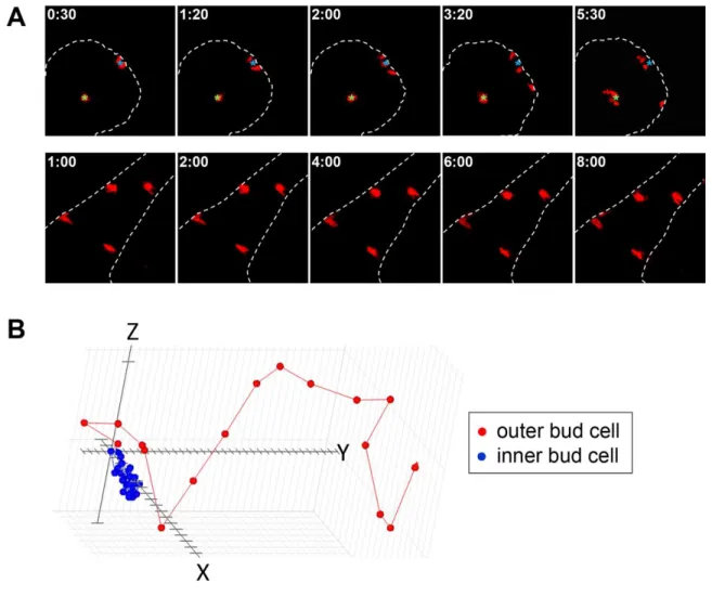
morphologically distinct regions of the epithelia -- the primary duct, inner bud, and outer bud (Fig. 2.1). The cells in the outer bud region and primary duct contacted the basement membrane, and the inner bud cells were at the center of the bud surrounded only by other epithelial cells. There were substantial differences in both the rates of migration and net displacement of the cells in the different regions (Fig. 2.2). This analysis also highlighted the random individual motility of the salivary gland epithelial cells, which contrasts with several other well-known developmental systems where the epithelia migrate collectively as a sheet in the Drosophila egg chamber and during gastrulation, and as groups of cells during neural crest development (Bilder and Haigo, 2012; Kerosuo and Bronner-Fraser, 2012; Nakaya and Sheng, 2009). The outer bud cells were highly motile, migrating an average of 20 µm/hr, which was significantly faster than the inner bud and duct cells that migrated at average rates of 8 and 5 µm/hr, respectively (Fig. 2.3A). The inner cells migrated in relatively circular motions at the middle of the bud, staying very close to the starting point throughout the tracking period (Fig. 2.2B). In comparison, the outer bud cells migrated significantly further than the inner bud cells, often traveling laterally along the basement membrane. Though the migration of the duct cells was calculated to be 5 µm/hr, the cells were almost completely static, translocating only with the overall expansion of the duct as a whole (Fig 2.2A).
27
visualize more clearly each individual epithelial cell; the outer bud cells were highly dynamic, frequently extending protrusions and flattening themselves in contact the basement membrane (Fig. 2.4). These outer bud cells were elongated and often migrated laterally along the edge of the basement membrane, frequently bumping up against it (Fig. 2.4B). They exhibited a strong affinity for the basement membrane, leaving the basal boarder only periodically and then immediately returning, often after dividing. The outer cells lost their elongated morphology when they moved away from the basement membrane. In contrast, the internal epithelial cells, particularly those at the center of the end bud, were more rounded and irregularly shaped (Fig. 2.3D). These inner epithelial cells almost never came into contact with the basement membrane and remained confined to the center of the bud. Consequently, the two cell populations within the epithelial bud rarely intermixed.
Distinct populations of epithelial cells use different modes of migration
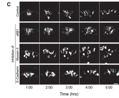
To determine whether this extensive individual cell migration is important for development of the salivary gland, we used inhibitory antibodies and pharmacological inhibitors of proteins integral to the cellular migration machinery, such as integrins and myosin II. Cells interact with the surrounding matrix via integrin attachments, which have been previously shown to be important for branching morphogenesis as a whole (Kadoya et al., 1998; Kadoya and Yamashina, 1993; Kadoya and Yamashina, 2010; Sakai et al., 2003). We hypothesized that the defects in branching after integrin inhibition could have resulted from a defect in cell migration. Inhibitory antibodies to the β1 and α6
28
with no effect on inner bud cell motility (Fig. 2.5). Branching morphogenesis was also significantly impaired as previously reported (Fig 2.6B). Immunostaining revealed a loss of organization of the outer bud cells and aberrant E-cadherin localization to the edge of the cell closest to the basement membrane (Fig. 2.6B). The basement membrane, as visualized by collagen IV, also appeared less condensed and less well-organized. These findings underscore the importance of cell-matrix interactions in branching morphogenesis.
29
Since the inner bud cells did not depend on integrins or myosin II for migration, we next inhibited the protein responsible for cell-cell adhesion, E-cadherin. The cortex of inner bud cells displayed an uninterrupted ring of E-cadherin, in contrast to the outer bud cells, which normally had E-cadherin localized to only three sides of the cell; the edge in contact with the basement membrane lacked any E-cadherin. We found that E-cadherin inhibition had very little effect on the motility of the outer cells, but it did increase the motility of the inner bud cells by more than two-fold (Fig. 2.5). Interestingly, the inhibitory E-cadherin antibody did not have any effect on branching morphogenesis after 20 hours of treatment, even though the inner bud cells were undergoing apoptosis (Walker et al., 2008) during this time, leaving large holes in the middle of the buds (Fig. 2.6C). In contrast, the outer cells were unaffected in their rates of motility and displayed only a slightly less-columnar morphology. However, after 24 hours in the presence of inhibitory E-cadherin antibody, branching slowed, and the size and number of buds were decreased significantly compared to control as previously described (Walker et al., 2008).
Discussion
Inhibition of α6/β1 integrin or myosin II decreased only outer cell motility,
30
restricts the individual motility of the inner bud cells. This finding is consistent with the observed decrease in cell motility as the gland matures and increases its cell-cell adhesions (Hieda et al., 1996; Larsen et al., 2006). Furthermore, both mammary glands and oral epithelial cells lack adherens junctions and tight junctions in early development (Ewald et al., 2008; Hieda et al., 1996; Kadoya and Yamashina, 1993), suggesting that the loss of strong cell-cell attachment to permit high rates of epithelial cell migration is integral to proper development of the glands. Although E-cadherin inhibition slightly decreased the velocity of the outer bud cells, the effect was not nearly as drastic as inhibition of either α6/β1 integrin or myosin II (Fig. 2.5), indicating that the outer cells
predominately use a cell-matrix mode of migration. Nevertheless, we have observed cells moving away from the basement membrane and then returning, which suggests that the outer bud cells have the ability to switch quickly these modes of migration.
31
The individual, random motility of the epithelial cells could also allow the developing gland to be more plastic and therefore more robust because the gland has the ability to adapt, ameliorating mistakes during development. While there is no standard branching pattern of the salivary gland, unlike the precisely stereotyped pattern of lung branching (Metzger et al., 2008), the gland has some undetermined mechanism to regulate cleft formation such that two clefts do not elongate too close to each other in order to prevent the formation of small, uneven end buds. On the rare occasion that we have observed the formation of more than one cleft, the gland adapted and the two clefts became one (Fig. 2.7); if the outer epithelial cells were not highly motile, this correction would not have been possible. In contrast, the duct exhibited very little morphogenic change over the course of 20 hours, which correlated with very low individual cell motility (Fig. 2.2A). We believe that high cell motility is indicative of plasticity of the tissue and that increased plasticity allows for major changes in tissue architecture during development.
32
In summary, we used transgenic mice expressing the photoconvertible protein KikGR to analyze single cell migration in the developing salivary gland epithelial bud. This approach was ideal, because it allowed us to track small sub-populations of cells anywhere within the intact gland without the potentially confounding effects of the viral infection approach used previously to track cell motility. We identified two distinct populations of cells in the salivary gland epithelium. One population, termed outer bud cells, consists of cells located at the periphery of the bud that are often in contact with the basement membrane. The cells in the middle of the bud termed inner bud cells are surrounded by other epithelial cells on all sides. We tracked the photoconverted cells in 3D and found that the outer bud cells were highly motile, migrating at an average speed of 20 µm/hr, the inner bud cells were significantly slower at 8 µm/hr, and the duct cells were not migratory, moving only due to overall duct expansion at 5 µm/hr. We found that the high rate of migration of the outer bud cells was dependent on both β1/α6 integrin and
33
Figure 2.1. Region-specific florescent highlighting in KikGR salivary gland epithelial cells.
34
Figure 2.2. The outer bud cells were highly migratory compared to the inner bud and duct cells.
35
Figure 2.3. Epithelial cells in different regions of the salivary gland exhibit different velocities.
36
Figure 2.4. The outer bud cells are highly pleomorphic.
37
Figure 2.5. Changes in the velocities of regional cell migration with the addition of various inhibitors.
(A) Average rates of outer bud cell migration (±SEM) in glands treated with a combination of α6 and β1 inhibitory antibodies at 100 mg/ml each, 100 mg/ml
38
39
Figure 2.6. Salivary gland morphological changes with various inhibitors.
Salivary glands at E14 after being treated with no inhibitor (A), a mixture of (Ha2/5) β1 and (GoH3) α6 inhibitory integrin antibodies at 100 mg/ml each (B), 100 mg/ml
40
Figure 2.7. The developing salivary gland is easily adaptable.
41 CHAPTER 3
Basement membrane remodeling and dynamics allows epithelial expansion during branching morphogenesis
Preface
The epithelial cells are highly motile, as we described in the previous chapter, and yet they stay confined within the basement membrane all throughout development. The basement membrane serves as an important physical barrier between the epithelium and mesenchyme compartments, which is particularly important when the epithelial cells are so migratory to prevent tissue mixing. We discuss the role of basement membranes in epithelial expansion this chapter.
For this third chapter, I have adapted parts of my first author paper entitled, “Local and global dynamics of the basement membrane during branching morphogenesis require protease activity and actomyosin contractility” that has been submitted and is
42 Introduction
43
Both the epithelial and mesenchymal cell populations of branching organs can be surprisingly motile during development (Ewald et al., 2008; Larsen et al., 2006; Metzger and Krasnow, 1999; Shakya et al., 2005). In the salivary gland, motility rates of epithelial cells immediately adjacent to the basement membrane can be even higher than in the interior of the buds, with cells migrating at average rates of 20 µm/hr (Hsu et al., 2013). This high rate of epithelial cell motility relies on integrin-dependent interactions with the basement membrane (Hsu et al., 2013). The substantial motility of the epithelial cells during early development underscores the importance of maintaining the basement membrane as a barrier to prevent intermixing of epithelial and mesenchymal cell compartments to permit normal morphogenesis. While many laboratories have investigated how cells invade through a basement membrane, particularly in pathological processes such as cancer metastasis (Rowe and Weiss, 2009), the fundamental question of how normal embryonic epithelia can expand rapidly while remaining enclosed by a basement membrane is poorly understood.
Results
The basement membrane surrounding several embryonic branched organs is perforated
44
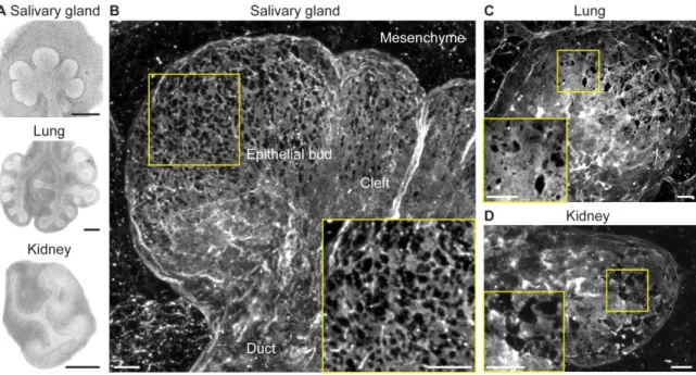
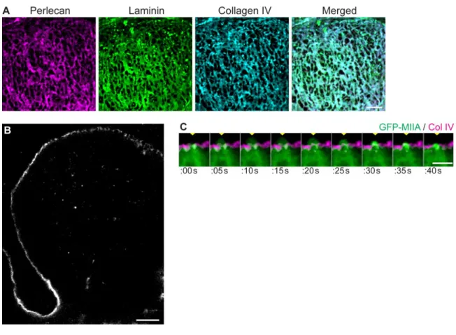
expansion and guide branching behavior. To determine if the basement membrane demonstrates local morphological and structural changes in these regions consistent with this hypothesis, we examined the basement membrane surrounding the end buds of salivary glands, lungs, and kidneys undergoing morphogenesis at stages of rapid expansion (Fig. 3.1A). By imaging using confocal microscopy and creating maximum-intensity projection images of organs immunostained for collagen IV, the characteristic structural collagen of basement membrane, as well as the glycoprotein laminin, revealed that the basement membrane was not uniform, but was instead perforated by numerous well-defined microscopic holes (Fig. 3.1B, 3.1C, 3.1D). The micro-perforations, or holes, were most prominent at regions of the basement membrane surrounding the tips of the expanding end buds and were absent from the cleft and duct areas where expansion was low (Fig. 3.1B).
The micro-perforations span the entire basement membrane
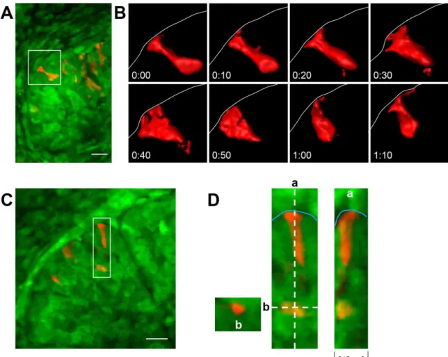
Focusing on developing salivary glands, we found that the holes appeared to penetrate through the entire basement membrane according to immunolocalization of three major constituents of basement membrane, collagen IV, laminin, and perlecan, a heparan sulfate proteoglycan (Yurchenco, 2011). A micro-perforation observed for each of these components could be matched exactly with a corresponding perforation involving each of the other two components (Fig. 3.2A). In single-plane, 0.38 µm optical Z-sections of the bud, the basement membrane appears physically thinner at the tip compared to the base of the cleft (Fig. 3.2B), consistent with previously published results.
45
cells were frequently found to extend cell surface protrusions through and beyond the basement membrane holes (Fig. 3.2C); these bleb-like protrusions could extend as far as 5 µm beyond the basement membrane. These numerous protrusions were highly dynamic and often extended repetitively through the same hole, confirming patency of the micro-perforations. These membrane protrusions were observed only at the tips of end buds, protruding through basement membrane perforations in both embryonic salivary gland and lung.
Basement membrane micro-perforation occurs while the tissue is rapidly expanding
The presence of micro-perforations in the basement membrane was associated with the stage of embryonic salivary gland development with the highest epithelial expansion and outgrowth (Fig. 3.3). We found that at E12.5, when the salivary gland is a single epithelial bud, the basement membrane had no perforations. At E13.5, when the gland was undergoing massive branching and epithelial expansion, the number and size of perforations reached their peak. At E14.5, when branching had slowed significantly and differentiation begins, the basement membrane appeared relatively smooth and free of perforations, as it does in the post-natal gland.
46
density was near the tip (Fig. 3.4B) where 27% of the basement membrane surface area was lost to holes. In terms of frequency, at the tip of the bud, there was an average of 60 holes per 500 µm2 or 3 perforations per cell (Fig. 3.4C). This number fell to only ~1 hole per cell toward the middle (equator) of the bud. The percent perforated area decreased dramatically closer to the center of the bud by almost 7-fold, as the number and size of the perforations decreased (Fig. 3.4C, 3.4D), so that the basement membrane was 94% intact near the center of the bud. The increased concentration of large basement membrane perforations near the tips of the end buds could potentially increase basement membrane flexibility to permit epithelial expansion.
Extensive perforation of the basement membrane increases its distensibility
To determine whether this extensive micro-perforation alters the mechanical flexibility or distensibility of the basement membrane, regions of E13 salivary glands were imaged live in the presence of fluorescently labeled, non-perturbing collagen IV antibody. We found the basement membrane to be highly dynamic (Fig. 3.5). Epithelial cells appeared to continuously “tug” on the matrix such that the micro-perforations
47
An additional method for evaluating basement membrane extensibility was to measure stretching of the basement membrane itself. We tracked pairs of fiduciary marks on the basement membrane surface and found distensibility, defined as the maximum distance between two points subtracted from the minimum distance within a 20 minute time period, was 2-fold higher near bud tips compared to the center of the bud (Fig. 3.5B, 3.5C). There was greater basement membrane stretching in the area of the gland with larger, more numerous basement membrane micro-perforations and high tissue expansion, a finding consistent with enhanced local basement membrane flexibility due to the perforations. The distensibility of the basement membrane was dependent on actomyosin contraction, since these basement membrane dynamics were inhibited by the addition of blebbistatin (Fig. 3.5A).
Both protease activity and myosin II contractility are required for perforation formation and maintenance
micro-48
perforations within four hours (Fig. 3.6C). Treatment with a slightly less broad protease inhibitor, GM6001 (Fig. 3.6C), had similar effects, to a lesser degree compared to glands treated with BB-94; tissue inhibitors of metalloproteases (TIMPs) had no effect (Fig. 3.6D). These data suggest that multiple proteases are involved in forming and maintaining basement membrane perforations.
Immunostaining for collagen IV in glands treated with the protease inhibitor BB-94 revealed enhanced basement membrane accumulation around the end buds (Fig. 3.6B), consistent with reversal of protease-mediated degradation of the basement membrane. Live-organ imaging revealed that treatment of salivary glands with BB-94 gradually inhibited basement membrane motion; after 12 hours, perforations were substantially diminished, collagen IV increased three-fold around the end buds (Fig. 3.7C), and distensibility was significantly reduced (Fig. 3.5B). Additionally, kymograph analysis (Fig. 3.7B) of this 12-hour time period revealed a two-fold reduced rate of bud outgrowth after prolonged protease inhibition (Fig. 3.7D). Although branching morphogenesis was strongly inhibited, this decreased expansion was not accompanied by altered cell proliferation as determined by EdU incorporation (Fig. 3.7E). These findings suggest that the accumulation and stiffening of basement membrane around the end buds after protease inhibition restricted expansion of the end buds and inhibited normal branching morphogenesis of the gland.
micro-49
perforations. Myosin IIA and IIB were found to be enriched at the epithelial cell cortex adjacent to the basement membrane (Fig. 3.8A). At E12.5, treatment with blebbistatin inhibited cleft progression and prevented micro-perforation formation (Fig. 3.8B, 3.8C). At E13.5, accompanying the loss of basement membrane dynamics, the size and numbers of perforations decreased progressively and were absent within four hours following treatment (Fig. 3.6C). Another inhibitor of actomyosin contraction, Y27632, had similar inhibitory effects on the basement membrane perforations (Fig. 3.6C). Combining treatment with blebbistatin and BB-94 accelerated closure of the basement membrane holes by two-fold (Fig. 3.8D), suggesting that the proteases and myosin II function in distinct, yet cooperative, ways to maintain the micro-perforations. Consequently, both broad protease activity and myosin II-mediated mechanical force are required for generating and maintaining regional basement membrane perforations and successful epithelial expansion.
Discussion
The basement membrane can be breached by migrating cells at specific stages of development in events involving mass outward movements of cells or local invasion by embryonic or tumor cells (Rowe and Weiss, 2008). This study addresses an alternative form of tissue remodeling in which motile epithelial cells and tissues expand rapidly during tissue morphogenesis while still remaining fully confined by a basement membrane barrier. This process allows for continued exposure of the epithelial cells to polarity cues and adhesive/signaling substrates while preventing tissue intermixing.
50
of salivary glands, lung, and kidney; (2) these micro-perforations are most numerous at the tips of buds where expansion is most rapid; (3) they appear at the developmental stage associated with high outward epithelial expansion; (4) these holes provide sites for epithelial cell protrusion through the basement membrane that extend out into the mesenchyme; (5) these micro-perforations and the basement membrane as a whole undergo rapid stretching movements that are myosin II-dependent and indicative of basement membrane flexibility; and (6) formation and maintenance of these perforations, as well as rapid tissue expansion, require contributions from both broad protease activity and myosin II contractility.
51
interactions affecting tissue architecture and specific movements (Daley and Yamada, 2013). In some cases, oriented matrix fibrils can direct tissue movements or rotation (Tanner et al., 2012; Wang et al., 2013), which contrasts with the process we describe involving a flat, sheet-like basement membrane barrier.
52
mechanical forces can play an additional role in the process of traversing the basement membrane by invading cells (Hagedorn et al., 2013; Hiramatsu et al., 2013).
We suggest that the micro-perforations may also contribute to epithelial-mesenchymal signaling by diffusible molecules, in addition to direct contact; specifically, growth factors such as FGFs (Patel et al., 2006) might diffuse more readily through these ~2 µm2 gaps rather than binding to the basement membrane (Patel et al., 2007). Additional growth factors are likely released as the cells degrade the matrix to form the micro-perforations contributing to epithelial proliferation and overall branching morphogenesis (Ortega and Werb, 2002; Rebustini et al., 2009). Consequently, the hundreds of micro-perforations per bud may play multiple roles in development by permitting trans-tissue contacts, increasing diffusion/release of key growth factors, and enhancing basement membrane flexibility to permit overall tissue expansion.
53
54
Figure 3.1. Micro-perforated basement membranes are present in multiple embryonic organs.
55
Figure 3.2. Micro-perforations penetrate through the entire basement membrane.
56
Figure 3.3. Basement membrane holes are associated with specific developmental stages.
57
Figure 3.4. Characterization of basement membrane micro-perforations.
58
Figure 3.5. Micro-perforations increase basement membrane distensibility.
59
Figure 3.6. Formation and maintenance of basement membrane micro-perforations require protease activity.
60
Figure 3.7. Salivary gland bud outgrowth decreases as basement membrane accumulates
61
Figure 3.8. Both protease and myosin II activity are required for the micro-perforations.
62 CHAPTER 4
Rearward translocation of the basement membrane helps to shape branching of the developing salivary gland
Preface
This chapter includes data from my first-authored paper entitled, “Local and global dynamics of the basement membrane during branching morphogenesis require protease activity and actomyosin contractility” that has been submitted and is under
63 Introduction
The current assumption in the field is that rapid matrix synthesis and degradation are tightly regulated by an unknown mechanism to maintain a basement membrane around the expanding epithelium during branching morphogenesis. We showed in the previous chapter that part of this remodeling process included the formation of micro-perforations in the basement membrane, which allowed the tissue to expand within the confines of the matrix. In seminal work from the 1980s, Bernfield and Banerjee radio-labeled native glycosaminoglycans and performed pulse-chase experiments in the salivary gland that showed this basement membrane component incorporated first at the tips of the end buds and later accumulated in the cleft regions. The authors concluded that the rates of synthesis and degradation of the basement membrane differ in various regions of the epithelium; the deposition and degradation were high at the distal ends of the buds and low in the clefts (Bernfield and Banerjee, 1982). This observation still dominates the field today.
64
guide cleft formation, but also to accumulate matrix to stabilize and sculpt the clefts and ducts.
Results
With our fluorescently-labeled, non-perturbing collagen IV antibody and the iLAS FRAP system, we bleached a small line, approximately 7x60 pixels, parallel to the tip of the bud on the basement membrane of intact glands as a reference point to explore whether the matrix translocated during development. The glands were incubated with the antibody in media for at least 2 hours, and then the antibody was washed out of the dish and replaced with imaging media that was free of antibody. In the presence of antibody in the media, the bleach spot recovers quickly, making it impossible to track; without the antibody in the media, the bleached area remains visible for several hours. Kymographs were created of the bleached area, perpendicular to the tip of the bud, to measure the translocation velocity. We captured several Z-planes starting at the bottom surface of the salivary gland end bud and created maximum intensity projections, which enabled us to visualize a flat surface instead of a single line outlining the bud we would have seen if we had imaged the middle section of the bud. Imaging the lower portion of the bud also allowed us to image closer to the coverslip for crisper images and cleaner edges of the bleached regions.
The epithelium translocates the basement membrane towards the duct, where it accumulates
65
membrane accumulates around the ducts, and its increase in thickness and loss of flexibility reduce the rate of translocation. The shape and size of a photobleached bleached region remains the same during translocation near the tip of the bud, but at the center of the bud, where the basement membrane has fewer micro-perforations, the bleached region appears to crumple and shrink (Fig. 4.1C). This observation suggests that the basement membrane compacts and builds up at the base of the buds. Immunostaining of perlecan, laminin, and collagen IV all showed increased staining at the center of the buds into the duct. In fact, the basement membrane appeared to accumulate into thick dense fibril-like structures beginning at the middle of the bud and extending into the ducts (Fig. 4.1D). We confirmed this observation with timelapse imaging. These fibrils were oriented perpendicular to the tip of the bud and parallel to the duct; similar accumulation was observed in the clefts. We also observed that the translocation speed decreased and the perforations disappeared at later stages of development. Preliminary data suggests similar basement membrane translocation and accumulation occur in the developing lung.
66
translocation was significantly slower compared to the intact gland, suggesting that mesenchymal growth factors may play a role in stimulating the rate of movement. However, the epithelium was still capable of pulling the basement membrane in a concerted manner towards the duct by itself without any mesenchyme.
Since the epithelium could translocate the basement membrane towards the duct on its own, the most obvious explanation was that the epithelial cells migrate together towards the duct, towing the basement membrane along with them in a mechanism similar to one described during mouse eyelid closure (Heller et al., 2014). We used the KikGR mice described in Chapter 2 to track the outer epithelial cells in conjunction with the labeled collagen IV antibody (Fig. 4.3A). We found that the individual epithelial cells migrate randomly along the basement membrane as we described in Chapter 2; they do not migrate collectively as an epithelial sheet (Fig. 4.3B). The cells were often associated with the basement membrane, but they do not migrate continuously or collectively towards the duct; in fact, we observe cells moving away from the duct as often as we observe cells moving toward it. Additionally, the migration speed of the epithelial cells associated with the basement membrane was much faster than the basement membrane translocation speed, 20 µm/hr (Fig. 2.3A) (Hsu et al., 2013) compared to 9 µm/hr, respectively. Thus, we conclude that the epithelial cells are not simply moving collectively towards the duct and towing the basement membrane along with them.
Basement membrane translocation requires actomyosin contractility and cell-matrix adhesion
67
rapid increases in fluorescent intensity in small areas of the basement membrane, suggesting that the epithelial cells beneath the basement membrane were contracting it. Contraction of the basement membrane in this area may be responsible for generating the pulling force required for its translocation. We speculate that a highly contractile population of cells, at the base of the bud where the basement membrane begins to accumulate, provide the pulling force for the translocation of the basement membrane.
Because contractility could be involved in the translocation of the basement membrane, we treated the glands with the myosin II ATPase inhibitor, blebbistatin, and found that the translocation stopped almost immediately. The bleached area actually moved forward slightly, towards the tip with bud expansion at a rate of 2.5 µm/hr (Fig. 4.1B and 4.4B). There also was less accumulation of the basement membrane in the duct following blebbistatin treatment. In addition to actomyosin contractility, preliminary data show that cell attachment to the basement membrane is required for the translocation, since inhibitory antibodies to β1 integrin significantly slow the speed of translocation (Fig. 4.4A). Both treatments greatly reduced branching of the salivary gland, as previously shown in Chapter 2 (Hsu et al., 2013).
68
allow controlled, directed expansion, as discussed in the previous chapter, and (2) to allow basement membrane translocation for structural stabilization.
Discussion
We have described novel cell-matrix dynamics in the developing salivary gland. In fact, we believe this dynamic movement can explain the findings reported by Bernfield and Bannerjee 1981. In their classic pulse-chase experiments, they found that the radioactivity was incorporated into the basement membrane at the tips of the buds first during the “pulse” and more slowly in the ducts and clefts during the “chase.” This
finding is exactly what we would expect if the basement membrane translocates into the duct regions. Our ability to perform florescent live organ imaging allowed us to visualize this process in 3D and in real time.
69
accumulate evenly throughout the gland, not just at the tip of the bud; this observation indicates that matrix deposition occurs everywhere (Fig. 4.4C).
The basement membrane appears to be pulled and not pushed, since the micro-perforations were elongated along the axis perpendicular to the tip of the bud (Fig. 4.2B). We eliminated the possibility of highly motile outer bud cells at the tip of the bud providing the cells responsible for the directional movement of the matrix when we found that the cells did not migrate in concerted fashion towards the duct with the basement membrane in tow (Fig. 4.3).
If the cells are not towing the basement membrane towards the duct as they migrate, there may be a subset of cells that act in a coordinated fashion to translocate the basement membrane evenly, suggesting some form of planar cell polarity. We hypothesize that a highly contractile, polarized population of cells near the base of the bud translocates the basement membrane rearward. We have already been demonstrated that there are separate populations of cells within the epithelium of the salivary gland with different modes of migration (Hsu et al., 2013); Perhaps this subpopulation of cells has specific dynamics as well. We have observed cells contracting the matrix in epithelial rudiment culture, and these cells are likely not migratory since they are positioned at the beginning of the duct (Walker et al., 2008). Planar cell polarity is required during