GREEN SYNTHESIS OF STABLE SILVER NANOPARTICLES
USING FLOWER EXTRACT OF ROSA DAMASCENA:
CHARACTERIZATION, ANTIMICROBIAL AND
ANTI-OXIDANT ACTIVITY STUDY
Mamatha Chekuri
[a], Sindhu Gangarudraiah
[a], Latha Bharadwaj Roopavatharam
[a], Anubhav
Kaphle
[a]and Anupama Chalimeswamy
[a]*Keywords:Rosadamascene; phytochemicals; silver nanoparticles; plasmon resonance; antimicrobial activity.
Biogenic synthesis of silver nanoparticles has attracted researcher’s attention in recent days because of the necessity to develop new clean, cost effective and efficient techniques. In our study, we report green biogenic approach to synthesize silver nanoparticles using flower extract of Rosa damascena as reducing and stabilizing agent. The phytochemicals present in the petal extract induced the reduction of Ag+ ions which resulted in the formation of conjugated nanoparticles. The formation and stability of the as-produced nanoparticles were monitored by UV-visible spectrophotometer that demonstrated an absorbance peak at around 412 nm corresponding to the plasmon resonance of silver nanoparticles. The average crystallite size of the particles calculated from the obtained XRD spectra using Debye Scherrer’s formula was 4 nm. TEM results confirmed the formation of nano-sized polydispersed quasi-spherical shaped particles having the average diameter size of approximately 20 nm. Analysis of FTIR spectra revealed that the chemical moieties containing mainly amine, carboxyl, alcohol, esters functional groups coated the surface of the nanoparticles. Obtained nanoparticles were highly stable with no agglomeration even it is preserved for a period of 3 months and showed ‘good’ antibacterial activity.
* Corresponding Authors
Mobile no: +91 9886027774
E-Mail: [email protected], [email protected] [a] Department of Biotechnology, Siddaganga Institute of
Technology, Tumakuru 572 103, Karnataka, India.
Introduction
Nanotechnology has immense potential to provide solutions to technological and environmental challenges in the diverse area of molecular medicine,1,2 bioremediation,3,4
catalysis,5,6 textile fabrics,7,8 waste water treatment,9-11
cosmetics.12,13 Nanomaterials exhibit completely new or
improved physicochemical properties due to their increased surface to volume ratio as well as the quantum confinement effects of size reduction, and thus have become an active area of research. Particularly, nanoparticles of noble metals such as silver have been explored tremendously due to their unique properties that can be easily tuned to confer them diverse utility.14,15 For example, Optical property of silver
nanoparticles known as surface plasmon resonance effects (SPR), which depends on the size and shape of the particles (and also on the dielectric of the medium16 can be tuned to
be used in various biomedical applications such as imaging orsensing.17-19 Crystalline silver nanoparticles (SNPs) have
also found significant applications in the field of high sensitivity biomarker detection and diagnostics20
antimicrobials and therapeutics,15,21 catalysis22 and
micro-electronics.23,24 Silver nanoparticles were also prominent
ingredients in Ayurvedic medical formulations; in the form of silver ash (‘Ragatbhasma’), used to cure many major ailments. Moreover, because of the long established biocompatibility of silver nanoparticles, issues of any apparent nanotoxicity contributed by them within a dosage window will not be of a concern therefore making them sustainable for multiple uses.25,26
Different approaches such as reduction in solutions,27
chemical reactions in reverse micelles,28 gamma
irradiation,29 UV light irradiation,30 microwave irradiation,31
protecting electrolytic techniques by controlling electrode potential,32 sonochemical,33 and thermal decomposition of
silver compounds34 etc. have been used for the preparation
of silver nanoparticles. However, these synthesis routes use hazardous chemicals, and/or extreme pressure and temperature conditions that are not very much desirable from the view of safety and economic viability. Also, trace amounts of unreacted precursor and aiding chemicals left behind as residues in the final product could pose potential harmful effects for end usages. Green synthesis approaches for the preparation of these kinds of nanomaterials can successfully eliminate toxic molecules involved in the synthesis process thereby making them more biocompatible. One green approach to synthesizing silver nanoparticles, also adapted in our study, is by using plant extracts that could avoid the use of synthetic stabilizing and capping agents as the plant materials can act alike.35-37 Also, as
compared to other biogenic approaches such as synthesis using microorganisms like bacteria38,39 or fungi40 plant
mediated synthesis is more advantageous as it eliminates tedious processes of maintaining sterile cell cultures environment, and other biohazard issues related to them.
In our study, a simple approach to green synthesis of SNPs using flower extract of Rosa demascena as reducing and stabilizing agent has been assessed. R. demascena belongs to the plant family Rosaceae. It has received widespread attention due to its pharmacological properties that include anti-diabetic, anti-HIV and potential cure for cardiovascular diseases.41,42 The petals of the flower have
these many potent anti-oxidants and reducing agents42 in the
extract, we expected that it can be used for the rapid and efficient reduction of silver ions to silver nanoparticles.
The synthesis of the silver nanoparticles was optimized by taking several process parameters such as holding time, pH, temperature and concentration of precursors into account and going through one-factor-a-time approach to narrow down into optimized parameters. The obtained silver nanoparticles were then characterized, and the shape, size, morphology were assessed. The silver nanoparticles were also used to study their anti-microbial properties against bacterial species and fungal species namely Escherichia coli,
Pseudomonas aeruginosa, Staphylococcus aureus,
Aspergillus niger and Fusariumsolani respectively
.
Experimental
Sample collection and preparation of extract
Fresh flowers of Rosa damascena were purchased from the KR market, Bengaluru and were authenticated at the Department of Botany, Tumkur University, Tumakuru, India. The flowers were washed with de-ionised water before use to remove dirt and impurities on the petals.
Aqueous extracts of Rosa damascena flower petals were obtained using the following three methods. Fresh aqueous extraction without homogenization under boiling condition (‘petal stripping’), homogenization of fresh petals followed by water extraction, and aqueous extraction from the dried flower powder under boiling. Among these approaches, fresh non-homogenized boiling method was adopted for our further studies because of the better extraction and quick reduction reaction observed based on the change in the solution color as well as the periodic UV test results.10 g of fresh flowers were washed for 4-5 times with deionized water and transferred to 250 ml of conical flask containing 100 ml of water and boiled with occasional stirring till the petals got decolorized, which took around 10-15 min. This solution was then cooled and filtered using Whatman No. 1 filter paper, and stored at 4o C for further use.
Phytochemical screening
Qualitative phytochemical screening was performed to obtain information about the presence of plant compounds such as saponins, alkaloids, flavanoids, tannins, proteins free amino acids, carbohydrates, vitamins and glycosides that may act as reducing and stabilizing agents. The protocol to perform the test was adapted from that of Bhandari et al.43
Biosynthesis of SNPs
1mmol of fresh AgNO3 solution was prepared, its pH was
adjusted to 10 using 1 % NaOH, kept on a magnetic stirrer and maintained at 60 oC. Fresh aqueous extract of Rosa
damascena prepared by the ‘petal stripping’ method was added to the AgNO3 solution drop wise. Change of the color
of the solution from pale yellow to dark brown indicated the reduction of silver ions (Ag+) into metallic silver
nanoparticles, Ag(0). It took nearly one hour to complete the
reaction. The completion of the reaction was confirmed by periodic UV-vis spectra checks for the surface plasmon resonance (SPR) peak at around 420 nm. The final reaction solution was dialyzed against water using cellulose membrane of 12 KD cut-off value to remove unbound plant materials. The concentrated solution was then suspended in deionized water and centrifuged at 7000 rpm for 15 min. The process was repeated for 3-4 times to remove residual plant materials. The final solution was then stored, dried and the obtained powder was used for material characterization and application. We preserved some of our samples to check for the stability of particles.
Characterization
Silver nanoparticles were characterized using UV-visible spectroscopy, Fourier transfer infrared spectroscopy (FTIR), Transmission electron microscopy (TEM) and X-ray diffraction (XRD). UV-Visible spectroscopy was acquired with double beam spectrophotometer Schimadzu -1800. A quartz cell with a path length of 1 cm was used, and spectra were collected over a range of 200 -700 nm. XRD measurement of silver nanoparticles was carried out employing the Shimadzu-7000 X-ray diffractometer with monochromatized Cu Kα radiation operating at 40 KV and a current of 30 A at a scan rate of 0.388 min-1. FTIR spectra
were recorded using Agilent technologies Cary 630 FTIR. The scan was performed in the range of 800 to 4000 cm-1.
The morphology of silver nanoparticles were analyzed by using high resolution images obtained with FEI Techani G2 S-Twin TEM operating at 200 KV.
Antibacterial studies
The nanoparticles were tested for their antibacterial activity against two gram negative bacteria namely
Escherichia coli (MTCC 1692) and Pseudomonas
aeruginosa (MTCC 1688) and one gram positive bacteria Staphylococcus aureus (MTCC 3160) using standard well diffusion method.44 Organisms were sub cultured on nutrient
broths at 37 °C on a rotary shaker at 200 rpm until the absorbance reached around 0.4 to 0.6 at 600 nm to ensure that cells were in exponential phase. Muller Hilton agar media was prepared and poured into sterile Petri plates and allowed to solidify. The above sub cultured broths of respective organisms were swabbed on the solidified MH agar plates and allowed to dry for about 10 min. Wells of diameter size 6 mm were punched into the plates using a cork borer. 5 mg of the nanoparticle sample were suspended in 1 mL of deionized water and used as working concentration. Different aliquot sizes of 100 µL, 150 µL and 200 µL from the samples were added to corresponding well. Standard antibiotic Streptomycin was taken in a volume of 100 µL (5 mg/mL of final concentration) as the positive control. The plates were then incubated at 37 °C for 18-24 h for the development of inhibition zones. After the incubation time the zones of inhibition were observed and diameters of the zones were measured and tabulated.
Antifungal assay
well diffusion method. The pure cultures of the organism were sub cultured on potato dextrose broths at 37 °C on a rotary shaker at 160 rpm until spores were observed. Potato Dextrose Agar media was prepared and poured into sterile Petri plates and allowed it to solidify. The above prepared/sub cultured broth of respective organisms was swabbed on the solidified PDA plates and allowed to dry for about 10 min. The wells were punched on the plates using cork borer of diameter size 6 mm. 5 mg of sample was dissolved with 1 mL of deionized water and used as working concentration. Different volumes: 100 µL, 150 µL and 200 µL of the sample were added to each well. Standard Fluconazole powder was taken with volume of 100µL (5 mg/mL of final concentration) as positive control. The plates were then incubated at 28 °C for 2-4 days for development of inhibition zones.
Antioxidant assay
The anti-oxidant activity of the synthesized nanoparticles were evaluated using 2,2- diphenyl-1-picryl-hydrazyl radical (DPPH) radical scavenging assay which was originally described by Blois in 195845 and protocol for this assay was
adopted from the work of Umesh et al.46 The stock reagent
solution was prepared by dissolving 24 mg of DPPH in 100 mL methanol and stored at -20 °C until use. The working standard solution was obtained by mixing 10 mL of stock solution with 45 m of methanol to obtain an absorbance value of ~1.1 at 517 nm. Aliquots of 50 µL, 100 µL, 150 µL and 200 µL of the nanoparticles were allowed to react with DPPH solution in the final reaction volume of 3 mL. 50 µL of methanol was made up to 3 mL by using DPPH solution. This was considered as negative control solut ion. The mixtures were shaken vigorously and allowed to stand in the dark condition at room temperature for about 30 min. The decrease in the absorbance of the resulting solution was then measured spectrophotometrically at 517 nm against methanol as blank. % of scavenging activity was calculated using the following formula,
where
% RSC depicts that free radical scavenging property of the sample,
As –absorbance of the sample,
Ac-absorbance of the control.
Results and discussion
Phytochemical screening
Preliminary qualitative phytochemical tests were performed on the extracts of Rosa damascena flowers prepared by all three methods of extraction as mentioned in the methodology part. All the extracts contained majority of group of secondary metabolite compounds namely saponins, alkaloids, flavonoids and tannins (phenolic compounds), carbohydrates, vitamin C and glycosides. Free amino acids
were absent in all the extracts. Proteins were present only in the extract obtained from fresh homogenization aqueous extraction. The absence of proteins in dried flowers extract may be due to the degradation of proteome over the drying period (Table 1).
Table 1. Phytochemical screening test.
S. No.
Phytochemical test
Method of extraction
Fresh petal strip extraction
Homogeni-zation
Dried flowers extraction
01 Saponins + + +
02 Alkaloids + + +
03 Flavonoids + + +
04 Tannins + + +
05 Proteins - + -
06 Free amino acids
- - -
07 Carbohydrates + + +
08 Vitamin C (ascorbic acid)
+ + +
09 Glycosides + + +
UV-Visible spectra
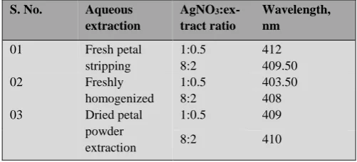
We did a series of experiments to optimize the production of silver nanoparticles with different concentration ratios of AgNO3 and the extracts. Those samples were analyzed with
the UV-visible spectroscopy. The rationale behind the optimization was; if any absorbance peak at around 420±10 nm was obtained from the synthesis solution for a given volume ratio of plant extract and silver nitrate solution, the ratio would be ‘good’ enough for the further synthesis process. It is showed the absorbance at 412 nm for the volume ratio of 1:0.5 corresponding to AgNO3: extract ratio
from ‘Fresh petal stripping’ extraction method. The ‘Petal strip’ method of extraction was selected for further synthesis because of the good stability and no agglomeration shown by the particles (Table 2).
Table 2. Optimization using different ratio of reactants.
Rosa damascena flower extract had an absorbance peak at
around 270 nm and silver nitrate solution at 250 nm. After adding the extract to the silver nitrate solution, the solution turned to brown color and the wavelength shifted to around 412 nm (Figure 1) indicating the reduction reaction. And reaction was completed within 50 min.
S. No. Aqueous extraction
AgNO3
:ex-tract ratio
Wavelength, nm
01 Fresh petal
stripping
1:0.5 412
8:2 409.50
02 Freshly
homogenized
1:0.5 403.50
8:2 408
03 Dried petal
powder extraction
1:0.5 409
8:2 410
s
% 100 1
c
A RSC
A
Figure 1. UV-Vis spectra of synthesized silver nanoparticles.
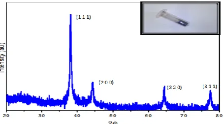
XRD studies
Nanocrystallinity of biosynthesized silver particles were confirmed using X-ray diffraction technique. The XRD pattern (Figure 2) showed numbers of Bragg reflections that may be indexed on the basis of the face-centered cubic structure of silver. It confirmed that the silver particles formed in our experiments were in the form of nanocrystals, as evidenced by the peaks at 2θ values of 38. 14 °, 44.46 °, 64.50 °, and 77.44 ° corresponding to (111), (200), (220), and (311) Bragg’s planes respectively. The result indicates that synthesized nanoparticles were face centered cubic in structure (JCPDS file No. 84-0713 and 04-0783). The average cyrstallite size of our nanoparticles was calculated using the Debye-Scherrer equation, and corresponds to around 3.7 nm.
Figure 2. X-ray diffraction pattern for the synthesized silver nanoparticle.
TEM Analysis
The average actual particle size was measured using TEM. TEM images (Figure 3) confirm the formation of ultra-small sized silver nanoparticles. The particles are quasi-spherical in shape, poly-dispersed in nature and correspond to an average size of 20 nm.
FTIR spectra
FT-IR analysis was carried out to identify chemical groups localized on the surface of particles that could have been responsible for the reduction of silver salts into silver nanoparticles. Figure 4 shows the FTIR absorption spectra of the synthesized silver nanoparticles. The stretch in 2 or more bands, one stronger and broader than others occurring
Figure 3. TEM image of prepared silver nanoparticles.
Figure 4. FTIR spectra for silver nanoparticles.
in the range of 1300-1000 cm-1 i.e., peaks centered at 1340,
1224, 1024, 1142 cm-1 are due to the stretching vibrations of
C-O groups of anhydrides, esters, ethers, alcohols and phenols, C-O-H of alcohols and phenols, and of C-N of amines. The stretch observed at 1603 cm-1 may be due to the
vibration of C=C group of alkenes, N-H of amides, and amine salts. The broad stretch occurring in the range of 3400-2400 cm-1, which is centered at 3283 cm-1, may well
be due to stretching vibrations of N-H of amines, amine salts, sulfonamides, C-H of alkenes and alkanes, and C=O of carboxylic acids. The other weak bands observed in the graph indicate the presence of nitriles, aromatic rings and aldehydes. Thus, FTIR study indicates that –C=O (carboxyl), -OH (hydroxyl) and N-H (amine) groups in the flower extract were mainly involved in reduction of Ag+ ions to Ag0 nanoparticles. The FTIR spectroscopic study also
suggest the role of carboxylic acid and ascorbic acid present in the flower extract as reducing and stabilizing agents for silver nanoparticles.
Biological activity assay
Considerable inhibition zone for fungal samples were not observed in the assay.The antifungal activity of SNPs depends on the type of fungal species,size and shape of nanoparticles.47
Figure 5. Antibacterial assay plates for S. auereus (S.a), P. aeruginosa(P.a) and E. Coli, respectively.
We tested anti-oxidant scavanging activity of both the plant extract as well as the silver nanoparticles using DPPH assay. Silver nanoparticles showed less scavanging activity compared to the flower extract with % of scavanging of 22.17 for 150 µL of the silver nanoparticles suspension. For the same volume of sample, rose extract showed 81.1 % of scavenging activity This may be due to the oxidation of the anti-oxidant molecules from the extract during the reduction of silver ions so that they could not donate electrons to the free radicals. All results confirmed that silver nanoparticles are synthesized successfully without agglommeration. Even it is stored for few months it is showing antibacterial activity against gram positive bacteria. Hence, by considering in view of cost effective production of silver nanoparticles by green method. It can be a promissing substitute route to use as a good antibacterial agent against multidrug resistant bacteria.
Conclusion
Silver nanoparticles with an average size of approximately 20 nm have been synthesized employing bio reduction method. Phytochemical screening of plant extract showed positive results for saponins, alkaloids, flavanoids, carbohydrates and glycosides. The synthesized silver nanoparticles were characterized by UV-vis spectrophotometry with absorbance wavelength at around 412 nm which is the suitable range for silver nanoparticles to exhibit surface Plasmon resonance. Multiple peak analysis of the XRD spectra confirmed the synthesis and indicated the crystallite size of our nanoparticles to be of ~4 nm. In addition, characterization done using FT-IR confirmed the presence of functional groups like alkanes, alkenes, alkynes, alcoholic and phenolic groups adhered on the surface of the particles. The conclusive evidence from phytochemical tests, UV-Visible spectroscopy and FT-IR revealed that the inherent action of reduction of silver nitrate to crystalline silver nanoparticles could be assigned to the compounds containing functional group C=O.
The prepared nanoparticles showed antibacterial effects with considerable inhibition in the growth of S. aureus. The particles did not show much effect on the fungal growth.
Rosa damascena flower extract acts as a good reducing and
stabilizing agent for the synthesis of silver nanoparticles in an eco-friendly and cost effective way. Particles were
observed to be highly stable for a period of 3 months. These biologically synthesized silver nanoparticles could be of immense use in medical field for their efficient antimicrobial function.
References
1Donner, A., Trends Mol. Med.,2010, 16, 551-552.
2Roco, M. C., Curr. Opin. Biotechnol. 2003, 14, 337-346.
3Duran, N., Rev. cienc. suelo. nutr. veg., 2008, 8, 33-38.
4Rajendran, P., Gunasekaran, P., Environmental Bioremediation
Technologies, Springer 2007, 211-221.
5Daniel, M.-C., Astruc, D., Chem. Rev.,2004, 104, 293-346.
6Zhou, B., Hermans, S., Somorjai, G. A. Nanotechnology in
Catalysis, Vol. 1, Springer, 2003.
7Perelshtein, I., Applerot, G., Perkas, N., Guibert, G., Mikhailov, S., Gedanken, A., Nanotechnology,2008, 19, 245705.
8Wang, R. R., Nasir, A., Nanotechnology in Dermatology, Springer,
2013, 41-49.
9 K Goyal, A., S Johal, E., Rath, G., Curr. Nanosci., 2011, 7(4), 640-654.
10 Kanchi, S., J. Environ. Anal. Chem., 2014, 1, e102.
11Theron, J., Walker, J., Cloete, T., Crit. Rev.Mmicrobio.,2008, 34, 43-69.
12 Katz, L. M., Dewan, K., Bronaugh, R. L., Food Chem. Toxicol.,
2015.
13Kogure, K., Hama, S., Nanomaterials for Cosmetics, (2014).
14Shan, J., Tenhu, H., Chem. Commun.,2007, 4580-4598.
15Sondi, I., Salopek-Sondi, B., J. Colloid Interface Sci.,2004, 275, 177-182.
16Miller, M. M., Lazarides, A. A., J. Phys. Chem. B, 2005, 109, 21556-21565.
17Jain, P. K., Huang, X., El-Sayed, I. H., El-Sayed, M. A.,
Plasmonics,2007, 2, 107-118.
18Lee, K.-S., El-Sayed, M. A., J. Phys. Chem. B, 2006, 110, 19220-19225.
19McFarland, A. D., Van Duyne, R. P., Nano lett., 2003, 3, 1057-1062.
20Minghui, Y., Jianxiu, W., Feimeng, Z., Functional Nanoparticles
for Bioanalysis, Nanomedicine, and Bioelectronic Devices Volume 1, 2012, 1112, 177-205.
21Rai, M., Yadav, A., Gade, A., Biotechnol. Adv., 2009, 27, 76-83.
22Xu, R., Wang, D., Zhang, J., Li, Y.,Chem. Asian J.,2006, 1, 888-893.
23Gittins, D. I., Bethell, D., Schiffrin, D. J., Nichols, R. J., Nature,
2000, 408, 67-69.
24Lee, K. J., Jun, B. H., Choi, J., Lee, Y. I., Joung, J., & Oh, Y. S.,
Nanotechnology,2007, 18, 335601.
25Lin, J.-J., Lin, W.-C., Li, S.-D., Lin, C.-Y., &Hsu, S.-h., ACS
Appl. Mater. Interfaces, 2013, 5, 433-443.
26Pauksch, L., Hartmann, S., Rohnke, M., Szalay, G., Alt, V., Schnettler, R., Lips, K. S., Acta biomate., 2014, 10, 439-449.
27López-Miranda, A., López-Valdivieso, A., Viramontes-Gamboa, G., J. Nanopart. Res.,2012, 14, 1-11.
29Soliman, Y., Radiat. Phys. Chem., 2014, 102, 60-67.
30Huang, L., Zhai, M. L., Long, D. W., Peng, J., Xu, L., Wu, G. Z., Wei, G. S. J. Nanopart. Res.,2008, 10, 1193-1202.
31Pal, A., Shah, S., Devi, S., Mater. Chem. Phys.,2009, 114, 530-532.
32Rodriguez-Sanchez, L., Blanco, M., Lopez-Quintela, M., J. Phys.
Chem. B,2000, 104, 9683-9688.
33Jiang, L.-P., Wang, A.-N., Zhao, Y., Zhang, J.-R., Zhu, J.-J., Inorg. Chem. Commun.,2004, 7, 506-509.
34Jeevanandam, P., Srikanth, C. K., Dixit, S., Mater. Chem. Phys.,
2010, 122, 402-407.
35Kotakadi, V. S., Gaddam, S. A., Rao, Y. S., Prasad, T., Reddy, A. V., Gopal, D. S., Eur. J. Med. Chem., 2014, 73, 135-140.
36Kotakadi, V. S., Rao, Y. S., Gaddam, S. A., Prasad, T., Reddy, A. V., Gopal, D. S., Colloids Surf., B: Biointerfaces,2013, 105, 194-198.
37Ponarulselvam, S., Panneerselvam, C., Murugan, K., Aarthi, N., Kalimuthu, K., Thangamani, S., Asian Pac. J. Trop. Biomed., 2012, 2, 574-580.
38Das, V. L., Thomas, R., Varghese, R. T., Soniya, E., Mathew, J., Radhakrishnan, E., 3 Biotech,2014, 4, 121-126.
39Deepak, V., Kalishwaralal, K., Pandian, S. R. K., Gurunathan, S.,
Metal Nanoparticles in Microbiology, Springer, 2011, 17-35.. 40Ahmad, A., Mukherjee, P., Senapati, S., Mandal, D., Khan, M. I.,
Kumar, R., Sastry, M., Colloids Surf., B: Biointerfaces,2003,
28, 313-318.
41Boskabady, M. H., Shafei, M. N., Saberi, Z., Amini, S., Iran J.
Basic Med. Sci.,2011, 14, 295.
42Sagdiç, O., Baydar, N., & Baydar, H., Food Sci. Technol. Int.,
2004, 10, 277-281.
43Bhandary, S. K., Kumari, N., Bhat, V. S., Sharmila, K., & Bekal, M. P., J. Health Sci., 2012, 2, 35-38.
44Palaksha, M., Ahmed, M., Das, S., J. Nat. Sci. Biol. Med., 2010,
1(1), 12.
45Blois, M. S. , Biochim. Biophys. Acta, 1955, 18, 165
46Jagtap, U. B., Waghmare, S. R., Lokhande, V. H., Suprasanna, P., Bapat, V. A., Ind. Crop Prod.,2011, 34, 1595-1601. 47Medda, S., Hajra, A., Dey, U., Bose, P., Mondal, N. K., Appl.
Nanosci., 2014, 1-6.