DOI: 10.22587/ajbas.2018.12.3.1
Research Article AENSI Publications
Studies on preparation, Characterization and Biodegradation Behavior of HDPE Natural
polymers Blends
1Fawzi Habeeb Jabrail, 2Alarqam Ziyad Taraq, 3Kailash Chandra Gupta
1Professor, Department of Chemistry, College of Science, University of Mosul, Iraq.
2Master Degree Student, Department of Chemistry, College of Science, University of Mosul, Iraq.
3Professor, Polymer Research Laboratory, Department of Chemistry, Indian Institute of Technology, Roorkee 247667, India.
Corresponding Author: Fawzi Habeeb Jabrail Professor, Department of Chemistry, College of Science, University of Mosul, Iraq.
E-mail: [email protected], [email protected]
Received date: 23 January 2018, Accepted date: 17 March 2018, Online date: 25 March 2018
Copyright: © 2018 Fawzi Habeeb Jabrailet al.This is an open-access article distributed under the terms of the Creative Commons Attribution License,
which permits unrestricted use, distribution, and reproduction in any medium, provided the original author and source are credited.
Abstract
Polyethylene (HDPE) is widely used in various applications due to its chemical, physical. and biological inertness but its durability presents a great challenge when it is released in the environment. To reduce its adverse effect on environment, currently various efforts are being made to modify its properties using naturally occurring biodegradable polymers but still these modifications found to be costly and required biodegradability in polyethylene is not yet achieved. Therefore, an attempt has been made to deveop biodegradable polyethylene blends using naturally occurring polymers. In this connection biodegradable high density polyethylene (HDPE) blends were prepared by thermally blending 2.0, 5.0 and 10.0 wt % amount of naturally occurring polymers such as; chitosan (CH), cellulose(CE), starch (ST), alginate (AL), pectin (PE), shellac (SH) and xanthan (XA). The observed biodegradabilility in HDPE blends might be due to the presence of hydrolysable linkages and stereo-favourable orientations of blended natural polymers. The added polymers have played a significant role in increasing the hydrophilicity in blended HDPE and acted as a bioassimilative nutrients for seeded microorganisms. The biodegradability of HDPE-polymer blends was evaluated in presence of various fungi such as; aspergillus niger, aspergillus terreus, fusarium solani, tricoderma hariziauum and tricoderrma viride. The disinfected films of pristine HDPE and polymer blened HDPE were inoculated with these fungi and the extend of biodegrdation was evaluated after a incubation period of three months at 28 ± 1˚C. The biodegradability of HDPE-polymer blends was compared with pristine HDPE by evaluating their molecular weights, and weight percent loss in samples incubated for three months along with selected fungus. The biodegrdation in pristine HDPE and its polymer blends was confirmed by comparing their FT-IR spectra and also by evaluating the variations in their mechanical and thermal properties. A significant variation in their morphologies in prsence of fungi has confirmed biodegradation in HDPE-polymer blends in comparison to pristine HDPE films. These studies have provided sufficient evidnces to confirm the role of added natural polymers in developing a biodegradable HDPE by blending various polymers such as chitosan (CH), cellulose(CE), starch (ST), alginate (AL), pectin (PE), shellac (SH) and xanthan (XA). Out of these polymers, the chitosan is found to be quite effective as it is acted better bioassimilative nutrient for microorganisms to cause biodegrdation of HPEF in comparison to other polymers.
Key words: HDPE, Natural polymers, Polymer blend, Biodegradation, Fungus bioassimilative nutrient polymers.
INTRODUCTION
Synthetic plastic has become an integral part of human lives due to its low cost and ease of synthesis. Polyethylene is synthesized easily without using expensive method or procedure (Honakar et al., 2005) and found to be useful in various applications such as; packaging materials for agricultural products (Li et al., 2015), food materials and other items (Fasoyiro et al., 2017). But non-judicious and unplanned usage of polyethylene or non-degradable polymers has generated hazardous non-degradable wastes in our surroundings, which become a threat to the very survival of human lives and biological systems (Singh and Sharma, 2008). In order to reduce the load of plastic waste accumaulation in our environment, it is highly desirable to develop biodegradable polymers (Arvanitoyannis et al., 1998) by modifying the properties of non-degradable synthetic polymers (Vallini et al.,1994). The biodegradable polymers are commonly produced either altering their structures (Sohel et al., 2018) or blending with naturally occurring biodegradable polymers (Arutchelvi et al., 2008; Kahar et al.,2017). Though naturally occurring polymers such as; 3-hydroxyl butyrate (PHB), its copolymers and some aliphatic polyester have been used frequently to develop biodegradable blends of polyethylene but these modifications found to be costly (Avella et al., 2005; Nowak et al., 2010) in comparison to natural polymers (Bandyopadhyay et.al. 2010). The best known and easily available natural polymers to develop biodegradable plastics are chitosan, pectin, starch, cellulose, shellac, and xanthane. The naturally occurring polymers are hydrophilic in nature (Kang et al., 1996), which is considered responsible for their biodegradability in comparison to hydrophobic synthetic polymers. Thus, considering these properties of naturally occurring polymers, the blends of polyethylene have been developed, which found to be quite susceptible for microbial biodegradation (Griffin, 1994) through enzymatic action of microorganisms. To study the microbial degradation of organic matters including polyetylene, the soil separated fungus has been used successfully (El-Shafei et al., 1998). The action of extracted enzymes in biodegradation of polyethylne is also evaluated by other workers (Mandels and Sternberg, 1976). In view of literature reports on application of naturally occurring polymers in developing biodegradable polymers, an effort has been made to prepare biodegradable polymer blends using commonly used synthetic high density polyethylene (HDPE) and naturally occurring biopolymers. In comparison to low density polythyelene (Satyalakshmi, 2016), the high density polyethylene (HDPE) is found to be highly resistant to microbiological degradation or disintegration; hence its blends were prepared using easily available
biopolymers such as; chitosan(CH), cellulose(CE), starch(ST), alginate(Al), pectin (PE), shellac(SH) or xanthane(XA), which themselves are biodegradable due to the presence of sufficient hydrolysable linkages. The enzymatic action of various fungi (Skariyachan et al., 2018; Al-Jailawi et al., 2015) such as: aspergillus niger, apergillus terrus, fusarium solani, tricoderma harziauum and tricoderma viride is found to be quite effective in biodegration of polymer blends; hence, these fungi have been used to test the biodegradability of prepared blend of HDPE with natural polymers. The extent of biodegradation in pristine HDPE and HDPE-Polymer blends before and after inoculation with chosen microorganisms has been evaluated as per ASTM standard.
2.Experimental: 2.1 Materials:
High density polyethylene (HDPE) was supplied by BDH Chemical Company, UK in a granular form. Chitosan, cellulose, starch, alginate, pectin, and xanthane were obtained from Sigma-Aldrich Chemicals Company, UK. Three stains of mesophilic and aerobic fungi were isolated from soil and preserved in presence of optimum humidity at 5˚C. The soil isolated fungi are aspergillus niger, aspergillus terreus, fusarium solani, tri coderma harziauum and tricoderma viride. Other chemicals used in expertimental work were of reagent grades in their purity and used without further purification.
2.2 Samples preparation:
High density polyethylene (HDPE) films of 0.5 mm thickness were prepared manually using thermo-press method at 110˚C. To measure the tensile strength, dog bone shaped samples were prepared from pristine HDPE and polymer blended-HDPE films. The melt-blended samples of HDPE and polymers were used to prepare HDPE-Polymer blended films. The melt-blended samples of HDPE-Polymers were prepared separately by mechanically mixing (Damrongsakkul and Ngamsinlapasathian, 2002) 2.0, 5.0, and 10 wt % of each polymers with 2.0 g of pristine HDPE. The HDPE blends were prepared using chitosan, cellulose, starch, alginate, pectin, shellac, and xanthane.
2.3 Culturing of pristine HDPE and HDPE-Polymer blend films:
To evaluate the biodegradability of pristine HDPE and its polymer blends, samples were incubated along with chosen fungus in a media for optimized time of incubation. To carry out these experiments, a media was prepared and HDPE-Polymer blended samples were cultured in it.
2.3.1 Preparation of potato dextrose agar media and disinfection of the films:
The potato dextrose agar media (PDA) was prepared by taking 200 g potato, 20 g D-glucose, and 15 g agar in 1.0 L distilled water. The mixture was boiled for 20 min and disinfected by autoclaving (BS2646,1988,AUX 750 OIOu) at 120 0C for about 30 min at 1kg/cm2 pressure. After autoclaving the mixture, the pH of potato dextrose agar media was adjusted to 7.4 using 0.1 M solution of NaOH and using pH meter (Hanna PH211, Instrument Microprocessor pH meter).The chemical disinfection of pristine HDPE and HDPE-Polymer blended films was carried out by immersing films in a solution prepared freshly by mixing 7.0 mL tween 80, 10 mL of 1.0 N sodium hypochlorite (bleach) in 1.0 L sterilised water. The films were kept for 30-60 min under mechanical stirring and after removing they were dried for about 60 min at room temperature before keeping for 30 min in 70 % (v/v) solution of ethanol. These disinfected films were incubated in a sterilised petri dish at 45˚C for overnight, and gradually cooled to ambient temperature before recording their weights (El-Shafei et al., 1998).
2.3.2 Culturing of microorganism and inoculation of HDPE-Polymer blended films:
The freshly germinated fungus was inculated homogenously using inoculation loop in a petri dish containing 2.0 mL potato dextrose agar media. After inoculation, the petri dish was incubated for one week in a autoclave maintained at 25 ± 1˚C. The cultured fungus was finally preserved at 5˚C (Kawai et al., 2004). The samples of other fungi were also cultured by following same procedure. The pre-weighed and disinfected HDPE-Polymer blended films were inoculated homogeneously on both sides with chosen fungus using disinfected loop. The fungus inoculated polymer films were kept vertically in potato dextrose agar media in a dish and then placed in an incubator. At the end of fixed period of incubation, the pristine and HDPE-Polymer blended films were harvested and washed gently using 70 % (v/v) solution of ethanol to remove adhered cell mass and biodegraded mass of the films. Finally, films were dried at 45˚C for 24 h in a hot air oven.
2.4 Characterization of pristine HDPE and HDPE-Polymer blended films:
The pristine HDPE and polymer blended HDPE films were charcterised for various parameters to confirm the enhanced biodegrdation in fungi inoculated HDPE-Polymer blended films in comparison to pristine HDPE films.
2.4.1 Determination of molecular weight:
The viscometric average molecular weights (
M
v
) of pristine HDPE and HDPE-Polymer blends were calculated by determing the intrinsic viscosity [η] of pristine HDPE and HDPE-Polymer blends in p-xylene using Ubbelohde type of capillary viscometer at 105˚C. The viscometric average molecular weights(
M
v
) of pristine HDPE and HDPE-Polymer blends were calculated (Dhiman et al., 2004) using following Mark-Houwink equation 1.
105
0c
K
M v
(1)Where, ‘K’ and ‘a’ are characteristic Mark-Houwink constants.
2.4.2 Determination of biodegraded weight loss in pristine HDPE and HDPE-Polymer blended films:
The observed weight loss (%) in pristine HDPE and polymer blended HDPE films after incubation with selected fungus has been used to evaluate the extent of biodegrdation and assimilation of films by incubated microorganisms (Ghazaki et al., 2005). The films weight loss (%) was calculated using following realation (Equation 2) (Krystyna et al., 2003; Labuzek et al.,2004) and by determining weight accurately using digital precision balance (AND HR-200).
Weight loss ( % )
W
i
W
f
.100
(2)
W
i
Where, Wi and Wf are the weights of the films before and after biodegradation by selected fungus.
2.4.3 FT-IR characterization of pristine HDPE and polymer plended HDPE-films:
2.4.4 Mechanical properties of pristine HDPE and polymer blended HDPE films:
Tensile strength of HDPE films was evaluated by using dog bone shaped samples at ambient temperature by following ASTM D638 standard and using universal testing machine (Gunt Wp 300-20). The samples with a gauge length of 38 mm, and a width of 8 mm were used for analysis. Young’s moduls (E) was calculated using Hooke's law (Equation 3) (Ghosh, 2003).
E
(3)Where, σ is stress (force per unit area, N/mm2), ε is strain (elongation in length, %) and E is Young's modulus corresponding to slope values of stress-strain curves.
2.4.5 Thermal studies of of pristine HDPE and polymer blended HDPE films:
To determine the extent of fungal biodegradation in polymer films, the variation in thermal properties of pristine HDPE and polymer blended HDPE films before and after incubation with selected fungus was determined by recording their TG and DTG thermograms under inert atmosphere using TG analyzer (Extra TG/DTA 6300) at a heating rate of 10˚C/min within a temperature range from 25-550˚C (Negi et al., 2009). To supplement the TG and DTG analysis, the differential scanning calorimetric (DSC) analysis of pristine HDPE and polymer blended HDPE films was also carried out.
2.4.6 Scanning electron micrograph of pristine HDPE and polymer blended HDPE films:
To analyse the effect of biodegrdation on morphological behavior of pristine HDPE and HDPE-polymer blends on incubation with selected fungus, the scanning electron micrographs (SEM) of pristine HDPE and HDPE-Polymer blends were recorded (LEO435VP-England). To record SEM micrographs, the samples were mounted on aluminum studs using double adhesive tapes and vacuum coated by exposing to a gold ion beam sputter (PELCO.S.C.6) at a current of 25 mA for about 40 s. To visualise the microstructural variation on microbial biodegradaion (Labuzek et al., 2004) in the samples, the SEM micrographs were recorded at an optimized magnification.
RESULTS AND DISCUSSION
HDPE is a indispensable material for domestic and industrial applications but its durability and chemical resistance is a main drawback for producing non-decomposable waste in the environment. Therefore, to sustain its domestic applications, its properties should be modified to make it eco-friendly and biodegradable. Considering the useful properties of HDPE it is difficult to discard its importance in commercial and industrial applications; hence, some efforts have been made to make biodegradable polyethylene using natural polymers (Avella et al., 2005). To produce biodegradable HDPE, various naturally occurring polymers have been used in making melt-mix blends of prestine HDPE and effect of blended natural polymers on biodegrdation of HDPE has been evaluated by studying their properties after seeding with selected fungus. The results of fungus seeded HDPE-Polymer blends have indicated that microbial biodegradation was first initiated on HDPE blended polymers and after that it continued to biodegrade of long chains of HDPE by enzymatic action. The biodegraded products were finally used up by seeded microorganism as bioassiminable nutrients. The extent of biodegradation of pristine
HDPE is found to be lower in comparison to polymer blended HDPE due to significant decrease in hydrophobicity of HDPE and due to the presence of hydrolysable linkages in HDPE-Polymer blends.
3.1 FT-IR Characterization:
FT-IR spectra of prestine HDPE and natural polymers were recorded separately to confirm the blening of polymers with HDPE (Table 1). FT-IR spectra of polymer blended HDPE samples having 10 wt% of polymers were recorded before and after (Fig. 1) seeding with selected fungus for three months to confirm biodegrdation in polymers blended HDPE by seeded microorganism. On comparing the FT-IR spectra of HDPE-Polymer blends before and after incubation with selected fungus for 3 months (Table 2 & Fig. 1), it is clear that incubation of HDPE-Polymer blends with selected microorganism has either shown a significant shift in characteristic functional groups of HDPE-Polymer blends (Table 2) or produced some new functional groups (Table 2 & Fig. 1) on biodegradation of polymer blended HDPE. The appearance of strong absorption band between 1650–1760 cm-1 (Fig. 1) after inoculation with selected fungus for three months has suggested the formation of new carbonyl compounds (Table 2 & Fig. 1) such as esters, aldehydes or carboxylic acid (Orhan and Buyukgungor, 2000).
Table 1: IR frequencies of HDPE and polymers Functional groups Wave number (cm)
HDPE PS CH CE ST AL PE SH XA
C-O -- -- 1075 1060 1081 1088 1080 --- 1055
C-H 2920-2850 2921 2922 2902 2932 2938 2926 2932 2930
O-H -- -- 3450 3445 3420 3409 3444 3420 3441
C=O -- -- 1655 amide --- -- 1622 1652 1696 1699
C---O -- 1543-1605 -- -- -- -- -- --
--The variation in stretching frequency at 3500 cm-1 for O–H bond and at 1100 cm-1 for C–O bond on incubation with selected fungus (Table 2 & Fig. 1) has further provided an evidence to confirm biodegradation in polymer blendd HDPE (Table 2 & Fig. 1).
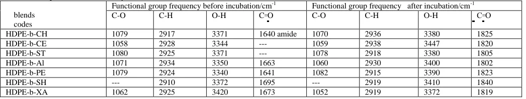
Table 2: IR frequencies of HDPE-Polymer blends
blends codes
Functional group frequency before incubation/cm-1 Functional group frequency after incubation/cm-1
C-O C-H O-H C꞊O C-O C-H O-H C꞊O
HDPE-b-CH 1079 2917 3371 1640 amide 1070 2936 3380 1825
HDPE-b-CE 1058 2928 3344 --- 1059 2938 3447 1820
HDPE-b-ST 1080 2925 3371 --- 1078 2918 3380 1805
HDPE-b-Al 1071 2934 3350 1663 1060 2930 3400 1802
HDPE-b-PE 1079 2924 3340 1641 1082 2915 3390 1823
HDPE-b-SH --- 2910 3372 1695 --- 2919 3410 1840
HDPE-b-XA 1062 2925 3420 1673 1052 2919 3372 1819
Seeding time, 3 months; Films thickness, 0.5 mm; HDPE blended with 10 wt% polymer.
Fig. 1a: FT-IR Spectra for HDPE blends with 10 % w/w cellulose recorded before and after seeding with selected fungus.
Fig. 1c: FT-IR Spectra for HDPE blends with 10 % w/w of xanthane recorded before and after seeding with selected fungus.
The HDPE chains were found to be a main source of nutrition for germinated colony of microorganism after enzymatic biodegradation of blended polymers in HDPE. The HDPE-b-CH blends (Table 2) before fungal inoculation have shown absorption band for C=O str at 1640 cm-1 corresponding to amide group but after inoculation with selected fungus for three months, the amide absorption band of chitosan was disappeared totally indicating a complete consumption of chitosan by inoculated fungus on HDPE-b-CH blends. The germinated colonies of fungi on HDPE-Polymer blends were subsequently able to cause enzymatic biodegrdation of long chains of HDPE. The enzymatic biodegrdation of long chains of HDPE and formation of oxidation byproducts with ketonic and alcoholic groups was confirmed by FT-IR analysis. The FT-IR spectra have shown new absorption bands at 1825 cm-1 and 3380 cm-1, corresponding to ketonic carbonyl (C=O str) and alcoholic (O–H str) bonds respectively. The HDPE-b-CE blends with 10 wt % cellulose after incubation for three months with selected fungus were totally free from characteristic bands of cellulose at 1060 cm-1, 2902 cm-1 and 3445 cm-1 (Table 1, Fig.1a), which suggested for a complete biodegradation of blended cellulose in HDPE-b-CE samples. The appearance of strong band at 1820 cm-1 (C=O str) and broad band at 3447cm-1 (O–H str) has further confirmed the formation of ketonic and alcoholic byproduct on biodegrdation of HDPE blended with cellulose. The FT-IR spectra of HDPE blended with other polymers have also provided sufficient evidences for the formation of new compounds and enzymatic biodegrdation of HDPE by germinated colonies of inoculated fungi. These results have also suggested that the extent of biodegradation has shown a significant dependance on type of blended polymers and microorganism used for seeding with polymer blended HDPE samples.
3.2 Molecular weight degradation:
The molecular weight (
M
v
) of pristine HDPE samples after seeding with fungi for three months is found to be almost same as it was before seeding with microorganism, which clearly suggested that pristine HDPE was highly resistant to microbial attacks. But on blending HDPE with polymers, the HDPE has showna significant decrease in its
M
v
. The initialM
v
of pristine HDPE was 654 kg mol-1, which remained almost constant after seeding with fungi for threemonths. But HDPE in polymer blended samples has shown a decreasing trend on seeding with different fungus for three months. The
M
v
of HDPE in polymer blended samples after seeding for three months has shown a systematic order such as HDPE-b-CH< HDPE-b-PE< HDPE-b-CE< HDPE-b-ST< HDPE-b-XA<HDPE-b-AL< HDPE-b-SH for the
M
v
of HDPE as 316, 350, 444,451, 480, 586, 598 kg mol-1 respectively in these blends. The seeded fungus has first used up the blended polymer as its nutrient and after that it caused biodegradation of HDPE chains through enzymatic action by germinated colonies of microorganism,which ultimately has reduced the
M
v
of HDPE in polymer blended HDPE. The HDPE blended with chitosan has shown a significant decrease inM
v
ofHDPE (316 kg mol-1), whereas
M
v
of HDPE in shellac blended HDPE (HDPE-b-SH) samples has shown least biodegradation (598 kg mol-1) after seededing with selected fungus. This has suggested that chitosan acted as a suitable nutrient to seded fungi and was able to germinate more colonies of microorganism to casue an effective enzymatic biodegrdation of HDPE in chitosan blended HDPE samples. The variation in extend of biodegrdation of HDPE in polymer blends might be due to the difference in chemical and stereostructutres of polymers used for blending with HDPE. The hydrophilicity, stereo-orientation and chain flexibility of polymers have all together played a significant role in controlling the permeability of fungus and their biodegradative action of polymers (Kawai et al., 2004). Amongst selected polymers, the chemical and stereo-structures of chitosan and pectin were found to be more effective in causing biodegradation or indecreasing the
M
v
of HDPE in polymer blends in comparison to other polymers used for blending with HDPE and seeded for three months with selected fungus. The molecules of chitosan and pectin are more flexible and able to form a layer of hydrogels through hydrogen bonding at HDPE surfaces that give rise an adhesive property to HDPE in the blends. This has provided a significant help to seeded fungi for their attachment, germination and in releasing a sufficientpectin, the cellulose, starch and xanthane have also shown a decreasing trend in
M
v
of HDPE on seeding with selected fungus due to the presence of microbial friendly functional groups in these biopolymers. However, alginate and shellac have shown poor biodegrdation of HDPE in comparison to other polymers, due to their acidic properties, which might have prevented the germination and colonization of fungus on the surface of HDPE in polymer blends, hencealginate and shellac have shown a minimal deceases in
M
v
of HDPE in polymer blends after incubation with selected microorganism.3.3 Effect of polymer amount and type of microorganism on biodegradation of polymer blended HDPE:
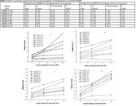
In order to determine the effect of weight percent of blended polymer on biodegrdation of HDPE by microorganism, the HDPE-Polymer blends with different amount (%) of polymers were prepared and incubated with selected fungus. The HDPE-Polymer blends having 2, 5, and 10 wt % of each polymer were prepared separately by melt-mix method and used to evaluate their biodegradability by seeding for three months with different fungi. The effect of weight percent of polymers on extent of biodegrdation of HDPE was evaluated by determining their weight loss by gravimetric method after incubation with different fungi and results are given in Table 3. The data shown in Table 3 have clearly indicated that the weight loss of HDPE in polymer blends has shown an increasing trend on increasing the weight percent of polymers from 2-5 wt% and shown a maximum weight loss in HDPE in blends prepared with 10 wt % of polymers. Therefore, HDPE blends with 10 wt% polymers were used to evaluate their properties.
Table 3: Effect of polymer amount and type of microorganism on biodegrdation of blended HDPE
Weight loss in HDPE (%) in blend with 2 wt % polymer Weight loss in HDPE (%) in blend with 5 wt % polymer
Blends A. A. T.harzi-auum F. A. A. T.harzi-auum F.
codes niger terreus solani niger terreus solani
HDPE-b-CH 1.64 3.08 0.84 3.80 2.33 4.25 1.85 4.35
HDPE-b-CE 1.52 1.73 0.78 2.53 2.22 2.31 1.59 3.87
HDPE-b-ST 0.94 1.64 0.65 2.42 1.32 1.86 1.14 3.45
HDPE-b-Al 0.32 1.12 0.28 2.26 0.74 1.78 1.10 3.08
HDPE-b-PE 1.62 2.55 1.54 2.73 2.21 3.96 1.82 3.24
HDPE-b-SH 0.71 0.95 0.45 1.92 0.83 1.04 0.65 2.33
HDPE-b-XA 1.05 2.38 0.82 3.61 1.94 3.52 0.94 4.02
Fig. 2:The percentage weight loss of HDPE polymer blended with different %(w/w) natural polymers after seeding for 3 months with (A) A. niger, (B) A. terreus, (C) T. harziauum, (D) F. solani.
The 10 wt % amount of polymer seems to be sufficient to cover the surface of blended HDPE and was sufficient to facilitate the germination and colonization of microorganism on blended HDPE. The HDPE blends with 10 wt% amount of polymer were able to show enhanced biodegradation of HDPE in comparison to HDPE blends with low (< 10 wt %) amount of polymers. The type of fungus has also caused a significant effect on weight loss (%) of HDPE in polymer blends (Table 3, Fig. 2). The variation in weight loss with type of fungus might be due to the differences in their β-oxidation system, which is an essential part of cell membranes (Kawai et al., 2004). The chitosan and pectin blends have shown maximum weight loss (%) in HDPE due to sufficient similarity in their chemical structures.
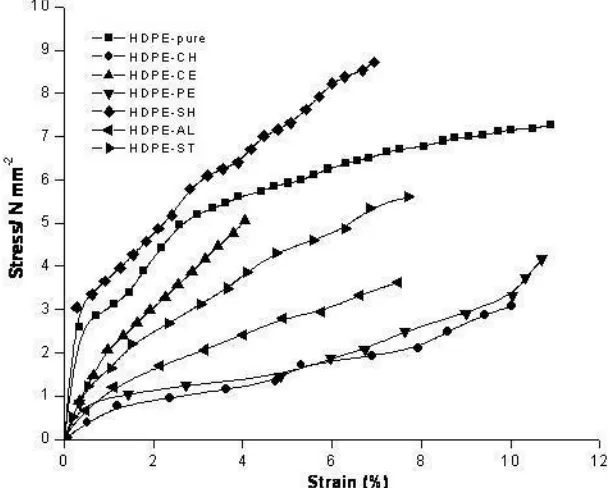
3.4 Mechanical properties of HDPE and polymer blended HDPE:
polymers in HDPE blends have played a significant role in overcoming the phase separation and controlling the mechanical properties of polymer blended HDPE (Dhiman et al., 2004; Lu et al., 2009). The lowering of Young’s modulus of blended HDPE films than pristine HDPE (Table 4) has suggested that blended polymers were homogenously intermingled with long chains of HDPE to cause an effective plasticization of HDPE. The mechanical properties of polymer blended HDPE has shown a significant variation with type of polymers used for blending with HDPE (Table 4 & Fig.3), which may be due to differences in interfacial thickness of anchored polymers with HDPE chains and compatibilization of HDPE in polymers blended HDPE. The properties of polymers and large surface area of HDPE have likely controlled the interfacial thickness in melt-mixed polymer blended HDPE (Wu and Liao, 2005).
Table 4: Mechanical properties of HDPE and polymer blended HDPE
Before incubation After incubation
Blends E/MPa Ultimate TS / MPa EB/% E/MPa Ultimate TS/ MPa EB/%
codes
Pristine-HDPE 15.2 11.3 19.6 15.0 11.2 19.4
HDPE-b-CH 13.9 10.4 17.5 2.4 1.6 4.7
HDPE-b-CE 11.0 7.6 16.4 7.9 4.8 9.5
HDPE-b-ST 8.8 6.8 17.6 7.1 4.0 10.2
HDPE-b-Al 11.5 8.1 15.1 6.3 5.8 8.3
HDPE-b-PE 14.2 10.7 18.0 3.6 2.4 5.1
HDPE-b-SH 18.5 13.8 1.0 17.4 13.0 2.5
HDPE-b-XA 12.7 9.5 15.7 4.0 3.1 5.5
Sample gauge length, 38 mm; Sample width,8 mm; Thickness, 0.5 mm; Temp., 22°C; Seeding time, 3 months; A. niger was used as microorganism.
The variation in mechanical properties of pristine HDPE and polymer blended HDPE after seeding with A. niger for three months was found to be significantly high in comparison to mechanical properties of samples before seeding with A. niger (Table 4 & Fig. 3).
Fig. 3: Stress-strain curves of pristine HDPE and its blends using 10 wt % of different polymers after seeding with fungi (A. niger) for three months.
In comparioson to polymer blends prepared with other polymer, the HDPE-Polymer blends prepared with chitosan, pectin and xanthane have shown a significant decrease in Young’s modulus, ultimate tensile strength (TS) and elongation at break (EB) after seeding with A.niger for three months (Table 4, Fig. 3). This might be due to the enhanced biodegrdation of HDPE chains and due to a significant loss in crystalline properties of HDPE after seeding for three months with selected fungus. The HDPE-Polymer blends with chitosan, pectin, and xanthan after seedingh for three months with selected fungus have produced highly amorphous HDPE due to enhanced enzymatic biodegrdation of HDPE than the HDPE blended with other polymers. Thus the enhanced decreasing trends in mechanical properties of HDPE blends prepared with chitosan, pectin and xanthan have clearly suggested that chitosan, pectin and xanthan have acted as suitable nutrients to selected fungus (A. niger) and helped in enzymatic degradation of HDPE due to enhanced germination and colonization during the seeding period of three months. The HDPE blends prepared with other polymers have also shown biodegradation of HDPE but inn these blends, the microorganism were less active in biodegradation of HDPE in comparison blends prepared with chitosan, pectin and xanthan. The shellac blended HDPE samples (Table 4, Fig.3) have shown insignificant decrease in their mechanical properties on seeding with fungus for three months, which indicated that shellac was not a suitable nutrient for selected fungus, hence it failed to cause significant enzymatic biodgradation of HDPE in shellac blended HDPE.
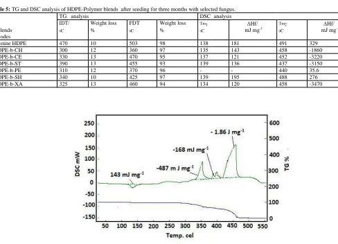
3.5 Evaluation of biodegaration of HDPE by thermal studies of polymer blended HDPE:
To confirm the biodegrdation of polymer blended HDPE on seeding with selected fungus, the thermal stability of seeded HDPE-Polymer blends was determined and compared with thermal stability of pristine HDPE. The thermal stability of polymer blended HDPE is evaluated as a loss in weight (%) at temperature by thermogravimetric (TG) analysis. The differential scanning calorimetric (DSC) analysis has been used to determine phase transition temperature and enthalpy of phase transition ( tr) in polymer blended samples before and after biodegradation. The HDPE-Polymer blends having 10 wt% amount of polymer
was used for seeding with selected fungus and after seeding their thermal properties was compared with pristine HDPE. The results of TG and DSC analysis of seeded pristine HDPE and polymer blended HDPE with different polymers are presented in Table 5. The pristine HDPE after seeding with selected fungus for three months is found to be thermally stable with an initial decomposition temperature (IDT) of 470 0C and with a weight loss of ~10%, whereas the final
decomposition temperature (FDT) was found to be 503 0C with a total weight loss of 98%. The rate of decomposition at maximum decomposition temperature (Tmax) of 491 0C (Table 5, Fig. 4) was found to be highest (4.3 mg min-1). The seeded pristine HDPE has shown a first phase transition at 138 0C with a heat of
Fig. 4: DSC and TG analysis of pristine HDPE after seeding with fungus for three months.
The weight loss (%) and phase transition temperatures (Tm1 and Tm2) of seeded HDPE were found to be almost same as were found with pristine HDPE without t seeding with selected fungi. These results have given an indication for a minimal biodegradation in HDPE after seeding with selected fungus due to the presence of high crystallinity in pristine HDPE (Thaweegan et al.,2005). But on comparing the weight loss decomposition temperatures (IDT and FDT) and heat of fusion (∆Htr) of fungus seeded polymer blended HDPE with fungus seeded pristine HDPE, it is clear that decomposition temperatures (IDT and FDT) of polymer blended HDPE were found to be significant low in comparison to pristine HDPE (Table 5).The HDPE-Polymer blends prepared with cellulose (HDPE-b-CE), starch (HDPE-b-ST), Schellac (HDPE-b-SH) and with xanthane (HDPE-b-XA) have shown high IDTs and almost a constant weight loss (~12 wt%) in comparison to HDPE-chitosan (PDPE-b-CH), and HDPE-pectin (HDPE-b-PE) blends (Table 5). This has suggested that chitosan and pectin were able to cause more biodegrdation of HDPE on seeding with selected fungus in comparison to other polymers (Table 5). The enhanced biodegrdation of HDPE in chitosan and pectin blended HDPE has caused a significant decrease in thermal stability of HDPE blends prepared with chitosan (HDPE-b-CH), and pectin (HDPE-b-PE), hence these blends were able to show thermal degradation at low IDT and FDT values in comparison to blends prepared with other polymers (Table 5 & Figs. 5 &6).
Table 5: TG and DSC analysis of HDPE-Polymer blendsafter seeding for three months with selected fungus.
TG analysis DSC analysis
IDT/ Weight loss FDT Weight loss Tm1 Hf/
mJ mg-1
Tm2 Hf/ mJ mg-1
Blends 0C % 0C % 0C 0C
codes
Pristine HDPE 470 10 503 98 138 181 491 329
HDPE-b-CH 300 12 360 97 135 143 458 -1860
HDPE-b-CE 330 13 470 95 137 121 452 -3220
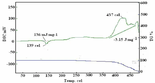
HDPE-b-ST 390 13 455 93 139 136 437 -3150
HDPE-b-PE 310 12 370 96 - - 440 35.6
HDPE-b-SH 340 10 425 97 139 195 488 276
HDPE-b-XA 325 13 460 94 134 120 458 -3470
Fig. 6: DSC and TG analysis of HDPE-b-PE blend after seeding with fungus for three months.
The thermogram (TG) for fungus seeded HDPE-b-CH blend (Fig. 5) has shown a maximum rate of decomposition (Tmax) at 300 oC for residual chitosan and at 360 0C for HDPE in biodegraded blends. On the other hand, the HDPE-b-PE blends were able to show maximum rate of decomposition (Tmax) at 310 0C and at 370 0C respectively for residual pectin and HDPE in seeded samples (Table 5 & Fig. 6).
Fig. 7: DSC and TG analysis of HDPE-b-SH blend after seeding with fungus for three months.
This has suggested that seeded fungus in pectin blended HDPE has consumed a very small amount of pectin, than the amount of chitosan consumed by fungus in chitosan blened HDPE, hence due to insufficent growth of fungi in pectin blended HDPE, the rate of biodegrdation of HDPE was slower (1.83 mg min-1)
than blends (HDPE-b-CH) prepared with chitosan (2.77 mg min-1). This has clearly suggested that the properties of blended polymer play important role in
conrolling the biodegradadtaion of chemically inert HDPE by seeded fungi. Like HDPE-b-PE blend, the HDPE blends prepared with schellac and starch have also shown maximum rate of decopmposition at 425 0C and 455 0C respectively as was confirmed from their TG curves (Table 5). The fungus seeded HDPE blends of
schellac and starch have also shown phase transitions as was shown by HDPE blends prepared with other polymers, which is clear from the observed heat of fusion ( tr) in their DSC thermograms (Table 5 & Figs. 7 & 8).
The rate of decomposition of polymer blended HDPE at Tmax is also used to understand the effect of seeded fungus on biodegradation of HDPE in HDPE-Polymer blends. The decreased rate of decomposition at Tmax in these smples is possibly due to the decrease in rate of decomposition of biodegradable shorter chains or oxidized long chains of HDPE as reported by other workers (Santonja et al., 2007). The long chains of HDPE decompose faster at Tmax in comparison to biodegraded shorter chains of HDPE, which decomposed at slower rate at low temperature as clear on comparing the rate of decomposition of seeded pristine HDPE (4.3 mg min-1) and of fungus seeded blends (HDPE-b-SH) of shellac (3.15 mg min-1) blends in which the rate of decomposition is found to be higher at 491 0
C and 437 0C respectively for HDPE in fungus incubated pristine HDPE and fungus seeded blends (HDPE-b-SH ) of shellac (Figs. 4 & 8). The trends in heats of fusion in fungus seeded HDPE-b-CH and HDPE-b-PE blends have suggested that chitosan and pectin were able to enhance the extend of biodegrdation of HDPE on seeding with selecyed fungus in comparision to HDPE blends prepared with other polymers (Table 5 & Figs. 8). In comparison to fungus seeded pristine HDPE, the fungus seeded blends (HDPE–b-CH ) of chitosan (Table 5 & Fig. 5) have shown exothermic phase transitions relatively at low temperatures (Tm1 and Tm2) for the decomposition of residual chitosan and fungal oxidized chains of HDPE. The TG and DSC analysis of fungus incubated blends of cellulose (HDPE-b-CE) and blends (HDPE-b-ST) of starch (Table 5) has indicated that cellulose was able to show enhanced biodegradation of HDPE in comparison to starch as ITD of fungus seeded HDPE-b-ST is found to be higher (390 0C) than ITD of fungus seeded blends (HDPE-CE) of cellulose (330 0C). Similar trends were
observed for the heat of phase transition in these blends (Table 5). The rate of decomposition is found to be low (1.37 mg min-1) at 437 0C for HDPE-b-ST in comparison to rate of decomposition for HDPE-b-CE (2.24 mg min-1) at 452 0C. This has indicated that HDPE in fungus seeded HDPE-b-CE blends were having
undecomposed long chains of HDPE in comparison to HDPE chains in fungus seeded blends (HDPE-b-ST) of starch. The appearance of single Tmax in fungus seeded HDPE blends of starch (HDPE-b-ST), pectin (HDPE-b-PE), and shellac (HDPE-b-SH) has given an indication that in these blends, the blended polymer was used up totally as nutrient by seeded fungus and enzymatically degraded residual amorphous HDPE subsequently decomposed relatively at a slower rate at Tamx during TG analysis. In comaprison to incubated blend of pectin (HDPE-b-PE), the incubated blend of shellac(HDPE-b-SH) and xanthane(HDPE-b-XA) have shown high Tmax and IDT suggesting that HDPE in fungus seeded blends of shellac and xanthane were less biodegraded than pectin; hence, HDPE in shellac (3.08 mg min-1) and xanthane (2.77 mg min-1) was able to show high rate of decomposition relatively at high tempaertures as 488 0C and 458 0C respectively. Thus,
considering the trends of Tmax and rate of decomposition of HDPE in polymer blends, the blends prepared with pectin is also found to be efficient in controlling the biodegrdation of HDPE as similar to chitosan, cellulose and starch. Though xanthane was able to induce biodegrdation in HDPE comparable to chitosan (Fig. 5) but lower than pectin and starch.
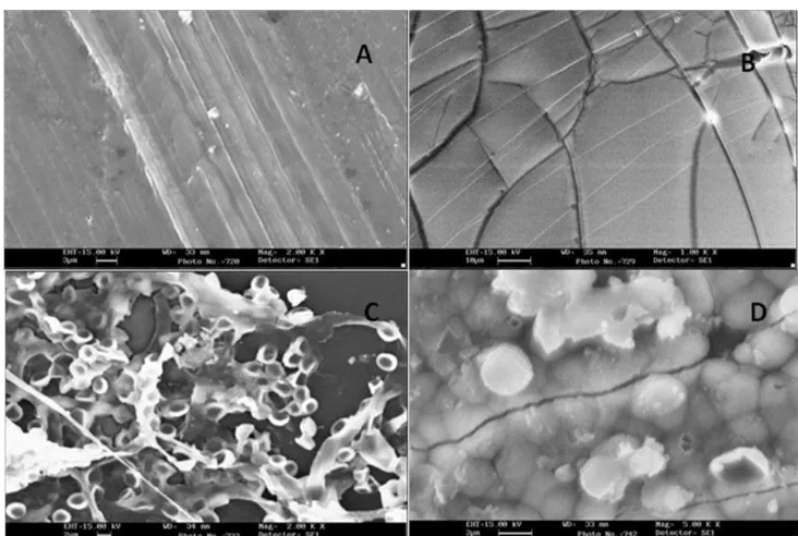
3.6 Morphological studies by scanning electron microscopy of chitosan blended HDPE films before and after seeding with fungi:
The HDPE-films blended with 10 wt % of chitosan were used to record their SEM micrographs for analysing the morphological properties before (Fig. 9A) and after seeding with T. harziauum fungus (Fig. 9 B, C, & D). The SEM micrographs of chitosan blended HDPE films have confirmed a homogeneous surface morphology (Wu,2003) before seeding with fungus (Fig. 9A) but after seeding with fungus for one month, the cracked surface morphology (Fig. 9B) was observed, which confirmed the biodegradation of blended chitosan (Fig. 9B). The SEM micrographs with white small sized spots morphology (Fig. 9 C) have suggested the biodegradation of long chains of HDPE in the blends by the enzymatic action of germinated colonies (Otake et al., 1995).
Fig. 9:SEM micrographs of HDPE blend with 10 wt % chitosan (A) before seeding. SEM micrographs of HDPE blends after seeding with fungus (T.
The SEM micrographs with large white spots correspond to advanced stage of biodegradation of HDPE (Fig. 9 D) by well developed colonies of seeded fungus. The increased amount of biodegraded HDPE in all direction is due to enhanced enzymatic action of fungus on chitosan blended HDPE (Wu,2005). The inhomogeneous large sized microbial colonies were responsible for the appearance of large size structures and cavities on the surfaces of chitosan blended HDPE films after seeding for three months (Pal et al., 2008; Luckachan et al., 2006). The surface morphology of chitosan blended HDPE films after seeding is found to be different than before seeding. Thus, surface morphological analysis of HDPE films by SEM analysis has provided enough supports to suggest microbial biodegrdation of HDPE in HDPE blends prepared using naturally occurring polymers. The SEM micrographs of other polymer blends have also shown similar morphological changes after seeding with selected fungi for three months.
4. Conclusions and future scope of the work:
The microbial biodegradation in high density polyethylene (HDPE) has been induced successfully by blending naturally occurring polymers such as, chitosan, cellulose, starch, alginate, pectin, shellac and xanthane. It has been seen that blending of these polymers has provided suitable environment to to fungi to germinate and colonise on hydrophobic crystalline HDPE. The evaluation of structures in HDPE by FT-IR spectra has been used successfully to confirm the blending of HDPE with polymers. The appearance of new absorption bands in FT-IR spectra after seeding with fungi has confirm the formation of new carbonyl compounds as degradation products of HDEPE and blended polymers. The reduction in molecular weight of HDPE after seeding with selected fungi has also been used successfully to suggest the biodegradation of HDPE in polymer blended HDPE. The variations in physical properties of HDPE such as mechanical, thermal and surface morphological changes after seeding with fungi have also been used to confirm the biodegradation of HDPE in polymer blended HDPE. The Young‘s modulus of HDPE before and after seeding with selected fungi has provided sufficient evidence to suggest biodegration in HDPE blends. There has been a significant decrease in Young‘s modulus, ultimate tensile strength and elongation at break in polymer blended HDPE in comparison to pristine HDPE after seeding fungi with polymer blended HDPE.The extend of biodegrdation in HDPE has varied with the type of polymers used for blending with HDPE. Amonst the selected polymers, chitosan and pectin were found to be quite effective in causing more biodegrdation of HDPE in comparison to other polymers such as; xanthane, starch and cellulose. Thermal studies of polymer blended HDPE before and after seeding with fungus has been used to confirm biodegradation of HDPE in polymer blended HDPE. The fungi seeded HDPE-Polymer blends have shown significant variation in their thermal stability and the rate of decomposition of HDPE in fungus seeded HDPE blends at Tmax has also provided useful information about the type of HDPE chains left for thermal decomposition after biodegrdation by selected fungi. The TG and DSC analysis has shown that chitosan and pectin were able to induce more biodegrdation in HDPE in comparison to HDPE blended with other polymers. The morphological analysis of HDPE blends at different time of seeding by SEM micrographs has also been used to confirm the biodegrdation of HDPE (Labuzek et al., 2004) in fungus seeded HDPE-Polymer blends in comparison to pristine HDPE. These studies have clearly indicated that the blending of HDPE with naturally occurring biodegradable polymers may be an useful effort to design a biodegradable materials to overcome the problem of waste disposal and environmental problems as caused by pure HDPE. The results of these studies have suggested the following important points to develop biodegradable HDPE for various applications.
1- The microbial biodegradation of synthetic polymer depends on its degree of hydrophility, hence blending of HDPE with chitosan and pectin has produced hydrophilic HDPE which is suitable for seeding of fungi. As natural polymers (Kang et al., 1996) are hydrophilic in nature, hence further studies may be carried out using other natural polymers having better chain flexilibity and structural orientation suitable for the growth of seeded fungi (Kawai et al., 2004).
2- The blended polymers with HDPE should also act as a better nutrient for the growth and colonization of seeded fungi. Out of selected polymers, the chitosan and pectin have acted as a suitable nutrient, hence the extent of biodegration of HDPE was more with these polymers in comparison to other polymers.
3- The microbial biodegradation of HDPE is only possible, if well developed colononies of fungi are formed on blended polymers. The biodegradation of long chains of HDPE takes place when optimum amount of enzymes are released by highly populated colonized microbes on the surface of HDPE.
4- The acidic environment may decrease the germination of fungi over the surface of HDPE, hence there should be no acidic by product to observe
biodegradation in HDPE.
5- The variation in degree of biodegration of HDPE with different type of fungi was due to the difference in β -oxidation system, which is an essential part
of cell membranes (Kawai et al., 2004).
6- The variation in Young’s modulus may be used to confirm the blending of polymers with HDPE on the basis of plasticization (Pal et al., 2008) as well as to confirm the biodegrdation of HDPE by seeded fungi.
7- The difference in thickness of interfacial layer of blended polymers on HDPE might also be a reason for the difference in biodegradation capacity of
blended polymers with HDPE. This might be the reason for low biodegradability of HDPE in presence of shellac.
8- Finally, the extent of biodegrdation might be evaluated by recording the variation in physical and thermal properties of fun gi seeded HDPE and also by
recording the morphological changes with SEM microgrpahs.
9- The results of these studies contribute significantly to the existing knowledge for the synthesis and characterization of biodegradable polymers by
bleding the naturally occurring polymers.
Conflict of Interests:
The authors declare no competing financial interest.
ACKNOWLEDGMENTS
Authors are thankful to University of Mosul-Iraq, for providing facilities to carry out the present work. Authors are also thankful to Prof. K. C. Gupta, Indian Institue of Technology Roorkee, India ([email protected]) for analysis of samples and for improving the discussion and interpretation of data reported in this paper.
REFERENCES
Al-Jailawi, M.H., R.S. Ameen and A.A. Al-Saraf, 2015. Polyethyelene degradation by pseudomonas putida S3A, International Journal of Advanced Research in Biological Sciences, 2: 90-97.
Arutchelvi, J., M. Sudhakar, A. Arkathar, M. Doble, S. Bhaduri and P. Veera Uppara, 2008. Biodegradation of polyethylene and polypropylene, Indian J.
Biotechnol.,7: 9-22.
Arvanitoyannis, I., C.G. Biliaderis, H. Ogawa and H. Kawasaki, 1998. Biodegradable films made from low-density polyethylene (LDPE), Rice starch and potato starch for food packaging application: Part I, Carbohydrate Polymers, 36: 89-104.
Avella, M., J.J. De Vlieger, M.E. Errico, S. Fischer, P. Vacca and M.G. Volpe, 2005, Biodegradable starch/ clay nanocomposite films for food packaging applications, Food Chemistry, 93: 467-474.
Bandyopadhyay, S., Zaeni, A., and Nath, D, 2010, Advanced utilization of as received and near whitened fly ash in polypropylene polymer to improve mechanical, notched impact and whiteness colour properties. Int J Plast Technol , 14: 51–56
Damrongsakkul, S., and S. Ngamsinlapasathian, 2002. Properties of post-used high-density polyethylene crates and its modification by ethylene vinyl acetate blending, J. Metal. Mater. Miner., 11: 19- 37.
El-Shafei, H.A., N.H. Abdel-Nasser, A.L. Kansoh and A.M. Ali, 1998. Biodegradation of disposable polyethylene by fungi and streptomyces species, Polym. Deg. Stab., 62: 361-365.
Fasoyiro, S.B.H., H. Gourama and C.N. Cutter, 2017. Stability and safety of maize-legume-fortified flours stored in various packaging materials, European Food Research and Technology, 243: 1861-1868.
Ghazaki, R., L.C. Mei, N.Z.K. Shaari, M. Yusof and S. Ahmed, 2005. Preliminary study on microbial degradation of flexible polyurethane foams-physico-michanical and weight changes during fungal deterioration, J. Oil Pal. Rese., 17: 103-109.
Ghosh, P., 2003. Polymer science and technology, plastics, rubbers, blends and composites, 2nd Edition, Tata McGraw;-Hill Publishing Company Limited, New Delhi, pp: 258.
Griffin, G.T.L., 1994. Starch polymer blends, Polym. Deg. Stab., 45: 241-247.
Honakar, H., M. Barikan and J. Moshedian, 2005. Preparation and study of degradable polyethylene based on starch, Iran. J. Polym. Sci. Tech. (Persian Ed.), 18: 107-114.
Kahar, A.W.M., N. Sarifuddin and H. Ismail, 2017. Structural, thermal and physico-chemical properties of high density polyethyylene density rubber /modified cassava starch blends, Iranian Polymer, 26: 149-159.
Kang, B.G., S.H. Yoon, S.H. Lee, J.E. Yie, B.S. Yoon and M.H. Suh, 1996. Studies on the physical properties of modified starch-filled HDPE film, J. Appl. Polym. Sci., 60: 1977-1984.
Kawai, F., M. Watanabe, M. Shibata, S. Yokoyama, Y. Sudate and S. Hayashi, 2004. Comparative study on biodegradability of polyethylene wax by bacteria and fungi, Polym. Deg. Stab., 86: 105-114.
Krystyna, W.T., E. Wesolowska, D. Wawro and H. Stuszczyk, 2003. Improvement of the enzymatic utilization of textile from cellulose/ polyester blends, Fib. Text. East. Europ., 11: 63-66.
Labuzek, S., B. Nowak and J. Pajak, 2004. The susceptibility of polyethylene modified with bionolle to biodegradation by filamentous fungi, Polish J. Environ. Stud.,13: 59-68.
Li, Y., W. Xu and G. Zhang, 2015. Freshness preservation of cheese with Ag-coated high density polyethylene membrane, Zhenkong Kexue Yu Jishu Xuebao, 35: 710-713.
Luckachan, G.E., L. Jose, V.S. Prasad and C.K.S. Pillai, 2006. Sugar end-capped polyethylene: Ceric ammonium nitrate initiated oxidation and melt phase grafting of glucose onto polyethylene and its microbial degradation, Polym. Deg. Stab., 91: 1484-1494.
Lu, D.R., C.M. Xiao and S.J. Xu, 2009. Starch-based completely biodegradable polymer materials, Expr. Polym. Lett., 3: 366-375. Mandels, M., and D. Sternberg, 1976. Recent advances in cellulose technology, J. Ferment. Technol., 54: 267-286.
Negi, H., A. Kapri, M.G.H. Zaidi, A. Satlewal and R. Goel, 2009. Comparative In-Vitro biodegradation studies of epoxy and its silicone blend by selected microbial consortia, Inter. Biodeter. Biodeg., 63: 553-558.
Nowak, B., J. Pajak and S. Labuzek, 2010. Biodegradation of compositions of polyethylene and poly(ethylene terephthalate) with bionollepolyesters or starch, Kompozyty, 10: 224-228.
Orhan, Y. and H. Buyukgungor, 2000. Enhancement of biodegradability of disposable polyethylene in controlled biological oil, Inter. Biodeter. Biodegr, 45: 49-55.
Otake, Y., T. Kobayashi, H. Asabe, N. Murukami and K. Ono, 1995. Biodegradation of low density polyethylene, polystyrene, polyvinyl chloride and urea-formaldehyde resin buried under soil for over 32 years, J. Appl. Polym. Sci., 56: 1789-1796.
Pal, J., A.K. Ghosh and H. Singh, 2008. Environmentally degradable LLDE/Esterified styrene-maleic anhydride (ESMA) blends, Europ. Polym. J., 44: 1261-1274.
Santonja, B., L. Contat-Rodrigo, R. Moriana-Torro and A. Ribes-Greus, 2007. Thermal characterization of polyethylene blends with a biodegradable masterbatch subjected to thermo-oxidative treatment and subsequent soil burial test, J. Appl. Polym. Sci., 106: 2218-2230.
Satyalakshmi, S., 2016. Isolation and identification of polyethyelene of bags degrading bacteria from visakhapatnam dumping yard, International Journal of Pharmaceutical Sciences and Research, 7: 4200-4203.
Singh B., and N. Sharma, 2008. Mechanistic implications of plastic degradation, Polym. Deg. Stab., 93: 561-584.
Skariyachan, S., A.A. Patil, A. Shankar, M. Manjunath, N. Bachappanavar and S. Kiran, 2018. Enhanced polymer degrdation of polyethylene and polypropylene by novel thermophilic consortia of brevibacillus species and aneurinibacillus species screened from waste management landfills and sewage treatment plants, Polymer Degradation and Stability, 23: 267-270.
Sohel, K.T., N. Gholam-Reza and M. Seyed Mohammad Mahdi, 2018. Oxo-biodegradability of high-density polyethylene films containing limited amount of isotactic polypropylene, Journal of Applied Polymer Science, 135: 233-238.
Thaweegan, N., T. Sukkamat and S. Trivijitkasem, 2005, Calorimetric studies of crystallization kinetics of degradable HDPE plastic film, 31st Congress on Science and Technology of Thailand at Suranaree University of Technology.
Vallini, G., A. Corti, A. Pera, R. Solaro, F. Cioni and E. Chiellini, 1994. Effects of intensive microbial metabolism on starch-filled polyethylene film in controlled composting windrows, Journal of General and Applied Microbiology, 40: 445-61.