Original Research Article
Elastography evaluation of normal thyroid gland and its comparison
with diffuse non nodular enlarged thyroid gland using strain
ultrasound elastography
Sanjay Kumar, Vishal Kumar Jain*, Ashutosh Gupta,
Satya Bhuvan Singh Netam, Nilesh Gupta
INTRODUCTION
The thyroid gland is located in the anterior part of neck, spanning between C5 and T1 vertebra and is located anterior to the thyroid and cricoids cartilage of larynx and first three tracheal rings. It is an endocrine gland, divided into two lobes connected by an isthmus. It is a butterfly or `H` shaped organ each with superior and inferior pole.
A swelling in neck due to enlarged thyroid gland is called a goiter.
Ultrasonography has been widely used for diagnosis of thyroid disease since, it was first introduced in clinical practice in the 1970’s. Used in clinical practice for more than 40 years, it is highly regarded for its ease of use, real-time capability, portability and low cost.
Department ofRadiodiagnosis, Pt. JNM Medical College, Raipur, Chhattisgarh, India
Received: 08 March 2019
Revised: 20 March 2019
Accepted: 29 March 2019
*Correspondence:
Dr. Vishal Kumar Jain, E-mail: [email protected]
Copyright: © the author(s), publisher and licensee Medip Academy. This is an open-access article distributed under the terms of the Creative Commons Attribution Non-Commercial License, which permits unrestricted non-commercial use, distribution, and reproduction in any medium, provided the original work is properly cited.
ABSTRACT
Background: The thyroid gland is located in the anterior part of neck, spanning between C5 and T1 vertebra and is located anterior to the thyroid and cricoids cartilage of larynx and first three tracheal rings. Ultrasonography has been widely used for diagnosis of thyroid disease it is highly regarded for its ease of use, real-time capability, portability and low cost.
Methods: The current observational study was conducted in Department of Radiology, Dr. BRAM Hospital, Pt. JNM Medical College, Raipur, India. Consecutive sampling method was used for the study. All the cases coming to radiology department during the study period were taken as study subjects.
Results: Of the normal subjects, author found 107 normal thyroid subjects and 53 patients were categorized into cases with diffuse non-nodular thyroid swelling groups. Of the diffuse thyroid swelling group about 85% of the study population is between 20-50 years of age group, 13.2% were <20 years of age and one patient is >50 years of age. Females to male ratio for diffuse thyroid disease is 3.4:1.
Conclusions: Ultrasound elastography (USE) is a newly developed non-invasive method to evaluate and compare the elasticity of the thyroid gland and other organs like liver, prostate, parotid, breast pathologies. Its use is based on the principle that pathological changes in a tissue also changes its elasticity.
Keywords: Elasticity contrast index, Strain ultrasound elastography, Thyroid gland, Ultrasound elastography
Since then, new ultrasound modalities have been developed, such as doppler imaging, which provides new information for diagnosis. Elastography was developed in the 1990’s to map tissue stiffness and reproduces/replaces the palpation performed by clinicians. Elastography, whose development started about 20 years ago, aims at imaging tissue stiffness, which provides an additional and clinically relevant information.1
Elastography is analogous to palpation, therefore wherever palpation has clinical value, elastography is relevant tool for diagnosis. Although palpation is limited to superficial organs, many elastography techniques can be applied to deeper organs opening new possibilities for diagnosis.
Ultrasound elastography is a non-invasive modern imaging technique which represents tissues and organs with evaluation of their elasticity, “stiffness”. It is based on the fact that pathological changes in tissues generally affect their stiffness. Strain elastography using internal physiologic force (pulsations from the adjacent carotid artery) to induce the displacement in thyroid tissue necessary to assess tissue elasticity. It is a semi-quantitative method that measures elasticity contrast index (ECI) that represents the stiffness of thyroid tissue. Ultrasonographic elastography is a new, developing method that shows increase in clinical practice.
Thyroid gland disorders include benign and malignant thyroid nodules and diffuse thyroid disorders. Studies had been done to differentiate between benign and malignant thyroid nodules using elastography showing significant difference in elasticity score.2
Studies had also been done with significant use in differentiating benign and malignant Breast neoplasm, prostate, some groups of lymph nodes and liver pathologies.3 Less have been known about the pattern of
changes in elastography in diffuse non-nodular thyroid swelling.
Author wished to conduct this study to get a reference value for normal thyroid tissue in the study population and compare the elasticity contrast index (ECI) in the subjects with diffuse non-nodular thyroid swelling using ultrasound strain elastography technique.
METHODS
The current observational study was conducted in Department of Radiology, Dr. BRAM Hospital, Pt. JNM Medical College Raipur, India during period August 2017-September 2018. Consecutive sampling method was used for the study.
Samsung RS-80 Ultrasound Machine with Linear Array Transducer (LA4-18B), Color Doppler and Strain Elastography Software used for the study purpose. As this is a clinical setting-based time bound study, all the
cases coming to radiology department and qualifying inclusion and exclusion criteria, during the study period will be taken as study subjects.
Inclusion criteria
• Patients between the age groups 20-50 years came for ultrasound abdomen with no significant abnormality detected on routine ultrasound neck for normal thyroid subjects,
• Patients with non-nodular diffuse thyroid swelling with or without symptoms of hypothyroidism or hyperthyroidism.
Exclusion criteria
• Patient with focal thyroid nodules, cystic lesion diagnosed on ultrasound,
• Patient with previous history of thyroid surgery,
• Patient who received radiation therapy,
• Pregnant women,
• Unwilling patients.
Protocol for thyroid elastography
Colour doppler diagnostic ultrasound instrument with linear probe having frequency 4-18 MHz and strain elastography software is used. Patient took supine position with neck in slight hyperextension for adequate exposure of thyroid. The probe was kept perpendicular to the skin over thyroid in transverse plane to routinely scan both the lobes and isthmus of thyroid to judge the type of thyroid gland.
Thyroid dimensions are recorded to categorize the patient into normal thyroid or diffuse thyroid swelling group. It is of importance to choose the shortest path for the exploratory US beam to the ROI, thus avoiding strain decay with distance. It is also important to exclude vessels (carotid), eso-tracheal structures, bones and muscles from ROI.4
Elastoscan mode is selected and the probe is hold still perpendicular to the thyroid lobe. No external compression with the transducer was needed as Elastoscan uses the pulsations of the carotid artery (in vivo compression) for the elastography response.5 Patient
is instructed to hold breath and swallowing for 10-15 seconds during scan.6 The image is paused after all the
colour bar turned green.
Elastogram was considered optimal when the screen quality indicator turned to green (multiple boxes that change color from red, to yellow or green). ROI is selected. The operator performed the examination in transverse plane and then drew the region of interest (ROI) of approximately 5 mm.
the screen, side by side with the B-mode image) the thyroid were color coded depending on their stiffness with a colorimetric scale on the screen indicating the corresponding hard and soft areas, color coded red and blue respectively.5 Within the ROI the software assessed
the elasticity contrast index (ECI), a semiquantitative stiffness evaluation.
The elasticity contrast index (ECI), which quantifies local strain contrast within a ROI, was computed and displayed on the screen.5 Total of 6 readings were obtained i.e. two
readings of ECI was obtained from each right and left lobe and from the isthmus. Average of the two was noted. ECI of each was obtained and interpreted separately. In few cases where isthmus was <2.5 mm, only readings from right and left lobes were obtained.
Elasticity Contrast Index (ECI)
Ultrasound elasticity contrast index (ECI) is the difference of tissue strain determined by elasticity imaging and can reflect the uniformity of strain in tissue. ECI is an ultrasonic quantitative index developed on the basis of ultrasonic elastography, avoids the limitation of the traditional triggered elastography, and can determine the tissue hardness as well as the difference and homogeneity of all pixel strain in the ROI at the same time.4,5,7-10
Statistical evaluation
Data was collected and analyzed with the help of excel and graph-pad in stat software for statistical analysis. Proportion as number and percentage was determined for categorical variable like gender.
RESULTS
Of the normal subjects, author found 107 normal thyroid subjects and 53 patients were categorized into cases with
diffuse non-nodular thyroid swelling groups. Of the normal subjects only the age group between 21-50 years were included to get ECI value of adult population. Of the diffuse thyroid swelling group about 85% of the study population is between 20-50 years of age group, 13.2% were <20 yrs of age and one patient is >50 years of age (Table 1).
Table 1:Age wise distribution of normal thyroid and diffuse thyroid swelling subjects.
Age (years)
Normal thyroid Diffuse thyroid swelling No. Percentage No. Percentage
Upto 20 NIL 0.00% 7 13.20% 21-30 49 45.80% 23 43.40% 31-40 32 29.90% 15 28.30% 41-50 26 24.30% 7 13.20% >50 NIL 0.00% 1 1.90%
Total 107 100% 53 100%
Out of 160 patients scanned, 112 patients were females and 48 were males. 63.4% out of total 112 females had normal thyroid dimensions and 36.6% had diffuse thyroid swelling. Out of 48 males studied, 75% were normal thyroid subjects and 25% had diffuse non-nodular thyroid swelling. Thus, in this study female patients had more diffuse thyroid disease (36.6%) compared to the male (25%) counterparts. Thus, females to male ratio for diffuse thyroid disease is 3.4:1 (Table 2).
Table 2:Gender wise distribution of patients with
diffuse thyroid swelling and normal thyroid dimensions.
Gender
Diffuse thyroid swelling
Normal thyroid dimensions
Total
Male 12 (25%) 36 (75%) 48 (100%) Female 41 (36.6%) 71 (63.4%) 112 (100%) Total 53 (33%) 107 (67%) 160 (100%)
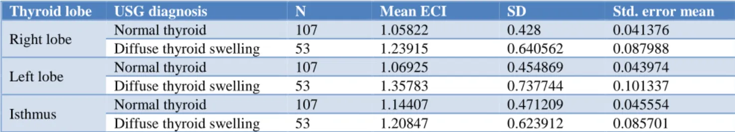
Table 3:Comparison of elasticity contrast index (ECI) value of normal thyroid vs diffuse thyroid swelling.
Thyroid lobe USG diagnosis N Mean ECI SD Std. error mean
Right lobe Normal thyroid 107 1.05822 0.428 0.041376
Diffuse thyroid swelling 53 1.23915 0.640562 0.087988
Left lobe Normal thyroid 107 1.06925 0.454869 0.043974
Diffuse thyroid swelling 53 1.35783 0.737744 0.101337
Isthmus Normal thyroid 107 1.14407 0.471209 0.045554
Diffuse thyroid swelling 53 1.20847 0.623912 0.085701
Author found elasticity contrast index (ECI) value of normal thyroid subjects with normal thyroid dimensions as ECI of right lobe=1.05±0.42 (range=0.63 to 1.47), Left lobe=1.06±0.45 (range=0.61 to 1.51) and Isthmus
groups of normal thyroid group and diffuse thyroid group using independent t-test, p-value of right lobe and left lobe was 0.035 and 0.003 respectively, which is
statistically significant, while p-value of isthmus was found to be 0.467 which is not statistically significant (Table 3, Table 4).
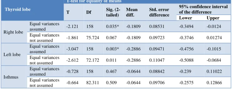
Table 4: Independent ‘T’ test result.
Thyroid lobe
T-test for equality of means
T Df Sig.
(2-tailed)
Mean diff.
Std. error difference
95% confidence interval of the difference
Lower Upper
Right lobe
Equal variances
assumed -2.121 158 0.035* -0.1809 0.08531 -0.3494 -0.0124 Equal variances
not assumed -1.861 75.724 0.067 -0.1809 0.09723 -0.3746 0.01274
Left lobe
Equal variances
assumed -3.047 158 0.003* -0.2886 0.09471 -0.4756 -0.1015 Equal variances
not assumed -2.612 72.172 0.011 -0.2886 0.11047 -0.5088 -0.0684
Isthmus
Equal variances
assumed -0.728 158 0.467 -0.0644 0.08842 -0.239 0.11022 Equal variances
not assumed -0.664 82.311 0.509 -0.0644 0.09706 -0.2575 0.12866
* Significant
The independent T - test result (Table 4 shows that there is a significant difference in mean value of right lobe (p=0.035) and left lo be (p =0.003) of thyroid between the groups at 95% confidence interval. However, the difference in the isthmus was found to be in significant (p- 0 .467).
DISCUSSION
Ultrasound elastography is a newer non-invasive modern imaging technique which measures the stiffness of the tissue examined. It is based on the fact that pathological changes in tissue also alters its stiffness. Diffuse thyroid swelling is caused by various diffuse thyroid diseases like Hahimoto`s disease, Graves`s disease, chronic autoimmune thyroiditis, de Quervain`s disease, Riedel`s struma, painless thyroiditis, acute suppurative thyroiditis, etc.11,7,12-14 In previously published papers, thyroid
elastography has been used to evaluate thyroid nodule stiffness in order to differentiate malignant from benign ones. Sebag FJ et al, used shear wave elastography (SWE) for differential diagnosis of benign and malignant thyroid nodules and compared SWE with ultrasound (US) and found that Elasticity Index (EI) was significantly higher in malignant nodules (150±95 (30-356 kPa) than in benign nodules (36±30 (0-200 kPa) and normal thyroid glands. T Rago et al, found promising results using strain elastography in differentiating nodules, which were of indeterminate significance in gray scale ultrasound.15,16,19
Choi WJ et al, did a study on 102 patients with indeterminate thyroid nodules using ultrasound elastography with carotid pulsation as the compression
source and found that, ECI values for malignant nodules (3.07±2.01) were significantly higher than those for benign nodules (1.44±0.47). The specificity of ECI (97.8%) was higher than that of the gray-scale ultrasound score (72.2%).2
autoimmune thyroiditis (CAT) and to verify the effect of fibrotic thyroid tissue on shear wave velocity (SWV).17
They scanned 229 patient’s normal thyroid lobes (controls) and 150 chronic autoimmune thyroiditis (CAT) lobes. They found that the mean SWV for CAT was 2.47±0.57 m/s, which was significantly higher than the mean shear wave velocity for normal controls (1.59±0.41 m/s) (P <0.001).
Another study done by Sporea I et al, on diffuse thyroid gland pathology using ARFI to assess whether acoustic radiation force impulse (ARFI) elastography can differentiate normal from pathological thyroid parenchyma.18 They evaluated 136 subjects (106 women
and 30 men), 44 (32.3%) without thyroid pathology, 48 (35.3%) with Graves’ disease (GD), 37 (27.2%) with chronic autoimmune thyroiditis (CAT), 4 (2.9%) with diffuse thyroid goiter and 3 (2.2%) cases with thyroid pathology induced by amiodarone. They found that thyroid stiffness (TS) assessed by means of ARFI in healthy subjects (2±0.40 m/s) was significantly lower than in GD (2.67±0.53 m/s) (P <0.0001) and CAT patients (2.43±0.58 m/s) (P=0.0002), but the differences were not significant between GD vs CAT patients (P=0.053). They concluded that ARFI seems to be a useful method for the assessment of diffuse thyroid gland pathology.
CONCLUSION
Ultrasound elastography (USE) is a newly developed non-invasive method to evaluate and compare the elasticity of the thyroid gland and other organs like liver, prostate, parotid, breast pathologies. Its use is based on the principle that pathological changes in a tissue also changes its elasticity.
Funding: No funding sources Conflict of interest: None declared
Ethical approval: The study was approved by the Institutional Ethics Committee
REFERENCES
1. Gennisson JL, Deffieux T, Fink M, Tanter M.
Available at:
https://www.sciencedirect.com/science/article/pii/S2 211570613000398. Élastographie ultrasonore: principes et procédés. J de Radiologie Diagnostique et Interventionnelle. 2013;94(5):504-13.
2. Choi WJ, Park JS, Koo HR, Kim SY, Chung MS, Tae K. Ultrasound elastography using carotid artery pulsation in the differential diagnosis of sonographically indeterminate thyroid nodules. Am J Roentgenol. 2015;204(2):396-401.
3. Giovannini M, Botelberge T, Bories E, Pesenti C, Caillol F, Esterni B, et al. Endoscopic ultrasound elastography for evaluation of lymph nodes and
pancreatic masses: a multicenter study. World J Gastroenterol: WJG. 2009;15(13):1587.
4. Dudea SM, Jid CB. Ultrasound elastography in thyroid disease. Medi Ultrasonography. 2015;17(1):74-96.
5. Cantisani V, David E, De Virgilio A, Sidhu PS, Grazhdani H, Greco A, et al. Prospective evaluation of Quasistatic Ultrasound Elastography (USE) compared with Baseline US for parotid gland lesions: preliminary results of elasticity contrast index (ECI) evaluation. Med Ultrasonography. 2017;19(1):32-8.
6. Cekic B, Parlak AE, Koksel Y, Toslak IE, Parlak M. Real-time ultrasound elastography evaluation of the thyroid gland in adolescent patients with hashimoto thyroiditis. Biomed Res. 2017;28(17).
7. Sheng JG, Wang B, Cao KK, Zhang S. Relationship of thyroid ultrasound elasticity contrast index with serum autoantibody and Th1/Th2 cytokine levels in patients with Hashimoto's thyroiditis. J Hainan Med University. 2016;22(19):147-50.
8. Menzilcioglu MS, Duymus M, Avcu S. Sonographic elastography of the thyroid gland. Polish J Radiol. 2016;81:152.
9. Zaleska-DorobiszA U, KaczorowskiB K, PawluśB A, PuchalskaB A, InglotB M. Ultrasound elastography-review of techniques and its clinical applications. Brain. 2013;6:10-4.
10. Franchi-Abella S, Elie C, Correas JM. Ultrasound elastography: advantages, limitations and artefacts of the different techniques from a study on a phantom. Diagnostic Interventional Imaging. 2013;94(5):497-501.
11. Rumack CM, Levine D. Diagnostic ultrasound E-book. Elsevier Heal Sci. 2017.
12. Prabhakar BS, Bahn RS, Smith TJ. Current perspective on the pathogenesis of Graves’ disease and ophthalmopathy. Endocrine Rev. 2003;24(6):802-35.
13. Castagnone D, Rivolta R, Rescalli S, Baldini MI, Tozzi R, Cantalamessa L. Color Doppler sonography in Graves' disease: value in assessing activity of disease and predicting outcome. AJR. Am J Roentgenol. 1996;166(1):203-7.
14. Desailloud R, Hober D. Viruses and thyroiditis: an update. Virol J. 2009;6(1):5.
15. Sebag F, Vaillant-Lombard J, Berbis J, Griset V, Henry JF, Petit P, et al. Shear wave elastography: a new ultrasound imaging mode for the differential diagnosis of benign and malignant thyroid nodules. J Clin Endocrinol Metab. 2010;95(12):5281-8. 16. Garra BS, Cespedes EI, Ophir J, Spratt SR, Zuurbier
RA, Magnant CM, et al. Elastography of breast lesions: initial clinical results. Radiol. 1997;202(1):79-86.
18. Sporea I, Sirli R, Bota S, Vlad M, Popescu A, Zosin I. ARFI elastography for the evaluation of diffuse thyroid gland pathology: Preliminary results. World J Radiol. 2012;4(4):174-8.
19. Rago T, Santini F, Scutari M, Pinchera A, Vitti P. Elastography: new developments in ultrasound for predicting malignancy in thyroid nodules. J Clin Endocrinol Metab. 2007;92:2917-22.
Cite this article as: Kumar S, Jain VK, Gupta A, Netam SBS, Gupta N. Elastography evaluation of normal thyroid gland and its comparison with diffuse non nodular enlarged thyroid gland using strain ultrasound elastography. Int J Res Med Sci