0270-6474/84/0411-2681$02.00/O The Journal of Neuroscience
Copyright 0 Society for Neuroscience Vol. 4, No. 11, pp. 2681-2688
Printed in U.S.A. November 1984
THE DISTRIBUTION
AND
CHROMATOGRAPHIC
CHARACTERIZATION
OF PHI
(PEPTIDE
HISTIDINE
ISOLEUCINE
AMIDE)-27-LIKE
PEPTIDES
IN RAT AND
PORCINE
BRAIN1
MARGERY C. BEINFELD,’ DIANA M. KORCHAK, BRYAN L. ROTH,* AND T. L. O’DONOHUES
Department of Pharmacology, St. Louis University, St. Louis, Missouri 63104; * Department of Psychiatry, Naval Medical Hospital, Bethesda, Maryland 20814 and Laboratory of Preclinical Pharmacology, National Institute of Mental Health, Washington, D.C. 20032; and $ Experimental Therapeutics Branch, National Institute of Neurological and Communicative
Disorders and Stroke, National Institutes of Health, Bethesda, Maryland 20205
Received October 28,1983; Revised February 27,1984; Accepted April 12, 1984
Abstract
This study was initiated to characterize PHI (peptide histidine isoleucine amide)-27-like peptides (PLPs) in rat and porcine brain in comparison with other members of the vasoactive intestinal polypeptide (VIP) family and to investigate their distribution by radioimmunoassay.
The peptidic nature of the rat brain PLP was indicated by its trypsin sensitivity. On Sephadex chromatog- raphy rat brain PLP has the same molecular weight as synthetic (porcine intestinal) PHI-27. High pressure liquid chromatographic separations revealed that PLP in rat and porcine brain extracts elutes as a single peak distinct from VIP or secretin. Porcine brain PLP elutes in the same position as synthetic PHI-27, whereas rat brain PLP immunoreactivity consistently separates from synthetic PHI-27. This suggests that porcine brain PLP is identical to synthetic PHI-27, in agreement with the reported sequence of Tatemoto et al. (Tatemoto, K., M. Carlquist, T. McDonald, and V. Mutt (1983) FEBS Lett. 153: 248-252), whereas PLP may have a different amino acid sequence (or may be post-translationally modified).
Using specific PHI and VIP radioimmunoassays, the distribution of PLP was found to parallel that of VIP in rat and porcine brain, being highest in cerebral cortex, amygdala, and hippocampus. PLP, like VIP, is abundant in rat retina and can be included in the growing list of retinal peptides. This highly correlated distribution of VIP and PLP may be explained by the recent discovery that they are derived from the same precursor (Itoh, N., K. Obata, N. Yanaihara, and H. Okamoto (1983) Nature 304: 547-549). In some brain regions, however, the distribution of the two peptides was less strongly correlated, which perhaps could be explained by differential local regulation of gene translation or peptide processing.
The closely correlated distribution of brain VIP and PLP and the known ability of PHI-27, like VIP, to activate adenylate cyclase (Jensen, R. T., K. Tatemoto, V. Mutt, G. Lemp, and J. Gardner (1982) Am. J. Physiol. 241: G498-G502; Watling, K. J., and J. E. Dowling (1983) J. Neurochem. 41: 1205-1213; Roth, B. L., M. C. Beinfeld, and A. C. Howlett (1984) J. Neurochem. 42: 1145-1152) and inhibit VIP binding (Jensen, R. T., K. Tatemoto, V. Mutt, G. Lemp, and J. Gardner (1982) Am. J. Physiol. 241: G498-G502; Robberecht, P., K. Tatemoto, P. Chatelain, M. Waelbroeck, M. Delhaye, G. Taton, P. DeNeff, J. Camus, J. C. Hause, and J. Christophe (1982) Regul. Pept. 4: 241-250) suggest that PLPs may play a role as endogenous modulators of VIP activity.
Peptide histidine isoleucine amide (PHI)-27 was isolated activities in several tissues. It stimulates amylase secretion and from porcine intestine (Tatemoto and Mutt, 1981) and the elevates levels of CAMP in pancreatic acinar cells (Jensen et identical sequence was found in porcine brain (Tatemoto et al., al., 1982), lung membranes (Robberecht et al., 1982), rat neu- 1983). This peptide has strong sequence homology with vaso- roblastoma cells (Roth et al., 1984), carp retinal pieces and active intestinal peptide (VIP), secretin, glucagon, gastric in- isolated horizontal cells (Watling and Dowling, 1983), and hibitory peptide, and newly discovered growth hormone-releas- intestinal epithelial cells, fat cells, and gastric glands (Bataille ing hormone (GHRH). PHI-27 has VIP- and secretin-like et al., 1980). Under some conditions PHI-27 is more potent
than VIP in releasing prolactin from dispersed anterior pitui- tary cells and hemipituitaries of the rat (Werner et al., 1983). 1 This work was supported by National Institutes of Health Grants Other VIP-like actions of PHI-27 have recently been summa- NS 18335 and NS 18667 and by a grant from the American Parkinson rized (Christofides et al., 1982a). PHI-27 can inhibit iz51-labeled Disease Association. We wish to thank Ms. Maggie Klevorn for expert VIP binding in these systems (although with lower affinity
secretarial assistance. than VIP) and appears to activate adenylate cyclase via the
* To whom correspondence should be addressed. VIP or secretin receptors in these tissues.
detailed descriptions of PLP’s distribution, chemical nature, chromatographic characteristics, and its relationship to other structurally related peptides in brain have not yet been re- ported. In preliminary reports, a striking similarity in the distribution of VIP and PLP was observed (Christofides et al., 1982a). Immunocytochemical staining of central and peripheral tissues indicated that VIP and PLP were often co-localized in the same neurons, although Hokfelt et al. (1982) found this not to be true for the paraventricular nucleus and the median eminence. This close association of VIP and PLP has led to the suggestion that PLP and VIP might originate from the same precursor peptide. The cloning of the mRNA for VIP from a human neuroblastoma cell line has revealed the presence of an amino acid sequence which codes for a peptide very similar to PHI (25 of 27 amino acids the same) (Itoh, et al., 1983). Whether the mRNA which codes for this PLP (called PHM-27) is the human equivalent of PHI or whether other mRNAs exit which code for PHI-27 in humans has not been
determined.
This study was initiated to examine in greater detail the
chemical composition and chromatographic characteristics of
PLP, its relationship to VIP and other chemically related brain
peptides, and its distribution in rat and porcine brain.
Materials and Methods
PHI antiserum
A specific antiserum against porcine PHI was obtained by immuniz- ing male New Zealand White rabbits with synthetic PHI-27 (Pennin- sula Laboratories) conjugated to bovine serum albumin with glutaral- dehyde. The conjugate was prepared by adding dropwise 3.5 ml of a solution of freshly prepared 22 mM glutaraldehyde to a mixture of 0.5 mg of PHI-27 dissolved in 0.5 ml of phosphate buffer (0.05 mM, pH 7.4) and 7.5 mg of bovine serum albumin dissolved in 1.2 ml of 0.38 M
borate buffer, pH 8.5. The rabbits were injected at approximately 2- week intervals with conjugate containing 50 fig of PHI in Fruend’s complete adjuvant and were bled periodically, 3 days after immuniza- tion. The bleed used in the study (bleed 5) was obtained about 6 months after the beginning of the immunizations.
PHI and VIP radioimmunoassays
PHI and VIP were iodinated by the same procedure, and the labeled peptides were separated using a fibrous cellulose column (Christophe et al., 1976). The labeled peptides were stable for 3 to 4 weeks at -20°C. The PHI antiserum was used at a final dilution of 1:20,000 in a final assay volume of 0.5 ml using phosphate-buffered saline (PBS; 10 mM
sodium phosphate, pH 7.4,0.15 M NaCl) containing 0.1% ethylmercur- ithiosalicylate and 25 mM EDTA. The assay employed a non-equilib- rium method in which antibody, buffer, and samples were incubated for 24 hr at 4°C iodinated peptide (4000 to 8000 cpm) was added, and the assay was stopped 16 to 18 hr later. Free and bound labeled peptide were separated by dextran-charcoal (5 gm of Decton-Dickinson RIA- grade Norit and 0.5 gm of Becton-Dickinson RIA-grade dextran/liter of PBS), 1 ml/tube, followed by centrifugation at 1,800 X g for 15 min. The supernatant was decanted and counted for 1 min in a Beckman 8000 gamma counter interfaced with an Apple II+ computer. Results were calculated utilizing the National Institutes of Health RIA program of Rodbard and Munson (1980), which employs a nonlinear least squares approximation of the log-logit transformation. The National Institutes of Health RIA program as provided by Biomedical Comput- ing Technology Information Center, Vanderbilt University, Nashville, TN.
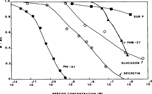
The PHI assay can reliably detect 10 to 20 pg/tube, with an EDs0 to 50 to 100 pg/tube. The antiserum cross-reacts weakly with secretin (0.8%), glucagon (0.05%), and PHM-27 (0.02%), but at 1 fig/tube it displays no cross-reactivity with any of the other peptides in the VIP- secretin family or with other unrelated neuropeptides tested (see Fig. 1).
VIP RIA. VIP RIA was performed as previously described (Eiden et al., 1982), using a VIP antiserum, N2-12 (kindly provided by Dr. G.
Tissue dissection and extraction
To determine the best extraction method for PLP from rat brain, several methods which work well for VIP were tested along with other methods which work well for other peptides. The results are shown in Table I.
The porcine brains were dissected at the slaughterhouse, frozen on dry ice, extracted in boiling water, and centrifuged at 10,000 x g for 10 min, and the pellets were re-extracted with 0.5 M acetic acid. The two supernatants were combined, dried down, resuspended, and assayed for PLP and VIP.
For the gross dissection of rat brain, the rat brain was placed in a Plexiglas brain block and sliced with razor blades into l-mm slices, placed on iced glass slides, and dissected free-hand into the areas described. The dissected areas were frozen on dry ice and stored at -20°C until they were extracted by sonication in 0.1 N HCl. An aliquot was removed from each homogenate for Lowry protein determination (Lowry et al., 1951); the extracts then were clarified by centrifugation at 10,000 x g for 10 min. The 0.1 N HCl extracts were neutralized with an equal volume of 0.1 N NaOH prior to the VIP or PHI RIA. The micropunches were prepared by the method of Palkovits (1973) and were extracted as previously described (Beinfeld and Palkovits, 1982).
Chromatography
Sephadex
A l.O-ml sample of 0.1 N HCl extract of rat brain (equivalent to one- tenth of a rat brain) was applied to a 1 x 100 cm column of Sephadex G-50 (Superfine) previously equilibrated with 1% (v/v) acetic acid. The elution of rat brain PLPs was compared with the elution of synthetic PHI. blue dextran (void volume marker). and Na? (included volume marker). Two-milliliter fractions were collected, and aliquots were removed and dried prior to the RIA.
HPLC
Three chromatographic systems were used to compare the elution of porcine and rat brain- VIP and PHI peptides. All employed a Varian 5000 eouinned with an LDC Snectrometer III variable wavelength UV detector and were run at 1 mljmin with l-ml fractions collected.
A. C18-TEAP system. This system utilized octadecylsilane (C18) column from Alltech (0.45 X 25 cm; 5-pm particle size) run with a linear gradient, lasting 60 min, from 20% to 60% acetonitrile in trieth- ylamine/phosphoric acid, pH 3.25.
II. Cl%TFA system. This system used column run with a linear gradient, lasting 60 min, of 20% to 80% buffer B (acetoni- trile:water:trifluoracetic acid (TFA), 900:99:1, v/v/v, buffer A (wa- ter:TFA, 999:1, v/v).
C. Phenyl-TFA system. This sytem utilized a Waters phenyl column (25 x 0.45 cm) and the same buffer system as in system B, with a gradient from 20 to 60% buffer B, lasting 60 min.
Fractions were dried in a Savant vacuum centrifuge and assayed for PHI and VIP. The elution of VIP and PHI immunoreactivity in rat and porcine brain was compared to the elution of synthetic (porcine) VIP and PHI-27 optical activity and immunoreactivity. Standards were run before and after the brain samples, with extensive washing and blank injections of water in between to ensure that no carryover of standards into sample occurred.
Protease digestion of PHI
Trypsin (Sigma, type III, bovine pancreas, 11, 250 N-cr-benzoyl-L- arginine ethyl ester units/mg of protein) was added at a final concen- tration of 5 mg/ml to 1 ng of synthetic PHI or a volume of rat extract containing 1.4 ng of PHI-like immunoreactivity, in a final volume of 1.6 ml containing 13 mM sodium phosphate, pH 7.6. The samples were incubated at 37”C, and aliquots were removed at 15, 30, and 45 min, boiled for 10 min, and assayed for PHI.
Results
The Journal of Neuroscience Brain PHI: Distribution and Characterization 2683
0.8
GLUCAGON f
-12 ..ll .I 0 -0 -8 -7 -6 -5
10 10 10 10 10 10 10 10
PEPTIDE CONCENTRATION (M)
Figure 1. Immunological characterization of the PHI antiserum (P3) used in this study. P3 cross-reacts weakly with secretin (0.8%), glucagon (0.05%), and PHM-27 (0.02%) but displays no cross-reactivity with other structurally related peptides (VIP, gastric inhibitory peptide, motilin, and GHRH) or unrelated peptides (Leu-enkephalin, gastrin, or cholecystokinin-8) tested to 1 pg/ml.
TABLE I
The effect of different extraction methods on the recovery of PHI-like immunoreactivity from rat brain
Rat brains were split sagittally into two equal pieces at the midline, frozen, weighed, and extracted individually by the different methods in groups of three, in a volume of 5.0 ml. All boiling steps were for 10 min. Homogenization was performed with a Tekmar Ultra-Turrax homogenizer. In method 3, the re-extraction was at room temperature. In method 5, the tissue was homogenized in 75% acetone/22% (4.5% v/v) acetic acid. In methods 4 and 5, the extraction was done at room temperature.
Extraction Method PHI
nglgm of wet wt. 1. Boiling water
2. Boiling 0.5 M acetic acid
3. Boiling water, re-extract pellet with 0.5 M
acetic acid
60 f 0.3 206 f 16
101 f 16
4. 0.1 N HCl 5. Acid/acetone
249 + 36 137 f 8
was completely eliminated in 15 min. This indicates that PLP in rat brain is a peptide and that, like synthetic PHI-27, which is cleaved by trypsin into five fragments (Tatemoto and Mutt, 1981), it has trypsin-sensitive basic residues in portions of the sequence where cleavage eliminates immunoreactivity.
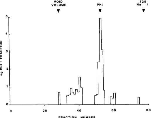
Sephudex chromatography of rat brain extracts. A rat brain extract was applied to a Sephadex G-50 column and the elution of PLP was determined (see Fig. 2). The majority of the PHI-
like material eluted in the same position as synthetic PHI,
although additional peaks appear with a higher apparent mo- lecular weight. These additional peaks could be either biosyn- thetic precursors to PLP, PLP aggregates, or other peptides
with some degree of cross-reactivity with the PHI antiserum.
Further experiments would be required to determine the iden- tity of this immunoreactive material.
High pressure liquid chromatographic (HPLC) separation of PLP from rat and porcine brain. Rat and porcine extracts were
subjected to HPLC separations as described under “Materials and Methods.” Three different systems were used, and since the data obtained on all of them were very similar, the data from only two systems are included. On all systems, rat and porcine brain PLP elutes as a single major peak separated from secretin and VIP. Porcine brain PLP co-elutes with synthetic
PHI-27 as would be expected, whereas rat brain PLP elutes
three to four fractions before synthetic PHI-27, indicating that it is less hydrophobic. VIP-like immunoreactivity in both por- cine and rat brain co-elutes with synthetic VIP, in agreement with sequence data on VIP in these two species (Said and Mutt, 1970; Dimaline et al., 1983) (Fig. 3). There is an additional smaller peak of VIP immunoreactivity which elutes near secre- tin in rat and porcine brain. Since the VIP antiserum has no cross-reactivity with secretin when tested at 1 pg/assay tube, it is unlikely that it is secretin. A minor peak of PLP also occurs in the same location in porcine brain but not in rat brain. Based on the secretin content of brain and the low cross-
reactivity of the PHI antiserum for secretin, it is unlikely that
this peak is secretin. The nature of this peak is unknown, although it could be modified VIP or PHI peptide, or perhaps is yet another member of the VIP family. The observation that it is detected by both VIP and PHI antisera suggests the latter possibility. On a phenyl HPLC column rat PLP also elutes as a single major peak separated from VIP, PLP, and secretin (see Fig. 4).
Distribution of PHI and VIP in rat and porcine brain. Based on the chromatographic characterization data, it is likely that rat PLP is different from PHI-27. Until the sequence of rat PLP is known and its degree of cross-reactivity with the PHI- 27 antiserum is determined, precise quantitation of PLP levels in rat brain cannot be performed. However, the PHI RIA can provide a good relative measure of the abundance of PLP peptides in different brain regions.
0 20 40 60 60 FRACTION NUMBER
Figure 2. Sephadex G-50 chromatography of a rat brain extract eluted with 1% (v/v) acetic acid. Fractions of 2.0 ml were collected. The elution of a rat brain extract and synthetic PHI measured by RIA is compared to the elution of blue dextran (void volume marker) and Nan51 (included volume marker).
MOT v
A
I
B
P SEC PHI
v v
MOT VIP SEC PHI
vvv v
100’
m PHI
Fi 0 VIP
Z :
; 60- \ z I 0
Figure 3. HPLC separation of VIP and PHI peptides of porcine (A) and rat (B) brain extracts in comparison with the elution of synthetic PHI or VIP detected opti- cally or by RIA, or secretin and motilin detected optically. This separation utilized the Cl&TEAP system described under “Materials and Methods.”
FRACTION NUMBER
The Journal of Neuroscience Brain PHI: Distribution and Characterization 2685
brain regions is very high, approaching that of cholecystokinin (Beinfeld et al., 1981). Like VIP, PLP is higher in cerebral cortex, hypothalamus, and the limbic system than it is in the thalamus and more caudal poritons of the brain. In most large areas of rat brain measured for PLP and VIP there is no statistical difference between either the concentration or the content of VIP and PLP. In the amygdala and cerebellum, the PLP concentration is higher than that of VIP, whereas in the mesencephalon, olfactory bulb, medulla, and retina the opposite is true.
When the PLP and VIP concentration is compared in micro- punches of rat brain, local differences in VIP and PLP concen-
ViP
SEC c PHI
0 10 20 30 40 50 60
FRACTION NUMBER
Figure 4. HPLC separation of a rat brain extract on a Waters phenyl column using the TFA system described under “Materials and Methods.” Elution of PHI and VIP were detected by RIA, secretin elution was detected optically.
tration become more apparent, as is shown in Table III. In more than half of the areas examined, there was a significant difference between the concentration of VIP and PLP. In some areas the difference is quite dramatic: in the suprachiasmatic nucleus, the PLP concentration was 3.6 + 0.6 and VIP was 0.88 f 0.05; in the posterior hypothalamus the VIP concentration was 0.34 + 0.04 and PLP was undetectable (<0.09). Neither VIP nor PLP is consistently the highest in all areas, although PLP is consistently higher than VIP in cerebral cortex and basal ganglia, whereas VIP is higher than PLP in thalamus and in most areas of the hypothalamus except the suprachias- matic and periventricular nuclei.
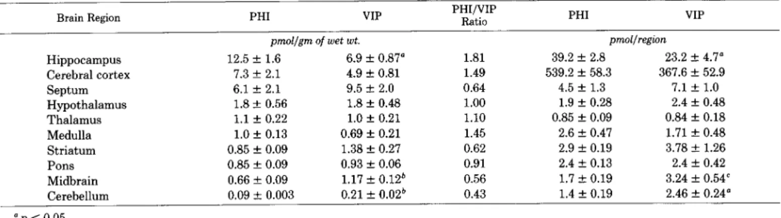
In porcine brain, also, PLP concentrations also closely par- allel VIP concentration, and both PLP and VIP are substan- tially lower in porcine brain than in rat brain (Table IV). That porcine brain has substantially lower VIP levels than does rat brain has been reported previously (Fahrenkrug et al., 1978), although differences in extraction methods between rat and porcine brain used in the present study also account for some of the difference.
Discussion
Rat and porcine brain contain substantial quantities of a PHI-like peptide (PLP). Based on the Sephadex and HPLC results reported here and on the sequence data of Tatemoto et al. (1983), this porcine brain peptide is identical to PHI. In rat brain, the PLP is similar in molecular weight but either has some alteration in its amino acid sequence or has been post- translationally modified in a way which has rendered it less polar than synthetic PHI-27. Rat PLP is unlikely to be VIP, secretin, glucagon, GHRH, or PHM-27 since the PHI antise- rum detects them so poorly. The sequence of this rat PLP may be quite similar to that of PHI-27 since it has similar trypsin sensitivity and can be detected efficiently with a PHI-27 anti- serum. Work is in progress to determine the sequence of rat PLP, to resolve questions about whether it is a PHI-27-related peptide or another new member of the diverse VIP family. It appears that the PHI-27-like sequence is not strongly conserved if porcine and human PLP are known to be different and rat PLP appears to be different than either human or porcine.
The PHI antiserum used in this study displays weak cross- reactivity with secretin (0.8%) and glucagon (0.5%); thus it is
TABLE II
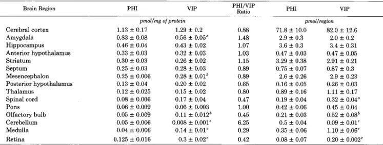
Comparison of the distribution of PHI- and VIP-like immunoreactivity in brains of 200-gm male rats
Data are expressed as picomoles of peptide per milligram of protein or as picomoles of peptide per brain region. Values are mean f SEM (N = $6). Statistical comparison between VIP and PHI values was performed with the two-tailed Student’s t test.
Brain Region PHI VIP PHI/VIP
Ratio PHI VIP
Cerebral cortex Amygdala Hippocampus
Anterior hypothalamus Striatum
Septum Mesencephalon Posterior hypothalamus Thalamus
Spinal cord Pons Olfactory bulb Cerebellum Medulla Retina
pnwl/mg ofprotein
1.13 + 0.17 1.29 + 0.2
0.83 + 0.08 0.56 + 0.05"
0.46 f 0.04 0.43 + 0.02
0.33 f 0.03 0.32 + 0.03
0.30 + 0.03 0.26 f 0.02
0.25 f 0.03 0.28 f 0.03
0.25 2 0.006 0.28 f 0.01*
0.13 f 0.04 0.20 f 0.02
0.12 + 0.025 0.15 k 0.02 0.08 f 0.006 0.17 zk 0.04 0.06 + 0.009 0.06 f 0.003 0.05 f 0.009 0.11 f 0.012* 0.05 f 0.006 0.008 f 0.001' 0.04 f 0.006 0.14 + 0.01’ 0.125 f 0.016 0.3 + 0.02'
0.88 1.48 1.07 1.03 1.15 0.89 0.89 0.65 0.80 0.47 1.00 0.45 6.25 0.29 0.42
pmol/regtin
71.8 + 10.0 82.0 + 12.6 2.9 f 0.3 2.0 f 0.2 3.6 + 0.3 3.4 f 0.31 0.47 + 0.03 0.47 f 0.05 3.29 + 0.38 2.91 * 0.21 0.75 + 0.07 0.87 + 0.3
2.6 + 0.26 2.9 + 0.23 0.16 k 0.05 0.26 & 0.03 0.89 + 0.16 1.11 + 0.17 0.19 f 0.04 0.32 + 0.04" 0.42 f 0.06 0.45 f 0.04 0.21 f 0.03 0.52 f 0.08'
0.5 f 0.04 0.09 f 0.01" 0.35 f 0.06 1.10 + 0.06' 0.08 + 0.07 0.20 + 0.002"
“p < 0.05. *p < 0.01.
Brain Region PHI VIP PHI/VIP Ratio
0.93 + 0.09 1.00 + 0.06 1.13 + 0.15 0.99 + 0.12 1.18 f 0.11” 0.75 + 0.10
1.03 f 0.046 0.71 + 0.03 0.93 + 0.1 0.61 -r- 0.03 0.76 2 0.13 0.58 f 0.02
0.82 + 0.08” 0.58 + 0.04
3.6 + 0.6” 0.88 +- 0.05
0.6 f 0.05 0.57 + 0.04 0.41 f 0.09 0.46 rt 0.04 0.46 f 0.14 0.42 + 0.07 Cerebral cortex
Striate cortex Temporal cortex Frontal, parietal motor
cortex
Cingulate cortex Piriform cortex Frontal, somatosensory
cortex Entorhinal cortex Hypothalamic and limbic
system
Suprachiasmatic nu- cleus
Medial amygdala Paraventricular nucleus Nucleus interstitialis
stria terminalis Arcuate nucleus Anterior hypothalamus Dorsomedial nucleus Posterior hypothalamus Periventricular nucleus Medial preoptic nucleus Lateral septum Median eminence Ventromedial nucleus Medial septum Subiculum
Supraoptic nucleus Hippocampus Dentate gyrus Preoptic lateral nucleus Lateral amygdala Mammillary bodies Nucleus tr. diag. Basal ganglia
Nucleus accumbens Caudate-putamen Globus pallidus Thalamus
Periventricular nucleus thal.
Habenula Brainstem
Interpeduncular nucleus Periaqueductal gray Medial geniculate nu-
cleus
Dorsal raphe nucleus Superior colliculus Inferior colliculus Substantia nigra pars
reticulata
0.14 f 0.03’ 0.40 f 0.08 0.25 + 0.04 0.37 f 0.05 0.09 + 0.02’ 0.36 + 0.05 ND, <O.Ogd 0.34 + 0.04 1.47 rf: 0.41’ 0.32 + 0.03 0.07 f o.oo* 0.30 f 0.02 0.24 f 0.10 0.27 IL 0.04 ND, CO.125 0.26 + 0.06 ND, co.05 0.23 + 0.02 0.13 2 0.02’ 0.21 + 0.02 0.18 + 0.02” 0.18 -t 0.05 0.08 + 0.02’ 0.17 f 0.02 0.18 + 0.02 0.17 + 0.02 0.24 f 0.08 0.14 f 0.02 0.08 + 0.02 0.12 + 0.01 0.06 + 0.002* 0.08 + 0.001
ND, co.02 0.06 -t 0.001 0.06 f O.OOl* 0.19 + 0.003
0.93 1.14 1.57 1.45 1.52 1.31 1.41 4.09 1.05 0.89 1.10 0.35 0.68 0.25 4.59 0.23 0.89 0.62 1.00 0.47 1.06 1.71 0.67 0.75 0.32 1.55 1.75 1.36 0.56 0.27 0.68 1.19 1.24 1.10 5.88 2.00 0.75 0.65 -t 0.05’ 0.42 f 0.02
0.07 f 0.006’ 0.04 f 0.006 0.15 f 0.04 0.11 + 0.04
0.18 +- 0.03” 0.32 _+ 0.04
0.03 + 0.006* 0.11 + 0.01
0.15 f 0.02” 0.22 + 0.02 0.25 f 0.02 0.21 +- 0.002 0.26 -t 0.03 0.21 f 0.002
0.23 f 0.06 0.21 + 0.002 0.47 f 0.046 0.08 f 0.006 0.12 +- 0.03 0.06 f 0.001 0.03 + 0.01 0.04 f 0.001
n’p < 0.05. *p < 0.001. = p < 0.02.
d ND, nondetectable. The value that follows was calculated based on sample size and detection limit.
‘p < 0.01.
pmol/mg ofprotein
be present in rat and porcine brain (O’Donohue et al., 1981). However, the levels of secretin are generally much lower than the levels of PHI, and areas which are high in secretin (e.g., thalamus) are low in PHI. Likewise the cerebral cortex, which is high in PHI, has the lowest secretin content of rat brain regions. Even in hypothalamus, where secretin is high, with 0.8% cross-reactivity between PHI-27 and secretin, only 16 of 2000 pg measured for PHI would be contributed by secretin. Glucagon can be expected to contribute even less to the tissue PHI levels measured because the cross-reactivity is lower than for secretin and the glucagon levels in the brain are even lower than those of secretin (Tager et al., 1980).
The possibility that we are detecting as yet unknown mem- bers of the VIP-PHI-secretin-glucagon peptide family with the PHI RIA cannot, of course, be excluded. However, the obser- vation that the rat and porcine brain PHI-like material elutes as a single major peak on t.hree different HPLC systems makes this possibility unlikely. Minor peaks of PHI-like and VIP-like immunoreactivity were observed in porcine brain which may represent new members of this peptide family. The possibility that one or more of the minor high molecular weight peaks
eluting from the Sephadex column is in fact a non-PHI, im- munologically related peptide cannot be excluded.
The distribution of VIP and PHI is positively correlated in rat and porcine brain, even though rat brain has at least 5 times more VIP than porcine brain. In older (400 gm) rats where VIP levels are reduced (Beinfeld et al., 1983) relative to 200-gm rats (as in Table II), the PHI levels are also reduced proportionately (data not shown). In some brain nuclei (the suprachiasmatic and periventricular nuclei of the hypothalamus) PHI is 3 to 5 times more abundant than VIP, whereas in other areas (pos- terior hypothalamus or ventromedial nucleus of the hypothal- amus) VIP is 3 to 4 times more abundant than PHI. Given the possibility that PHI and VIP occur in the same neurons and that they may arise from the same precursor, this difference in concentration could reflect local differential regulation of gene products, as in the case of the pro-opiomelanocortin precursor (Mains and Eipper, 1981), or differential regulation of mRNA splicing and processing or differential metabolism of the proc- essed peptides. Another possible explanation is that addition, as yet undiscovered, members of the VIP family of peptides exist which cross-react differentially with the VIP and PHI RIA in different brain regions, causing an apparent nonuniform distribution of VIP and PHI.
Whether PHI and VIP are co-localized in the same neurons is still under investigation, and there have been positive (Chris- tofides et al. 1982a, b) and negative (Hokfelt et al., 1982) reports in specific brain areas. Hokfelt et al. (1982) report dense PHI fiber staining in the median eminence whereas VIP stains weakly there; also, PHI but not VIP antiserum stains cells in the paraventricular nucleus of the hypothalamus. We report here that, by RIA, the median eminence has measurable VIP levels (0.85 + 0.21 ng/mg of protein), whereas PHI is below the detection limit (cO.25). The VIP and PHI levels in the para- ventricular nucleus are similar (-1.5). The reasons for this discrepancy are not apparent, but since the PHI RIA is mark- edly less sensitive than the VIP RIA, the immunocytochemical technique of Hokfelt et al. (1982) may be more sensitive than the VIP RIA and can more easily detect PHI in the median
eminence. The immunocytochemical techniques used by Hok-
felt et al. (1982) may also have lacked the sensitivity to detect VIP and PHI in all brain regions. Differences in detectability
between RIA and immunocytochemistry have been frequently
The Journal of Neuroscience Brain PHI: Distribution and Characterization 2687
TABLE IV
Comparison of VIP and PHI immunoreactivity in porcine brain regions
Data are expressed as picomoles of peptide per gram of tissue or as picomoles of peptide per brain region. Values are mean f SEM (N = 3 to 5). Statistical comparison was performed with a two-tailed Student’s t test.
Brain Region PHI VIP PHI/VIP
Ratio PHI VIP
pmol/gm of wet wt. pmol/region
Hippocampus 12.5 + 1.6 6.9 + 0.87” 1.81 39.2 + 2.8 23.2 f 4.7” Cerebral cortex 7.3 + 2.1 4.9 5 0.81 1.49 539.2 + 58.3 367.6 f 52.9 Septum 6.1 f 2.1 9.5 + 2.0 0.64 4.5 f 1.3 7.1 -t 1.0 Hypothalamus 1.8 -c 0.56 1.8 f 0.48 1.00 1.9 + 0.28 2.4 + 0.48 Thalamus 1.1 + 0.22 1.0 + 0.21 1.10 0.85 f 0.09 0.84 + 0.18 Medulla 1.0 + 0.13 0.69 + 0.21 1.45 2.6 f 0.47 1.71 f 0.48 Striatum 0.85 f 0.09 1.38 Z!I 0.27 0.62 2.9 + 0.19 3.78 -t 1.26 Pons 0.85 + 0.09 0.93 f 0.06 0.91 2.4 31 0.13 2.4 + 0.42 Midbrain 0.66 + 0.09 1.17 + 0.12* 0.56 1.7 ir 0.19 3.24 f 0.54’ Cerebellum 0.09 -+ 0.003 0.21 f 0.02b 0.43 1.4 f 0.19 2.46 f 0.24”
np < 0.05. *p < 0.01. ‘p < 0.02.
Preliminary binding data indicate that PHI-27 can potently inhibit the binding of iz51-labeled VIP to rat brain membranes, with an affinity in the nanomolar range. Whether rat brain PLP can also displace labeled VIP from its binding sites has not been determined but is under investigation. The ability of PHI-27 to competitively inhibit VIP binding has been reported in a number of other tissues (Bataille et al., 1980; Jensen et al., 1982; Robberecht et al., 1982), which suggests that PHI could be an agonist or antagonist for VIP receptors. That PHI-27 has VIP- and secretin like actions in carp retina (Watling and Dowling, 1983) and in a neuroblastoma cell line (Roth et al., 1984) is suggestive of agonist rather than antagonist activity. These observations are suggestive of a possible role of PLPs as an endogenous VIP-like agonist or antagonist in the central
nervous system.
References
Bataille, D., C. Gespach, M. Laburthe, B. Amiranoff, K. Tatemoto, N. Vauclin, V. Mutt, and G. Rosselin (1980) Porcine peptide having N- terminal histidine and C-terminal isoleucine amide (PHI). FEBS Lett. 114: 240-242.
Beinfeld, M. C., and M. Palkovits (1982) Distribution of cholecystoki- nin (CCK) in the rat lower brain stem nuclei. Brain Res. 238: 260- 265.
Beinfeld, M. C., D. K. Meyer, R. L. Eskay, R. T. Jensen, and M. J. Brownstein (1981) The distribution of cholecystokinin immunoreac- tivity in the central nervous system of the rat as determined by radioimmunoassay. Brain Res. 212: 51-57.
Beinfeld, M. D., D. M. Korchak, and G. Nilaver (1983) The develop- ment of motilin, choleystokinin, and vasoactive intestinal peptide immunoreactivity in the forebrain and hindbrain of the rat, as determined by radioimmunoassay. Dev. Brain Res. 10: 146-150. Christofides. N. D.. Y. Yianeou. M. A. Blank. K. Tatemoto. J. M.
Polak, and S. R. Bloom (1982aj Are peptide histidine isoleucine and vasoactive intestinal peptide co-synthesized in the same pro-hor- mone? Lancet 2: 1398.
Christofides, N. D., Y. Yiangou, P. McGregor, E.Aarons, P. Woodhams, K. Tatemoto, and S. R. Bloom (1982b) Distribution of PHI in the rat brain. Biomed. Res. 3: 573-574.
Christofides, N. D., Y. Yiangou, E. Aarons, G. Ferri, K. Tatemoto, J. Polak, and S. R. Bloom (i983) Radioimmunoassay and intramural distribution of PHI-IR in human intestine. Dia. Dis. Sci. 28: 507- 512.
Christophe, J. P., T. P. Conlon, and J. D. Gardner (1976) Interaction of porcine vasoactive intestinal peptide with dispersed pancreatic acinar cells from the guinea pig. J. Biol. Chem. 251: 4629-4634.
Dimaline, R., J. R. Reeve, D. Hanke, J. Shively, J. H. Walsh, and G. J. Dockray (1983) Amino acid sequence of rat VIP and partial sequence of a VIP variant. Regul. Pept. 6: 298.
Eiden, L. E., G. Nilaver, and M. Palkovits (1982) Distribution of vasoactive intestinal peptide (VIP) in the rat brain stem nuclei. Brain Res. 231: 472-477.
Fahrenkrug, J., and 0. B. Schaffalitzky de Muckadell (1978) Distribu- tion of vasoactive intestinal polypeptide (VIP) in the porcine central nervous system. J. Neurochem. 31: 1445-1451.
Hokfelt, T., J. Fahrenkrug, K. Tatemoto, V. Mutt, and S. Werner (1982) PHI, a VIP-like peptide, is present in the rat medial eminence. Acta Physiol. Stand. 116: 469-471.
Itoh, N., K. Obata, N. Yanaihara, and H. Okamoto (1983) Human preprovasoactive intestinal polypeptide contains a novel PHI-27-like peptide, PHM-27. Nature 304: 547-549.
Jensen, R. T., K. Tatemoto, V. Mutt, G. Lemp, and J. Gardner (1982) Actions of a newly isolated intestinal peptide, PHI, on pancreatic acini. Am. J. Physiol. 241: G498-G502.
Lowry, 0. H., N. J. Rosebrough, A. L. Farr, and R. J. Randall (1951) Protein measurement with the Folin phenol reagent. J. Biol. Chem. 193: 265-275.
Mains, R. E., and B. A. Eipper (1981) Differences in the post-transla- tional processing of fl-endorphin in rat anterior and intermediate pituitary. J. Biol. Chem. 256: 5683-5688.
O’Donohue, T. L., C. G. Charlton, R. L. Miller, G. Boden, and D. M. Jacobowitz (1981) Identification, characterization, and distribution of secretin immunoreactivity in rat and pig brain. Proc. Natl. Acad. Sci. U. S. A. 78: 5221-5224.
Palkovits, M. (1973) Isolated removal of hypothalamic or other brain nuclei of the rat. Brain Res. 59: 449-459.
Robberecht, P., K. Tatemoto, P. Chatelain, M. Waelbroeck, M. Del- haye, G. Taton, P. DeNeff, J. C. Camus, D. Heuse, and J. Christophe (1982) Effects of PHI on vasoactive intestinal peptide receptors and adenylate cyclase activity in lung membranes. A comparison in man, rat, mouse, and guinea pig. Regul. Pept. 4: 241-250.
Rodbard, D., and P. J. Munson (1980) RIA data processing. In Manual
of Clinical Immunology, Ed. 2, N. R. Rose and H. Friedman, eds., pp. 343-349, American Society of Microbiology, Washington, D.C. Roth, B. L., M. C. Beinfeld, and A. C. Howlett (1984) Secretin receptors
on neuroblastoma cell membranes: Characterization of iZ51-labeled secretin binding and association with adenylate cyclase. J. Neuro- them. 42: 1145-1152.
Said, S. I., and V. Mutt (1970) Polypeptide with broad biological activitv: Isolation from small intestine. Science 169: 1217-1218. Tager, H., M. Hohenboken, J. Markese, and R. J. Dinerstein (1980)
Identification and localization of glucagon-related peptides in rat brain. Proc. Natl. Acad. Sci. U. S. A. 77: 6229-6233.