Original Research Article
What should be the PSA threshold value? 2.5 or 4 ng/mL?
Caner Ediz
1*, Serhan Cimen
2, Serkan Akan
1, Muhammed Cihan Temel
1,
Omer Yilmaz
1, Ozlem Koksal
3INTRODUCTION
Prostate cancer (PCa) is the most common cancer in elderly men and can be evaluated as highly effective by screening methods. Among these methods, the most commonly used is PSA. PSA is produced by prostatic ductal epithelium and acinar cells and is responsible for the liquefaction of the ejaculate.1
It is extremely successful in screening and diagnosing, as well as following the PCa. However, it is not specific to cancer tissue. The possibility of high PSA levels due to many causes (infection, catheterization, digital rectal examination etc.) outside of cancer makes it difficult to perform a good cancer screening. 4 ng/ml, which is considered to be a threshold value of PSA, has been replaced by 2.5 ng/mL for the last few years.2
1Department of Urology, Sultan Abdulhamid Han Education and Research Hospital, Istanbul, Turkey 2Department of Urology, Malatya Education and Research Hospital, Malatya, Turkey
3Department of Biostatistics, Sultan Abdulhamid Han Education and Research Hospital, Istanbul, Turkey
Received: 07 January 2019
Accepted: 30 January 2019
*Correspondence:
Dr. Ediz Caner,
E-mail: [email protected]
Copyright: © the author(s), publisher and licensee Medip Academy. This is an open-access article distributed under the terms of the Creative Commons Attribution Non-Commercial License, which permits unrestricted non-commercial use, distribution, and reproduction in any medium, provided the original work is properly cited.
ABSTRACT
Background: In this study, author aimed to detect of threshold value of prostate-specific antigen (PSA) to distinguish malignant or benign prostatic lesions in PSA evaluation.
Methods: A total of 61 patients underwent TRUSBP due to high PSA values (2.5-4 ng/mL) at the clinic between 2012-2017. Digital rectal examinations of all patients were normal. Cases with PSA elevation were divided into groups according to the pathology by benign (group 1) or malign (group 2). Author evaluated the predictive factors with the exception of digital rectal examination findings in two groups.
Results: Benign prostate hyperplasia was detected in 35 patients (57.4%) and prostate adenocarcinoma was detected in 26 patients (42.6%). The patient’s age, tPSA, fPSA and PSA density were 62.07 years, 3.55 ng/mL, 0.65 ng/mL and 0.09 ng/ml2 in group 1 and 58.54 years, 3.55 ng/mL, 0.74 ng/mL and 0.10 ng/ml2 in group 2, respectively.
Patient’s age was statistically significant between in two groups (p<0.05). Number of received cores and rate of f/tPSA were 12.24-12 and 20.51-18.45% in group 1 and 2, respectively. tPSA, fPSA and PSA density, number of received cores and rate of f/tPSA were similar in both groups. In group 2, prostate adenocarcinoma was most common detected with Gleason score 3+3 in 19 of 26 patients (73.1%).
Conclusions: There is a need different assessment to distinguish of malignant lesions from benign lesions. Nowadays, it was impossible to make this difference in patients without digital rectal examination findings, so accepted threshold of PSA should be 2.5 ng/mL.
Keywords: Prostate cancer, Prostate biopsy, Prostate specific antigen
Description of sextant biopsy by Hodge et al, was opened a new era in 1989, Transrectal ultrasound-guided biopsy of the prostate (TRUSBP) has a vital place in the determination of PCa.3 PCa has been increasing in recent
years because PSA has easy accessibility and prostate biopsies can be performed more frequently than in previous years. For this reason, the increase in the frequency of clinically insignificant prostate cancer diagnoses is the basis of the discussion on the PSA threshold value. Author aimed to assess the relationship between PSA (2.5-4 ng/mL) and PCa in patients with benign digital rectal examination (DRE) and determine the cut-off value of PSA in our prostate biopsy cohort.
METHODS
Study population
Between January 2012 and December 2017, patients with high PSA level in our clinic were retrospectively enrolled. Patient’s Age, grading and findings of digital rectal examination, total (tPSA) and free (fPSA) serum PSA was assessed before biopsy. TRUSBP was then performed in 61 men during the study period. For men with multiple biopsy sessions only the initial session was included in analysis.
The criteria for inclusion in the study were as follows: patients with a PSA value of between 2.5 and 4 ng/mL were included in the study. DRE results were normal. Patients who had an abnormal finding on the digital rectal examination, disease of coagulopathies, urinary tract infections, individuals who have had surgery in the past year and patients who had previous anti-androgen, 5-alfa reductase inhibitory treatment were also excluded from the study.
The patient's medical records were reviewed, patients with inadequate data were not included the study. Individuals age, grading and findings of digital rectal examination, TRUS calculated PVs with the ellipse method (length X depth X width X π/6), number of received cores, tPSA and fPSA before biopsy, rate of percentage of free to total prostate specific antigen (f/tPSA) and PSA density were noted. PSA density was calculated as total PSA (ng/mL) divided by prostate volume (ml).
One day before the procedure, oral administration of 500-mg levofloxacin and 400-500-mg etodolac was started and it was continued until the end. The day of biopsy a rectal enema (250 mL) was performed before the biopsy. The procedure was performed while the patient was in the left lateral position with the thighs flexed. The procedure was performed under the guidance of ultrasound device with a 7.5 mHz biplanar probe.
The biopsy was performed on an outpatient basis in a room equipped with all material necessary for emergency intervention. Sedation and anesthesia were not achieved.
10 minutes before the procedure, periprostatic nerve blockade was performed in addition to perianal intrarectal lidocain gel. Injections were delivered at the angle between the seminal vesicle and prostate on each side using 5 cc of 2% lidocaine.
The biopsies were performed by multiple experienced urologists. Standard 12 (both lateral and medial biopsies from the base, medial and apex on the right and left side of the prostatic peripheral zone) or 14 core biopsies was performed for all prostate volumes.
Pathological specimens were reviewed by a single genitourinary pathologist based on the 2005 International Society of Urological Pathology Consensus Conference on Gleason Grading of Prostatic Carcinoma4. Cases were
divided into groups according to the pathology by benign (group 1) or malign (group 2).
Patients with high grade prostatic intraepithelial neoplasia (HGPIN) and atypical small acinary proliferations (ASAP) were excluded for the sake of clarity of the results. The detection of clinically significant or clinically insignificant disease by targeted over 12-14 core biopsy was separated with Gleason score.
Statistical analysis
All data was analysed with SPSS 16 Windows package (SPSS Inc. Chicago, II, USA) and Microsoft excel computer programs. In the analysis of the data, the normality hypothesis was first investigated using the Kolmogorov-Smirnov test, followed by Mann-Whitney U test, chi-square as the statistical method. P<0.05 was accepted as statistically significant.
RESULTS
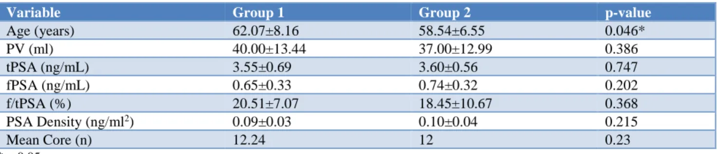
Benign prostate hyperplasia was detected in 35 patients (57.4%) and prostate adenocarcinoma was detected in 26 patients (42.6%). The mean ages were statistically significant between two groups: 62.07±8.16 for group 1, 58.54±6.55 for group 2 (p<0.05).
The tPSA level was found 3.55±0.69 ng/mL and 3.60±0.56 ng/mL in group 1 and 2, respectively. Although it was higher in the group 2, there was no statistical difference (p:0.747). The risk of malignancy increases as it correlates with tPSA level. The fPSA level was found 0.65±0.33 ng/mL and 0.74±0.32 ng/mL in group 1 and 2, respectively. Although it was higher in the group 2, there was no statistical difference (p:0.202). The f/tPSA level was found 0.20±0.07% and 0.18±0.01 in group 1 and 2, respectively.
2, respectively. Although it was higher in the group 2, there was no statistical difference (p:0.215).
The tPSA, fPSA, f/tPSA and PSA density levels were not statistically significant between two groups (Table 1).
Table 1: Descriptive characteristics and results of patient groups.
Variable Group 1 Group 2 p-value
Age (years) 62.07±8.16 58.54±6.55 0.046* PV (ml) 40.00±13.44 37.00±12.99 0.386 tPSA (ng/mL) 3.55±0.69 3.60±0.56 0.747 fPSA (ng/mL) 0.65±0.33 0.74±0.32 0.202 f/tPSA (%) 20.51±7.07 18.45±10.67 0.368 PSA Density (ng/ml2) 0.09±0.03 0.10±0.04 0.215
Mean Core (n) 12.24 12 0.23
*p<0.05
In group 2, prostate adenocarcinoma was most common detected with Gleason score 3+3 in 19 of 26 patients (73.1%) (Table 2). Patients with a PSA level of 2.5 to 4 ng/mL have more clinical insignificant prostate cancer and lower Gleason scores. However, high Gleason scores are rarely detected
.
Table 2: Distribution of prostate cancer results according to Gleason score.
Gleason Score (Group 2) n (Overall) %
3+3 19 73.1
3+4 3 11.5
5+3 1 3.8
ASAP 3 11.5
DISCUSSION
The use of PSA as a serum marker has reformed PCa diagnosis.5 As an independent variable, PSA is a better
predictor of cancer than either DRE or transrectal ultrasound (TRUS).6 There are no agreed standards
defined for measuring PSA.7 Higher PSA levels
indicating greater likelihood of PCa. Numerous men may harbor PCa notwithstanding having low serum PSA.8 The
PSA threshold value is still being discussed up-to-date and there is no clear consensus. With the frequent use of PSA in practice, the diagnosis of PCa is increasing. Clinically insignificant prostate cancer diagnosis is the most important question mark in PSA threshold value. The low PSA threshold value leads to unnecessary biopsy and diagnosis of clinically insignificant prostate cancer. Conversely, a high threshold value leads to the skipping or delay of clinically significant prostate cancer diagnosis.
The current threshold value for tPSA is assumed to be 2.5 ng/mL and varies with age. TPSA thresholds are available in different age groups from 2.5 to 6.5 ng/mL.9
Another reliable factor for low PSA values is digital rectal examination findings. However, digital rectal
examination findings that is specific to prostate cancer is not detected in every patient. Without digital rectal examination, the diagnostic efficacy of PSA variations is extremely important.
Catalona et al, were found the great majority of cancers detected in men with serum PSA levels of 2.6 to 4.0 ng/mL.10 Free serum PSA measurements may reduce the
number of additional biopsies required by the lower PSA cut-off. Thompson et al, were found in patients with lower PSA value (<4 ng/mL) prostate cancer was diagnosed in 15.2% and they said that biopsy-detected prostate cancer, including high-grade cancers, is not rare.8
Gilbert et al, were found prostate cancer in 27.4% in patients with PSA value (2.5-4 ng/mL) and they have suggested that 2.5 ng/mL may be a more appropriate cutpoint than 4.0 ng/mL.11 Rashid et al, in 2.5-4 serum
PSA range, 28.26% of all malignancy were found, which would be missed if we take cut off value 4.12 In this
study, PCa was detected in 37.7% (23/61) and it was found at a higher rate than the literature. Author think that the lower values of tPSA are more useful in diagnosis of PCa than estimated percentage.
To improve the biopsy/PCa ratio, the consensus recommends fPSA measurement. Cut-off of 1.5 seems to be the most appropriate. Veneziano et al, were found 1.5 value is recommended for patients with PSA levels between 4 and 10 ng/ml.13 Faria et al, were suggested this
value in men with normal DRE and PSA 2.5-4.0 ng/mL in a PCa screening programme.14 In a similar work,
Kitagawa et al, were demonstrated that the f/t PSA ratio was a strong predictor of future cancer detection the probabilities of prostate cancer detection in men with total PSA levels of 2.1-10.0 ng/ml.15 The differences in
ratios were higher than literature in both groups. The amount of serum tPSA is a parameter that can be influenced by the prostate volume. tPSA and prostate volume associations were first reported by Veneziano, Pavlica, Querze, Viglietta, Trenta in 1991.17 Threshold of
greater than 0.15 PSA density for benign lesions and less than 0.15 for malign lesions are still being used nowadays.
Yamamoto et al, found to based on PSA density decided prostate biopsy may be further improved, with fewer unnecessary biopsies.18 Contrary to common belief Pepe
et al, was found moreover PSA density accuracy was of poor value in diagnosing PCa in comparison with f/t PSA ratio.19 In present study, threshold of PSA density could
not help us in predicting of prostate cancer.
The relationship between low prostate volume and prostate cancer has been previously described in the literature. The reliability of PSA decreases in patients with prostate volume over 50 ml. Demura et al, was found that increase in prostate volume (PV) is associated with a decrease in size and detectability of cancer lesions resulting in a decrease in biopsy yield.20 In this study, PV
in the PCa detected group was found less than the benign group.
CONCLUSION
Many factors influence prostate biopsy decision. PSA value is one of these factors. Standard tPSA, fPSA, f/tPSA rates and PSA density cutoffs are not sufficient for a proper diagnosis of prostate cancer by ultrasound guided transrectal biopsies. Values of PSA from 2.5 to 4 ng/mL may suggest more benign lesions in advancing age. There is a need different assessment to distinguish of malignant lesions from benign lesions. Nowadays, it was impossible to make this difference in patients without digital rectal examination findings, so accepted threshold of PSA should be 2.5 ng/mL.
Funding: No funding sources Conflict of interest: None declared
Ethical approval: The study was approved by the Institutional Ethics Committee
REFERENCES
1. Wang MC, Valenzuela LA, Murphy GP, Chu TM. Purification of a human prostate specific antigen. Invest Urol. 1979;17(2):159-63.
2. Ng TK, Vasilareas D, Mitterdorfer AJ, Maher PO, Lalak A. Prostate cancer detection with digital rectal examination, prostate-specific antigen, transrectal ultrasonography and biopsy in clinical urological practice. BJU Int. 2005;95(4):545-8.
3. Hodge KK, McNeal JE, Terris MK, Stamey TA. Random systematic versus directed ultrasound guided transrectal core biopsies of the prostate. J Urol. 1989;142(1):71-4.
4. Epstein JI, Allsbrook WC, Jr., Amin MB, Egevad LL, Committee IG. The 2005 International Society of Urological Pathology (ISUP) consensus conference on Gleason grading of prostatic carcinoma. Am J Surg Pathol. 2005;29(9):1228-42. 5. Stamey TA, Yang N, Hay AR, McNeal JE, Freiha
FS, Redwine E. Prostate-specific antigen as a serum marker for adenocarcinoma of the prostate. N Engl J Med. 1987;317(15):909-16.
6. Catalona WJ, Richie JP, Ahmann FR, M’Liss AH, Scardino PT, Flanigan RC, et al. Comparison of digital rectal examination and serum prostate specific antigen in the early detection of prostate cancer: results of a multicenter clinical trial of 6,630 men. J Urol. 1994;151(5):1283-90.
7. Semjonow A, Brandt B, Oberpenning F, Roth S, Hertle L. Discordance of assay methods creates pitfalls for the interpretation of prostate-specific antigen values. Prostate Suppl. 1996;7:3-16.
8. Thompson IM, Pauler DK, Goodman PJ, Tangen CM, Lucia MS, Parnes HL, et al. Prevalence of prostate cancer among men with a prostate-specific antigen level <or =4.0 ng per milliliter. N Engl J Med. 2004;350(22):2239-46.
9. DeAntoni EP. Age-specific reference ranges for PSA in the detection of prostate cancer. Oncology (Williston Park). 1997;11(4):475-82.
10. Catalona WJ, Smith DS, Ornstein DK. Prostate cancer detection in men with serum PSA concentrations of 2.6 to 4.0ng/mL and benign prostate examination. Enhancement of specificity with free PSA measurements. JAMA. 1997;277(18):1452-5.
11. Gilbert SM, Cavallo CB, Kahane H, Lowe FC. Evidence suggesting PSA cutpoint of 2.5ng/mL for prompting prostate biopsy: review of 36,316 biopsies. Urol. 2005;65(3):549-53.
12. Rashid MM, Alam AK, Habib AK, Rahman H, Hossain AK, Salam MA, et al. Efficacy of lower cut off value of serum prostate specific antigen in diagnosis of prostate cancer. Bangladesh Med Res Counc Bull. 2012;38(3):90-3.
13. Veneziano S, Pavlica P, Compagnone G, Martorana G. Usefulness of the (F/T)/PSA density ratio to detect prostate cancer. Urol Int. 2005;74(1):13-8. 14. Faria EF, Carvalhal GF, dos Reis RB, Tobias‐
Machado M, Vieira RA, Reis LO, et al. Use of low free to total PSA ratio in prostate cancer screening: detection rates, clinical and pathological findings in Brazilian men with serum PSA levels <4.0 ng/mL. BJU Int. 2012;110(11 Pt B):E653-657.
15. Kitagawa Y, Ueno S, Izumi K, Kadono Y, Konaka H, Mizokami A, et al. Cumulative probability of prostate cancer detection in biopsy according to free/total PSA ratio in men with total PSA levels of 2.1-10.0 ng/ml at population screening. J Cancer Res Clin Oncol. 2014;140(1):53-9.
antigen levels of 4.0 ng/ml or less. Iran J Public Health. 2015;44(11):1466-72.
17. Veneziano S, Pavlica P, Querze R, Viglietta G, Trenta A. Importance of specific prostatic antigen to prostatic volume ratio in the selection of patients for ultrasonography-guided biopsy of the prostate. Radiol Med. 1991;81(6):857-60.
18. Yamamoto S, Kin U, Nakamura K, Hamano M, Nishikawa Y, Takenouchi T, et al. Transperineal ultrasound-guided 12-core systematic biopsy of the prostate for patients with a prostate-specific antigen level of 2.5-20 ng/ml in Japan. Int J Clin Oncol. 2005;10(2):117-21.
19. Pepe P, Candiano G, Fraggetta F, Galia A, Grasso G, Allegro R, et al. Is PSA density still useful in
diagnosing prostate cancer? Arch Ital Urol Androl. 2009;81(4):199-202.
20. Demura T, Takada T, Shimoda N, Hioka T, Iwaguchi Y, Ichihara S, et al. Mechanism underlying the negative effect of prostate volume on the outcome of extensive transperineal ultrasound-guided template prostate biopsy. Cancer Med. 2018;7(2):336-43.