REGULATION AND ROLES OF RAL GTPASE SIGNALING COMPONENTS IN ONCOGENESIS
Timothy D. Martin
A dissertation submitted to the faculty of the University of North Carolina at Chapel Hill in partial fulfillment of the requirements for the degree of Doctor of Philosophy in the
Curriculum of Pharmacology
Chapel Hill 2013
Approved by:
Channing J. Der, PhD Christopher Counter, PhD Adrienne Cox, PhD Lee Graves, PhD
ABSTRACT
TIMOTHY MARTIN: Regulation and roles of Ral GTPase signaling components in oncogenesis
(Under the direction of Channing J. Der)
Since their discovery in 1986, Ral (Ras-like) GTPases have emerged as critical regulators of diverse cellular functions. Like Ras, the Ral proteins cycle between an inactive GDP-bound state and an active GTP-bound conformation. Ral guanine nucleotide exchange factors (RalGEFs) facilitate the exchange of GDP for GTP thus activating the Ral proteins. When bound to GTP, Ral can interact with an array of downstream effector proteins and mediate numerous biological processes. Ral GTPase-activating proteins (RalGAPs) catalyze the hydrolysis of the bound GTP returning Ral to an inactive, GDP-bound conformation. RalGEFs function as downstream effectors of the Ras oncoprotein that is mutationally active in approximately one-third of human cancers. The RalGEF-Ral signaling network comprises the third best-characterized effector of Ras-dependent human oncogenesis. The two Ral isoforms, RalA and RalB, have been found to play key roles in both normal and tumor cell biology including regulation of vesicular trafficking, migration and invasion, tumor formation, metastasis, and gene expression.
anchorage-iv
independent growth. Specifically, RalA was necessary for anchorage-independent growth while RalB functioned to suppress anchorage-independent proliferation. We determined that RalA and RalB utilized common and distinct effector proteins to drive their respective growth properties. Lastly, we found that depletion of one Ral isoform resulted in the upregulation of the activity of the remaining isoform indicating that RalA may be a viable therapeutic target to curb the growth of CRC.
Previous efforts to understand small GTPase signaling has found that phosphorylation in the membrane-targeting region of a number of small GTPases results in profound changes in their signaling properties. We found that RalB is phosphorylated
by PKC! on serine 198 and that this phosphorylation event results in the relocalization of RalB from the plasma membrane to endomembranes concurrent with a change in RalB GTP-loading. Phosphorylation of RalB also results in a change in RalB effector utilization where non-phosphorylated RalB interacting with the exocyst and phosphorylated RalB associating with RalBP1. Interestingly, we found that phosphorylation of RalB controls vesicular trafficking and that the surface expression of
!5-integrin is dependent upon RalB phosphorylation/dephosphorylation cycling.
serum stimulation results in a RalB-dependent translocation of mTORC1 to the plasma
membrane. Surprisingly, we found that the tumor suppressor Tsc1/2 complex also
regulates Ral GTPase activity and that RalGAP expression can restore mTORC1
signaling in Tsc-deficient cells. In pancreatic cancer (PDAC) cells, where RalB is known
to drive invasion and metastasis, loss of RalGAP signaling enhanced RalB activation and
led to an increase in cellular invasion. This increase in invasion upon RalGAP loss was
blocked by treatment with the mTOR inhibitor rapamycin. Together, these studies have
further defined Ral signaling in Ras-driven tumor cells by identifying key signaling
events that regulate or are regulated by Ral GTPase signaling. This work provides a
more in depth framework for potentially targeting Ral for the treatment of diseases
vi
ACKNOWLEDGEMENTS
First and foremost I need to thank my parents, Tim and Lou Martin, for their
unwavering love and support that has made every thing I’ve done possible. Without their
guidance, this dream would not have been realized. I am thankful for Jen Jen Yeh’s
support during my time as a technician when I was just learning about small GTPases and
cancer biology. Thanks for sticking with me and giving me a chance even when I
couldn’t get my Ral pulldowns to work. In addition, I’d like to thank all members of the
Der lab who made it an enjoyable experience during my time as part of the lab. In
particular, I want to thank Dave Reiner who was always available and willing to discuss
new research ideas and directions. Also, I’d like to thank Danielle Cook for being a great
friend, an ear to bounce ideas off of, and for pushing me to be the best I could be. I want
to thank all members of my committee and other faculty members who have helped guide
me to the completion of this dissertation. I’d like to thank Adrienne Cox for always
being available to chat and for asking insightful questions that helped me keep focus.
Lastly, I want to thank my mentor Channing Der. I am especially grateful for his support
during my time in the lab and for allowing me the freedom to try out new ideas while I
TABLE OF CONTENTS
LIST OF FIGURES………x
LIST OF ABBREVIATIONS……….………..xii
Chapter I. INTRODUCTION………...1
Evolutionary Conservation of Ral………...3
Ral-selective GEFs and GAPs: regulators of GDP-GTP cycling…………4
Ral effectors: regulators of vesicular trafficking, actin cytoskeletal organization and gene expression………...9
Post-translational regulation of Ral by protein kinases……….12
Ral signaling in oncogenesis……….15
Rationale for studies………..21
II. ACTIVATION AND INVOLVEMENT OF RAL GTPASES IN COLORECTAL CANCER………..24
Overview..……….24
Introduction………...25
Materials and methods………..28
Results………...29
viii
III. PHOSPHORYLATION BY PKC! REGULATES RALB SMALL GTPASE ACTIVATION, SUBCELLULAR
LOCALIZATION AND EFFECTOR UTILIZATION………...49
Overview.………...49
Introduction………..50
Materials and methods……….52
Results………...55
Discussion………69
IV. RAL AND RHEB GAPS INTEGRATE MTOR AND GTPASE SIGNALING IN AGEING, AUTOPHAGY, AND TUMOR CELL INVASION………75
Overview……….……….75
Introduction………...76
Materials and methods………..79
Results………...82
Discussion………....102
V. CONCLUDING REMARKS AND FUTURE DIRECTIONS…………107
Does Ral GTPase signaling contribute to colorectal tumorigenesis in vivo?...107
How might we therapeutically target Ral signaling for the treatment of CRC?...108
How are Ral proteins becoming activated in CRC and are RalGEFs or K-Ras required for activation?...109
Is RalB phosphorylation required for RalB-driven cancer phenotypes?...110
Does RalB phosphorylation affect novel effector interactions?...111
Does C. elegans HGAP-1/HGAP-2 RalGAP complex require
RHEB-1 or Ral-1 to modulate ageing?...113 Are there changes in metabolic signaling upon loss of RalGAP?...114 Is mTOR inhibitor tumor response associated with RalB activity?...115 Are there novel mTORC1 substrates that
are dependent upon RalB signaling?...116 Why does RalB aid in localizing mTORC1 to
x
LIST OF FIGURES Figure
1-1. RalA and RalB are members of the Ras superfamily of small GTPases...2
1-2. Regulation of the Ral GDP-GTP cycle by RalGEFs and RalGAPs...4
1-3. Ral guanine nucleotide exchange factors (RalGEFs)...6
1-4. Regulation of Ral signaling by RalGAP proteins...8
1-5. Ral GTPase effectors...9
1-6. The Role of the exocyst in exocytosis...11
1-7. RalA and RalB are regulated by distinct protein kinases...13
1-8. RalA activity is regulated by Aurora-A kinase dependent phosphorylation...14
1-9. The roles of Ral GTPases in a variety of cancer phenotypes...17
2-1. Response of CRC tumor cells to MAPK and PI3K Inhibition...32
2-2. Ral-GTP formation in CRC cell lines and patient tumors...34
2-3. RalA and RalB activity show opposing roles in regulation of CRC anchorage-independent growth...36
2-4. RalA activity is dominant over RalB in regulation of CRC anchorage-independent growth...39
2-5. RalA and RalB require interaction with RalBP1 but distinct exocyst subunits for regulation of anchorage-independent growth...42
2-6. Model for role and effector involvement in CRC anchorage-independent growth...48
3-1. RalB is phosphorylated on serines 192 and 198 and PKC! is necessary for phosphorylation...58
3-3. RalB phosphorylation regulates vesicular fusion...65 3-4. Phosphorylation of RalB regulates the trafficking of !5-integrin...68 3-5. Model for RalB-regulated vesicular fusion. RalB is
associated with the exocyst through its interaction with Sec5...74 4-1. RalGAP loss reduces the increased longevity of
DAF-2 deficient C. elegans...84 4-2. RalGAPs regulate mTORC1 signaling...87 4-3. RalB associates with mTORC1 and regulates mTORC1 signaling...90 4-4. The Tuberous-Sclerosis complex (TSC)
regulates Ral GTPase signaling...93 4-5. RalB regulates serum-induced mTORC1
plasma membrane relocalization…...95 4-6. The exocyst is the RalB effector responsible for mTORC1 activity...98 4-7. RalGAP signaling suppresses the invasive
properties of PDAC tumor cells…...101 4-8. Working model for Ral and Rheb GAP integration of
xii
LIST OF ABBREVIATIONS
4E-BP1 Eukaryotic translation initiation factor 4E-binding protein 1 Akt RAC serine/threonine-protein kinase
APC Adenomatous Polyposis Coli Arf ADP ribosylation factor
ATCC American Type Culture Collection ATPase Adenosine triphosphatase
Blast Blasticidin Bryo Bryostatin-1
BSA Bovine Serum Albumin C-terminus Carboxy terminus C. elegans Caenorhabditis elegans
CAAX Cysteine-Aliphatic-Aliphatic-Any amino acid CDC25 Cell Division Cycle 25
Cdc42 Cell Division Cycle 42 CDK5 Cyclin-dependent kinase 5 cDNA Complementary DNA
CHAPS 3-[(3-cholamidopropyl)dimethylammonio]-1-propanesulfonate CMV Cytomegalovirus
COSMIC Catalog of Somatic Mutations in Cancer CRC Colorectal Cancer
DMEM Dulbecco's Modified Eagle Medium
DNA Deoxyribonucleic Acid
Drp1 Dynamin 1-like
dsRNA double-stranded Ribonucleic Acid
E-Cadherin Epithelial Cadherin
EGF Epidermal Growth Factor
EGFR Epidermal Growth Factor Receptor
Eps EH domain-binding mitotic phosphoprotein
Erk Mitogen-activated protein kinase 1
Exo70 Exocyst complex component 7
Exo84 Exocyst complex component 8
FCS Fetal Calf Serum
FIP FH protein interacting protein
FOXO Forkhead Box Transcription Factor
GAP GTPase activating protein
GAPDH Glyceraldehyde 3-phosphate dehydrogenase
GDP Guanosine diphosphate
GEF Guanine nucleotide exchange factor
GFP Green fluorescent protein
GGTI Geranylgeranyl transferase inhibitor
GPCR G-protein coupled receptor
Grb10 Growth factor receptor-bound protein 10
xiv GTP Guanosine triphosphate
GTPase Guanosine triphosphatase HCl Hydrogen Chloride HEK Human embryonic kidney
HV Hypervariable
IgG Immunoglobulin G
InsR Insulin Receptor
IRES Internal ribosomal entry site JNK Mitogen-activated protein kinase 8
LC3 Microtubule-associated protein 1 light chain 3 LET-60 Lethal protein 60
MAPK Mitogen-activated protein kinase 1
mCh mCherry
MDCK Madin-Darby canine kidney MEFs Mouse embryonic fibroblasts
MEK Mitogen-activated protein kinase kinase MMP Matrix metalloprotease
mTOR mechanistic target of rapamyin
mTORC mTOR complex
Myc c-Myc proto-oncogene N-terminal Amino terminal
NS Non-specific
p70S6K Ribosomal protein S6 kinase, 70kDa, polypeptide 1 pAkt Phospho-Akt
PBS Phosphate buffered saline
PDAC Pancreatic ductal adenocarcinoma
PDK 3-phosphoinositide dependent protein kinase pERK phospho-Erk
PH Pleckstrin homology
PI3K Phosphatidylinositol 3-kinase PKA Protein kinase A
PKC Protein kinase C
PLC! Phospholipase C, epsilon PLD1 Phospholipase D1
PP2A Protein phosphatase 2A pS6K phospho-S6K
PTEN Phosphatase and tensin homolog Puro Puromycin
PVDF Polyvinylidene difluoride RA Ras association
Rab Ras-associated binding
Rac Ras-related C3 botulinum toxin substrate Raf v-raf murine sarcoma viral oncogene homolog
xvi RalBP1 RalA binding protein 1
RalGDS Ral guanine nucleotide dissociation stimulator
RalGPS Ral GEF with PH domain and SH3 binding motif
Rap Ras-related protein Rap-1A
Raptor Regulatory associated protein of MTOR, complex 1
Ras Rat sarcoma viral oncogene homolog
RBD Ras or Ral binding domain
REM Ras exchange motif
Reps RalBP1-associated Eps domain-containing protein
Rgl Ral guanine nucleotide dissociation stimulator-like
Rheb Ras homolog enriched in brain
Rho Ras homolog
Rictor Raptor independent companion of mTOR
Rit Ras-like without CAAX 1
RLIP76 76 kDa Ral-interacting protein
RNA Ribonucleic acid
RNAi RNA interference
RPMI Roswell Park Memorial Institute medium
S. cerevisia Saccharomyces cerevisiae
S. pombe Saccharomyces pombe
SDS-PAGE Sodium dodecyl sulfate polyacrylamide gel electrophoresis
Sec10 Exocyst complex component 5
Sec3 Exocyst complex component 1 Sec5 Exocyst complex component 2
Sec6 Exocyst complex component 3 Sec8 Exocyst complex component 4 SEM Standard error of the mean
SEP Superecliptic pHluorin SH3 SRC Homology 3 Domain
siRNA Small interfering RNA
SNAP-23 Synaptosomal-associated protein, 23kDa
SNARE Soluble N-ethylmaleimide-sensitive factor activating protein receptor
HA Hemagglutinin
SQSTM1 Sequestosome 1
SREBP Sterol regulatory element binding t-SNARE target-SNARE
TBK1 TANK-binding kinase 1
TBST Tris buffered saline with tween-20 TC21 Teratocarcinoma oncogene 21
TfR Transferrin Receptor
Tiam1 T-cell lymphoma invasion and metastasis 1 TRC The RNAi Consortium
Tsc Tuberous-Sclerosis TULIP1 Tuberin-like protein 1
xviii v-SNARE vesicular-SNARE
VAMP3 vesicle-associated membrane protein 3
VPC Vulval precursor cell
WT Wildtype
CHAPTER 1
INTRODUCTION
The Ral proteins are members of the Ras superfamily of small GTPases (Figure 1-1A). RalA was initially identified using oligonucleotide probes to find genes that were similar to Ras in a cDNA library of immortalized simian B-lymphocytes [1]. Three years later, screening performed on a pheochromocytoma library with RalA oligonucleotide probes identified RalB [2]. These two proteins were found to contain 82% sequence identity including identical effector binding regions comprised of switches I and II (Fig. 1-1B) with the majority of sequence divergence occurring near the C-terminus in the membrane-targeting hypervariable region. This sequence diversity present in the C-terminus is largely the reason for the different functions attributed to RalA and RalB. This is likely due to this region being critical in determining subcellular localization and potential effector interactions of the Ral proteins. Striking evidence for this comes from chimeric RalB that possesses a RalA C-terminus which shows that simply swapping the hypervariable region can confer RalA-like activity on RalB and vice versa [3].
2
examination in human cells, it was found that Ral GTPase signaling is the main Ras effector pathway necessary for cellular transformation underscoring distinct requirements of transformation between rodent and human cells and setting the stage for last decade of research into the contribution of Ral signaling to oncogenesis [5].
A.
B.
Evolutionary conservation of Ral. Similar to Ras, functional orthologs of Ral are found in Drosophila and C. elegans as well as all vertebrate species. However, unlike Ras, no Ral orthologs are found in S. cerevisia or S. pombe.
In Drosophila Ral signaling has been implicated in cell morphology during development. Expression of constitutively-active RalA in the fly eye results in a rough eye phenotype with altered actin organization leading to changes in cell shape during pupal development[6]. It was later determined that RalA signaling through JNK pathway led to the changes in cell shape during eye development [7]. Work done by Camonis and colleagues showed that instead of canonical Ras-RalGEF-Ral signaling described in mammalian cells, Drosophila signaling is bifurcated with Ras and the Rap small GTPase both binding and activating the RalGEF-Ral pathway [8]. More recently the Rap-RalGEF-Ral signaling pathway was shown to regulate spindle orientation in drosophila neural stem cells.
4
Ral-selective GEFs and GAPs: regulators of GDP-GTP cycling. Like many other small GTPases, RalA and RalB have low intrinsic GTPase activity and families of other proteins regulate GTP-to-GDP cycling. Guanine nucleotide exchange factors (GEFs) catalyze GDP-for-GTP exchange to activate small GTPases and GTPase activating proteins (GAPs) help to accelerate GTP hydrolysis by small GTPases (Figure 1-2).
The original RalGEF to be identified was RalGDS (Ral guanine nucleotide dissociation stimulator) [11]. Large numbers of yeast two-hybrid screens were performed in the early 1990s to identify Ras effectors and these screens identified not only the Raf serine/threonine kinase but also RalGDS [12, 13]. RalGDS was found to catalyze nucleotide exchange on both RalA and RalB but not other small GTPases including members of the Ras, Rho, and Rab families. Yeast two-hybrid screening of H-Ras, R-Ras, TC21/R-Ras2, and Rit identified three additional RalGEF proteins that were named Rgl (RalGDS-like), Rgl2, and Rgl3 [14-16]. All four human RalGEFs contain a common domain architecture including an N-terminal REM domain followed by a CDC25 GEF domain and a C-terminal RA domain (Figure 1-3).
6
8 A.
B.
Ral effectors: regulators of vesicular trafficking, actin cytoskeletal organization and
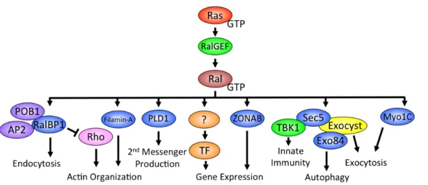
gene expression. Like Ras and other small GTPases, Ral interacts with a number of effector proteins when bound to GTP (Figure 1-5).
Figure 1-5. Ral GTPase effectors. Ral GTPases can interact with a number of effector proteins involved in various biological functions. The interaction of Ral with the exocyst compenents Sec5 and Exo84 is the most understood and is involved in many of the cancer phenotypes associated with Ral signaling including tumor cell survival and invasion/metastasis.
10
done by many groups has shown that Ral is involved in the endocytic recycling of surface expressed proteins including E-Cadherin and EGFR [28]. Whether or not this occurs through Ral-RalBP1 interaction is unclear.
12
Less characterized Ral effector signaling pathways include phospholipase D1 (PLD1), Filamin, and ZONAB. PLD1 is best known for its role in converting phosphotidylcholine to phosphatidic acid and choline in response to PKC signaling downstream of G-protein coupled receptor (GPCR) stimulation [37]. Recent evidence shows that RalA is necessary for the PLD1-mediated stimulation of mTORC1 signaling [38]. Additionally, the interaction of both RalA and RalB with PLD1 has been shown to be critical for HeLa cell cytokinesis [18]. Filamin is an important component of the actin cytoskeleton and is involved in actin crosslinking and lamellipodia formation [39]. The association of RalA with Filamin was found to be important for filopodia formation in Swiss-3T3 cells [40]. Lastly, active RalA has been shown to engage the transcription factor ZONAB (zonula occludens 1-associated nucleic acid binding protein) in a cell density dependent manner in MDCK cells [41]. At high cell densities, RalA engages ZONAB unlocking the transcription of ZONAB targets but it is unclear which genes are regulated [41].
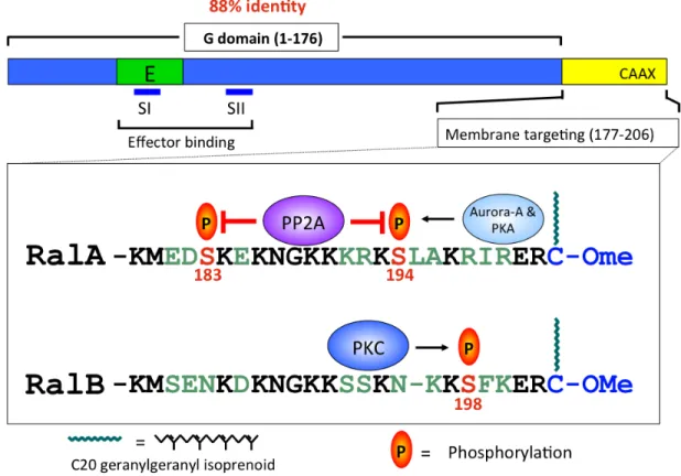
Figure 1-7. RalA and RalB are regulated by distinct protein kinases. RalA and RalB sequence divergence is concentrated in their C-terminal hypervariable membrane-targeting region. Though both RalA and RalB are modified by geranylgeranylation, each can be phosphorylated by distinct protein kinases. Aurora-A and PKA phosphorylate RalA on S194 and this is opposed by PP2A phosphatase activity. RalB is phosphorylated by PKC on S198. Phosphorylation serves as a key signal to regulate Ral subcellular localization and function.
14
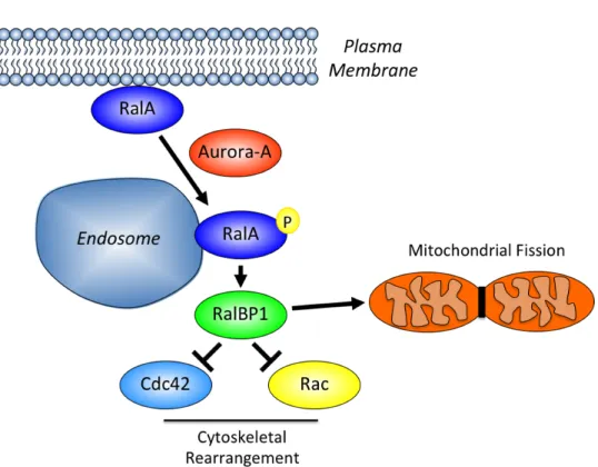
RalBP1 and the large GTPase Drp1 where it can promote mitochondrial fission (Figure 1-8).
Figure 1-8. RalA activity is regulated by Aurora-A kinase dependent phosphorylation. RalA is normally found at the plasma membrane of cells but when phosphorylated by Aurora-A, it relocalizes to both endosomes and mitochondria. On endosomes, phosphor-RalA interacts with RalBP1 to negatively regulate Ccd42 and Rac signaling to control the actin cytoskeleton. At the mitochondria, RalA recruits RalBP1 and Drp1, which is important for the proper fission of mitochondria that occurs during cytokinesis.
proper exocytic vesicle trafficking and fusion at the plasma membrane and the delivery of surface alpha-5 integrin is regulated by dynamic RalB phosphorylation [47]. Theodorescu and colleagues found that phosphorylation of RalB on S198 was critical in regulating the ability of RalB to promote the metastatic growth of bladder cancer cells in a nude mouse model [44].
Like most small GTPases, RalA and RalB contain a lipid modified C-terminus that assists in membrane anchoring [48]. Both Ral proteins are modified by the addition of a geranylgeranyl moiety on cysteine 203 and this lipid modification is critical for both RalA and RalB function. Studies by Sebti and colleagues found that RalA and RalB were both targets of geranylgeranyl transferase inhibitors (GGTI) [49]. They went on to show that GGTI treatment of pancreatic tumor cells leads to a decrease in anchorage-dependent and anchorage-independent proliferation and increased apoptosis [49].
16
survival while RalA was necessary for the anchorage-independent growth of cancer cells [50]. Importantly, this also marked the first time RalA and RalB were found to have non-overlapping functions [50]. Work over the subsequent years has elucidated unique roles for RalA and RalB in a variety of human cancers including bladder, colorectal, melanoma, and pancreatic as described below.
Ral and bladder cancer
Activating Ras mutations occur in a small percentage of bladder cancers and Ral activation is frequently seen in both human tissue samples and tumor cell lines [51]. Using RNAi and overexpression of mutationally activated Ral, Theodorescu and colleagues found that RalA and RalB play antagonistic roles in the migratory activity of bladder cancer cell lines with RalA suppressing and RalB enhancing motility [52]. Further analysis of the contribution of RalB to bladder cancer found that the phosphorylation of RalB on S198 was critical for the ability of bladder tumor cells to metastasize in a mouse tail-vein injection model [44].
18 Ral and colorectal cancer
Oncogenic KRAS mutations occur in approximately 40-50% of colorectal cancer (CRC) tumors and we’ve shown Ral signaling to be a critical regulator of the anchorage-independent growth properties of CRC tumor cells [55]. We found that RNAi-mediated suppression of RalA resulted in a decrease in soft agar colony growth while loss of RalB had the opposite effect leading to an enhancement of anchorage-independent growth [55]. Despite RalA and RalB interacting with a common set of downstream effectors, we found that RalA and RalB utilize both common and distinct effector proteins in regulating CRC anchorage-independent growth [55]. Using well-defined mutants of Ral that are selectively uncoupled from either Exo84, Sec5, or RalBP1 we’ve shown that RalA requires Exo84 and RalBP1 binding to promote the anchorage-independent growth of CRC cells [55]. Conversely, RalB requires Sec5 and RalBP1 to suppress soft agar colony formation [55]. Intriguingly, loss of one Ral isoform was found to increase the activation of the alternate isoform suggesting crosstalk between RalA and RalB at least in CRC cells [55]. What specifically mediates this RalA and RalB crosstalk is unknown but could be through either enhanced RalGEF accessibility for the remaining Ral protein or a downregulation of RalGAP activity upon single Ral isoform depletion.
Ral and Melanoma
N-Ras mutation status when injected subcutaneously in mice [57]. This study also showed that in a panel of human melanoma cells with diverse mutational backgrounds, there is a consistently high level of RalA but not RalB activation [57]. Experiments using Arf-deficient immortalized mouse melanocytes to investigate the contributions of Ras downstream signaling to melanomagenesis has also indicated a role for Ral signaling [58]. In this system, expression of the RalGEF Rgl2 with a membrane localization sequence to mimic Ras-RalGEF activation was sufficient to promote the anchorage-indpendent growth and invasion of these melanocytes similar to what is observed with oncogenic N-Ras [58].
Ral and pancreatic cancer
20
maintenance [60]. In this study, the use of inducible RNAi to stably deplete RalA from established primary tumors resulted in the regression of the tumor indicating a necessity for persistent RalA signaling in established PDAC tumors. There is also recent evidence that active K-Ras signaling to RalB but not RalA plays a critical role in the formation of invadopodia in PDAC cells [61]. Invadopodia are actin-rich membrane protrusions that are known to be involved in the secretion of matrix metalloproteases (MMP) during tumor cell invasion [62]. RalB requires the ability to interact with RalBP1 to mediate this process and RalBP1 itself is necessary for the formation of invadopodia in PDAC cells [61]. Surprisingly, the RhoGAP activity of RalBP1 is not necessary for invadopodia formation while the ATPase activity is required [61]. Why the ATPase activity is necessary for RalBP1 to mediate invadopodia formation is unclear.
Rationale for studies. Oncogenic mutations in Ras proteins are found in approximately 33% of all cancers and Ras signaling is implicated in driving both tumorigenesis and metastasis. Direct antagonism of Ras has proved to be unsuccessful thusfar and efforts to block downstream Ras effector signaling are currently underway. Recent evidence suggests a key role for RalGEF-Ral GTPase signaling network in the pathogenesis of a variety of cancer types. Since oncogenic KRAS muations occur in a large percentage of colorectal cancer (CRC) tumors, I hypothesized that RalGEF-Ral signaling would play a key role in maintaining the oncogenic properties of CRC tumor cells. As discussed in Chapter 2, I found a striking difference in the requirements for RalA and RalB in the regulation of CRC tumor cell anchorage-independent growth. Specifically, RalA was required for soft agar colony formation while RalB actually suppressed the soft agar growth of CRC tumor cells. Interestingly, RalA and RalB required both common and unique effector proteins to mediate their effects on CRC anchorage-independent growth. More in vivo studies are needed to better characterize and address the importance of Ral signaling to CRC tumorigenesis and metastasis.
22
regulator of exocytosis. I found that phosphorylation on S198 allows for a switch in the ability of RalB to associate with Sec5/exocyst and RalBP1 and that phosphorylation signals for the internalization of RalB from the plasma membrane to endomembranes. Future studies are needed to address whether preventing the phosphorylation of RalB will be therapeutically tractable to prevent RalB-driven cancer phenotypes such as invasion and metastasis.
CHAPTER 2
Activation and Involvement of Ral GTPases in Colorectal Cancer1
Overview
Current approaches to block KRAS oncogene function focus on inhibition of K-Ras downstream effector signaling. We evaluated the anti-tumor activity of selumetinib (AZD6244, ARRY-142886), a potent and selective MEK1/2 inhibitor, on a panel of colorectal carcinoma (CRC) cells and found no inhibition of KRAS mutant CRC cell anchorage-independent growth. While AKT activity was elevated in KRAS mutant cells, and PI3K inhibition did impair the growth of MEK inhibitor-insensitive CRC cell lines, concurrent treatment with selumetinib did not provie additional anti-tumor activity. Therefore, we speculated that inhibition of the Ral guanine exchange factor (RalGEF) effector pathway may be a more effective approach for blocking CRC growth. RalGEFs are activators of the related RalA and RalB small GTPases and we found activation of both in CRC cell lines and patient tumors. Interfering RNA stable suppression of RalA expression reduced CRC tumor cell anchorage-independent growth, but surprisingly, stable suppression of RalB greatly enhanced soft agar colony size and formation frequency. Despite their opposing activities, both RalA and RalB regulation of
anchorage-independent growth required interaction with RalBP1/RLIP76 and components of the exocyst complex. Interestingly, RalA interaction with the Exo84 but not Sec5 exocyst component was necessary for supporting anchorage-independent growth, whereas RalB interaction with Sec5 but not Exo84 was necessary for inhibition of anchorage-independent growth. Our results suggest that anti-RalA-selective therapies may provide an effective approach for KRAS mutant CRC.
Introduction
Recent sequence analysis verified that KRAS is the most frequently mutated oncogene in colorectal cancer (CRC) [64, 65]. With considerable experimental evidence that mutant KRAS is required for the maintenance of CRC cell growth [66-68], it is believed that K-Ras inhibition will provide an effective therapeutic strategy for CRC. However, since efforts to directly block mutant K-Ras have not been met with success, current strategies have focused on inhibitors of Ras effector signaling [69].
The best studied Ras effector pathways are the Raf-MEK-ERK mitogen-activated protein kinase and phosphotidylinositol 3-kinase (PI3K)-AKT serine/threonine protein kinase effector pathways [70], with inhibitors of components of both pathways currently under clinical evaluation2. Further support for the functional role of these effectors in cancer growth comes from the identification of mutationally activated B-Raf [71, 72] or
the p110! catalytic subunit of PI3K [73] in CRC. However, mouse model analyses have validated essential roles for other effectors in Ras-mediated oncogenesis: Ral guanine nucleotide exchange factor (RalGEF; RalGDS), the Tiam1 Rac-specific GEF and
26
phospholipase C epsilon (PLC!) [74-76]. Tiam1 [77] or PLC! [78] deficiency impaired colon tumorigenesis in APC mutant Min mice. Tiam1 overexpression has also been implicated in colon cancer metastatic growth [76]. These observations raise the question of whether inhibition of Raf or PI3K effector signaling, or another effector pathway, will be the most effective approach for treatment of KRAS mutant CRC.
Recent studies established that Ral small GTPases, activated by RalGEFs, are critical drivers of human oncogenesis [79]. Our recent studies found more frequent activation of Ral, rather than the Raf or PI3K effector pathways, in KRAS mutant pancreatic ductal adenocarcinoma (PDAC) tissue and cell lines [3, 80]. Ral has also been found to contribute to the growth of cancer types where RAS mutations are seen infrequently [81].
PC3 prostate tumor cells inhibited bone metastasis but not subcutaneous tumor growth [83].
RalA and RalB interact with functionally diverse effectors. Less clear is the role of specific effectors in Ral-mediated oncogenesis. Perhaps the best-validated effector in tumor cell growth is the Sec5 subunit of the octomeric exocyst complex that regulates vesicle trafficking, where Sec5 activation of the TBK1 atypical IkappaB kinase is essential for tumor but not normal cell survival [33]. The second best studied Ral effector is RalBP1/RLIP76 that functions as a GAP for Rho small GTPases [84]. RalBP1 overexpression has been found in a variety of tumor cells and RalBP1 inhibition suppressed tumor xenograft tumor growth in mice [85]. Whether these reported tumor promoting activities of RalBP1 are relevant for mutant Ras-activated RalGEF-Ral signaling has not been addressed.
28 Materials and Methods
Cell culture
Human CRC cell lines were obtained directly from ATCC and maintained in either DMEM-H or RPMI-1640 supplemented with 10% fetal calf serum, and frozen down to maintain limited passage history. All lines are reconstituted from an original freeze down and used for limited cell passage and discarded. Cell lines were treated with either selumetinib (provided by AstraZeneca) or LY294002 for 24 h for inhibition of ERK or AKT, and soft agar analyses were done as described previously [86]. Mutation status for KRAS, BRAF and PIK3CA was derived from the COSMIC database3.
Plasmids
Short hairpin RNA (shRNA) sequences for human RalA or RalB were subcloned into pSuper.retro.blast (provided by Dr. John Minna) and pSuper.retro puro (Oligoengine), pBabe-puro retrovirus expression vectors encoding wild-type human RalA and RalB and RNAi-insensitive Ral cDNA sequences for RalA or RalB have been described previously [80], and were used to generate cDNA sequences encoding effector binding mutants. shRNA sequences for human Exo84 were cloned into pSuper.retro puro and the sequences are as follows: shExo84 1 – GGTGCCACTTTACTCTATA and shExo84 2 – ACAATATAATTTGAATGGCTAA. RalBP1 shRNA has been described previously (42). RNAi-insensitive RalBP1 was cloned into pBabeHAII puro with the following silent mutations (in italics): GTAGAACGTACGATGATGT. pLKO.1 puro lentiviral vectors encoding Sec5 shRNA (TRCN0000116102- TRCN0000116106) were
obtained from OpenBiosystems TRC shRNA library and pLKO.1 puro NS shRNA
(CAACAAGATGAAGAGCACCAA) was obtained from Sigma.
Immunoblotting
Blot analyses were done with antibodies for RalA (BD Laboratories), RalB
(Millipore), !-actin and vinculin (Sigma), GAPDH and RalBP1 (Abcam), phosphorylated
ERK1 and ERK2 T202/Y204, phospho-AKT S473, and total AKT (Cell Signaling), total
ERK1/2 (Santa Cruz Biotechnology), and Exo84 (Orbigen). A Sec5 monoclonal
antibody was a kind gift from Dr. Charles Yeaman (University of Iowa). Activated
Ral-GTP was determined by pull down analyses as described previously [80].
Statistical analysis
The Fisher’s exact test was used to analyze associations between two variables
and the Pearson Chi-square test was used to analyze association between more than two
variables.
Results
KRAS mutant CRC cell lines are insensitive to growth inhibition by blocking MEK
Our previous studies with two MEK1/2 inhibitors, U0126 and CI-1040
(PD184352), found that KRAS mutation status and ERK1/2 activation (pERK) did not
correlate strongly with MEK inhibitor sensitivity [87]. However, both inhibitors possess
off-target activities. Therefore, we extended these analyses using a more potent and
30
trial analyses. The majority of previous studies evaluated MEK1/2 inhibitor activity
against tumor cell lines grown as two-dimensional anchorage-dependent cultures [88-91].
When compared, it was found that tumor cell growth in three-dimensional suspension
cultures was more resistant to MEK inhibitor treatment [92, 93]. Therefore, we evaluated
colony formation in soft agar, since this assay is widely regarded as a more accurate
indicator of tumorigenic growth in vivo. One previous cell culture study determined that
selumetinib inhibited pERK with a half-maximal inhibition (IC50) at a dose of <40 nM [87], whereas a second study found selumetinib inhibition of Raf activation of pERK in
different cells ranging from 10 to 100 nM [91]. Therefore, we treated cells with one set
high concentration (200 nM) to assess sensitivity, determined the degree of pERK
reduction and evaluated its effect on growth.
As we described previously [86], elevated pERK levels did not correlate with
KRAS or BRAF mutation status (Figure 2-1A-C). Similar to our previous observations with the U0126 and CI-1040 MEK1/2 inhibitors [86], there was wide variation in
selumetinib treatment reduction in pERK, with some cell lines showing high sensitivity
and others with relative insensitivity when evaluated in adherent cultures. Interestingly,
selumentinib treatment caused limited to no inhibition of growth in all six KRAS mutant CRC cell lines (p=0.031) (Fig. 2-1 D). This pattern was distinct from what we observed
with U0126 and CI-1040, where KRAS mutant SW480 growth was sensitive to both U0126 and CI-1040 [86]. Four of five BRAF mutant CRC lines were inhibited by selumetinib, with only NCI-H508 showing insensitivity. NCI-H508 cells were also
insensitive to U0126 and CI-1040 treatment [86]. The different activities seen with each
inhibitors, selumetinib does not exhibit inhibition of MEK5 [87, 91]. Nevertheless, despite inhibitor-specific differences in sensitivity, we reached the same conclusion with all three MEK inhibitors, that neither elevated pERK levels nor the degree of pERK inhibition was predictive of sensitivity to selumetinib growth inhibition. Unlike U0126 and CI-1040 however, none of the six KRAS mutant cell lines were sensitive to selumetinib.
32
alone or together with MEK and PI3K, may be required to effectively block the growth of KRAS mutant CRC cells. Since we recently identified a role for a third Ras effector pathway, leading to Ral GTPase activation, for pancreatic cancer growth [80], the remainder of this study focused on validating a role for Ral GTPases in CRC growth.
RalA and RalB are activated CRC cell lines
34
A.
B.
RalA and RalB exhibit opposing activities in regulation of CRC cell line anchorage-independent growth
In our previous analyses of 9 of 9 PDAC cell lines, we found that sustained shRNA depletion of RalA but not RalB reduced anchorage-independent growth as determined by colony formation efficiency in soft agar [80]. To determine if these two related isoforms also served similar roles in CRC anchorage-independent growth, we established CRC cell lines stably-infected with the same retrovirus vector-based shRNA vectors used to selectively silence RalA or RalB expression as used in our PDAC studies.
For these analyses, we evaluated KRAS or BRAF mutant or KRAS/BRAF WT CRC cell lines and additionally two PDAC cell lines from our previous study [80]. Mass populations of PDAC and CRC cell lines stably-infected with each shRNA vector were characterized by western blot analyses to verify steady-state reduction in endogenous RalA or RalB protein (Figure 2-3A and data not shown).
36
RalA and RalB have been shown to have distinct functions in a number of cellular processes or biological activities [81, 94, 95]. However, when concurrently suppressed, the phenotype associated with RalA has typically been dominant over that of RalB [81, 82, 96]. To address this possibility, we extended our analyses to a total of six KRAS mutant CRC cell lines with concurrent shRNA suppression of RalA and RalB, verified reduction in RalA and RalB protein (Figure 2-4A) and then evaluated colony formation in soft agar (Figure 2-4B). For five of six cell lines, colony formation was similar to that of the negative control scramble shRNA. However, for one cell line (LS174-T), concurrent suppression caused a more significant reduction in colony formation than was seen with suppression of RalA alone. Thus, it appears that co-depletion of RalA and RalB reverses the RalB-depletion phenotype to a level similar to that of control shGFP.
38
RalB-GTP levels in the two KRAS mutant cell lines. Thus, the reduced soft agar growth
caused by RalA suppression may be mediated by the concurrent loss of RalA function
together with increased RalB activation. Conversely, suppression of RalB in KRAS
mutant cell lines was associated with a modest 1.3- to 1.5-foldincreased RalA-GTP that
may contribute to the observed increased colony formation. For the BRAF mutant HT29
cells, a converse result was seen, where RalA suppression caused only a 2.0-fold increase
in RalB-GTP formation, whereas RalB suppression caused a greater 9-fold increase in
40
RalA and RalB both utilize RalBP1, but distinct exocyst subunits, to regulate CRC
anchorage-independent growth
The opposing activities of RalA and RalB seen in CRC anchorage-independent growth suggests that these related isoforms may utilize different effectors in CRC cells. To address this possibility, we utilized well-characterized effector domain mutants of Ral that cause differential impairment in effector binding. We first evaluated the activities of the D49E and D49N missense mutants, which are impaired in exocyst and RalBP1/RLIP76 effector binding, respectively [97-99]. It is also possible that these mutants are defective in binding to unknown or recently described Ral effectors such as ZONAB. For these analyses, we first suppressed endogenous RalA or RalB expression in SW480 CRC cells, and then we compared the ability of ectopic expression of WT or effector binding mutant RalA or RalB to rescue the growth effects caused by loss of the endogenous protein (Figure 2-5A and B). The levels of the ectopically expressed proteins were comparable to the endogenous levels. The reduced soft agar growth caused by RalA shRNA was reversed and further enhanced by ectopic expression of WT RalA when expressed from an shRNA-insensitive cDNA expression vector (Figure 2-5C). In contrast expression of either the D49E or D49N mutant of RalA did not restore colony formation activity, suggesting that both the exocyst and RalBP1 contribute to RalA promotion of CRC soft agar growth.
RalBP1 and Sec5, but is impaired in Exo84 binding. RalA E38R but not A48W
expression restored soft agar colony forming activity, indicating that Exo84 binding is
important for RalA promotion of anchorage-independent growth.
To determine what effector(s) may be required for RalB suppression of soft agar
growth, a similar analysis was done (Figure 2-5D). RalB shRNA enhancement of soft
agar growth was reversed by ectopic expression of WT RalB expressed from an
shRNA-resistant cDNA expression vector. However, neither ectopic expression of the D49E or
D49N mutant of RalB was able to suppress soft agar colony formation activity. Thus,
similar to RalA, both effectors appear to be required for RalB suppression of soft agar
colony formation. To further delineate the role of each exocyst component, we evaluated
the ability of E38R and A48W to reverse the colony stimulating activity of RalB shRNA.
Ectopic expression of A48W but not E38R suppressed soft agar colony formation,
indicating that RalB required Sec5 binding to suppress CRC anchorage-independent
growth. Thus, RalA and RalB utilize different exocyst subunits to regulate their
opposing actions on CRC anchorage-independent growth.
42
Discussion
Currently, the most promising and vigorously pursued anti-Ras approaches are
inhibitors of the Raf-MEK-ERK or PI3K-AKT effector signaling [69]. However, these efforts are complicated by the likelihood that Ras-mediated oncogenesis involves the combined action of these and other effector pathways. In this study, we extended our
previous evaluation of MEK inhibitors [86] to the selumetinib MEK1/2-selective inhibitor and concluded that KRAS mutation status but not pERK activity may be a
marker to define selumitinib resistance in CRC. Although, pAKT activity was weakly associated with inhibitor insensitivity, PIK3CA mutation status was not. We also found Ral activation in CRC cell lines and tumors. However, in contrast to our recent
observations in KRAS mutant PDAC, where RalA but not RalB promoted PDAC anchorage-independent and tumorigenic growth, we found that RalA and RalB exhibited
opposing roles for CRC anchorage-independent growth. These results reveal the striking cell context functional differences that these GTPases may have in KRAS mutant cancers.
Our analyses with selumetinib reached the same conclusion that we had for our
previous study with other MEK1/2-selective inhibitors [86]; pERK activation did not reliably predict MEK inhibitor sensitivity. However, we did find a different pattern of
sensitivity to selumetinib when compared to U0126 and CI-1040. Whereas we found previously that a subset of KRAS mutant CRC cells did exhibit sensitivity to U0126 and CI-1040, we saw that all KRAS mutant CRC lines were resistant to treatment with
selumetinib. Perhaps this different activity reflects the more specific nature of this MEK1/2 inhibitor and different off-target activities of the other inhibitors (e.g.,
44
activity was determined on adherent cultures, whereas growth inhibitory activity was determined in nonadherent three-dimensional colonies. One recent study found that KRAS or BRAF mutation status did not correlate with selumetinib sensitivity, but did find
that inhibitor resistance correlated with weak ERK and/or strong AKT activity [91]. Consistent with their findings, we did find elevated pAKT in all KRAS mutant CRC cell lines and overall found a weak association of elevated pAKT with selumitinib resistance. Although KRAS mutant cell lines showed partial sensitivity to PI3K inhibition, we found that concurrent PI3K inhibition did not further enhance MEK inhibitor sensitivity. Our results are consistent with another recent study that found that selumetinib response did not correlate with RAS mutation or PI3K activation. Instead, they identified a 13-gene signature that implicates the existence of compensatory signaling from RAS effectors other than PI3K [101]. Finally, we found that a subset of KRAS mutant pancreatic carcinoma cell lines are sensitive to selumetinib inhibition of soft agar growth (unpublished), suggesting that response to selumetinib will be tumor type dependent. Taken together, these results support the need to assess the importance of other effectors in RAS mutant cancers.
a significant RalB functional difference in KRAS mutant tumor cells that arise from different tissues. While we presently do not have a mechanistic explanation for this cell context difference, we speculate that it may reflect differences in RalB subcellular localization or posttranslational modifications, leading to different activation of effectors, in each tumor type.
The different functional roles of RalA and RalB in the growth of different tumor types complicate the issue of whether isoform-selective or pan-Ral therapeutic approaches will be the most effective. For five of six KRAS mutant CRC cell lines, we found that concurrent suppression of both RalA and RalB resulted in statistically insignificant reduction in colony formation when compared to the control shGFP cells. These results contrast with previous studies in different cancer types where the phenotype of RalA is dominant over that of RalB [81, 82, 96]. These observations argue that a RalA-selective therapeutic approach may be the best approach for inhibiting the growth of CRC and PDAC cells. However, we also found that RalB was necessary for PDAC Matrigel invasion and lung colonization metastasis [80]. Whether RalB will have opposing actions and RalB loss will promote CRC invasion and metastasis will need to be established to better understand the consequences of RalA and RalB ablation for tumor growth in the CRC patient.
46
observed a modest 1.3- to 1.5-fold increase in the steady-state level of RalA-GTP was
increased by RalB suppression in KRAS mutant CRC lines that may contribute to the enhancement of growth. However, we suspect that additional more significant
compensatory events must also contribute. In contrast, we observed a 59- to 70-fold
increase in RalB-GTP levels by RalA suppression in KRAS mutant cells. Our
observation that steady-state expression of constitutively activated RalB impaired CRC
growth argues that this increase contributes to RalA suppression-associated growth
inhibition. Since it is generally accepted that targeted therapies focused on signal
transduction molecules will require chronic therapy to maintain persistent suppression of
target activity, we believe that our observations with sustained Ral suppression are
relevant and important for understanding the potential consequences of Ral targeted
therapies for CRC treatment.
In light of our observed opposing functions of sustained RalA and RalB depletion
in CRC anchorage-independent growth, we were surprised to find that both RalA and
RalB activities were dependent on RalBP1 binding. Since RalA and RalB exhibit
different subcellular localizations, perhaps each GTPase engages RalBP1 in
spatially-distinct locations, leading to spatially-distinct cellular outcomes. In any case, our implication of
RalBP1 in Ral-dependent oncogenesis contrasts with other studies where RalBP1 has not
been involved. Furthermore, while both RalA and RalB required association with
exocyst components to regulate CRC growth, RalA required association with Exo84 but
not Sec5 whereas RalB required Sec5 but not Exo84 binding (Figure 2-6). One possible
explanation for this result is that the differential requirements for Sec5 and Exo84 are
involves the TBK1 protein kinase [33]. Similarly, it was suggested that Exo84 also
exhibits an exocyst-independent function required for growth transformation [102].
In summary, our results, while supporting the value of targeting Ral GTPases for
KRAS mutant CRC, also indicate that Ral targeted therapies may need to be tailored
differently for different cancers. For example, since we found that RalB was important
for PDAC invasion and metastasis, a RalB-selective therapy may be ideally suited for
advanced PDAC. In contrast, a RalB-selective therapy may enhance CRC tumor growth.
Future studies with genetic ablation of RalA or RalB in KRAS-driven mouse models of
PDAC and CRC will provide a more comprehensive understanding of the most effective
48
CHAPTER 3
Phosphorylation by protein kinase C alpha regulates RalB small GTPase activation,
subcellular localization and effector utilization1
Overview
Ras-like (Ral) small GTPases are regulated downstream of Ras and the
noncanonical Ral guanine nucleotide exchange factor (RalGEF) effector pathway.
Despite RalA and RalB sharing 82% sequence identity and utilization of shared effector
proteins, their roles in normal and neoplastic cell growth have been shown to be highly
distinct. Here we determined that RalB function is regulated by protein kinase C alpha
(PKC!) phosphorylation. We found that RalB phosphorylation on S198 in the C-terminal
membrane targeting sequence resulted in enhanced RalB endomembrane accumulation
and decreased RalB association with its effector, the exocyst component Sec5.
Additionally, RalB phosphorylation regulated vesicular trafficking and membrane fusion
by regulating v- and t-SNARE interactions. RalB phosphorylation regulated vesicular
traffic of alpha(5)-integrin to the cell surface and cell attachment to fibronectin. In
summary, our data suggest that phosphorylation by PKC! is critical for RalB-mediated
vesicle trafficking and exocytosis. 1
50 Introduction
Ral GTPases function as molecular switches that toggle between an effector
binding GTP-bound “on” state and an inactive GDP-bound “off” state. Ral-GTP
preferentially interacts with a spectrum of functionally distinct effectors that regulate
diverse cellular processes such as actin cytoskeleton rearrangement, autophagy, migration
and invasion, and vesicle trafficking [79, 103]. Despite sharing 82% sequence identity
and interaction with a common set of effector proteins, RalA and RalB exhibit distinct
and sometimes opposing roles in normal and cancer cell growth. For example, in
pancreatic ductal adenocarcinoma (PDAC) cells, we determined that RalA but not RalB
was necessary for anchorage-independent growth and tumorigenic growth in vivo while
RalB was instead required for invasion and metastasis in vivo [59]. In contrast, we
showed that in colorectal carcinoma (CRC) cells, RalA was necessary to support
anchorage-independent growth whereas RalB antagonized anchorage-independent growth
[104]. Surprisingly, both RalA and RalB required binding to a common effector,
RalBP1/RLIP76, to control soft agar growth, but additionally needed interaction with
distinct components of the exocyst to support their phenotypes. RalA required interaction
with Exo84 but not Sec5 to promote anchorage-independent growth, whereas RalB
required association with Sec5 but not Exo84 to antagonize anchorage-independent
proliferation.
One basis for the functional differences between RalA and RalB can be attributed
to their divergent C-terminal hypervariable (HV) sequences (residues 177-202). The HV
sequences, together with the CAAX prenylation signal motif, determines the subcellular
specific effector interactions. Ral GTPases can be found at the plasma membrane or
associated with endosomes and other endomembrane compartments. Furthermore, there
are cell context differences in Ral localization and their activation state, and
posttranslational modifications can dynamically regulate Ral subcellular localization [3,
18, 45, 105]. For example, we determined that phosphorylation of S194 regulated both
RalA subcellular localization and specific effector association, and was necessary for
RalA to support the anchorage-independent and tumorigenic growth of PDAC cells [45].
Furthermore, Hahn and colleagues showed that RalA S183 and S194 phosphorylation
was regulated by the PP2A phosphatase and that this activity is necessary for the tumor
suppressor activity of PP2A [106].
The RalA S183 and S194 phosphorylation sites are not found in the RalB HV
sequence. Instead, the presence of other, evolutionarily conserved, serine residues in the
RalB C-terminus suggests the possibility that RalB may be a substrate for other protein
kinases. In this study, we determined that protein kinase C alpha (PKC!) phosphorylated
RalB on S192 and S198 in the HV sequence and that S198 phosphorylation promoted
activation of RalB and caused RalB relocalization from the plasma membrane to late
endosomes. The novelty of our study is that we completed a systematic dissection of the
consequences of phosphorylation, where it was necessary for RalB to engage RalBP1,
whereas an interaction with Sec5 and the exocyst complex was negatively regulated by
S198 phosphorylation. This phosphorylation-dependent regulation of RalB association
with the exocyst also regulated cellular adhesion of CRC cells to fibronectin, presumably
52
phosphorylation cycle for the proper delivery of !5-integrin to the cell surface by
regulating SNARE engagement at the plasma membrane and ultimately vesicular fusion.
Materials and Methods
Cell lines
SW480 CRC cells were obtained from the ATCC and were maintained in
RPMI-1640 supplemented with 10% fetal calf serum (FCS). 293T cells were obtained from the
ATCC and were maintained in DMEM-H supplemented with 10% FCS. To establish
stably infected mass populations of cells expressing shRNA or cDNA expression vectors,
293T cells were either transfected with pLKO.1 lentiviral or pBabe-puro retroviral
expression constructs along with their respective packaging plasmids. Viral supernatants
were isolated and then added to SW480 cells followed by selection in 1.0 µg/mL
puromycin or 1.0 µg/mL blasticidin-S HCl. Bryostatin-1 (Calbiochem) treatment was done as indicated.
DNA constructs
RalB retrovirus expression constructs have been described previously [104]. RalB
cDNA sequences encoding S192 and/or S198 phosphodeficient (S to A) or putative
phosphomimetic (S to D) mutants were prepared by site-directed mutagenesis. A
lentiviral shRNA vector targeting human RalB was prepared by subcloning a previously
described shRNA sequence [104] into pLKO.1 blasticidin. An expression vector
encoding RalB with an N-terminal mCherry (mCh) fluorescent reporter (mCh-RalB) was
the EcoRI and XhoI sites where the mCh DNA sequence was inserted into the BamHI
and EcoRI sites. The TfR-mCh-SEP expression vector encodes the transferrin receptor
tagged with both mCherry and the pH-sensitive green fluorescent protein (GFP) variant
superecliptic pHluorin (SEP) was provided by Michael Ehlers (Duke University) and has
been described previously [108]. PLKO.1 shRNA vectors targeting PKC! were obtained from the UNC-Chapel Hill lentiviral core facility with the following TRC numbers:
TRCN0000001691 and TRCN0000001692. Full length rat PKC! has been described previously [109]. Full length human VAMP3 cDNA sequences were subcloned into
pEGFP-N3 between the XhoI and HindIII sites. Full length human Sec5 was subcloned
into pEGFP-C3 between XhoI and ApaI. Full length human RalBP1 was subcloned into
pEGFP-C3 between the XhoI and BamHI sites. All sequences were verified by
automated sequencing (UNC genome analysis facility). Alpha 5 integrin-GFP encodes
human !5-integrin with a C-terminal GFP tag and was obtained from Addgene (Rick
Horwitz; plasmid #15238) [110].
Immunoprecipitations and immunoblotting
Lysates were obtained from the indicated cell lines, were resolved by SDS-PAGE
and transferred to PVDF filters and blotted with anti-RalB and anti-phosphoserine
(Millipore), anti-VAMP3 (Synaptic Systems), anti-SNAP23 (Abcam), anti-Sec8
(Stressgen), anti-"-actin (Sigma), anti-GFP (Clontech), anti-HA (Covance), anti-PKC!
(BD biosciences), and anti-!5 integrin (Cell Signaling) antibodies. Immunoprecipitations
54
rabbit IgG coupled to protein G Dynabeads for 2 h at 4°C. Precleared lysates were then
immunoprecipitated for 3 h to overnight at 4°C. RalB-GTP activity assays were
performed by pulldown analyses as we described previously [104].
Immunofluorescence
Cells were grown on glass coverslips and were fixed with 4% paraformaldehyde
in PBS. Cells were permeabilized with 0.2% Triton X-100 followed by blocking with 5%
goat serum and 5% BSA in PBS. Primary antibodies were incubated for one h at room
temperature followed by incubation with an Alexa Fluor-conjugated secondary antibody
(Invitrogen) for two h at room temperature. Coverslips were mounted with Fluorsave
(Calbiochem). Images were acquired using a Zeiss LSM 510 or Leica SP2 confocal
microscope, and then processed using ImageJ (NIH).
Live-cell imaging
Cells were grown in 35-mm glass bottom MatTek dishes to subconfluency and
were transfected with the indicated fluorescent constructs using Lipofectamine 2000
(Invitrogen). To maintain pH, prior to imaging the SW480 cells were switched to
DMEM/F12 medium containing HEPES (Gibco) supplemented with 10% FCS. Cells
were kept in an environmental chamber set to 37°C (Zeiss TempControl 37-2 digital heater controller) and images were acquired using a Zeiss LSM 510 confocal microscope using sequential scanning for dual channel images. Cells were examined with an inverted
laser scanning confocalmicroscope (Zeiss 510 LSM) using an oil immersion 63x NA 1.4
543 nM HeNe1 laser (488 nM and 543 nM for 2-color staining), and the BP 505-530 (for
Alexa 488) and BP 585-615 (for Alexa 568 and mCherry),emission filters. To monitor
vesicle fusion events, TfR-mCh-SEP was transfected into subconfluent SW480 cells.
Cells were then split onto MatTek dishes coated with 5 µg/mL fibronectin (BD Bioscience) and allowed to attach overnight. Single cells were imaged on a Zeiss LSM
510 confocal microscope and images were collected every two sec. Fusion events were
quantitated in ImageJ by applying a grid to each cell (n=10) and monitoring for an
increase in SEP fluorescence, indicating fusion.
Statistical analysis
Data were analyzed using an unpaired t-test using Microsoft Excel and the
Pearson's correlation coefficient was determined. Values are shown as means ± SEM. A
p-value of <0.05 was considered significant.
Results
RalB is phosphorylated at S192 and S198 by protein kinase C alpha
RalA and RalB share 82% overall sequence identity, with 100% sequence identity
in the switch I and II sequences involved in effector interaction. However, the C-terminal
HV membrane-targeting domain of RalA and RalB (Figure 3-1 A) contains the majority
of the sequence diversity between the two proteins (~50% identity) and dictates distinct
subcellular localization and function. RalA contains a well-conserved S194 residue that is
a target of Aurora-A kinase phosphorylation [111]. Our recent studies showed that this
56
tumorigenic growth of pancreatic ductal adenocarcinoma (PDAC) cells [112]. To
determine whether RalB may be similarly regulated, we performed ScanSite analyses of
human RalB, which identified S192 and S198 (Figure 3-1A) as putative targets of protein
kinase C (PKC) isoforms !, ", #, $ and %. To determine if S192 and S198 of RalB are
phosphorylated in vivo, we stably suppressed endogenous RalB expression in SW480 cells by retrovirus vector-expressed shRNA and ectopically expressed wild type or
S192A and/or S198A point mutants of RalB from RNAi-resistant cDNA sequences
(Figure 3-1B). Since these mutations disrupted anti-RalB antibody recognition of these
mutant proteins, we utilized HA epitope-tagged RalB proteins for these analyses. Blot
analyses with anti-HA and anti-RalB antibody indicated that these ectopically-expressed
proteins were expressed at steady-state levels comparable to endogenous RalB levels.
Mutation of S192 or S198 alone significantly reduced phosphorylation (~50%) while
concurrent mutation (S192/198A) essentially abolished RalB serine phosphorylation
(Figure 3-1C). Thus, both S192 and S198 are sites of steady-state phosphorylation in vivo.
We next wanted to determine the kinase(s) responsible for RalB phosphorylation.
Based on our ScanSite analysis we identified S198 as a putative recognition site for
conventional (!, " and #) and novel ($ and %) PKC isoforms. To address this possibility,
we treated SW480 cells with the conventional and novel PKC isoform activator
bryostatin-1 to examine potential changes in endogenous RalB phosphorylation. A
transient increase in RalB serine phosphorylation was observed 10 min post-stimulation
with bryostatin-1 indicating that PKC activation can stimulate the phosphorylation of
RalB in CRC cells (Figure 3-1D). Next, treatment of SW480 cells with the broadly active
steady-state RalB serine phosphorylation (data not shown). These results support a PKC-dependent RalB phosphorylation mechanism in CRC cells.
We next wanted to identify the specific PKC isoform(s) involved in RalB
phosphorylation. A previous study identified PKC! and PKC! but not PKC", PKC# or
PKC$ protein expression in SW480 and other CRC cell lines [113]. Another study found
that PKC% antagonized colonic tumorigenesis [114], consistent with RalB antagonism of
anchorage-independent growth in CRC cells. We therefore focused on addressing a role for PKC% in RalB phosphorylation. To identify whether PKC! is the isoform responsible
for RalB phosphorylation, shRNA was used to stably deplete SW480 cells of PKC!. Two independent shRNAs effectively knocked down endogenous PKC! without affecting the total levels of RalB (Figure 3-1E). Loss of PKC! expression resulted in a near-complete loss of RalB serine phosphorylation. We also found that the serine phosphorylation of
ectopically expressed RalB was abolished in PKC%-deficient but not wild type mouse embryo fibroblasts (data not shown). Finally, our in vitro analyses showed recombinant
PKC% phosphorylated purified RalB in vitro, suggesting that RalB is a direct substrate of