High-throughput screening of a GlaxoSmithKline protein kinase
inhibitor set identifies an inhibitor of human cytomegalovirus
replication that prevents CREB and histone H3
post-translational modification
Amina S. Khan,1†Matthew J. Murray,2Catherine M. K. Ho,1William J. Zuercher,3Matthew B. Reeves2and Blair L. Strang1,4,
*
†Abstract
To identify new compounds with anti-human cytomegalovirus (HCMV) activity and new anti-HCMV targets, we developed a high-throughput strategy to screen a GlaxoSmithKline Published Kinase Inhibitor Set. This collection contains a range of extensively characterized compounds grouped into chemical families (chemotypes). From our screen, we identified compounds within chemotypes that impede HCMV protein production and identified kinase proteins associated with inhibition of HCMV protein production that are potential novel anti-HCMV targets. We focused our study on a top‘hit’in our screen, SB-734117, which we found inhibits productive replication of several HCMV strains. Kinase selectivity data indicated that SB-734117 exhibited polypharmacology and was an inhibitor of several proteins from the AGC and CMCG kinase groups. Using Western blotting, we found that SB-734711 inhibited accumulation of HCMV immediate-early proteins, phosphorylation of cellular proteins involved in immediate-early protein production (cAMP response element-binding protein and histone H3) and histone H3 lysine 36 trimethylation (H3K36me3). Therefore, we identified SB-734117 as a novel anti-HCMV compound and found that inhibition of AGC and CMCG kinase proteins during productive HCMV replication was associated with inhibition of viral protein production and prevented post-translational modification of cellular factors associated with viral protein production.
INTRODUCTION
Disease associated with human cytomegalovirus (HCMV) infection affects a range of immunodeficient individuals [1]. As yet, there is no widely available vaccine against HCMV [2], and disease management largely rests on the use of anti-HCMV drugs [1, 3]. The most widely used anti-anti-HCMV drugs [including the frontline drug ganciclovir (GCV)] target the viral DNA polymerase, thereby inhibiting HCMV replication [3]. However, there are drawbacks to the use of GCV and other currently available anti-HCMV drugs, including the development of drug-resistant virus [3]. Furthermore, HCMV not only undergoes productive replication but can also enter a latent state from which the virus can reactivate. Currently, there is no effective treatment to clear latent HCMV infection.
Several novel anti-HCMV drugs are under development [1, 3, 4]. One strategy to expand the range of anti-HCMV drugs available is to identify existing compounds with hitherto unappreciated anti-HCMV activity. A large number of cur-rently available compounds inhibit protein kinases in each of the groups that comprise the human kinome. Protein kinases are involved in many aspects of HCMV replication and pathogenesis, including intracellular signalling that results in transcription from the HCMV major immediate-early promoter (MIEP), which stimulates a transcriptional cascade (immediate-early to early to late gene transcription) required for productive HCMV replication and reactivation from latency [1]. Therefore, protein kinase inhibitors could inhibit productive HCMV replication or reactivation from latency, and a number of kinase inhibitors with anti-HCMV
Received 2 August 2016; Accepted 16 January 2017
Author affiliations:1Institute of Infection & Immunity, St George’s, University of London, London, UK;2Institute of Immunity and Transplantation, University College London, London, UK;3Eshelman School of Pharmacy, University of North Carolina, Chapel Hill, NC, USA;4Department of Biological Chemistry & Molecular Pharmacology, Harvard Medical School, Boston, MA, USA.
*Correspondence:Blair L. Strang, [email protected]
Keywords:human cytomegalovirus; screening; kinase; CREB; histone.
Abbreviations: CAMK, calcium/calmodulin-dependent protein kinase; CREB, cAMP response element-binding protein; GCV, ganciclovir; GPCR, G-protein-coupled receptor; GSK PKIS, GlaxoSmithKline protein kinase inhibitor set; HCMV, human cytomegalovirus; HFF, human foreskin fibroblast; MIEP, major immediate-early promoter; NIH, National Institutes of Health; p.i., post-infection; S-T-PK, serine/thronine protein kinase; TK, tyrosine kinase.
†These authors contributed equally to this work.
activity have been identified [3]. Furthermore, it is possible that the full complement of protein kinases that are required for HCMV replication has yet to be identified. Therefore, kinase inhibitors could be used as chemical probes to iden-tify kinase proteins required for HCMV replication, many of which could be novel anti-HCMV drug targets. However, an important consideration when using kinase inhibitors is that compounds targeting the conserved ATP-binding site of a kinase protein can display polypharmacology and are capable of inhibiting several kinase proteins or proteins out-side the kinome, such as G-protein-coupled receptors (GPCRs) [5–7]. Therefore, knowledge of kinase selectivity is important when discussing the use of kinase inhibitors as drugs or chemical probes [5].
We utilized a high-throughput screening methodology to assess the ability of compounds within a GlaxoSmithKline published kinase inhibitor set (GSK PKIS) [8] to inhibit HCMV protein production. This compound library con-tained a range of extensively characterized compounds organized into structurally related collections of compound families (chemotypes) [7, 8]. Known characteristics of com-pounds within this GSK PKIS collection include kinase selectivity, compound structures and off-targets effects. Therefore, screening of this compound library allowed iden-tification of both compounds and chemotypes with anti-HCMV activity, identification of novel anti-anti-HCMV drug targets and permitted the on- and off-target effects of com-pounds identified in the screening process to be considered. These data lead to investigation of the anti-HCMV activity of a top‘hit’in our screen, SB-734117.
RESULTS
High-throughput screening of a GSK PKIS library to identify protein kinase inhibitors with anti-HCMV activity
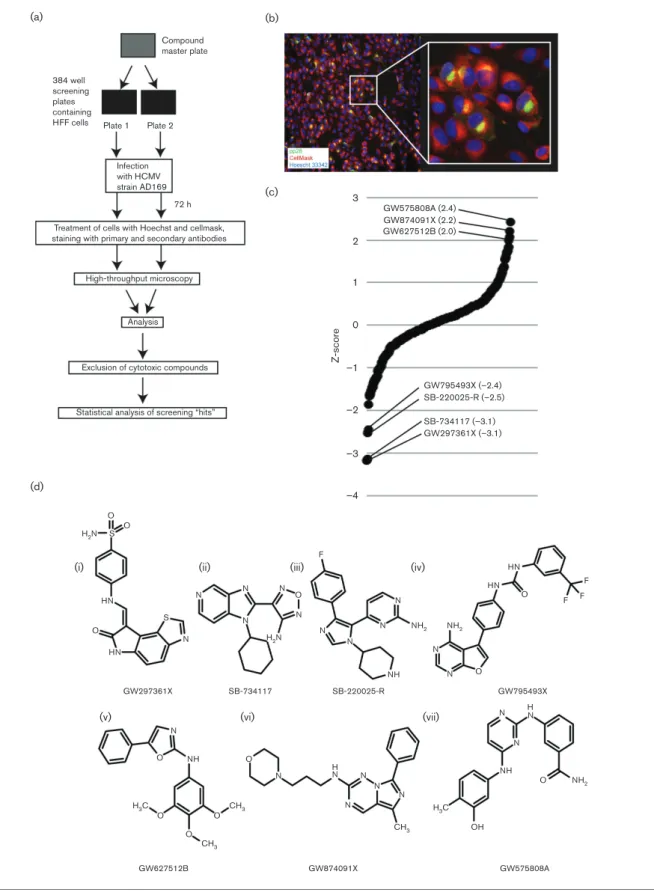
To identify compounds with anti-HCMV activity, we utilized a high-throughput screening methodology (Fig. 1a), similar to the approach that we have previously used to screen small interfering RNAs in HCMV-infected cells [9]. Briefly, high-passage HCMV strain AD169 and compounds from the GSK PIKS collection (listed in Table S1, available in the online Sup-plementary Material) were added to duplicate 384-well plates seeded with human foreskin fibroblast (HFF) cells. As negative and positive controls for compound treatment, several wells in each plate were treated with either DMSO or heparan sulphate (a small molecule that inhibits HCMV entry into cells) [10], respectively. At 72 h post-infection (p.i.), cells were stained with Hoechst 33342 to detect nuclear DNA and CellMask to detect the area of the cell, and were treated with antibodies to detect the cytoplasmic HCMV antigen pp28. An automated microscopy system was then used to assay both the number of cells in each well and the number of infected cells in each well expressing pp28. An image of infected cells treated as described above and captured using automated microscopy is shown in Fig. 1(b).
The mean number of cells in each well per plate was deter-mined. Where the number of cells in any well was less than two-fold below the mean number of cells of the plate, the compound in that well was judged to be cytotoxic (listed by chemotype in Table 1 and by compound in Table S2). Data from the remaining wells on duplicate plates were combined and converted to a Z-score (the number of standard devia-tions from the mean of the data) [11, 12] to demonstrate the increase (positive score) or decrease (negative Z-score) in the number of pp28 positive cells in the presence of each compound [shown in Fig. 1(c), listed by chemotype in Table 1 and by compound in Table S3].
Analysis of cytotoxic compounds
We first investigated what compounds within chemotypes were judged to be cytotoxic. Approximately 40–50 % of compounds in the benzimidazole N-thiophene, 2H-3 pyri-midinyl pyrazolopyridazine, 3-amino pyrazolopyridine and 6-phenyl isoquinoline chemotypes contained compounds judged to be cytotoxic to HCMV-infected cells (Tables 1 and S2). Therefore, compounds from these chemotypes are, generally, not suitable for further use.
We then utilized kinase selectivity data to investigate which kinase proteins were inhibited by each compound judged to be cytotoxic (Table S4). Kinase selectivity data [7] lists the ability of each compound to inhibit a panel of 224 kinase proteins from several protein groups of the human kinome [including TK (tyrosine kinase), STE (homologues of yeast Sterile 7, Sterile 11, Sterile 20 kin-ases), AGC (containing PKA, PKG, PKC families), S-T-PK (serine/threonine protein kinase), CAMK (calcium/cal-modulin-dependent protein kinase) and CMCG (contain-ing CDK, MAPK, GSK3, CLK families) groups]. We found that all cytotoxic benzimidazole N-thiophenes were potent inhibitors of PLK-1 (polo-like kinase 1), whose inhibition can lead to apoptosis, and nearly all other cyto-toxic compounds from a number of chemotypes were potent inhibitors of a range of CDK (cyclin-dependent kinase) proteins, which are involved in regulation of the cell cycle. In our screen cytotoxicity was judged by the number of cells detected in each well of the screening plate. Therefore, we concluded that compounds were gen-erally judged to be cytotoxic due to apoptosis associated with inhibition of PLK-1 or due to lack of cell division associated with inhibition of CDK function.
Analysis of compounds with assigned Z-scores
384 well screening plates containing HFF cells
Compound master plate
(a) (b)
(c)
(d)
Infection with HCMV strain AD169
72 h 3 GW575808A (2.4)
2
1
0
Z-score
–1
–2
–3
–4
O S H2N
H2N
HN
(i)
(v) (vi) (vii)
(ii) (iii) (iv)
O
H3C
NH
O O
N N
N
NH N N HN
OH N
N N H
N
CH3
CH3
H3C
NH2
O
CH3
O
O O
O
GW874091X (2.2) GW627512B (2.0)
GW795493X (–2.4)
GW297361X (–3.1) SB-220025-R (–2.5)
SB-734117 (–3.1)
GW575808A GW874091X
GW627512B
GW795493X GW297361X SB-734117 SB-220025-R
Treatment of cells with Hoechst and cellmask, staining with primary and secondary antibodies
High-throughput microscopy
Analysis
Exclusion of cytotoxic compounds
Statistical analysis of screening “hits”
HN S
N
N N
F
N N
N
N
N O N
NH2
NH
NH2
HN HN
O
F F F
N N
N O Plate 1 Plate 2
Fig. 1.High-throughput screening of the PKIS collection. (a) Diagram of the screening process. (b) A representative example of a
with negative and positive effects on pp28 production, respectively (Tables 1 and S3). We sought to further charac-terize the results of our screen and judged any compound with a Z-score between 1 and 1 to have little or no effect on pp28 production, whereas compounds with Z-scores of 1 to 2 and 1 to 2 had modest negative or positive effects on pp28 production, respectively. Thus, compounds with Z-scores of less than 2 or more than 2 had the greatest nega-tive or posinega-tive effect on pp28 production, respecnega-tively. Therefore, three compounds (GW575808A, GW874091X and GW627512B) from three different chemotypes (2,4-diamino pyrimidines, imidazotriazines and 2-amino oxa-zoles, respectively) had strong positive effects on pp28 production, while four compounds (GW297361X, SB-734117, SB-220025-R and GW795493X) from four chemo-types (oxindoles, furazan benzimidazoles, 4-pyrimidinyl
ortho-aryl azoles and furopyrimidines, respectively) had strong negative effects on pp28 production (Fig. 1c, d).
Examination of kinase protein inhibition by compounds with assigned Z-scores
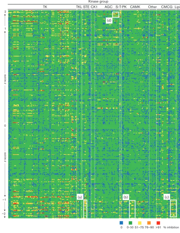
We then examined kinase selectivity data of compounds with assigned Z-scores (Table S5). The data from Table S5 are presented in Fig. 2 as a ‘heatmap’ of kinase inhibition. Nearly all compounds with assigned Z-scores exhibited polypharmacology and could inhibit more than one kinase protein. Consistent with our analysis of compounds judged to be cytotoxic (Table S2), we found that less than 5 % of all compounds either potently inhibited PLK-1 or were potent inhibitors of several different CDK proteins (Table S5). Compounds with positive or negative Z-scores were inhibitors of a wide range of kinase proteins in the TK group (Fig. 2 and Table S5). Therefore, inhibition of TK proteins alone was unlikely to positivity or negatively influence pp28 production. However, a number of kinases in the STE [including MAP4K4 (mitogen-activated protein kinase kinase kinase kinase 4) and MNK (MAP kinase-interacting serine/threonine-protein kinase)], CAMK [including PRKD1
Table 1.Results of screening listed by chemotype
Chemotype Total no. compounds
in chemotype
No. compounds excluded due to cytotoxicity
No. compounds with assigned Z-scores
4-Pyrimidinylortho-aryl azoles 31 0 31
Oxindoles 30 3 27
Furazan benzimidazoles 25 1 24
4-Anilino quinazolines and related 25 0 25
BenzimidazoleN-thiophenes 21 9 12
4-Pyridylortho-aryl azoles 18 0 18
2H-3 Pyrimidinyl pyrazolopyridazines 16 7 9
2-Amino oxazoles 15 0 15
4-Hydrazinyl pyrazolopyrimidines 15 0 15
2,4-Dianilino pyrrolopyrimidines 15 1 14
Biaryl amides 14 0 14
3-Vinyl pyridines 13 0 13
Anilino thienopyrimidines 12 0 12
Benzimidazolyl diaryl ureas 12 2 10
2-Aryl 3-pyridimidinyl pyrazolopyridazines 12 0 12
2,4-Diamino pyrimidines 12 0 12
Maleimide 11 0 11
Furopyrimidines and related 9 0 9
Indazole-3-carboxamides 7 1 6
3-Amino pyrazolopyridines 7 3 4
2-Pyridinyl imidazoles and related 7 0 7
4-Anilino 5-alkynyl pyrimidines 7 0 7
3-Cyano thiophenes 6 2 4
Phenyl carboxamides 6 0 6
Indazole-5-carboxamides 6 0 6
3-Amino pyrazolopyridazines 4 1 3
3-Amino pyrazoles 3 0 3
Imidazotriazine 3 0 3
4-Anilino quinolones 2 0 2
6-Phenyl isoquinolines 2 1 1
Kinase group
TK TKL STE CK1 AGC
(d)
2
0
–1
–2
(a)
0 0–5051–75 76–90 >91 % inhibition
(b) (c)
1
z-score
z-scor
e
S-T-PK CAMK Other CMCG Lipid
Fig. 2.Kinase selectivity of compounds with assigned Z-scores. The full list of kinase selectivity data is shown in Table S5. Table S5
(protein kinase D1), PRKD2 and PRKD3] and CMCG [including CLK2 (cyclin-dependent kinase-like kinase 2), HIPK1 (homeodomain-interacting protein kinase 1), HIPK4, DYRK1A (dual-specificity tyrosine phosphorylation-regu-lated kinase 1A), DYRK1B, DYRK2] groups were inhibited by compounds with assigned Z-scores of less than 1 from eight, four and three different chemotypes, respectively (Fig. 2a–c, respectively, and Table S5). Therefore, these kinase proteins, alone or in combination, were likely to be important for HCMV replication and could represent future anti-HCMV drug targets. A number of compounds from two different chemotypes that inhibited kinases in AGC kinase family [including PRKG1 (protein kinase, cGMP dependent 1), PRKG2, PRKX (protein kinase, x-linked), PKA, ROCK1 (rho-associated, coiled-coil-containing protein kinase 1), ROCK2] were assigned Z-scores over 1 (Fig. 2d). Therefore, these kinase proteins, alone or in combination, were likely to be inhibitory to HCMV replication. Com-pounds targeting these kinase proteins are likely to be of lit-tle value as anti-HCMV drugs.
Compounds with assigned Z-scores of less than 2 (GW297361X, SB-734117, SB-220025-R and GW795493X) each had a different kinase selectivity profile (Fig. 2 and Table S5). Therefore, it was likely each compound inhibited pp28 production by a different mechanism. Each was a potent inhibitor of several kinase proteins from several groups, except for SB-220025-R, which was a potent inhibi-tor of only two kinase proteins: CK1a (casein kinase 1a) and p38a(Fig. 2 and Table S5).
Kinase inhibition of compounds with assigned Z-scores of greater than 2 was also examined. GW575808A and GW627512B had similar kinase selectivity profiles and were inhibitors of several TK and S-T-PK group kinases (Fig. 2 and Table S5). As inhibition of these TKs and S-T-PKs can result in either positive or negative Z-scores (Fig. 2 and Table S5), it was unlikely that inhibition of these TK and S-T-PK proteins had a direct effect on pp28 production. Moreover, we found that GW874091X was not a potent inhibitor of any kinase assayed (Fig. 2 and Table S5). There-fore, GW874091X was either an inhibitor of kinase proteins not assayed in the kinase selectivity data or exerted an effect on pp28 production that did not involve inhibition of cellu-lar kinase proteins. It therefore remains unclear from this analysis which kinase proteins should not be targeted in the development of future anti-HCMV drugs.
We further considered the polypharmacology of compounds tested in our screen. It has been reported that ATP-competi-tive kinase inhibitors can inhibit the function of proteins other than kinases, including aminergic GPCRs [6]. GPCR agonism and antagonism of the compounds in the GSK PKIS collection has been investigated elsewhere [7]. No compound within the GSK PKIS collection is a GPCR ago-nist, but several are GPCR antagonists [7]. However, we observed no obvious correlation between compounds judged to be cytotoxic, compounds assigned a Z-score and GPCR antagonism (data not shown).
Inhibition of HCMV replication by SB-734117
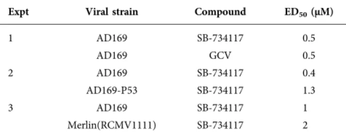
We chose to focus our studies on SB-734117, a compound from the furazan benzimidazole chemotype that had a low Z-score in our screen (Fig. 1c, d). First, we used viral yield reduction assays to assess the ability of SB-734117 to inhibit replication of HCMV strain AD169 compared to the front-line anti-HCMV drug GCV (Table 2, experiment 1) at up to 96 h p.i. The ED50of both SB-734117 and GCV was 0.5 µM,
indicating that SB-734117 inhibited AD169 replication as efficiently as the current frontline anti-HCMV drug. To complement and confirm this data, we analysed AD169 rep-lication over time and found an approximately twofold decrease in AD169 replication from 72–96 h p.i. in the pres-ence of 1 µM SB-734117 (Fig. 3a).
We also found that SB-734117 could inhibit replication of a GCV-resistant virus (AD169-P53) and a low-passage HCMV strain [Merlin(RCMVR1111)], whose genomic con-tent is similar to a clinical sample [13], at low or sub-micromolar ED50 values (Table 2, experiments 2 and 3,
respectively) at up to 96 h p.i. Therefore, SB-734117 was an effective inhibitor of different HCMV strains.
To ensure that the anti-HCMV activity of SB-734117 was not due to cellular cytotoxicity, we used an MTT dye-uptake assay to assess cell viability and cell division in uninfected cells in the presence of SB-734117. We found that the 50 % cellular cytotoxicity of SB-734117 after 96 h treatment with SB-734117 was greater than 10 µM (data not shown). Thus, the 50 % cellular cytotoxicity values in uninfected cells were greater than 10 µM at the ED50values for all HCMV strains
tested. Therefore, inhibition of HCMV replication by SB-734117 observed in experiments shown in Table 2, or in the other experiments presented here, was unlikely to be the result of cellular cytotoxicity or inhibition of cell division.
Examination of HCMV immediate-early protein and mRNA production in HCMV-infected cells treated with SB-734117
We next sought to understand how HCMV replication was inhibited by SB-734117. Therefore, western blotting was used to analyse the accumulation of HCMV proteins in the pres-ence or abspres-ence of SB-734117 (Fig. 3b). Compared to treat-ment of infected cells with DMSO (Fig. 3b, lanes 2–4), the treatment of infected cells with SB-734117 (Fig. 3b, lanes 5–7) reduced the accumulation of immediate-early proteins IE1 and IE2, and IE2 proteins expressed late in infection (IE2-60 and IE2-40) [14]. In this and subsequent western blots, the amount of b-actin in each sample was also assayed, which demonstrated equivalent loading of samples in each lane. We then quantified the relative density of the Western blot-ting bands shown in Fig. 3(b), by determining the intensity of bands corresponding to viral proteins relative to the intensity of theb-actin band in the same lane (Fig. 3c). We found an approximately two- to fourfold decrease in the accumulation of IE1 in the presence of 1 µM SB-734117, which is consistent with an ED50 of 0.5–1 µM shown in
2- to 20-fold, depending on which antibody was used Fig. 3c) was greater than IE1. Consistent with loss of IE pro-tein production and our screening results, we also observed using western blotting that treatment of cells with SB-734117 resulted reduced accumulation of the HCMV early and late proteins UL44 and pp28, respectively, compared to infected cells treated with DMSO (data not shown). To complement these findings, we assayed for differences in IE1 and IE2 mRNA expression in infected cells treated with SB-734117 compared to infected cells treated with DMSO using quantitative PCR against the two major IE RNA spe-cies (Fig. 3d). This analysis revealed that no obvious defect in IE1 mRNA levels was evident in the presence of SB-734117. The analysis of IE2 mRNA again did not show any overt phenotype, although typically a twofold reduction in IE2 mRNA was observed in SB-734117-treated cells when compared with DMSO control. However, taken together, the data suggest that the decrease in IE1 and IE2 protein production shown in Fig. 3(b, c) was unlikely to be the result of decreasedIEgene expression alone.
Our studies thus far could not rule out that SB-734117 impacted events occurring prior to IE gene expression. Thus, we addressed whether the presence of SB-734117 may affect virus entry into the cell or translocation of the HCMV genome to the nucleus. Pre-exposure of cells to SB-734117 before infection or incubation of virus with SB-734117 before infection did not increase the inhibitory effects of the compound (data not shown). However, when we treated AD169-infected HFF cells with 1 µM SB-734117 at 24 h p.i., we found a two-fold decrease in HCMV replication at 120 h p.i., compared to infected cells treated with DMSO at 24 h p.i. (Fig. 4a). Quantitative analysis of Western blotting of infected cells treated as described above (Fig. 4a, b) showed that, similar to data presented in Fig. 3, treatment of infected cells with SB-734117 resulted in approximately two-fold decrease in IE2 protein production depending on which antibody was used. However, there was no obvious decrease in production of IE1 protein.
Therefore, SB-734117 had no obvious effect on cells or virus before infection but could inhibit HCMV replication after entry of the HCMV genome and did so by reducing IE pro-tein production. Thus, SB-734117 may not inhibit events during infection before expression of IE proteins, but could have inhibitory effects on HCMV replication after the initiation of IE protein production.
Inhibition of AGC and CMCG kinase proteins by SB-734711
Next, we investigated what kinase proteins are inhibited by SB-734117. SB-734117 has been reported to inhibit MSK1 (mitogen and stress kinase 1) [15]. However, using the kinase selectivity data shown in Fig. 2 and Table S5, we found that SB-734117 inhibits several AGC kinase group proteins, including MSK1 [MSK1, MSK2, RSK1 (ribosomal s6 kinase 1), RSK2, RSK3, p70S6K1, PKC-h(protein kinase C-h), PRKG2, ROCK1, ROCK2] and several CMCG kinase group proteins (GSK3A, GSK3B, DYRK1A and DYRK1B). However, in our screen, potent and selective inhibitors of GSK3A and GSK3B had no obvious negative effect on pp28 production (Table S5), and compounds with either positive or negative Z-scores were potent inhibitors of PKC-h, PRKG2, ROCK1 and ROCK2 (Table S5). Therefore, inhibition of these kinase proteins may not be related to inhibition of pp28 production. Rather, analysis of SB-734117 kinase selectivity data compared to other assigned Z-scores argued that potent inhibition of MSK1, MSK2, RSK1, RSK2, RSK3, p70S6K1, DYRK1A and DYRK1B was related to inhibition of pp28 production.
A kinase inhibitor that is structurally unrelated to SB-734117, H-89, inhibits a similar range of AGC and CMCG kinase proteins [16]. We found that H-89 inhibited productive HCMV replication and immediate-early protein production (data not shown). Therefore, inhibition of AGC and CMCG kinase proteins, not an unknown function of SB-734117, is likely to be responsible for the observed defects in HCMV replication and protein production. Fur-thermore, using western blotting [17], we found that SB-734117 did not inhibit autophosphorylation of the HCMV-encoded kinase UL97 (data not shown). Therefore, the anti-HCMV effects of SB-734117 were unlikely to be due to inhibition of UL97.
Analysis of cAMP response element-binding protein and histone H3 phosphorylation in HCMV-infected cells
We then considered how inhibition of AGC and CMCG kinase proteins by SB-734114 would affect post-transla-tional modification of cellular proteins thought to be involved in HCMV replication. We focused our investiga-tion on phosphorylainvestiga-tion of the cellular transcripinvestiga-tion factor cAMP response element-binding protein (CREB) and his-tone H3.
CREB is thought to directly or indirectly facilitate tran-scription from the MIEP [18, 19] and other viral promoters [20] during productive HCMV replication, and it has been reported that phosphorylation of CREB at serine residue 133 (CREB-Ser133) by MSK1 is involved in promoting changes to chromatin required for activation of the MIEP during HCMV reactivation from latency [21]. In preliminary experiments, we could not detect either total cellular CREB or CREB-Ser133 before 72 h p.i. using western blotting (data not shown). How-ever, both proteins could only be detected at 72 h p.i. when we
Table 2.Viral replication assays
Expt Viral strain Compound ED50(µM)
1 AD169 SB-734117 0.5
AD169 GCV 0.5
2 AD169 SB-734117 0.4
AD169-P53 SB-734117 1.3
3 AD169 SB-734117 1
increased the amount of cell lysate assayed (see Methods). Therefore, we used western blotting to assay total cellular CREB and CREB-Ser133 phosphorylation in HCMV-infected cells treated with either DMSO or SB-734117 at 72 h p.i. (Fig. 5a). We observed a decrease in accumulation of CREB-Ser133 and an increase in the accumulation of CREB in the presence of SB-734117 (Fig. 5a, lane 5), compared to infected
cells treated with DMSO (Fig. 5a, lane 3). Analysis of relative band intensities (Fig. 5b) indicated that there was approxi-mately a two-fold decrease in CREB-Ser133 in infected cells treated with SB-734117, compared to those treated with DMSO and a modest increase in CREB. The two-fold decrease in the accumulation of CREB-Ser133 in the presence of 1 µM SB-734117 was consistent with the observed ED50of 0.5–1 µM
(c)
(d)
3
24 48 72 h p.i.
UL123 RNA (IE1)
UL122 RNA (IE2) 2.5
2 1.5
∆∆
Ct
(D
M
S
O vs S
B-734117)
1 0.5 0
IE2 IE2(86) IE2(60) IE2(40) IE1
Protein 24 48
h p.i. h p.i.
DMSO SB-734117
72 2.25 4.19 6.28 6.98 5.72 1.65 1.46 0.21 0 0.03 0.51
0 0 0 0
24 0.16
0 0 0 0
48 72
1.38 1.50 0.37 0 1.27 0.52
0 0 0
0 DMSO
(a) (b)
140 000
120 000
100 000
80 000
Plaque-forming units (p.f.u.) ml
–1
60 000
40 000
20 000
0
24 48 72
Hours post-infection (h p.i) 96
40
1 2 3 4 5 6 7
40 63 75 75
0 24 48 72 24 48 72 IE2
IE2 (86) IE2 (60) IE2 (40)
b-Actin IE1 SB-734117 DMSO
h p.i. SB-734117
Fig. 3.Analysis of HCMV replication and protein production in infected HFF cells treated with DMSO or SB-734117 at the time of
infec-tion. (a) HFF cells were infected at an m.o.i. of 1 with AD169 and then treated with 1 µM SB-734117 or the equivalent volume of DMSO. Viral titre (p.f.u. ml 1) was determined at the indicated time points (hours p.i.). Data points and error bars represent the mean and
and two- to four-fold decrease in production of immediate-early HCMV protein production (Table 2 and Fig. 3). There-fore, the effect of SB-734117 on HCMV replication correlated with a loss of CREB-Ser133 phosphorylation. Similar observa-tions were made when infected cells were treated with H89 (data not shown), indicating the AGC and CMCG kinase pro-teins were involved in phosphorylation of CREB.
Phosphorylation of histone H3 by MSK1 or another kinase, IKKa, is required for binding of transcription factors to DNA in uninfected cells [22, 23]. We have previously demonstrated that phosphorylation of histone H3 at serine residue 10 (H3S10p) by IKKais associated with immediate-early protein production during productive HCMV replication [17]. Also, it has been demonstrated that H3S10 phosphorylation by MSK1 is associated with immediate-early gene expression during reactivation of HCMV from latency [21]. We decided to assay H3S10 phosphorylation in the presence of SB-734117. Using Western blotting, we analysed accumulation of H3 and H3S10 phosphorylation in uninfected HFF cells (Fig. 5c, lane 1) and AD169-infected HFF cells treated with either DMSO or SB-734117 (Fig. 5c, lanes 2–4 and 5–7, respectively, at 24–72 h p.i.). Similar levels of H3 were found in each sample; however,
over time we observed a decrease in H3S10p in infected cells treated with SB-734117 to near undetectable levels, compared to infected cells treated with DMSO. Therefore, inhibition of H3S10 phosphorylation during productive HCMV replication may have contributed to the anti-HCMV activity of SB-734117. Similar results were found when infected cells were treated with H89 (data not shown), indicating that AGC and CMCG kinase proteins were involved in phosphorylation of H3S10.
Phosphorylation of histone H3 at serine residue 28 (H3S28) by MSK1 is also known to be associated with gene expres-sion in uninfected cells [22]. We also used western blotting to investigate H3S28 phosphorylation during HCMV repli-cation. However, we could not detect H3S28p in uninfected HFF cells, AD169-infected cells HFF treated with either DMSO or SB-734117, or uninfected HFF cells treated with either anisomycin, which can stimulate H3S28 phosphoryla-tion, or phosphatase inhibitor okadaic acid, which can pre-vent dephosphorylation of histones (data not shown). Therefore, we concluded that inhibition of H3S28 phos-phorylation did not contribute to the anti-HCMV activity of SB-734117.
0 IE1
Protein (c)
h p.i.
24 24 48 72 24 48 72
h p.i. h p.i.
DMSO SB-734117
IE2 IE2(86) IE2(60) IE2(40)
0.11
1.10 1.02 0.81 0.87
1.74 2.64 2.92 2.14 1.32 0.61 0 0.90 0.62
0 0 0 1.10 1.00 1.02
1.08 2.06 3.66 2.06 1.32 1.10 0.23 1.86 0.81
0 0 0.09 0
0
Hours post-infection (h p.i.)
75
24 48 72 96 48 72 96 h p.i.
IE2
IE2 (86) IE2 (60) IE2 (40)
b-Actin IE1
75 50 40
40
1 2 3 4 5 6 7
100 000
(a) (b)
80 000
60 000
Plaque-forming units (p.f.u.) ml
–1
40 000
20 000
0
SB-734117 DMSO
SB-734117 DMSO
Fig. 4.Analysis of HCMV replication and protein production in infected HFF cells treated with DMSO or SB-734117 at 24 h p.i. (a) HFF
Analysis of histone H3 post-translational modifications in HCMV-infected cells
H3S10 phosphorylation by either MSK1 or IKKais associated with the presence of acetyl modifications of H3, including acetylation (ac) of lysine 14 (H3K14ac) [24–26]. There is a relationship during transcriptional activation between the presence of H3S10p and H3K14ac and the association of transcription factors with DNA [23, 27]. As the presence of H3K14ac is associated with transcriptional activation in HCMV-infected cells [28], H3K14ac may be required for HCMV replication. We have previously demonstrated that, during HCMV replication, inhibition or depletion of IKKa
leads to loss of total cellular H3S10 phosphorylation, but not loss of total cellular H3K14ac [17]. Thus, we assayed whether treatment of HCMV-infected cells by SB-734117 would lead to loss of H3S10p or H3K14ac. We used Western blotting to assay total cellular levels of H3, H3S10p and acetylation of H3 on a number of commonly studied H3 lysine residues includ-ing K14 (H3K9ac, H3K14ac, H3K18ac, H3K27ac) in either
uninfected HFF cells (Fig. 5c, lane 1) or HFF cells infected with HCMV and treated with either DMSO or SB-734117 (Fig. 5c, lanes 2–4 and 5–7, respectively). Treatment of infected cells with SB-734117 had a slight effect [less than twofold (data not shown)] on accumulation of H3K14ac and no detectable effect on detection of H3K9ac, H3K18ac or H3K27ac. Therefore, treatment of infected cells with SB-734117 was associated with loss of total cellular H3S10p, but not loss of the total cellular H3 acetylation modifications we assayed, including H3K14ac.
The relationship between H3S10p and dimethylation (me2) and trimethylation (me3) of H3 and H3 phosphorylation is not well characterized, but it has been reported that, in a murine model, there is a relationship between the presence of H3S10p and the presence of H3K36me3 [29] and in
Drosophila melanogaster loss of the MSK1/2 homologue JIL-1 results in loss of H3S10p, H3 acetylation and H3 methylation, including H3K36me3 [30]. Therefore, we
42
(a) (c)
(e) (b)
CREB-Ser133
CREB
b-Actin
– –
Uninfected DM
S
O
S
B-734117
42
40
1 2 3 4
17
0 24 48
DMSO SB-734114 72 24 48 72 h p.i.
H3K9ac
H3K14ac
H3K18ac
H3K27ac
H3S10p
H3
b-Actin 17
17
17
17
17
48
1 2 3 4 5 6 7
17 (d)
0 24 48
DMSO SB-734114 72 24 48 72 h p.i.
H3K4me2
H3K27me2
H3K36me2
H3S10p
H3
b-Actin 17
17
17
17
48
17
17
17
17
17
17
48
1 2 3 4 5 6 7
0 24 48
DMSO SB-734114 72 24 48 72 h p.i.
H3K9me3 H3K4me3
H3K27me3
H3K36me3
H3S10p
H3
b-Actin
1 2 3 4 5 6 7
5
Protein Uninfected DMSO SB-734117
0.42 1.24 0.94
1.02 1.71
0.73
CREB-Ser133 CREB
Fig. 5.Inhibition of phosphorylation of cellular proteins by SB-734117. (a–c, lane 3) HFF cells were infected with AD169 at an m.o.i. of
asked if loss of total cellular H3S10p in HCMV-infected cells was associated with me2 and me3 modification of H3 lysine residues. Western blotting was used to assay the pres-ence of H3 and H3S10p, as well as me2 (H3K4me2, H3K27me2, H3K36me2) or me3 (H3K4me3, H3K9me3, H3K27me3, H3K36me3) modifications of H3 in uninfected HFF cells (lane 1, Fig. 5d, e, respectively) or HFF cells infected with HCMV and treated with either DMSO or SB-734117 (lanes 2–4 and 5–7, Fig. 5d, e, respectively). We observed that SB-734117 had no effect on total cellular accumulation of any me2 modification of H3 (Fig. 5d) or accumulation of H3K4me3, H3K9me3 or H3K27me3 (Fig. 5e). However, we observed a near total loss of detect-able H3K36me3 over time in infected cells treated with SB-734117 (Fig. 5e), lane 7) compared to infected cells treated with DMSO (Fig. 5e, lane 4). Similarly, a near total loss of detectable H3K36me3 was observed in infected cells treated with H89 (data not shown), suggesting that loss of H3K36me3 is related to inhibition of AGC and CMCG kinase proteins. Thus, inhibition of H3K36me3 was associ-ated with loss of total cellular H3S10p and was likely the result of inhibition of AGC and CMCG kinase proteins inhibited by SB-734117. Loss of both total cellular H3S10p and total cellular H3K36me3 may have contributed to the anti-HCMV activity of SB-734117.
Investigation of HCMV MIEP transcriptional activation
Loss of CREB and H3S10 phosphorylation (Fig. 5a–e) sug-gested that SB-734117 acted by inhibiting activation of the HCMV MIEP. However, our analysis of IE1 and IE2 gene expression (Fig. 3d) indicated that transcription from the MIEP was not obviously compromised in the presence of SB-734117. To investigate this in more detail, we utilized chromatin immunoprecipitation to assay the presence of H3K14ac, a marker of MIEP transcriptional activation [28],
at the MIEP in the presence of either DMSO or SB-734117 (Fig. 6). The data showed no overt impact on H3K14ac at the MIEP between 24 and 72 h p.i. in DMSO- or treated cells. Indeed, we noted that SB-734117-treated cells actually showed higher levels of H3K14ac at the MIEP at late times p.i. when compared to control. There-fore, in agreement with our analysis of IE1 and IE2 gene expression (Fig. 3d), there was no obvious defect in MIEP transcriptional activation in the presence of SB-734177. Thus, the observed defects in HCMV immediate-early pro-tein production (Figs 3, 4) could not be explained by defects in transcription from the MIEP.
DISCUSSION
Our overall analysis of the chemotypes screened indicated that each chemotype contained compounds with anti-HCMV activity, however modest that anti-anti-HCMV activity may have been. As the structure of each compound in each chemotype is known, structure–activity relationships derived from our data could form the basis of future studies in the discovery of compounds with anti-HCMV activity from each chemotype.
Our survey of the PKIS kinase selectivity data argued that several proteins from several kinase groups, alone or in combination, were required for pp28 production. These protein kinases include those from the STE (including MAP4K4 and MNK), CAMK (including PRKD1, PRKD2 and PRKD3) and CMCG (including CLK2, HIPK1, HIPK4, DYRK1A, DYRK1B, DYRK2) kinase groups. The function of these protein kinases in productive HCMV replication is unclear or unknown. Therefore, it is possible that our data identified novel cellular factors required for productive HCMV replication. However, the polypharmacology of the compounds tested makes it difficult to identify specific kin-ases required for productive HCMV replication. Thus, each
3
2.5
2
1.5
1
0.5
0
C K14 C
M
IE
P P
C
R (% input)
K14
24 48 72 h p.i.
DMSO
SB-734117
C K14 C K14 C K14 C K14
Fig. 6.H3K14ac at the MIEP is unaffected in SB-734117-treated cells. DNA was immune-precipitated from infected cells (24–72 h p.i.)
of the aforementioned kinases will have to be tested individ-ually to identify their roles in HCMV-infected cells. With this information, these kinases could be exploited as novel anti-HCMV drug targets.
We chose to pursue studies of SB-734117 as this compound had one of the greatest negative effects on pp28 production in our screen, with no obvious cytotoxic effects, and was an effective inhibitor of a number of HCMV strains. Moreover, the function of the AGC and CMCG kinase proteins inhib-ited by SB-734117 in productive HCMV replication was unknown or unclear.
SB-734117 was originally described as an inhibitor of MSK1 [15]. However, like other MSK1 inhibitors [16], SB-734117 displays polypharmacology and can inhibit several kinases whose roles in productive HCMV replication are unknown or unclear. Thus, to understand how SB-734117 inhibits HCMV replication, it will be necessary to under-stand if a particular kinase, or a combination of kinase proteins, is required for productive HCMV replication. A truly selective inhibitor of MSK1 has yet to be found. Structure–activity relationships involving SB-734117 and other furazan benzimidazole compounds could be explored to generate compounds with improved anti-HCMV activ-ity and kinase selectivactiv-ity. However, we could discern no obvious relationship among inhibition of HCMV replica-tion, kinase selectivity and the structures of compounds within the furazan benzimidazole chemotype analysed here due to the small number of furazan benzimidazole com-pounds that returned low negative Z-scores in our screen (data not shown). An improved compound related to SB-734117 would have value as an anti-HCMV drug, as it would have the potential to inhibit both productive HCMV replication and reactivation of HCMV from latency. In addition, based on our observations, an improved compound should be as effective an inhibitor of HCMV replication as GCV and be able to inhibit replica-tion of GCV-resistant HCMV strains.
Perhaps the most intriguing observations we make concern the modification of histone H3 in the presence of SB-734117. Previous observations from our laboratory have indicated that IKKawas required for H3S10 phosphoryla-tion in AD169-infected HFF cells [17], which is consistent with data presented elsewhere indicating that H3S10 is a substrate of IKKa[24, 25, 31–33]. We note that inhibition of IKKaresults in loss of H3S10p early in HCMV replica-tion (24 h p.i. onwards) [17], whereas treatment with SB-734117 leads to a loss of H3S10p later in HCMV replication (48–72 h p.i.). SB-734117 does not inhibit IKKa(Table S5). Therefore, we propose that, in HCMV-infected cells, a mechanism exists wherein IKKa does not phosphorylate H3S10 during treatment with SB-734117. Conversely, kin-ases inhibited by SB-734117 do not phosphorylate H3S10 when IKKa is inhibited or depleted. This mechanism may ensure appropriate regulation of H3S10 phosphorylation that is necessary for productive HCMV replication.
It has been reported that, in uninfected cells from humans and mice, loss of H3S10p can lead to loss of H3K14ac [24–
26]. However, we did not observe loss of total cellular H3K14ac upon treatment of HCMV-infected cells with SB-734117 or in our previous study where inhibition or deple-tion of IKKaresulted in loss of H3S10p [17]. We speculate, as we have done previously [17], that an as yet unrecognized mechanism exists in HCMV-infected cells that maintains total H3K14ac when total H3S10p levels are lowered to near undetectable levels.
We observed that treatment of HCMV-infected cells with SB-734117 resulted in loss of H3K36me3. We have previ-ously found that depletion of IKKain HCMV-infected cells leads to loss of H3S10p, but not H3K36me3 [17]. Therefore, the loss of H3K36me3 in HCMV-infected cells is related to loss of H3S10p during inhibition of AGC and CMCG kin-ases, but not during inhibition of IKKa. This may be related to regulation of H3S10 phosphorylation by different kinases, as we discuss above. We propose that, as in mice and
Drosophila[29, 30], the presence of H3K36me3 in HCMV-infected cells is related to the presence of H3S10p, poten-tially via phosphorylation of H3 by MSK1. Alternatively, there may be a substrate of kinase proteins inhibited by SB-734117 whose phosphorylation directly or indirectly medi-ates H3K36 tri-methylation.
Treatment of HCMV-infected cells with SB-734117 impacted on immediate-early protein production and caused loss of total cellular levels of post-translational modi-fication of cellular factors potentially involved in transacti-vation of the HCMV MIEP. However, in the presence of SB-734117, we did not find obvious defects in activation of transcription from the HCMV MIEP or defects in immedi-ate-early gene transcription. Therefore, the loss of total cel-lular levels of CREB and H3S10 phosphorylation or H3K36me3 had no direct impact on transcription from the HCMV MIEP. This interpretation would be consistent with previous studies that have shown that the deletion of CREB-binding sites from the MIEP has little impact on productive HCMV replication [21, 34]. During reactivation, it is hypothesized the H3S10p is important as it drives the tran-sition of the MIEP from a repressed to active promoter, a mechanism also proposed for herpes simplex virus reactiva-tion [35] Thus, during productive HCMV replicareactiva-tion at high m.o.i., where the MIEP is associated with active, not repressed, chromatin very early p.i. [28], H3S10p may not be essential for transcription. However, it remains possible that H3S10p has a role in the release of early and late HCMV promoters from repression as infection proceeds. Thus, future challenges will include mapping of CREB, H3S10p and H3K36me3 to viral and cellular promoters to understand in more detail their possible involvement in productive HCMV replication.
production of both proteins. Post-translational modification of both IE1 and IE2 is not yet completely characterized. Thus, SB-734117 could directly or indirectly inhibit phos-phorylation of these proteins, which leads to their loss. Fur-ther investigation of IE protein production and the roles of IE post-translational modification is required in order to fully understand the mechanism of action of SB-734117 during productive HCMV replication.
METHODS
Compounds
The PIKS library (version 1) was supplied to the Institute of Chemistry and Chemical Biology-Longwood at Harvard Medical School by GlaxoSmithKline. SB-734117 was a kind gift from GlaxoSmithKline. GCV was obtained from SIGMA. H89 and heparan sulphate were obtained from Cal-biochem. All drugs were resuspended in DMSO.
Cells and viruses
HFF cells (clone Hs29) were obtained from American Type Culture Collection no. CRL-1684 and maintained in Dul-becco’s modified Eagle’s medium (Gibco) containing 5 % (v/v) FBS (Gibco), as well as penicillin and streptomycin. High-passage HCMV strain AD169 was a gift from Don Coen (Harvard Medical School). Low-passage strain Merlin R1111 (derived from BACmid pAL1111, which does not express RL13 and UL128) [36] was a gift from Richard Stan-ton (Cardiff University). GCV-resistant virus AD169-P53 was supplied by the National Institutes of Health (NIH) AIDS Reagent Program.
High throughput screening
For screening of PKIS collection and analysis of screening data, as well as characterization of compounds within the PKIS collection, see Supplementary Material.
Viral yield reduction assays
Assays were performed essentially as described previously [37]. HFF cells were plated at 5104cells per well in 24-well plates. After overnight incubation, cells were infected with HCMV at an m.o.i. of 1. After virus adsorption for 1 h at 37
C, cells were washed and incubated with 0.5 ml medium containing DMSO or compounds at a range of concentra-tions in duplicate. Plates were incubated for 4 days at 37C.
Titres were determined by serial dilution of viral superna-tant onto HFF monolayers that were covered in Dulbecco’s modified Eagle’s medium containing 5 % (v/v) FBS, 0.6 % methylcellulose and antibiotics. Cultures were incubated for 14 days, cells were stained with crystal violet and plaques were counted. Data shown represent the mean value of duplicate plaque counts. The final concentration of DMSO in all samples was maintained at <1 % (v/v).
MTT cytotoxicity assays
Assays were performed essentially as described previously [37]. HFF cells were seeded at 1104cells per well into
96-well plates. After overnight incubation to allow cell
attachment, cells were treated for the time indicated in the text at range of concentrations in duplicate. The highest concentration of compound tested was 10 µM. Cell viability was then determined with an MTT assay according to the manufacturer’s instructions (GE Healthcare). Data shown represent the mean value of duplicate readings. The final concentration of DMSO in all samples was maintained at <1 % (v/v). As a positive control, in all experiments, a two-fold dilution series of HFF cells starting at 1104cells per
well was included. In each experiment, we found a linear relationship between the number of cells per well and out-put from the MTT assay (data not shown).
Western blotting
At time points indicated in the text, cells were washed once with PBS and resuspended in 100 µl Laemmli buffer con-taining 5 % (v/v) b-mercaptoethanol. Proteins were separated on 8 or 10 % polyacrylamide gels. Typically, a vol-ume of cell lysate corresponding to 1104HFF cells was analysed, except when blotting for CREB or CREB-Ser133 when a volume of cell lysate corresponding to 5105HFF
cells was analysed. Antibodies used are listed in Supplemen-tary Material. Relative band intensity (band intensity rela-tive tob-actin signal in the same lane) was analysed using ImageJ software, obtained from the NIH (USA).
RNA analysis and quantitative PCR
Quantitative PCR was performed using a SYBR green qPCR kit (Qiagen) and analysed using theDDCt method to
com-pare DMSO- versus SB-734117-treated cells. Briefly, cDNA was prepared from RNA extracted from infected cells at time points indicated in the figure. cDNA and no RT con-trols were amplified in technical duplicates from multiple experiments using a constant primer in exon 3 (UL122-123) and a primer from exon 4 (UL123) or exon 5 (UL122). Cel-lular RNA was amplified using 18S RNA primers.
Exon 3, ACG AGA ACC CCG AGA AAG ATG; exon 4, CGC CAG TGA ATT TCT CTT C; exon 5, CCG GGG AGA GGA GTG TTA GT; 18S for, GTA ACC CGT TGA ACC CCA; 18S rev, CCA TCC AAT CGG TAG TAG CG. qPCR was performed using cycling conditions: 95C (15 s)
and then 40 cycles of 94
C (15 s), 55
C (30 s) and 72
C (30 s).
Chromatin immunoprecipitation
Specific immunoprecipitation of sequences was expressed as enrichment from input.
Funding information
This work was supported by New Investigator funds from St. George’s, University of London, a St. George’s Impact and Innovation Award and a PARK/WestFocus Award (all to B. L. S.). B. L. S. was also supported by grants from the NIH (R01 AI019838 and R01 AI026077) awarded to Don Coen (Harvard). M. J. M. is supported by a Medical Research Coun-cil (UK) PhD studentship.
Acknowledgements
We would like to express our thanks to Don Coen for his encour-agement during this study and his support of B. L. S. We also ac-knowledge Simon Arthur (Dundee), Nathanael Gray (Harvard), GlaxoSmithKline, Gloria Komazin-Meredith (Harvard), Andrew Macdon-ald (Leeds) and Richard Stanton (Cardiff) for providing reagents and insightful discussions, as well as I’ah Z Donovan-Banfield for technical assistance and Lisa Rickelton for assistance with preparation of fig-ures. Special thanks go to all members of the Institute of Chemistry and Chemical Biology-Longwood for their assistance in all aspects of the screening process.
Conflicts of interest
The authors declare that there are no conflicts of interest.
References
1. Mocarski ES, Shenk T, Griffiths PD, Pass RF.Cytomegaloviruses. In: Knipe DM and Howley PM (editors).Fields Virology, 6th ed. vol. 2, New York: Lippincott, Williams & Wilkins; 2015. pp. 1960–2015. 2. Krause PR, Bialek SR, Boppana SB, Griffiths PD, Laughlin CAet al.
Priorities for CMV vaccine development.Vaccine2013;32:4–10. 3. Coen DM, Schaffer PA.Antiherpesvirus drugs: a promising
spec-trum of new drugs and drug targets.Nat Rev Drug Discov2003;2: 278–288.
4. Rogers N.A dormant danger: new therapies target a ubiquitous path-ogen known as cytomegalovirus.Nat Med2015;21:1104–1105. 5. Müller S, Chaikuad A, Gray NS, Knapp S.The ins and outs of
selective kinase inhibitor development. Nat Chem Biol 2015;11: 818–821.
6. Paolini GV, Shapland RH, van Hoorn WP, Mason JS, Hopkins AL. Global mapping of pharmacological space.Nat Biotechnol2006;24: 805–815.
7. Elkins JM, Fedele V, Szklarz M, Abdul Azeez KR, Salah Eet al.
Comprehensive characterization of the published kinase inhibitor set.Nat Biotechnol2016;34:95–103.
8. Drewry DH, Willson TM, Zuercher WJ.Seeding collaborations to advance kinase science with the GSK published kinase inhibitor set (PKIS).Curr Top Med Chem2014;14:340–342.
9. Polachek WS, Moshrif HF, Franti M, Coen DM, Sreenu VBet al.
High-throughput small interfering RNA screening identifies phosphatidylinositol 3-kinase class II alpha as important for production of human cytomegalovirus virions. J Virol 2016;90: 8360–8371.
10. Compton T, Nowlin DM, Cooper NR.Initiation of human cytomega-lovirus infection requires initial interaction with cell surface hep-aran sulfate.Virology1993;193:834–841.
11. Birmingham A, Selfors LM, Forster T, Wrobel D, Kennedy CJ
et al. Statistical methods for analysis of high-throughput RNA
interference screens.Nat Methods2009;6:569–575.
12. Zhang JH, Chung TD, Oldenburg KR.A simple statistical parame-ter for use in evaluation and validation of high throughput screen-ing assays.J Biomol Screen1999;4:67–73.
13. Wilkinson GW, Davison AJ, Tomasec P, Fielding CA, Aicheler R
et al. Human Cytomegalovirus: taking the strain. Med Microbiol Immunol2015;204:273–284.
14. White EA, Del Rosario CJ, Sanders RL, Spector DH.The IE2 60-kilodalton and 40-60-kilodalton proteins are dispensable for human
Cytomegalovirusreplication but are required for efficient delayed early and late gene expression and production of infectious virus. J Virol2007;81:2573–2583.
15. Bamford MJ, Alberti MJ, Bailey N, Davies S, Dean DKet al. (1H-imidazo[4,5-c]pyridin-2-yl)-1,2,5-oxadiazol-3-ylamine derivatives: a novel class of potent MSK-1-inhibitors. Bioorg Med Chem Lett 2005;15:3402–3406.
16. Naqvi S, Macdonald A, Mccoy CE, Darragh J, Reith ADet al. Char-acterization of the cellular action of the MSK inhibitor SB-747651A.Biochem J2012;441:347–357.
17. Ho CM, Donovan-Banfield IZ, Tan L, Zhang T, Gray NS et al.
Inhibition of ikkaby BAY61-3606 reveals IKKa-dependent histone H3 phosphorylation in human cytomegalovirus infected cells. PLoS One2016;11:e0150339.
18. Chia YL, Ng CH, Lashmit P, Chu KL, Lew QJet al.Inhibition of
human cytomegalovirus replication by overexpression of CREB1. Antiviral Res2014;102:11–22.
19. Stinski MF, Thomsen DR, Stenberg RM, Goldstein LC. Organiza-tion and expression of the immediate early genes of human cyto-megalovirus.J Virol1983;46:1–14.
20. Rodems SM, Clark CL, Spector DH.Separate DNA elements con-taining ATF/CREB and IE86 binding sites differentially regulate the human cytomegalovirus UL112-113 promoter at early and late times in the infection.J Virol1998;72:2697–2707.
21. Kew VG, Yuan J, Meier J, Reeves MB.Mitogen and stress acti-vated kinases act co-operatively with CREB during the induction of human cytomegalovirus immediate-early gene expression from latency.PLoS Pathog2014;10:e1004195.
22. Arthur JS. MSK activation and physiological roles. Front Biosci 2008;13:5866–5879.
23. Macdonald N, Welburn JP, Noble ME, Nguyen A, Yaffe MBet al.
Molecular basis for the recognition of phosphorylated and phos-phoacetylated histone h3 by 14-3-3.Mol Cell2005;20:199–211. 24. Yamamoto Y, Verma UN, Prajapati S, Kwak YT, Gaynor RB.
His-tone H3 phosphorylation by IKK-alpha is critical for cytokine-induced gene expression.Nature2003;423:655–659.
25. Anest V, Hanson JL, Cogswell PC, Steinbrecher KA, Strahl BD
et al. A nucleosomal function for IkappaB kinase-alpha in NF-kappaB-dependent gene expression.Nature2003;423:659–663. 26. Vicent GP, Ballare C, Nacht AS, Clausell J, Subtil-Rodríguez A
et al.Induction of progesterone target genes requires activation of ERK and MSK kinases and phosphorylation of histone H3.Mol Cell 2006;24:367–381.
27. Lo WS, Trievel RC, Rojas JR, Duggan L, Hsu JYet al. Phosphoryla-tion of serine 10 in histone H3 is funcPhosphoryla-tionally linked in vitro and in vivo to GCN5-mediated acetylation at lysine 14.Mol Cell 2000;5: 917–926.
28. Cuevas-Bennett C, Shenk T.Dynamic histone H3 acetylation and methylation at human cytomegalovirus promoters during replica-tion in fibroblasts.J Virol2008;82:9525–9536.
29. Gr€aff J, Woldemichael BT, Berchtold D, Dewarrat G, Mansuy IM. Dynamic histone marks in the hippocampus and cortex facilitate memory consolidation.Nat Commun2012;3:991.
30. Karam CS, Kellner WA, Takenaka N, Clemmons AW, Corces VG. 14-3-3 mediates histone cross-talk during transcription elonga-tion inDrosophila.PLoS Genet2010;6:e1000975.
31. Dong W, Li Y, Gao M, Hu M, Li Xet al.IKKacontributes to
UVB-induced VEGF expression by regulating AP-1 transactivation. Nucleic Acids Res2012;40:2940–2955.
32. Lubin FD, Sweatt JD.The IkB kinase regulates chromatin struc-ture during reconsolidation of conditioned fear memories.Neuron 2007;55:942–957.
34. Meier JL, Keller MJ, Mccoy JJ.Requirement of multiple cis-acting elements in the human cytomegalovirus major immediate-early distal enhancer for viral gene expression and replication.J Virol 2002;76:313–326.
35. Cliffe AR, Arbuckle JH, Vogel JL, Geden MJ, Rothbart SBet al.
Neuronal stress pathway mediating a histone methyl/phospho switch is required for herpes simplex virus reactivation.Cell Host Microbe2015;18:649–658.
36. Stanton RJ, Baluchova K, Dargan DJ, Cunningham C, Sheehy O
et al. Reconstruction of the complete human cytomegalovirus
genome in a BAC reveals RL13 to be a potent inhibitor of replica-tion.J Clin Invest2010;120:3191–3208.
37. Loregian A, Coen DM.Selective anti-cytomegalovirus compounds discovered by screening for inhibitors of subunit interactions of the viral polymerase.Chem Biol2006;13:191–200.
Five reasons to publish your next article with a Microbiology Society journal 1. The Microbiology Society is a not-for-profit organization.
2. We offer fast and rigorous peer review–average time to first decision is 4–6 weeks. 3. Our journals have a global readership with subscriptions held in research institutions around
the world.
4. 80% of our authors rate our submission process as‘excellent’or‘very good’.
5. Your article will be published on an interactive journal platform with advanced metrics.