REPORT
Mutations in
KDSR
Cause Recessive
Progressive Symmetric Erythrokeratoderma
Lynn M. Boyden,1 Nicholas G. Vincent,2 Jing Zhou,3 Ronghua Hu,3 Brittany G. Craiglow,3 Susan J. Bayliss,4 Ilana S. Rosman,4 Anne W. Lucky,5 Luis A. Diaz,6 Lowell A. Goldsmith,6 Amy S. Paller,7 Richard P. Lifton,1 Susan J. Baserga,1,8,9 and Keith A. Choate1,3,10,*
The discovery of new genetic determinants of inherited skin disorders has been instrumental to the understanding of epidermal function, differentiation, and renewal. Here, we show that mutations inKDSR(3-ketodihydrosphingosine reductase), encoding an enzyme in the ceramide synthesis pathway, lead to a previously undescribed recessive Mendelian disorder in the progressive symmetric erythrokerato-derma spectrum. This disorder is characterized by severe lesions of thick scaly skin on the face and genitals and thickened, red, and scaly skin on the hands and feet. Although exome sequencing revealed several of theKDSRmutations, we employed genome sequencing to discover a pathogenic 346 kb inversion in multiple probands, and cDNA sequencing and a splicing assay established that two mutations, including a recurrent silent third base change, cause exon skipping. Immunohistochemistry and yeast complementation studies demon-strated that the mutations cause defects in KDSR function. Systemic isotretinoin therapy has achieved nearly complete resolution in the two probands in whom it has been applied, consistent with the effects of retinoic acid on alternative pathways for ceramide generation.
Mendelian disorders of cornification (MEDOCs) are severe skin disorders that feature localized or generalized scaling and redness and can be associated with significant morbidity and mortality. Genetic investigation has revealed MEDOC-causing mutations in over 50 genes; despite diverse function, they share a common pathobiologic feature of increased transepidermal water loss. Included among MEDOCs are progressive symmetric erythrokerato-derma (PSEK [MIM: 133200], which shows symmetrically distributed hyperkeratotic and erythematous plaques on the face, buttocks, and groin and palmoplantar kerato-derma in about half of affected individuals) and erythroker-atoderma variabilis (which exhibits similar features in conjunction with transient patches of erythema). Both dis-orders display substantial phenotypic heterogeneity, and cutaneous involvement can vary markedly from localized to generalized disease, even within kindreds. Previously described Mendelian forms of erythrokeratoderma are usu-ally dominant and often result from heterozygosity for a de novo mutation. These include erythrokeratoderma variabi-lis (MIM: 133200; caused by heterozygosity for a mutation in GJB3 [MIM: 603324], GJB4 [MIM: 605425], or GJA1 [MIM: 121014], which encode connexins 31, 30.3, and 43, respectively1–3), Vohwinkel syndrome (MIM: 604117; caused by heterozygosity for a mutation in LOR [MIM: 152445], encoding loricrin4), and a syndrome of erythro-keratoderma, ichthyosis, and cardiomyopathy (EKC syn-drome [MIM: 615821], caused by heterozygosity for a missense mutation in a single domain of DSP [MIM: 125647], encoding desmoplakin5).
We recruited a large cohort of 750 kindreds with disor-ders of cornification, including subjects with ichthyosis and erythrokeratoderma phenotypes. The Yale Human Investigation Committee approved the study in accor-dance with institutional and national ethical standards, and subjects provided verbal and written informed con-sent. We screened probands for 48 genes in which muta-tions cause MEDOCs via targeted amplification and high-throughput sequencing. Subjects without patho-genic mutations identified in this gene panel make up a subset that facilitates the discovery of new genetic causes of MEDOCs, typically via exome sequencing.3,5
In the course of exome sequencing of the MEDOC dis-covery cohort, two probands described by their referring physicians as having an unusual keratinization or PSEK-like disorder (and unaffected parents) were each found to be compound heterozygous for a deleterious mutation and a silent mutation in the same gene, KDSR (MIM: 136440; GenBank: NM_002035.2 and NP_002026.1; Ta-bles S1andS2 andFigures S1A and S1B). Subject 429 is heterozygous for the previously unreported mutation c.164_166delAAG (p.Gln55_Gly56delinsArg), and subject 101 is heterozygous for the previously unreported splice-site mutation c.2562A>C. Both subjects are heterozy-gous for the silent mutation c.879G>A (p.Gln293Gln) (mi-nor allele frequency 0.00003).
The silent mutation shared by both probands is at the last base of exon 9. Because guanine is highly conserved at this position,6 we hypothesized that this mutation might alter splicing. We used skin tissue from subject 429
1
Department of Genetics, Yale University School of Medicine, New Haven, CT 06510, USA;2Department of Microbiology, Yale University School of Med-icine, New Haven, CT 06510, USA;3Department of Dermatology, Yale University School of Medicine, New Haven, CT 06510, USA;4Division of
Derma-tology, Washington University School of Medicine, Saint Louis, MO 63110, USA;5Dermatologists of Southwest Ohio, Cincinnatti, OH 45247, USA; 6Department of Dermatology, University of North Carolina School of Medicine, Chapel Hill, NC 27516, USA;7Department of Dermatology, Northwestern
University Feinberg School of Medicine, Chicago, IL 60611, USA;8Department of Therapeutic Radiology, Yale University School of Medicine, New Haven,
CT 06510, USA;9Department of Molecular Biophysics and Biochemistry, Yale University School of Medicine, New Haven, CT 06510, USA;10Department of
Pathology, Yale University School of Medicine, New Haven, CT 06510, USA *Correspondence:[email protected]
to examineKDSRtranscripts, which revealed an in-frame deletion of exon 9, leading to p.Gln260_Gln293del ( Fig-ures 1A and S1C). Using a splicing assay, we also found that c.2562A>C results in an in-frame deletion of exon 4, leading to p.Val86_Gln107del (Figures 1B andS1D).
Ongoing exome sequencing of subjects new to our MEDOC cohort subsequently revealed two probands, each born to unaffected parents, with single inherited
KDSR mutations (Tables S1 and S2). Subject 1107 is heterozygous for the mutation shown to result in p.Gln260_Gln293del, and subject 438 is heterozygous for the previously unreported mutation c.557A>T (p.Tyr186Phe). Although exome sequencing did not show any otherKDSRmutations in these two probands, examina-tion of aligned reads with the Integrative Genomics Viewer (IGV)7 revealed that an intronic SNP (c.108þ166C>T, rs62098681, minor allele frequency 0.01) was most likely present in both subjects, despite low exome coverage. In both kindreds, this SNP was inherited from the parent in whom exome sequencing did not reveal a damagingKDSR mutation (Tables S2andS3). Because the minor allele fre-quency is too high to be plausibly pathogenic for this disor-der but also too infrequent for this to be a coincidental observation, we hypothesized that the SNP was a marker for anotherKDSRmutation that is shared by both subjects but is not observable by exome sequencing.
To investigate the possibility of an intronic mutation affecting KDSR, we obtained a skin biopsy from subject 1107 to examine KDSR transcripts. Although subject 1107 is heterozygous for the mutation that causes exon 9 skipping, exon 9 was omitted in all KDSR transcripts detectable in this experiment (Figure 1C). This result indi-cated that the other allele harbored either an intronic mutation also affecting splicing of exon 9 or a mutation that abolishes production of KDSRtranscripts altogether. Sequencing of the entire intronic sequence flanking exon 9 revealed no rare or novel mutations, effectively ruling out the former possibility.
Subsequent genome sequencing of subject 1107 revealed a novel 346 kb inversion on chromosome 18 (g.63,361,789_63,707,612inv), which flips the genomic sequence flanked by intron 2 ofKDSR(coded on the () strand) and intron 1 ofSERPINB11(a gene distal toKDSR and coded on the (þ) strand) (Figures 1D and S2). This inversion replaces KDSR’s upstream promoter and 5,485 bp of gene sequence, including the 50 UTR, start codon, and first two exons, with unrelated sequence. PCR with primers at the flanking boundaries confirmed the presence of this inversion in both subjects 1107 and 438. Similar amplification of parental DNAs affirmed that in both kindreds this previously unreported mutation is on the same allele as SNP rs62098681 (as predicted) and Figure 1. KDSR Mutations in PSEK Sub-jects Include Single-Nucleotide Changes That Affect Splicing and a 346 kb Inversion That Abolishes Expression
(A) Reverse transcription and amplification of a portion ofKDSRRNA spanning exons 6–10 from a wild-type control sample produced a band of the expected size (543 bp, right lane), whereas RNA from subject 429, a subject heterozygous for a substitution at the last base of exon 9 (c.879G>A), also produced a smaller band (441 bp, left lane) in which exon 9 had been skipped (r.778_879del [p.Gln260_ Gln293del]). Corresponding Sanger se-quences are shown inFigure S1C. (B) Reverse transcription and amplification of a portion of KDSR RNA generated by a wild-type KDSR construct expressed in HEK cells produced a single product including exons 3–5 (450 bp, middle lane), whereas RNA from aKDSRconstruct with the exon 4 splice acceptor muta-tion found in subject 101 (c.2562A>C) produced a smaller band (384 bp, left lane) in which exon 4 had been skipped (r.256_321del [p.Val86_Gln107del]). Corresponding Sanger sequences are shown inFigure S1D.
(C) When the same experiment shown in (A) was performed with RNA from subject 1107, who, like subject 429, is heterozygous for the KDSRc.879G>A mutation (causing skipping of exon 9), all amplification products lacked exon 9, demonstrating that his otherKDSR allele harbors a mutation that either also affects exon 9 splicing or ablates expression ofKDSRentirely.
(D) Genome sequencing of DNA from subject 1107 revealed a 346 kb inversion on chromosome 18 (g.63,361,789_63,707,612inv), which flips the genomic sequence between intron 2 ofKDSRand intron 1 ofSERPINB11, as shown.KDSRis in purple, andSERPINB11 is in green; there are six additional genes between these (not shown). Amplification of genomic DNA across the boundaries of the inver-sion with the red or blue primer set (arrows) should produce a product only when the inverinver-sion is present.
consequently that both subjects 1107 and 438 are com-pound heterozygous for deleteriousKDSRmutations ( Fig-ures 1E,S1E, and S1F). The inversion abolishes expression ofKDSR(Figures 1C and 1D). Although fusion transcripts of KDSR and SERPINB11 are theoretically possible, the consistent phenotype of subjects with and without the inversion strongly suggests that such transcripts do not significantly contribute to the phenotype and that disease pathobiology results fromKDSRloss of function.
All four of the probands compound heterozygous for deleterious coding mutations inKDSR(Table 1) were found to exhibit a similar and previously undescribed skin phenotype (Figure 2). All presented either at birth or in the perinatal period with thickened red skin with vernix (subjects 429 and 101), thickened skin in the diaper area (subject 1107), or tight red skin with deep fissures and
collodion membrane (subject 438). Erythema faded in infancy, and by 4 months all four probands developed well-demarcated, thickened, scaly skin lesions on the cheeks and periocular areas and erythema and thickening of the palms and soles. Most experienced thickened scaly plaques on the genitals, and subject 438 also had well-demarcated scaly plaques on the torso, legs, and arms. All reported exacerbation with cold weather. Subject 438’s sister, who died of pneumonia at 9 days of age, showed similar skin findings, consistent with inheritance of a recessive Mendelian disorder.
KDSR encodes 3-ketodihydrosphingosine reductase (KDSR), an enzyme in the ceramide synthesis pathway (Figure 3A). After palmitate and serine are condensed by serine palmitoyl transferase (SPT), KDSR reduces 3-ketodi-hydrosphingosine (KDS) to di3-ketodi-hydrosphingosine (DHS),
Table 1. Compound Heterozygosity for DamagingKDSRMutations in PSEK Subjects
DNA Change Exon(s) Protein Change Method(s)
Subject 429
Subject 101
Subject 1107
Subject 438
g.63,361,789_63,707,612inv 1, 2 expression lost genome sequencing, cDNA analysis
– – maternal paternal
c.164_166delAAG 2 p.Gln55_Gly56delinsArg exome sequencing paternal – – –
c.2562A>C 4 p.Val86_Gln107del exome sequencing, splicing assay
– maternal – –
c.557A>T 6 p.Tyr186Phe exome sequencing – – – maternal
c.879G>A 9 p.Gln293Gln, p.Gln260_Gln293del
exome sequencing, cDNA analysis
maternal paternal paternal –
Shown for each mutation are the DNA change, affected exon(s), effect on the protein, method(s) of discovery, and inheritance in subjects (on the maternal or paternal allele). The genomic position is from the UCSC Genome Browser build hg38. Accession numbers for cDNA, protein, and exon positions are GenBank: NM_002035.2, NP_002026.1, and NG_028249.1, respectively.
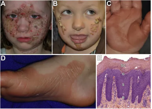
Figure 2. Clinical and Histologic PSEK Features Due toKDSRMutations
The face, palms, soles, and genitals are the most severely affected areas in all subjects. (A) Image of the face of subject 101 shows well-demarcated pink-red plaques, with overlying thick yellow-white scale, which are prominent on the cheeks, central fore-head, and neck. There are focal, denuded, red areas that showed no evidence of bacte-rial superinfection but could be the result of over-grooming.
(B) Subject 1107 has similar focal plaques with thick scale on the nose, cheeks, and chin.
(C and D) Images of the palm of subject 1107 (C) and sole of subject 101 (D) show erythematous palmoplantar hyperkeratosis with peeling scale.
and either DHS or its hydroxylated product PHS is acylated by ceramide synthases (CERS1–CERS6) for the generation of ceramides.8,9Ceramides are central to cutaneous barrier function and are secreted by keratinocytes with cholesterol and free fatty acids to form ordered lipid lamellae as cells transition from the stratum granulosum to the stratum corneum.10In addition to playing a role in barrier func-tion, ceramides regulate cutaneous proliferation and differ-entiation.11Recessive mutations inCERS3(MIM: 615276) cause ichthyosis characterized by encasement in a collo-dion membrane at birth, which sheds in infancy to reveal whole-body erythroderma and fine or coarse scaling.12,13 CERS3mutations have been found to alter the ceramide content of epidermis and to lead to defects in cutaneous differentiation.13
The domain structure of KDSR is homologous to that of other reductase enzymes (Figure 3B). The canonical TyrXXXLys reductase active site in KDSR is at amino acids 186–190. Conserved Asn and Ser residues at amino acids 145 and 173, respectively, form the canonical catalytic
triad within a larger hydrophilic domain that extends from residues 22 to 270. A structurally conserved NAD-binding Rossman fold (an alpha-beta fold with a central beta sheet) extends from the putative NAD binding site ThrGlyXXXGlyXGly at amino acids 38–45 to residue 222. There are predicted homodimer and homotetramer interfaces at amino acids 208–268 and 98–268, respec-tively.14 Immunofluorescence studies have shown that KDSR localizes to the endoplasmic reticulum (ER), and proteinase K digestion has demonstrated that the large hydrophilic domain faces the cytosol.14,15 There are putative transmembrane domains at residues 1–21, 271–291, and 294–314.14
The KDSR variants p.Val86_Gln107del and p.Gln260_ Gln293del most likely have profound effects on protein structure. Both affect residues within the hydrophilic enzy-matic domain. The former occurs within the NAD-binding domain and the homotetramer interface, and the latter is within both the homodimer and homotetramer interfaces and would omit one of the putative transmembrane do-mains, thereby affecting membrane topology at the C-ter-minal end of the protein. The p.Gln55_Gly56delinsArg variant alters highly conserved residues within the hydro-philic enzymatic domain. The p.Tyr186Phe variant affects the active-site tyrosine, which is completely conserved in both orthologs and paralogs and is the most conserved res-idue in reductase proteins (Figures 3B andS3). SIFT16and PolyPhen17predict that p.Tyr186Phe is highly damaging with scores of 0 and 1, respectively.
To investigate the consequence of KDSR mutations in the skin, we immunostained affected tissue from subject 1107 with antibodies to KDSR and to keratin 14 (KRT14, a marker of basal keratinocytes), keratin 10 (KRT10, a marker of suprabasal keratinocyte differentiation), and filaggrin (FLG, a marker of terminal differentiation in the granular layer of the epidermis). KDSR immunostaining intensity and localization were similar between normal tissue and affected tissue from this subject (Figures 4A and 4D). Although programs of epidermal differentiation can be grossly disrupted in some disorders of keratinization, including expansion of basal cell markers and loss of differ-entiation markers,18,19 affected tissue from subject 1107 showed basal distribution of KRT14 and suprabasal distribu-tion of KRT10, as was found in normal tissue (Figures 4B, 4C, 4F, and 4G). Notably, despite histologic absence of a granular layer (Figure 2E), affected tissue from subject 1107 showed expansion of filaggrin immunostaining (Figures 4D and 4H). Together, these results suggest a defect in keratinocyte terminal differentiation. Similar findings have been seen in tissue from affected individuals with widespread recessive congenital ichthyosis due toCERS3mutations, which also affect ceramide synthesis.12,13
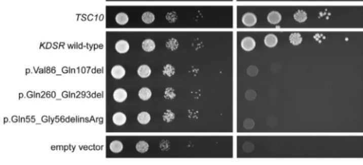
It has been previously shown that expression of wild-type humanKDSRrescues the growth defect in yeast null for the orthologousTSC10, which otherwise cannot grow without supplementation with KDSR product DHS or PHS.14To demonstrate the effect of theKDSRmutations Figure 3. KDSREncodes 3-Ketodihydrosphingosine Reductase,
an Enzyme in the Ceramide Synthesis Pathway
(A) The three major metabolic pathways capable of producing ce-ramides (the de novo, sphingomyelinase, and salvage pathways) are shown in purple, aqua, and brown, respectively. KDSR (in red) is critical to the creation of ceramides via de novo synthesis but is not intrinsic to the other two pathways.
observed in our subjects on the function of KDSR, we ex-pressed both wild-type and mutant forms of human
KDSR, including all of the mutant forms found in our initial two subjects, in yeast withTSC10 under a galac-tose-inducible and glucose-repressible conditional pro-moter. As expected, wild-type KDSR complemented the depletion of endogenous Tsc10, but the mutant forms of
KDSR failed to complement, demonstrating their detri-mental effect on KDSR function (Figures 5 and S4). We did not test the two other mutations identified. The inver-sion abolishes expresinver-sion of KDSR, and the active-site Tyr186 has been characterized in other studies. This site is crucial to reductase function in both KDSR and paralo-gous members of the large reductase family of enzymes. Tyr-to-Phe substitutions at this site in several other reduc-tases abolish enzymatic activity in vitro.23–25Additionally, a temperature-sensitive p.Tyr186Phe KDSR variant ex-pressed in Tsc10-null yeast failed to complement at 37C.15
In complex organisms, three major pathways are capable of producing ceramides (Figure 3A). The de novo pathway generates ceramides ‘‘from scratch’’ via smaller building-block molecules, whereas the other pathways release ce-ramides via breakdown of larger molecules, either sphingo-myelin from cell membranes (sphingosphingo-myelinase pathway) or sphingolipids that are converted to sphingosine and then acylated to form ceramides (salvage pathway).8,9,26 KDSR is critical to the creation of ceramides via de novo synthesis but is not intrinsic to the other two pathways.
Two of the probands (subjects 429 and 438) have been treated with a systemic retinoic acid derivative (isotreti-noin), which remarkably resulted in nearly complete resolution of their scale and erythema (Figure S5). A third subject (1107) has been advised to start systemic isotreti-noin therapy as a result of this work. Interestingly, retinoic acid has been shown to increase sphingosine acylation and to upregulate sphingomyelinase.27–29These effects serve to stimulate the ceramide salvage pathway and sphingomye-linase pathway, which produce ceramides independent of KDSR (Figure 3A). This suggests that retinoic acid therapy is effective, at least in part, by compensating for a genetic defect in the ceramide de novo synthesis pathway via pharmacologic induction of alternative pathways for cer-amide generation.
The identification of four unrelated probands compound heterozygous for previously unreported or extremely rare damaging mutations inKDSR, all of whom exhibit a unique skin disorder with well-demarcated symmetric scaling, erythema, and mild palmoplantar keratoderma, provides definitive evidence that recessive mutations inKDSRcause progressive symmetric erythrokeratoderma. This discovery underscores the central importance of ceramides in epithe-lial differentiation. The finding that systemic retinoids lead to nearly complete resolution of the skin findings in two subjects, most likely in part through activation of the salvage pathway for ceramide synthesis, provides an impor-tant therapeutic opportunity. Finally, RNA analysis and genome sequencing were essential to the detection of Figure 4. Filaggrin Distribution Is Expanded in Affected PSEK Tissue
Tissue from a normal donor and affected tissue from subject 1107 was immunostained with primary antibodies to KDSR, KRT14, KRT10, and FLG (Santa Cruz Biotechnology sc-366781, sc-53253, sc-53252, and sc-25897, respectively) and corresponding Cy2 (green) or Cy3 (red) secondary antibodies. DAPI was used as a nuclear counterstain (blue).
(A and E) Positive immunostaining for KDSR (green) was observed in all layers of the epidermis in normal (A) and affected (E) tissue. (B and F) Positive immunostaining for KRT14 (red) was limited to the basal layer of the epidermis in normal (B) and affected (F) tissue. (C and G) Positive immunostaining for KRT10 (red), a marker of epidermal differentiation, was limited to the suprabasal epidermis in normal (C) and affected (G) tissue. Normal tissue showed typical autofluorescence of the stratum corneum.
pathogenic mutations. Three of the four subjects are hetero-zygous for a silent third base change, for which cDNA sequencing was required to establish its damaging effect on the transcript, and two of the four are heterozygous for an inversion undetectable by exome sequencing. Conse-quently, at least one of the mutations in each of the four subjects would have been undetected by standard methods of exome sequencing and filtering. This work highlights the importance of comprehensive genetic evaluation in the study of severe, rare disorders such as erythrokeratoderma.
Supplemental Data
Supplemental Data include five figures and three tables and can be found with this article online athttp://dx.doi.org/10.1016/j.ajhg.
2017.05.003.
Acknowledgments
We thank the study subjects, their families, and the health-care professionals whose participation made this work possible. We also thank Erin Loring, Carol Nelson-Williams, Irina Tikhonova, Christopher Castaldi, Kaya Bilguvar, James Knight, and Bill Rizzo for technical assistance. This work was supported in part by a Clinician Scientist Development Award from the Doris Duke Char-itable Foundation to K.A.C., the Foundation for Ichthyosis and Related Skin Types, and the National Institutes of Health (R01 AR068392 to K.A.C., R01 GM115710 to S.J. Baserga, and the Yale Center for Mendelian Genomics U54 HG006504).
Received: February 24, 2017 Accepted: May 8, 2017 Published: June 1, 2017
Web Resources
1000 Genomes,http://www.internationalgenome.org/
ANNOVAR,http://annovar.openbioinformatics.org/en/latest/
BWA-MEM,http://bio-bwa.sourceforge.net/index.shtml
Database of Genomic Variants,http://dgv.tcag.ca/dgv/app/home
dbSNP,https://www.ncbi.nlm.nih.gov/projects/SNP/
Exome Aggregation Consortium (ExAC) Browser, http://exac. broadinstitute.org/
ExonPrimer, https://ihg.helmholtz-muenchen.de/ihg/ExonPrimer. html
GenBank,https://www.ncbi.nlm.nih.gov/genbank/
Genome Analysis Toolkit (GATK),https://software.broadinstitute. org/gatk/
Integrative Genomics Viewer (IGV),http://software.broadinstitute. org/software/igv/
OMIM,https://www.omim.org/
SNPmasker,http://bioinfo.ebc.ee/snpmasker/
UCSC Genome Browser,https://genome.ucsc.edu/index.html
Variant Effect Predictor,http://useast.ensembl.org/info/docs/tools/ vep/index.html
References
1. Richard, G., Smith, L.E., Bailey, R.A., Itin, P., Hohl, D., Epstein,
E.H., Jr., DiGiovanna, J.J., Compton, J.G., and Bale, S.J. (1998). Mutations in the human connexin gene GJB3 cause
erythro-keratodermia variabilis. Nat. Genet.20, 366–369.
2. Macari, F., Landau, M., Cousin, P., Mevorah, B., Brenner, S.,
Panizzon, R., Schorderet, D.F., Hohl, D., and Huber, M. (2000). Mutation in the gene for connexin 30.3 in a family
with erythrokeratodermia variabilis. Am. J. Hum. Genet.67,
1296–1301.
3. Boyden, L.M., Craiglow, B.G., Zhou, J., Hu, R., Loring, E.C.,
Morel, K.D., Lauren, C.T., Lifton, R.P., Bilguvar, K., Paller, A.S., Choate, K.A.; and Yale Center for Mendelian Genomics (2015). Dominant De Novo Mutations in GJA1 Cause Erythro-keratodermia Variabilis et Progressiva, without Features of
Ocu-lodentodigital Dysplasia. J. Invest. Dermatol.135, 1540–1547.
4. Maestrini, E., Monaco, A.P., McGrath, J.A., Ishida-Yamamoto,
A., Camisa, C., Hovnanian, A., Weeks, D.E., Lathrop, M., Uitto, J., and Christiano, A.M. (1996). A molecular defect in loricrin, the major component of the cornified cell envelope,
underlies Vohwinkel’s syndrome. Nat. Genet.13, 70–77.
5. Boyden, L.M., Kam, C.Y., Herna´ndez-Martı´n, A., Zhou, J.,
Crai-glow, B.G., Sidbury, R., Mathes, E.F., Maguiness, S.M., Crumr-ine, D.A., Williams, M.L., et al. (2016). Dominant de novo DSP mutations cause erythrokeratodermia-cardiomyopathy
syn-drome. Hum. Mol. Genet.25, 348–357.
6. Zhang, M.Q. (1998). Statistical features of human exons and
their flanking regions. Hum. Mol. Genet.7, 919–932.
7. Robinson, J.T., Thorvaldsdo´ttir, H., Winckler, W., Guttman,
M., Lander, E.S., Getz, G., and Mesirov, J.P. (2011). Integrative
genomics viewer. Nat. Biotechnol.29, 24–26.
8. Hannun, Y.A., and Obeid, L.M. (2008). Principles of bioactive
lipid signalling: lessons from sphingolipids. Nat. Rev. Mol.
Cell Biol.9, 139–150.
9. Kihara, A. (2016). Synthesis and degradation pathways,
func-tions, and pathology of ceramides and epidermal
acylcera-mides. Prog. Lipid Res.63, 50–69.
10. Borodzicz, S., Rudnicka, L., Mirowska-Guzel, D., and
Cud-noch-Jedrzejewska, A. (2016). The role of epidermal
sphingo-lipids in dermatologic diseases. Lipids Health Dis.15, 13.
11. Uchida, Y. (2014). Ceramide signaling in mammalian epidermis.
Biochim. Biophys. Acta1841, 453–462.
Figure 5. MutantKDSR Does Not Complement Depletion of Endogenous YeastTSC10
A yeast strain with galactose-inducible and glucose-repressible expression of the KDSR orthologTSC10 was created,20–22 and
12. Eckl, K.M., Tidhar, R., Thiele, H., Oji, V., Hausser, I., Brodesser,
S., Preil, M.L., Onal-Akan, A., Stock, F., Mu¨ller, D., et al. (2013).
Impaired epidermal ceramide synthesis causes autosomal recessive congenital ichthyosis and reveals the importance
of ceramide acyl chain length. J. Invest. Dermatol. 133,
2202–2211.
13. Radner, F.P., Marrakchi, S., Kirchmeier, P., Kim, G.J., Ribierre, F.,
Kamoun, B., Abid, L., Leipoldt, M., Turki, H., Schempp, W., et al. (2013). Mutations in CERS3 cause autosomal recessive
congenital ichthyosis in humans. PLoS Genet.9, e1003536.
14. Kihara, A., and Igarashi, Y. (2004). FVT-1 is a mammalian
3-ke-todihydrosphingosine reductase with an active site that faces the cytosolic side of the endoplasmic reticulum membrane.
J. Biol. Chem.279, 49243–49250.
15. Gupta, S.D., Gable, K., Han, G., Borovitskaya, A., Selby, L.,
Dunn, T.M., and Harmon, J.M. (2009). Tsc10p and FVT1: to-pologically distinct short-chain reductases required for
long-chain base synthesis in yeast and mammals. J. Lipid Res.50,
1630–1640.
16. Sim, N.L., Kumar, P., Hu, J., Henikoff, S., Schneider, G., and
Ng, P.C. (2012). SIFT web server: predicting effects of amino
acid substitutions on proteins. Nucleic Acids Res.40, W452–
W457.
17. Adzhubei, I.A., Schmidt, S., Peshkin, L., Ramensky, V.E.,
Gera-simova, A., Bork, P., Kondrashov, A.S., and Sunyaev, S.R. (2010). A method and server for predicting damaging
missense mutations. Nat. Methods7, 248–249.
18. McAleer, M.A., Pohler, E., Smith, F.J., Wilson, N.J., Cole, C.,
MacGowan, S., Koetsier, J.L., Godsel, L.M., Harmon, R.M., Gruber, R., et al. (2015). Severe dermatitis, multiple allergies, and metabolic wasting syndrome caused by a novel mutation in the N-terminal plakin domain of desmoplakin. J. Allergy
Clin. Immunol.136, 1268–1276.
19. Mirza, H., Kumar, A., Craiglow, B.G., Zhou, J., Saraceni, C.,
Torbeck, R., Ragsdale, B., Rehder, P., Ranki, A., and Choate, K.A. (2015). Mutations Affecting Keratin 10 Surface-Exposed Residues Highlight the Structural Basis of Phenotypic
Varia-tion in Epidermolytic Ichthyosis. J. Invest. Dermatol.135,
3041–3050.
20. Mumberg, D., Mu¨ller, R., and Funk, M. (1995). Yeast vectors
for the controlled expression of heterologous proteins in
different genetic backgrounds. Gene156, 119–122.
21. Sikorski, R.S., and Hieter, P. (1989). A system of shuttle vectors
and yeast host strains designed for efficient manipulation of
DNA in Saccharomyces cerevisiae. Genetics122, 19–27.
22. Longtine, M.S., McKenzie, A., 3rd, Demarini, D.J., Shah, N.G.,
Wach, A., Brachat, A., Philippsen, P., and Pringle, J.R. (1998). Additional modules for versatile and economical PCR-based gene deletion and modification in Saccharomyces cerevisiae.
Yeast14, 953–961.
23. Albalat, R., Gonza´lez-Duarte, and Atrian, S. (1992). Protein
en-gineering of Drosophila alcohol dehydrogenase. The hydroxyl group of Tyr152 is involved in the active site of the enzyme.
FEBS Lett.308, 235–239.
24. Obeid, J., and White, P.C. (1992). Tyr-179 and Lys-183
are essential for enzymatic activity of 11 beta-hydroxyste-roid dehydrogenase. Biochem. Biophys. Res. Commun.
188, 222–227.
25. Ensor, C.M., and Tai, H.H. (1994). Bacterial expression and
site-directed mutagenesis of two critical residues
(tyrosine-151 and lysine-155) of human placental NAD(þ)-dependent
15-hydroxyprostaglandin dehydrogenase. Biochim. Biophys.
Acta1208, 151–156.
26. Kitatani, K., Idkowiak-Baldys, J., and Hannun, Y.A. (2008). The
sphingolipid salvage pathway in ceramide metabolism and
signaling. Cell. Signal.20, 1010–1018.
27. Kale´n, A., Borchardt, R.A., and Bell, R.M. (1992). Elevated
cer-amide levels in GH4C1 cells treated with retinoic acid.
Bio-chim. Biophys. Acta1125, 90–96.
28. Riboni, L., Prinetti, A., Bassi, R., Caminiti, A., and Tettamanti,
G. (1995). A mediator role of ceramide in the regulation of neuroblastoma Neuro2a cell differentiation. J. Biol. Chem.
270, 26868–26875.
29. Kraveka, J.M., Li, L., Bielawski, J., Obeid, L.M., and Ogretmen,
B. (2003). Involvement of endogenous ceramide in the inhibi-tion of telomerase activity and inducinhibi-tion of morphologic dif-ferentiation in response to all-trans-retinoic acid in human