CONVERGENCE OF ABERRANT ELECTROPHYSIOLOGICAL CORRELATES OF SALIENCE, AFFECTIVE PROCESSING AND STRESS REACTIVITY IN PATIENTS WITH SCHIZOPHRENIA
Elizabeth H. Andersen
A dissertation submitted to the faculty at the University of North Carolina at Chapel Hill in partial fulfillment of the requirements for the degree of Doctor of Philosophy in the Curriculum of Neurobiology in the
School of Medicine.
Chapel Hill 2017
© 2017 Elizabeth H. Andersen ALL RIGHTS RESERVED
ABSTRACT
Elizabeth H. Andersen: Convergence of Aberrant Electrophysiological Correlates of Salience, Affective Processing and Stress Reactivity in Patients with Schizophrenia.
(Under the direction of Aysenil Belger)
Patients with schizophrenia exhibit debilitating deficits in attention and affective processing, which are often resistant to treatment and associated with poor functional outcomes. Attentional and affective processing relies on a distributed neural network of fronto-limbic circuits, which enable cognitive control and affective processing, and assist in their interaction to regulate emotional responses. Despite evidence of intact affective valence processing, schizophrenia patients are often unable to employ cognitive change strategies to reduce attentional capture by emotionally salient stimuli, or modulate neurophysiological responses to aversive stimuli. Aberrant neurophysiological correlates of orienting to task-relevant emotional stimuli are also present in unaffected first-degree relatives of schizophrenia patients, suggesting they may represent vulnerability markers. However, less is known about the attentional processing of emotionally salient, task-irrelevant information in these groups, which is examined in Experiment 1 (Chapter 2). Results suggest that despite intact novelty detection, schizophrenia patients and relatives shared deficiencies in attentional processing of emotionally salient information. First-degree relatives exhibited a unique enhancement of the electrophysiological correlate underlying salience evaluation, possibly indicating a compensatory engagement of neural circuitry.
demonstrated aberrant fronto-limbic oscillatory indices of affective processing, as indicated by
exaggerated neural excitability and inefficient frontal cognitive control, and maladaptive stress function. This imbalance between heightened neural responsivity and inefficient frontal regulation may reflect an atypical arousal state that may in turn interfere with fronto-limbic processing and promote
ACKNOWLEDGEMENTS
This dissertation would not have been possible without the unwavering support I have received from my family, friends, colleagues, and mentorship team. I am incredibly grateful for the graduate education and training that I have received at the University of North Carolina under the mentorship of Dr. Aysenil Belger. The curriculum in neurobiology offered me a unique opportunity to interact, research and learn amongst distinguished neuroscience researchers, while establishing an essential neurobiological foundation for comprehending and appreciating the elaborate construction of neural networks. Dr. Belger was my top choice PI that I wanted to work for out of every school I applied to across the country and I am so grateful that she accepted me into her lab. Even after almost 6 years in the Belger lab, I am still amazed and impressed by Dr. Belger’s expansive knowledge of the human brain and the neurobiological correlates of neuropsychiatric disorders and her passion for sharing her expertise with those around her. Dr. Belger has been an outstanding mentor over the last five and a half years. She has always been patient and encouraging, and my two children were immediately welcomed into the lab and grew to love their extended lab family. Payton helped me with presentations and Keaton was always the first to smile when we started the “data blitz”. I recognize that this was a unique arrangement and I am thankful that Dr. Belger was so understanding and supportive.
their friendship and support. Furthermore, I would like to acknowledge my high school anatomy teacher, Mr. Bradley, for igniting my initial interest in science and for believing I could succeed at it.
The Belger Lab has been a welcoming, supportive, collaborative and stimulating environment and I have enjoyed getting to know all of the amazing people that are here now and have passed through. Mariko Weber and Joe Shaffer were imperative to my success early in my graduate career. They were always there for me to offer advice or just listen. We traveled to conferences together and became great friends. I am forever grateful for their friendship and support. Franc Donkers, Carolyn Bellion and Anna Evans helped to smooth my transition to graduate school and I am thankful for everything they showed me in maneuvering the lab and providing my foundation for the next 5 years. Alana Campbell has been there for me since my undergraduate research experience and has been an excellent mentor and friend over the last ten years. She always knows how to cheer me up and motivate me; if it’s not chocolate then the “data dance.” Sarah Schipul was always the first to respond to emails, requests, manuscript edits, and her upbeat spirit was contagious throughout the lab. I have had the opportunity to work with an amazing research team, including Hannah, Candace and Mae. Hannah keeps the lab cheerful and productive with her sense of humor and amazing baking skills. Furthermore, I am thankful for my students and assistants, including Ally, Kelly, Erin, Jesse and Kohrissa. They never complained about the tedious tasks that made the research possible.
I am grateful for Dr. Karen Graham’s recruitment efforts, and I would not be here today if it were not for her assistance in getting patient volunteers. Jennifer Nieri provided fundamental training and guidance on conducting the clinical interviews. Maria Davila and Greg Lewis offered their expertise in collecting and analyzing the physiological stress measures, and became good friends. I appreciate all of the schizophrenia patients and healthy volunteers who went through five hours of grueling interviews and assessments, viewed disturbing images and suffered through a stressor for the research cause. I enjoyed interacting with all of the participants and feel that I can truly appreciate the heterogeneity of
schizophrenia and the debilitating symptoms that often distort their reality, interfere with living independently and prevent them from experiencing personal relationships.
my research endeavors. I am forever grateful for my parents, George and Mary, my sister, Lindsay, my husband, Eric, and my children, Payton and Keaton for loving me through the difficult times, picking me up when I needed a little help, and being understanding when I was absorbed in graduate school obligations. I am thankful for my uncle Tom, who has schizophrenia, for giving my research profound meaning and relevance. My childhood friend, Mia, has also offered meaningful support and friendship every step of the way.
PREFACE
Chapter 1
Chapter 1 is an introduction to the topics of cognitive and affective salience processing, fronto-limbic-mediated stress and emotion regulation, the interaction between stress and neurophysiological correlates of affective processing, and how these processes are disrupted in schizophrenia. It provides the background and justification for the two projects presented in Chapters 2 and 3.
Chapter 2
Chapter 2.2 was published in the Clinical EEG and Neuroscience journal (Andersen et al., 2016), and formatted to meet the editing standards while keeping the integrity of the manuscript intact, per the request of Sage Publishing. In addition to performing the analyses and interpreting the results, I drafted the manuscript and critically revised the manuscript with help from the other authors.
Chapter 3
Chapter 3 is a manuscript in preparation. I administered the clinical and neurocognitive interviews and assessments, collected the EEG and ECG data, and performed the analyses.
Chapter 4
Chapter 4 is the Conclusions chapter, where I summarize important findings, discuss implications and significance, and propose future directions.
TABLE OF CONTENTS
LIST OF TABLES ... xiii
LIST OF FIGURES ... xiv
LIST OF ABBREVIATIONS ... xv
CHAPTER 1: INTRODUCTION ... 1
1.1 Clinical Overview of Schizophrenia ... 1
Introduction ... 1
Neurobiology of Schizophrenia and Potential Risk Factors ... 1
Disparities in Treatment Efficacy and Symptom Management ... 5
Prognosis and Quality of Life ... 7
1.2 Schizophrenia as a Dysregulated Salience Syndrome ... 7
Introduction ... 7
Neural Indices of Salience and Affective Processing ... 8
Electrophysiological Studies of Salience and Affective Processing ... 11
Salience Disturbance and Cognitive/Behavioral Consequences ... 14
1.3 Fronto-Limbic Circuitry Underlying the Relationship Between Stress and Affective Processing in Schizophrenia ... 15
Introduction ... 15
Overview of the Stress Response ... 16
Studying the Stress Response in Patients ... 19
Stress Cascade and the Affective Pathway to Psychosis ... 22
Stress Influences on Neurophysiology ... 23
Interaction of Stress and Affective Processing ... 24
2.1 Context ... 26
2.2 Electrophysiological Correlates of Aberrant Motivated Attention and Salience Processing in Unaffected Relatives of Schizophrenia Patients ... 27
Introduction ... 27
Method ... 31
Results ... 34
Discussion ... 37
2.3 Future directions ... 41
2.4 Significance ... 42
Chapter 2.2 Tables ... 43
Chapter 2.2 Figures ... 46
Chapter 2.3 Figures ... 49
CHAPTER 3: STRESS EFFECTS ON EEG CORRELATES OF AFFECTIVE PROCESSING ... 51
3.1 Context ... 51
3.2 Stress Modifies the Electrophysiological Correlates of Affective Processing In Patients with Schizophrenia and Healthy Controls ... 51
Introduction ... 51
Method ... 57
Results ... 65
Discussion ... 70
3.3 Future Directions ... 79
3.4 Significance ... 81
Chapter 3.2 Tables ... 82
Chapter 3.2 Figures ... 85
CHAPTER 4: CONCLUSIONS ... 98
4.1 Support of Schizophrenia as a Salience Syndrome ... 98
4.2 Insights into Fronto-Limbic–Dependent Affective Processing Impairments in Patients with Schizophrenia ... 99
4.4 Disruption of Fronto-Limbic Oscillatory Indices of Salience and
LIST OF TABLES
Table 2.1. Demographics ... 43
Table 2.2. Results for Oddball Detection Paradigm ... 44
Table 3.1. Demographic and Clinical Characteristics of Study Groups ... 82
Table 3.2. Neurocognitive Assessments and Questionnaires ... 83
LIST OF FIGURES
Figure 2.1. Novelty Detection of Novel Distractor Stimuli and Directed Attention to Targets ... 46
Figure 2.2. Late Positive Potential (LPP) Underlying Motivated Attention and Salience Processing ... 47
Figure 2.3. Relationship between SOPS, Neurocognitive Assessments, and Oddball ERP Amplitudes ... 48
Figure 2.4. Enhanced Beta Activity in FDR in Response to Aversive Stimuli ... 49
Figure 2.5. Disrupted Neural Oscillatory Activity in SCZ and FDR ... 50
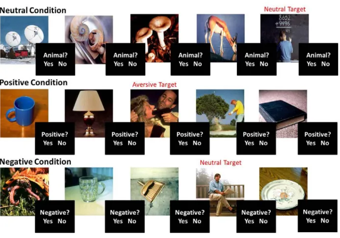
Figure 3.1. Emotional Oddball Framing Paradigm ... 85
Figure 3.2. Diagram of Study Design ... 86
Figure 3.3. Summary of Neurocognitive and Clinical Measures ... 87
Figure 3.4. Electrode Map of Parietal Montage ... 88
Figure 3.5. Subjective Stress and Affect Ratings ... 89
Figure 3.6. Physiological Measures of Stress ... 90
Figure 3.7. Valence Discrimination During Neutral (Animal) Framing Condition ... 91
Figure 3.8. ERP Amplitudes Elicited During the Different Framing Conditions Before and After Stress.... 92
Figure 3.9. Early Frontal Theta Activity Before Stress ... 93
Figure 3.10. Stress Reactivity Induced Changes in Early Frontal Theta ERSP ... 94
Figure 3.11. Late Parietal Beta Activity Before Stress ... 95
Figure 3.12. Changes in Late Parietal Beta Activity Following Stress ... 96
LIST OF ABBREVIATIONS
5HT2A 5-hydroxytryptamine 2A serotonin receptor
ACTH adrenocorticotropic hormone
AIC anterior insular cortex ANOVA analysis of variance
ANS autonomic nervous system
AVLT Auditory Verbal Learning Test
BCAET Baron-Cohen “Reading the Mind in the Eyes” Test–Revised
BNM brain network modulation
BNST bed nucleus of the stria terminalis
BPM beats per minute
CAINS Clinical Assessment Interview for Negative Symptoms
CAN central autonomic network
CBT cognitive behavioral therapy
CEN central executive network
CPT-IP Continuous Performance Test-Identical Pairs CRH corticotropin-releasing hormone
CSF cerebral spinal fluid
CT computed tomography
D2 D2 subtype of dopamine receptor dACC dorsal anterior cingulate cortex
DKEFS-CWIT Delis-Kaplan Executive Function System–Color-Word Interference Test DLPFC dorsal lateral prefrontal cortex
DMN default mode network
DSI Daily Stress Inventory
ECG electrocardiogram
EEG electroencephalography
EP evoked power
ER emotion regulation
ERD event-related desynchronization
ERP event-related potential
ERQ emotion regulation questionnaire ERSP event-related spectral perturbation
FA fractional anisotrophy
FDR first-degree relative
FEIT Facial Emotion Identification Test
FRN feedback-related negativity ERP component
GABA gamma-Aminobutyric acid
GR glucocorticoid receptors
GWAS genome-wide association study H-MRS proton magnetic resonance imaging
HP heart period
HPA hypothalamic-pituitary-adrenocortical axis
HR heart rate
HRV heart rate variability
IAPS International Affective Picture System ITC inter-trial phase coherence LPP late positive potential
LTP long-term potentiation
MEG magnetoencephalography
mGluRs metabotropic glutamate receptors MIST Montreal Imaging Stress Test
MRI functional magnetic resonance imaging NAART North American Adult Reading Test
NMDA N-methyl-D-aspartate
PANAS Positive and Negative Affect Schedule PANSS Positive and Negative Syndrome Scale
PCP phencyclidine
PET positron emission tomography
PFC prefrontal cortex
PSS Perceived Stress Scale
PTSD post-traumatic stress disorder PVN paraventricular nucleus of the hypothalamus RDoC Research Domain Criteria Initiative
RSA respiratory sinus arrhythmia
RT reaction time
sAA salivary alpha amylase
SAM self-assessment manikin
SCID structured clinical interview for DSM
SCI-PANSS Structured Interview for Positive and Negative Syndrome Scale
sCORT salivary cortisol
SCZ schizophrenia
SES socioeconomic status
SNPs single nucleotide polymorphisms SOPS Scale of Prodromal Symptoms
SSR subjective stress and affect rating TSST Trier Social Stress Test
CHAPTER 1: INTRODUCTION
1.1 Clinical Overview of Schizophrenia
Introduction
Schizophrenia is a complex heterogeneous disorder with dynamic symptom manifestations spanning cognitive, psychological and behavioral processing domains. As the most common type of psychosis, schizophrenia affects twenty-one million people worldwide (World Health Organization [WHO]) and represents an overwhelming economic burden on society (Chaiyakunapruk et al., 2016). It is
recognized as a chronic and severe mental illness, characterized by debilitating disruptions in disorganized thinking and behavior, distortions of reality, and profound deviations in cognitive and
affective function (American Psychiatric Association, 2013). Patients experience periods of remission and relapse throughout their lifetime, with some individuals experiencing intermittent symptoms and some following a course of progressive deterioration. The Diagnostic and Statistical Manual of Mental
Disorders, 5th Edition (DSM-V) criteria for a schizophrenia diagnosis involves persistent symptoms in two of five symptom domains, including delusions, hallucinations, disorganized speech, disorganized behavior and negative symptoms. Patients diagnosed with schizoaffective disorder experience depression or bipolar symptoms in parallel with schizophrenia symptoms, while a schizophreniform diagnosis is dependent on symptoms lasting less than six months. Symptoms of schizophrenia typically emerge during young adulthood (18-35), with approximately 1.4:1 incidence ratio for males and females and a female dominated older onset (Abel et al., 2010). While female schizophrenia patients express greater depression symptoms, males typically experience heightened negative symptoms contributing to a more severe course of illness.
Neurobiology of Schizophrenia and Potential Risk Factors
of neural circuitry (Garey, 2010; Penzes et al., 2011). The cortical deterioration observed in schizophrenia is accompanied by a collection of symptoms encompassing an extensive range of behavioral and
cognitive abnormalities. Nevertheless, schizophrenia is first and foremost an information processing disorder in which patients experience a notable decline in numerous domains of cognition (Aas et al., 2014), including attention (Cornblatt and Erlenmeyer-Kimling, 1985), working memory (Manoach, 2003), response selection and inhibition (Kiehl et al., 2000), and attribution of salience (Kapur, 2003).
Schizophrenia patients typically experience their first psychotic episode during late adolescence or early adulthood, consistent with the significant neural network refinement that occurs during this period (Penzes et al., 2011). As a neurodevelopmental disorder, environmental factors during fetal life are proposed to modify neurodevelopmental trajectories and promote the manifestation of symptoms during adolescence and adulthood (Wójciak et al., 2016). The following sections summarize the pertinent neural mechanisms promoting the pathophysiology of schizophrenia, including genetic and environmental interactions, disruptions in neurotransmitter transmission, and structural and functional brain abnormalities.
Environment
A convergence of environment and genetic influences is thought to promote vulnerability for psychosis (Caspi and Moffitt, 2006). Early developmental insults, including infections in utero (i.e., rubella, influenza, and poliovirus) (Brown and Susser, 2002) and increased prenatal stress, may disrupt the maternal cytokine response leading to obstetric complications in the mother (Patterson, 2002), which are thought to predispose vulnerability for schizophrenia (Ross et al., 2006). These early neurodevelopmental lesions may establish vulnerability to atypical late neurodevelopmental processes which interact with environmental factors, including social adversity accompanying immigration (Weiser et al., 2008), urbanicity (Krabbendam, 2005), season of birth (Mortensen et al., 1999), and exposure to psychoactive substances such as cannabis (Arseneault et al., 2002), to promote the emergence of psychosis (Pantelis, 2005). While numerous environmental influences have been proposed to contribute to the manifestation of schizophrenia, the validity of the claims remains inconclusive. Nevertheless, genetic and environmental influences likely play a synergistic role in promoting disease onset through the genetic control of
Stress aggravates synaptic pathologies observed in schizophrenia and is considered a significant environmental trigger in the etiology of schizophrenia (Corcoran et al., 2003). A more in depth description of the stress system and implications of stress in psychosis can be found in section 1.3. Briefly, the cascade of events following stress exposure stimulates structural changes in the brain, including reduced dendritic arborization and spine density (Radley, 2005), and exposure to repeated, chronic stress
associated with extreme life events, such as childhood trauma, may provoke the development of psychotic symptoms (Liston et al., 2009). Stress interferes with already vulnerable circuitry in
schizophrenia patients to intensify cortical atrophy and threaten neural network integrity (Arnsten, 2011). Because prefrontal cortical circuits are disproportionately affected by the stress-induced cortical
disturbances, processes governed by the prefrontal cortex, including working memory, attentional control, and behavioral flexibility are particularly susceptible to disruption in patients (Arnsten, 2009, 2011). Given the substantial neural modifications and disruptions in cognitive processes inflicted by stress exposure, it is not surprising that stress is considered a precipitating factor for psychosis in vulnerable individuals.
Disruptions in Neurotransmitter Transmission
Hyperdopaminergic transmission is thought to play a pivotal role in the etiology of schizophrenia and is considered one of the most prevailing theories in psychiatry (Howes and Kapur, 2009). The dopamine hypothesis of schizophrenia emerged concurrently with the introduction of the conventional antipsychotic drugs which target the D2 dopamine receptor subtype and were found to alleviate positive symptoms. Moreover, psychostimulants that trigger dopamine release cause psychosis symptoms or exacerbate symptoms in patients (Ross et al., 2006). Studies implementing positron emission tomography (PET) report that patients exhibit presynaptic dysregulation of dopamine, with increased dopamine release in response to impulse and elevated dopamine synthesis, and confirm that the hyperdopaminergic state is associated with psychotic symptoms in patients (Kapur, 2003; Soares and Innis, 1999). Furthermore, genome-wide association studies (GWAS) have identified the D2 receptor gene (DRD2) as a potential candidate gene of interest in schizophrenia, further signifying a fundamental role of dopamine in the pathophysiology of schizophrenia.
small piece of the puzzle. In addition to atypical dopamine activity, disruptions in glutamatergic
transmission may underlie prominent symptoms of schizophrenia. Proton magnetic resonance imaging (H-MRS) has permitted the study of glutamate function in vivo in schizophrenia patients by measuring glutamate and its metabolites (Poels et al., 2014). Aberrant glutamate activity and hypofunction at N-methyl-D-aspartate (NMDA) and metabotropic glutamate receptors (mGluRs) resulting in elevated glutamate may also contribute to the pathophysiology of schizophrenia, as phencyclidine (PCP) and ketamine block NMDA receptors and produce schizophrenia-like psychotic symptoms (Coyle, 2006).
Furthermore, while deficiencies in inhibitory gamma-Aminobutyric acid (GABA) neurotransmission have been associated with cognitive dysfunction and have a proposed role in dopamine regulation, the influence of GABA in the neuropathology of schizophrenia remains controversial (Chiapponi et al., 2016). The integration and synergistic interaction of neurotransmitter systems underlies the neurophysiology of schizophrenia and motivates a circuit based framework, systems-level approach to understanding schizophrenia (Lisman et al., 2008).
Structural Brain Abnormalities in Schizophrenia
Global deficits in structural brain morphology have been reported in schizophrenia. Early
structural MRI and computed tomography (CT) studies of schizophrenia confirmed post-mortem findings of a widening of lateral and third ventricles in patients, though these findings are not specific to
schizophrenia (Ross et al., 2006). Cortical atrophy is consistently observed in schizophrenia patients, with dramatic reductions in frontal lobe, amygdala, insula, thalamus and hippocampal gray matter volume (Wójciak et al., 2016), and gray matter may decline with illness progression (Haijma et al., 2013). Furthermore, a disproportionate deterioration of frontal neural integrity, particularly the dorsal lateral prefrontal cortex (DLPFC), and frontal network architecture is thought to underlie devastating
psychological, cognitive and behavioral symptom pathology in schizophrenia patients (Arnsten, 2011; Weinberger, 1987).
Schizophrenia as a Disconnection Syndrome
elucidating the compromised structural and functional integrity of neural network construction subserving schizophrenia. Anatomical connectivity can be assessed using diffusion tensor imaging (DTI), which concentrates on the fractional anisotrophy (FA) metric to evaluate microstructural properties of white matter tracts. DTI studies have revealed that patients with schizophrenia demonstrate reduced FA in frontal and temporal lobes, along with aberrations within the fiber bundles connecting these regions (Wheeler and Voineskos, 2014), suggesting alterations in structural connectivity in schizophrenia.
In addition to structural connectivity assessments using DTI, functional MRI studies are useful for investigating functional connectivity among brain regions by assessing synchronized blood oxygenation levels, an indirect measure of functional integration. The inefficiency of network connectivity and functional integration, especially between frontotemporal brain regions, is a fundamental feature of schizophrenia, with hypo and hyper task-related connectivity reported (Friston, 2011; Lynall et al., 2010). Aberrant activity in the default mode network (DMN), which is active during rest and suppressed during attention-demanding tasks, is one of the most robust findings of network disruptions in schizophrenia, and impaired DMN suppression is related to abnormalities in neural networks associated with language, attention, and working memory (Fitzsimmons et al., 2013; Garrity et al., 2007).
Along with neuroimaging studies, EEG measures of oscillatory activity offer valuable insight into the integrity of functional networks, as the integration of information among distributed brain regions and efficiency of connections relies on oscillations and their synchronization. Oscillatory indices of cognitive and affective processing are disrupted in patients with schizophrenia, and provide additional evidence of deficient network activity and dysconnectivity. Measurement and interpretation of neural oscillatory activity is described in more detail in section 1.2.
Disparities in Treatment Efficacy and Symptom Management
The combination of pharmacotherapy, along with psychosocial and psychotherapeutic interventions, is often the most effective method to ameliorate symptoms of schizophrenia. However, pharmacological agents have only progressed modestly since the first antipsychotic medication was introduced 60 years ago, and offer minimal symptom relief with considerable adverse side-effects (Bruijnzeel et al., 2014; Ross et al., 2006). Consequently, advancing the understanding of the
anti-psychotic medications targeting D2 dopamine receptors are typically effective at dampening positive and disorganization symptoms, the side-effects are sometimes intolerable and lead to reduced medication compliance. Furthermore, these first-generation drugs, including chlorpromazine and haloperidol, introduce devastating neurological side effects such as tremor, rigidity, dystonia and dyskinesia, along with metabolic and gastrointestinal adverse effects. In addition to targeting the D2 dopamine receptors, second generation antipsychotics (atypical), including clozapine and olanzapine, block serotonin 5-hydroxytryptamine 2A (5HT2A) receptors and have demonstrated efficacy at minimizing the neurologic side effects; however, they are not free of adverse side-effects and are associated with substantial weight gain and risk for type-II diabetes (Bruijnzeel et al., 2014). With the exception of clozapine, which has been shown to be more effective in reducing negative symptoms, there are no major differences in efficacy between first and second generation antipsychotics.
Negative Symptoms and Treatment Considerations
pharmacological treatment progression and only partially effective and intolerable medications available, identifying novel endophenotypes and treatment targets is critical.
Prognosis and Quality of Life
Schizophrenia is a chronic debilitating disorder that has profound psychological, behavioral, emotional and financial impact on the individual and family. Cognitive and negative symptoms, along with poor hygiene and low education levels, interfere with patients’ ability to live productive and meaningful lives and often promote occupational and social dysfunction (Bobes et al., 2007). Patients tend to self-medicate with drugs and alcohol, which along with comorbid medical conditions including obesity, diabetes, hypertension and coronary heart disease, contribute to an abbreviated life span. Extensive support and supervised housing is required, adding to the financial burden on the patients and their families (Kitchen et al., 2012). With this dismal prognosis, advancing the understanding of the pathophysiological mechanisms promoting vulnerability and neurobiological correlates underlying
schizophrenia is crucial for devising more effective treatment approaches and improving the quality of life for patients with schizophrenia.
1.2 Schizophrenia as a Dysregulated Salience Syndrome
Introduction
Salience processing involves the reactive cognitive elaboration of unique stimuli or stimulus features which facilitates directed attention and goal-oriented motivational behaviors (Kapur, 2003). The convergence of psychological state, previous experiences, goals, and autonomic and homeostatic functions of the brain influence the perception of a stimulus as “salient”. The emotional response to novel salient stimuli is context dependent and malleable in order to adapt to situational demands. Inappropriate attribution of salience and aberrant perceptual integration are proposed to subserve clinically relevant domains of cognitive and affective processing.
reactions to evocative stimuli in laboratory settings (Cohen and Minor, 2008; Horan et al., 2010; Kring and Moran, 2008; Strauss and Gold, 2012). Nevertheless, patients have difficultly disengaging attention from unpleasant environmental cues, demonstrate exaggerated devotion of attention to negative stimuli, and exhibit both hedonic and aversive emotions when exposed to positive and neutral stimuli (Cohen and Minor, 2008). The inappropriate salience attribution, impaired sensory filtering and elevated reactivity to irrelevant neutral information observed in patients (Anticevic and Corlett, 2012) may promote disruptions in response selection and regulating negative emotional responses, further exasperating negative symptom severity (Strauss et al., 2011).
Aberrant salience impacts the development and maintenance of psychotic symptoms through insula dysfunction and atypical dopaminergic transmission. Appropriate salience attribution is critical for the selection of task-relevant information, and involuntary orientation to salient, motivationally-relevant and potentially significant stimuli in the sensory environment. Disruptions in salience attribution interfere with attentional and cognitive processing of emotional information, suggesting that abnormalities in affective processing and regulation may revolve around a central deficit in salience processing. The following sections describe the neurobiological correlates of salience processing, including a discussion of the insula-centered salience network, dopamine transmission, and how these systems are disrupted in psychosis. In addition, the utility of electroencephalography (EEG) in elucidating salience and affective processing disparities in patients with schizophrenia is discussed.
Neural Indices of Salience and Affective Processing
optimize behavior. The down-regulation of the emotional response can be achieved by implementing antecedent-focused strategies, such as cognitive reappraisal, or response-focused strategies, including expressive suppression (Goldin et al., 2008). Antecedent-focused strategies involve modifying internal (e.g., increasing or decreasing certain thoughts) or external (e.g., physical environment where emotions are more likely to occur) emotion eliciting factors prior to the emotion being experienced, and by changing the way evocative stimuli are appraised (i.e., directing attention to certain features of the environment, or framing the environment in a positive or negative emotional context) (Gross and Muñoz, 1995). While antecedent-focused strategies can manipulate both the experience and expression of emotion, response-focused strategies, such as masking sadness with a smile, only impact the emotional expression, and are therefore less effective in generating ideal emotional outcomes (Livingstone et al., 2009).
The successful implementation of emotion regulation strategies requires the enhanced
Salience Network
The neural processing of salient information synthesizes external sensory and internal emotional and visceral state information to motivate attention and behavioral outcomes, and relies on the integration of information among distributed regions, comprising a salience network. This salience neural network revolves around the insular cortex, along with involvement of the dorsal anterior cingulate cortex (dACC), subcortical, and limbic brain regions (Uddin, 2015). The insular cortex is the first cortical target of
interoceptive and visceromotor inputs, with multiple functionally distinct regions, and plays an integral role in integrating information as a hub of the salience network. Additionally, the anterior insular cortex (AIC) serves diverse roles in visceral and somatic sensory processing, autonomic regulation of the heart, has structural connections with the amygdala, orbitofrontal cortex and ACC, and exerts influence over the DMN and central executive network (CEN) (Palaniyappan et al., 2012b; Uddin, 2015).
Insula and Salience Dysfunction in Schizophrenia
Atypical insular connectivity and activation patterns accompany the aberrant salience processing observed in schizophrenia. Schizophrenia patients exhibit reduced gray matter volume in bilateral insular cortex (Fornito et al., 2009; Glahn et al., 2008; Kasai et al., 2003; Shepherd et al., 2012), and deficient activation in response to subjectively salient stimuli and emotion regulation tasks (Li et al., 2010; van der Meer et al., 2014). Because of AIC’s crucial role in influencing CEN and DMN network engagement, it is proposed that impaired insular connectivity could contribute to the alternative reality experienced by patients with the difficulty discriminating between self-generated internal salience and external information (Manoliu et al., 2014; Palaniyappan et al., 2012b; Wang et al., 2014; Wylie and Tregellas, 2010). Given these essential functions in physiological arousal and stimulus appraisal, deviations in insular connectivity and activation may subserve the disruption of salience in neuropsychiatric disorders.
Role of Dopamine in Aberrant Salience
system subserves motivational salience attribution by signifying a neural representation of external stimuli as attractive or aversive to promote appropriate cognitive and behavioral actions (Kapur, 2003).
Consequently, abnormal dopamine transmission, particularly the hyperdopaminergic state observed in psychosis, may promote aberrant salience by generating stimulus-independent attribution of motivational salience (Kapur, 2003). The inappropriate assignment of valence to external objects and internal
representations amplifies the significance of unimportant, irrelevant perceptions and ideas which are experienced as hallucinations and ultimately result in delusions following cognitive conceptualization of the aberrant salience representations (Kapur, 2003; Uddin, 2015). Accordingly, improved salience processing accompanies the administration of antipsychotic medications targeting dopaminergic D2 receptors.
Electrophysiological Studies of Salience and Affective Processing
are more restricted to localized circuits due to conduction properties of the brain (Moran and Hong, 2011). The P3 and late positive potential (LPP) ERP components and theta and beta oscillations are particularly relevant for studying salience and affective processing and are discussed in the following sections.
ERP Correlates of Salience and Affective Processing
The P3 ERP component is thought to reflect the automatic orienting to salient environmental information and allocation of attention to task demands (Polich, 2007). Accordingly, deviations in the P3 amplitude may underlie abnormalities in numerous domains of cognition, including attention, working memory, response selection and inhibition, and attribution of salience to task-relevant stimuli exhibited by schizophrenia patients. In fact, a diminished P3 ERP amplitude response during auditory and visual modalities in schizophrenia patients is one of the most robust, replicated findings in schizophrenia research (Kidogami et al., 1991; Mathalon, 2000; Pritchard, 1986; Roth and Cannon, 1972; van der Stelt et al., 2004), and has proposed utility in elucidating intermediate neurobiological processes influenced by genetic variation (Meyer-Lindenberg and Weinberger, 2006). Emotional, motivationally relevant stimuli should automatically capture attention and generate a midline P3a response approximately 300 ms following stimulus onset reflecting the attribution of stimulus significance or salience (Johnson, 1984). The P3b, on the other hand, is elicited between 300 and 500 ms over medial central and parietal scalp
locations following target stimulus presentation and can serve as an index for the amount of attention devoted to the stimulus event (Falkenstein et al., 1999; Johnson, 1984; Katayama and Polich, 1998; Polich, 2007).
2012). Specifically, it is possible to relieve the heightened LPP response to aversive stimuli by simply introducing cognitive framing strategies (i.e., positive or negative contextual cues), thereby assessing a key aspect of emotion regulation (Kisley et al., 2011). Furthermore, schizophrenia patients have reported using cognitive reappraisal strategies less often (O’Driscoll et al., 2014) and demonstrate neural
deficiencies in emotion regulation, with diminished PFC activation (Morris et al., 2012) and inability to down-regulate the emotional response (LPP amplitude) to unpleasant images using cognitive change strategies (Strauss et al., 2013).
Oscillations and Their Synchronization Underlying Salience and Affective Processing
Schizophrenia is recognized as having profound disruptions in neural connectivity and can be conceptualized as a disconnection syndrome, suggesting that investigating EEG oscillatory activity can provide critical information for understanding the neurobiological correlates of symptom pathology. Deviations in working memory, attention and affective processing expressed in schizophrenia have been connected to widespread deficiencies in oscillatory activity, including activity in theta and beta frequency bands (Basar and Guntekin, 2013; Uhlhaas et al., 2008; Uhlhaas and Singer, 2014). A discussion of theta and beta oscillations and how they are disrupted in schizophrenia is presented in the following sections.
Probing Fronto-Limbic Circuitry Using EEG
Theta oscillatory activity occurs in the frequency range of approximately 4 to 8 Hz, is primarily generated by glutamatergic and GABAergic neurons, and is prominent in the hippocampus where it plays a pivotal role in locomotion, spatial navigation, and memory. The efficient interaction and integration of information between frontal and limbic brain regions can be represented by the synchronization of neural oscillations in low frequencies, including theta (Javitt et al., 2008). Therefore, disruptions in low frequency oscillations may produce impairments in long-distance functional connectivity between frontal and limbic brain regions critical for affective processing (Lesting et al., 2011). Additionally, theta activity supports prefrontal cortex dependent top-down cognitive control and working memory function, with increases in theta oscillatory activity, localized to midline frontal scalp locations, corresponding to greater task
increase in frontal theta activity has also been reported to accompany successful reappraisal of emotional events, representing enhanced frontal recruitment (Ertl et al., 2013).
Atypical Theta Oscillatory Activity in Schizophrenia Patients
Deficiency in theta activity is proposed to be of particular interest in patients, as theta oscillations are prominent in the hippocampus, a brain region that demonstrates volumetric reductions in SCZ (Mondelli et al., 2010), and because of theta’s involvement in affective processing, working memory and cognitive control, which are all impacted in SCZ (Berger et al., 2016; Schmiedt et al., 2005; Uhlhaas et al., 2008). In addition, reduced theta coherence between frontal and temporal regions has been shown to be associated with auditory hallucinations (Ford et al., 2002).
Aberrant Beta Oscillatory Activity in Schizophrenia Patients
Beta oscillations in frequencies between 12 and 30 Hz are generated by glutamate, N-methyl D-aspartate (NMDA) and GABAergic systems and are found in all cortical and some subcortical structures, including the hippocampus and basal ganglia, where they serve to coordinate motor function, and mediate top-down activity in learning, novelty detection, sensory gating and reward evaluation (Uhlhaas et al., 2008). Given the dopaminergic modulation of beta activity in the basal ganglia, it is possible that dysregulated dopaminergic activity may subserve the deficits in beta synchronization observed in SCZ patients.
Salience Disturbance and Cognitive/Behavioral Consequences
Disturbances in the salience network, particularly the insula, may promote prodromal symptoms such as perceptual and cognitive abnormalities, which progressively decline into distortions of reality, aberrant affective processing and disorganization of speech and behavior in established illness
(Palaniyappan et al., 2012b). Atypical activity in the salience network is not found in first-degree relatives, suggesting that environmental insults may trigger the deterioration of salience network architecture and function in vulnerable individuals (Palaniyappan et al., 2012a). Consequently, salience network
pharmacology to improve plasticity of connections and cognitive training that could enhance salience network activity to provide potential symptom relief for patients with schizophrenia (Palaniyappan et al., 2012b). Mindfulness training (Tang et al., 2012) and neurofeedback (Johnston et al., 2010) strategies have also demonstrated efficacy in improving salience network activity and connectivity. Identifying EEG correlates of salience network efficiency will provide valuable biomarkers that can be used to test the effectiveness of novel treatment strategies and intervention approaches.
1.3 Fronto-Limbic Circuitry Underlying the Relationship Between Stress and Affective Processing in Schizophrenia
Introduction
The regulation of stress and affective systems both rely on fronto-limbic circuitry. Projections from the prefrontal cortex, amygdala, and hippocampus provide feedback for regulating the stress response (Herman et al., 2005). These same regions are integral to appropriately processing emotionally salient stimuli and regulating affective responses. As a result, the disruptions in fronto-limbic circuitry observed in schizophrenia patients may contribute to aberrant stress and affective processing (Dedovic et al., 2009; Zhang et al., 2014). Stress exposure disrupts the dynamic balance between frontal and limbic brain regions, causing reduced frontal engagement and enhanced recruitment of limbic regions, especially the amygdala (van Marle et al., 2010). This in turn heightens the emotional salience and arousal for aversive stimuli, causing greater arousal interference on performance, and makes the implementation of emotion regulation strategies more challenging (Raio et al., 2013). Stress is proposed as an important
environmental trigger for psychosis and aberrant stress reactivity may disrupt cognitive treatment efforts reliant on fronto-limbic dependent processes (Corcoran et al., 2003; Garner et al., 2011;
Venkatasubramanian et al., 2010). The interaction between fronto-limbic mediated stress and affective processing is described in more detail in the following sections, with particular focus on the hypothalamic-pituitary-adrenocortical (HPA) axis, the autonomic nervous system, and how these systems are
Overview of the Stress Response
Hypothalamic-Pituitary-Adrenocortical (HPA) Axis
Exposure to stress, whether it is reactive, anticipatory, physical or psychological, initiates a cascade of events to prepare an organism for the perceived increase in demand of cognitive and physiological resources (Dedovic et al., 2009; Herman et al., 2005). The activation of the HPA axis ultimately releases glucocorticoids to mobilize stored energy, to augment autonomic function, and to provide a negative feedback mechanism to restrict the magnitude and duration of glucocorticoid (cortisol) release (de Kloet et al., 2005; Herman et al., 2005; Ulrich-Lai and Herman, 2009). In response to stress, neurons in the paraventricular nucleus (PVN) of the hypothalamus secrete corticotropin-releasing hormone (CRH), as well as other factors including arginine vasopressin (Ulrich-Lai and Herman, 2009). Together these hormones act on the anterior pituitary to promote adrenocorticotropic hormone (ACTH) secretion, which further stimulates the synthesis and release of glucocorticoids from the adrenal cortex (de Kloet et al., 2005; Dedovic et al., 2009; Herman et al., 2005; Ulrich-Lai and Herman, 2009).
Negative Feedback Systems
Converging projections from the medial prefrontal cortex (mPFC), amygdala, and hippocampus integrate at subcortical relay sites, including the bed nucleus of the stria terminalis (BNST), to dynamically coordinate autonomic and neuroendocrine stress responses and accommodate the physical and
heavily implicated in the development of maladaptive stress responses and consequently, the pathophysiology of psychiatric disorders.
Autonomic Stress Response
In addition to the HPA response, the autonomic nervous system (ANS) rapidly responds autonomously to stress by mobilizing energy resources, increasing heart rate, and elevating catecholamine secretion for immediate assessment and reaction to physiological disruptions in
homeostasis (Appelhans and Luecken, 2006). The ANS innervates internal organs through the excitatory sympathetic “fight or flight” and the inhibitory parasympathetic “rest and digest” systems, which offer opposing mechanisms to optimize physiological and behavioral responses (Appelhans and Luecken, 2006; Porges, 2007). While the sympathetic division responds to stress exposure by increasing heart rate, dilating airways to facilitate breathing, and releasing stored energy, the parasympathetic system is suppressed to prevent interference of stress-inappropriate processes, such as digestion and urination. The parasympathetic (vagal) and sympathetic branches of the ANS exert competing regulatory influences on the heart rate by influencing the activity of the sinoatrial ‘pacemaker’ node of the heart. During
stressful conditions, vagal tone to the sinoatrial pacemaker node of the heart is suppressed or the “vagal brake” is removed in order to support mobilization, defensive “flight or fight” strategies, while the vagal influence is disinhibited and the vagal brake is maintained or increased to foster calm, engaging social behaviors (Porges, 2007). Deficient neural regulation of the vagal brake promotes maladaptive physiological reactivity and is proposed to account for compromised social engagement and affect expressivity in patients (Porges, 2007). Sympathetic influences, mediated by norepinephrine
neurotransmission, offer an excitable effect on the sinoatrial node to slowly increase heart rate; whereas, the parasympathetic system exerts a rapid inhibitory influence on the heart rate through acetylcholine transmission. Consequently, parasympathetic and sympathetic ANS divisions exert antagonistic
influences on heart rate, differentially regulating the interval between consecutive heartbeats (heart rate variability (HRV)), and generating heart rate oscillations at distinct frequencies (Porges, 2007). The rapid parasympathetic vagal response is uniquely modified by respiration, with inhalation preventing
Respiratory sinus arrhythmia (RSA) refers to the parasympathetic mediated heart rate oscillations in synchrony with respiration, reflected in the high-frequency component of HRV, and it is thought to index the efficiency of vagal brake regulation (Lewis et al., 2012; Porges, 2007).
Physiological Arousal and Emotional Response
Emotion regulation and coping strategies rely on the appropriate management of metabolic resources and physiological arousal to initiate situationally-relevant behaviors and emotional responses (Appelhans and Luecken, 2006). The assessment of external stimuli is governed by the central autonomic network (CAN) comprised of brain regions spanning executive and limbic neural networks (including the PFC, ACC, insula, hypothalamus, amygdala, and BNST) along with brainstem regions, which coordinates with visceral afferents conveying information about internal physiological state to adjust arousal and impact emotional expression. The CAN output is transmitted by the ANS to the sinoatrial node to regulate heart rate, reflected in HRV (Appelhans and Luecken, 2006). Furthermore, efficient use of emotion regulation and coping strategies have been reported to be associated with higher levels of
parasympathetic mediated HRV, or RSA (Fabes and Eisenberg, 1997; O’Connor et al., 2002), while lower resting RSA levels were related to elevated negative emotional arousal in response to stress (Fabes and Eisenberg, 1997).
Psychophysiological Theories of HRV
dissolution and response hierarchy, when the ventral vagal complex is insufficient to manage the
metabolic demand, other lower systems dominate to adapt to the situation (Wiest, 2012). Social behavior disturbances, therefore, are proposed to result from a compromised ventral vagal system, resulting in phylogenetically older neural system-dominated mobilization or immobilization behaviors, which are incompatible with appropriate social engagement (Porges, 2007). In contrast, the neurovisceral integration model, introduced by Thayer and colleagues (2000), propose that the CAN is the
neurophysiological hub of a dynamical system, through which interactions among lower level elements, such as valence and arousal, give rise to specific behavioral, cognitive and physiological emotional states.
Studying the Stress Response in Patients
The stress response can be evaluated experimentally using psychosocial, pharmacological and physiological challenges that induce fluctuations in heart rate parameters and hormone release.
Trier Social Stress Test
The Trier Social Stress Test (TSST) is a well-established acute psychosocial stressor which has been used in diverse populations (with slight modifications) and activates the HPA axis with well-defined endocrine, immune and central nervous system stress responses. The TSST combines a public speaking task with challenging mental arithmetic (serial subtraction) to reliably induce a moderate stress response associated with a subjective negative experience (Allen et al., 2014; Kirschbaum et al., 1993). In
the stress response in clinical populations, including schizophrenia (Brenner et al., 2009; Ciufolini et al., 2014; Foley and Kirschbaum, 2010).
Cortisol Collection Parameters
During a stress manipulation, cortisol is typically collected at distinct intervals to capture the characteristic stress-induced cortisol response curve. While cortisol can be assayed from cerebral spinal fluid (CSF), urine, and plasma, salivary cortisol is the preferred measurement of bioavailable cortisol, primarily because it is non-invasive and easily collected in patient populations (Allen et al., 2014). Cortisol has a natural diurnal rhythm which peaks 30 minutes upon awakening and plateaus in the early
afternoon. Taking the natural fluctuations into consideration, experimental assessments of cortisol are typically conducted during the early afternoon hours to most accurately evaluate HPA function and stress reactivity. In response to an experimental stressor, cortisol peaks approximately 20 minutes following the introduction of stress, and recovers back to baseline levels about 70 minutes after the termination of the stress manipulation (Allen et al., 2014; Foley and Kirschbaum, 2010). Schizophrenia patients have demonstrated a blunted cortisol response in reaction to the TSST (Brenner et al., 2009; Jansen et al., 1998, 2000).
Heart Rate Parameters
The balance of parasympathetic and sympathetic nervous systems and autonomic flexibility can be evaluated using heart rate variability measurements, including parasympathetic mediated HRV or RSA (Appelhans and Luecken, 2006; Porges, 2007). TSST stress exposure elicits increased heart rate and blood pressure while reducing HRV, consistent with reduced HRV supporting mobilization behaviors in healthy individuals. Despite reports of resting acute atypical heart rate variability, HRV has not been examined in SCZ patients in reaction to the TSST.
Alternative Stress Induction Protocols
administered to study negative feedback to the HPA axis and suppression of cortisol and
adrenocorticotropic hormone (ACTH) secretion (Allen et al., 2014; Carroll, 1982). The dex test can also be combined with corticotrophin releasing hormone (CRH) application for a more sensitive experimental approach to elucidate HPA axis function in neuropsychiatric disorders (Heuser et al., 1994). While a large release of cortisol and ACTH is prevented following the dex/CRH test in healthy individuals, indicating appropriate HPA axis activity, an abnormal dex/CRH test would suggest a failure to suppress cortisol, therefore indexing deficiencies in negative feedback mechanisms (Heuser et al., 1994). Additionally, the physiological stress response can be examined using the short Synacthen test which involves ACTH stimulation to assess HPA axis function at similar time intervals to TSST, and inhaling carbon dioxide is used to induce panic attacks and study physiological stress (Abdu et al., 1999). Furthermore, the cold pressor task is often used to induce a stress response by submerging the hand in cold water for a couple of minutes; however, it has a relatively short stress exposure, assesses pain tolerance in addition to stress, and is less efficient at inducing cortisol changes (McRae et al., 2006). Alternatively, a social evaluation component can be added to the cold pressor task, or the cold pressor task can be combined with the mental arithmetic component of the TSST (e.g., the Maastricht Acute Stress Test) to elicit a more robust stress response (Allen et al., 2014).
Stress Cascade and the Affective Pathway to Psychosis
Effect of Stress on the Brain
Chronic exposure to stress and associated elevations in cortisol has detrimental effects on neural structural and functional integrity. Prefrontal cortical and hippocampal brain regions, which are
fundamental to the neuropathology of schizophrenia, are disproportionally effected by the deleterious effects of stress exposure (Arnsten, 2009). Stress-induced neurotoxicity in the hippocampus and PFC causes cell atrophy and loss of glucocorticoid receptors, resulting in impaired cognitive functions reliant on these brain regions (Fuchs and Flügge, 2003). Furthermore, rodents with heightened glucocorticoids (corticosterone) exhibited diminished neurogenesis (Cameron and Gould, 1994) and weakened long-term potentiation (LTP) in the dentate gyrus of the hippocampus (Pavlides et al., 1993), and demonstrated behavioral deficits in hippocampal-dependent spatial memory (Luine, 1994). Additionally, endogenous (i.e., Cushing’s disease) or exogeneous (experimenter-delivered) high levels of cortisol are associated with reduced hippocampal volume and memory function (Belanoff et al., 2001; Lupien et al., 2007), which can be reversed with the normalization of cortisol levels (Starkman et al., 1999). The effect of stress on volumetric changes observed in neuropsychopathology is further supported by the fact that mental disorders with stress as a defining feature, such as post-traumatic stress disorder (PTSD), present with reduced hippocampal volume, similar to schizophrenia patients (Bremner et al., 1995). Developmental insults may subserve hippocampal dysfunction leading to HPA dysregulation and impaired negative feedback, further exasperating stress-induced hippocampal impairment. Stress is also proposed to stimulate hyperresponsivity of subcortical dopamine activity through impaired PFC-inhibition and disrupted frontal connections with stress-related circuitry (Moghaddam, 2002).
HPA Dysfunction in Schizophrenia
Converging evidence points to a relationship between the HPA axis and psychosis, with SCZ patients exhibiting elevated baseline diurnal cortisol levels (Garner et al., 2011) and volumetric reductions in brain regions important for HPA regulation, including the hippocampus (Mondelli et al., 2010).
(Buchman, 2001; Warrington and Bostwick, 2006). An impaired stress response may result in chronically elevated cortisol levels and heightened negative emotional reactivity, as well as promote cognitive deficits and increased symptom severity by modulating vulnerable fronto-limbic circuitry, and associated neural synchrony. Schizophrenia patients report greater subjective levels of stress (Horan et al., 2005; Renwick et al., 2009), heightened stress sensitivity (Yuii et al., 2007) and emotional reactivity to stressful events (Docherty et al., 2009), and appraise positive and negative events as being less well managed and less controllable than healthy individuals (Horan et al., 2005). Previous research has established that symptom severity and inferior coping tendencies are related to an aberrant stress response (Belvederi Murri et al., 2012; Corcoran et al., 2003; Quirin et al., 2011; Walder et al., 2000; Walker et al., 2013), and symptom severity has been associated with abnormalities in frontal and limbic brain activation (Goghari et al., 2010).
Stress Influences on Neurophysiology
Beta activity has also been found to increase during a stressful Stroop task (Alonso et al., 2015). Consistent with enhanced beta activity during stress, Chapotot and colleagues (1998) propose a
significant coupling between natural cortisol fluctuations and beta power that could potentially coordinate the regulation of arousal and alertness. Moreover, the relationship between resting state slow wave (theta) and fast wave (beta) oscillatory activity is proposed to underlie PFC network efficiency and be useful as a biomarker for PFC-mediated attentional control. Exaggerated limbic activity and weakened frontal control is reflected in an increased theta/beta ratio (relatively greater theta than beta activity), and is related to a greater decline of subjectively experienced attentional control following an acute
psychosocial stressor (Putman et al., 2014). Additionally, stress is reported to shift resting frontal EEG asymmetry from greater left frontal alpha to predominately right frontal alpha during the high stress condition (Lewis et al., 2007), further demonstrating stress induced disruptions of oscillatory activity. Fronto-limbic oscillatory indices provide valuable information about the integrity of the fronto-limbic circuitry and the resiliency of the system in response to stress.
Interaction of Stress and Affective Processing
Emotion regulation strategies rely on fronto-limbic brain regions, which are disproportionately susceptible to the deleterious effects of stress (i.e., hippocampus, amygdala, PFC), indicating a critical role of stress in modifying emotional responses. Stress exposure interferes with emotion regulation and working memory processing by disrupting fronto-limbic circuitry engagement, causing an exaggerated recruitment of ventral affective regions and diminished dorsal frontal control (Oei et al., 2012). The shift towards a ventral affective dominated response may explain why it is more difficult to disengage from emotional images (Kinner et al., 2014) and to utilize emotion regulation strategies to minimize conditioned fear responses following stress exposure (Raio et al., 2013). Additionally, the amplified amygdala
(Kinner et al., 2014), who also demonstrate increased limbic, amygdala, and superior temporal gyrus (mediating attention) recruitment during stress relative to males (Kogler et al., 2014). However, the threat of an acute stressor has also been found to disrupt sensitivity in facial emotion identification to a greater extent in females relative to males (DeDora et al., 2011).
The heightened cortisol levels that accompany stress exposure may aid in suppression and reappraisal emotion regulation approaches, and relieve some interference by emotional distractors on working memory performance (Lam et al., 2009; Oei et al., 2012). Yet, the advantageous role of cortisol in emotion regulation and working memory appears to be sex-dependent (Kinner et al., 2014; Kogler et al., 2014; Smeets et al., 2009). Cortisol levels following stress exposure had opposing effects on social cognition performance in males and females, as elevated cortisol associated with superior social cognition in males and weaker social cognition in females (Smeets et al., 2009).
While emotion regulation strategies are more difficult to implement under stressful conditions, they are essential for controlling the negative reaction to stress. Accordingly, the successful
CHAPTER 2: SALIENCE AND AFFECTIVE PROCESSING IN SCHIZOPHRENIA PATIENTS AND FIRST-DEGREE RELATIVES
2.1 Context
Despite intact electrophysiological responses to attended evocative, emotional stimuli, it is unclear whether schizophrenia patients respond appropriately to extraneous task information or motivationally relevant distractor emotional stimuli (Dichter et al., 2010; Horan et al., 2010, 2012). This study examined differential electrophysiological responses to task-relevant and irrelevant emotional images in schizophrenia patients, first-degree relatives and healthy controls to determine whether patients are more susceptible to task interference from irrelevant, salient emotional stimuli.
Elucidating the pathophysiological mechanisms underlying familial risk for neuropsychiatric disorders is critical for identifying potential biomarkers for psychosis. Accordingly, the P3 component has demonstrated valuable utility as a biological trait or intermediate phenotype, as reduced P3 amplitudes are observed for medication naïve first episode schizophrenia patients along with first-degree family members (Kidogami et al., 1991). However, the P3 amplitude does not necessarily distinguish
schizophrenia patients from other neuropsychiatric disorders, and may instead represent a trait marker for more general cognitive symptom pathologies shared among psychiatric disorders (Bestelmeyer et al., 2009; Johannesen et al., 2012). This is the first study to examine the LPP component in response to task-irrelevant emotional distractors as a biomarker for schizophrenia. Investigating the electrophysiological correlates of affective processing in schizophrenia and first-degree relatives may facilitate the discovery of genetic vulnerability markers and biological traits that represent pathophysiological mechanisms and cognitive dysfunctions evident in schizophrenia, and aid in the investigation of novel genes of interest (Meyer-Lindenberg and Weinberger, 2006).
2.2 Electrophysiological Correlates of Aberrant Motivated Attention and Salience Processing in Unaffected Relatives of Schizophrenia Patients1
Introduction
Schizophrenia (SCZ) is characterized by symptoms spanning psychological, cognitive, and behavioral domains. Disturbances in attentional and affective processing are particularly debilitating (Keefe and Harvey, 2012), considered core deficits (Nuechterlein et al., 2004), are associated with poor functional outcomes (Green et al., 2004; Keefe and Harvey, 2012; Williams et al., 2008), and remain difficult symptoms to treat (Erhart et al., 2006). Affective symptoms, including avolition and anhedonia, and attentional deficiencies, including impaired vigilance and sustained attention, are not mutually exclusive, and collectively rely on frontal and limbic distributed neural networks (Anticevic and Corlett, 2012). External salience, such as task-relevance, and internal salience, such as emotional or motivational aspects of stimuli, compete for frontolimbic engagement to orient attention and facilitate the processing of context-appropriate goal-relevant information, while disengaging from salient but task-irrelevant
distractors. Affective information is particularly salient, and the inability to disengage from emotionally salient stimuli may lead to the over-allocation of sustained attention to, and impaired filtering of, irrelevant affective stimuli. Similarly, the inability to process external salience may lead to a bias towards affective yet task-irrelevant stimuli, further compromising the ability to orient attention to task-relevant stimuli and initiate goal-oriented behaviors (Anticevic and Corlett, 2012). Accordingly, the system is left more susceptible to interference from salient emotional stimuli. Imbalance in the dynamic interface between complementary affective and attention systems may affect numerous aspects of functioning, including social engagement, motivation, and overall quality of life (Anticevic and Corlett, 2012).
There is considerable evidence of aberrant information filtering deficits in SCZ, which potentially contribute to aberrant salience detection and an over-evaluation of task-irrelevant information, resulting in an inability to stay on task. Reported information filtering deficits in SCZ range from impairments in early sensory gating (Patterson et al., 2008), to disruptions in attention orienting (Laurens et al., 2005), to
abnormalities in higher order, goal-directed, and context-driven information processing (Barch and Dowd, 2010; Heerey and Gold, 2007). Aberrant orienting to task-relevant target information and salience detection have also been demonstrated across multiple processing domains in first-degree relatives (FDRs), including aberrant working memory (Krabbendam et al., 2001), episodic memory (Toulopoulou et al., 2003), executive functioning (Sitskoorn et al., 2004; Staal et al., 2000), and attention (Cornblatt and Keilp, 1994), suggesting that deviations in cognitive processing precede psychosis onset and may represent vulnerability markers. Elucidating the neurophysiological properties of attentional disturbances in patients with SCZ and individuals at familial high risk is critical for advancing our understanding of treatment-resistant attentional and affective symptoms, and for identifying novel biomarkers of SCZ to facilitate early intervention approaches and treatment strategies.
The goal of this study was to elucidate the neural correlates underlying the convergence of task-directed attention and automatic motivated attention to salient emotional distractors in patients with SCZ, clinically unaffected FDRs, and healthy control participants. Because of its high temporal resolution, electroencephalography (EEG), particularly event-related potentials (ERPs), can differentiate between perceptual and cognitive stages of information processing (Hajcak et al., 2010; Luck, 2005; van der Stelt and Belger, 2007). As such, ERP measures enable the isolation of distinct attentional and affective processing components, including novelty detection and motivated and sustained attention, and can inform whether impaired sensory filtering and ineffective salience detection and attribution are associated with specific clinical dimensions of SCZ.
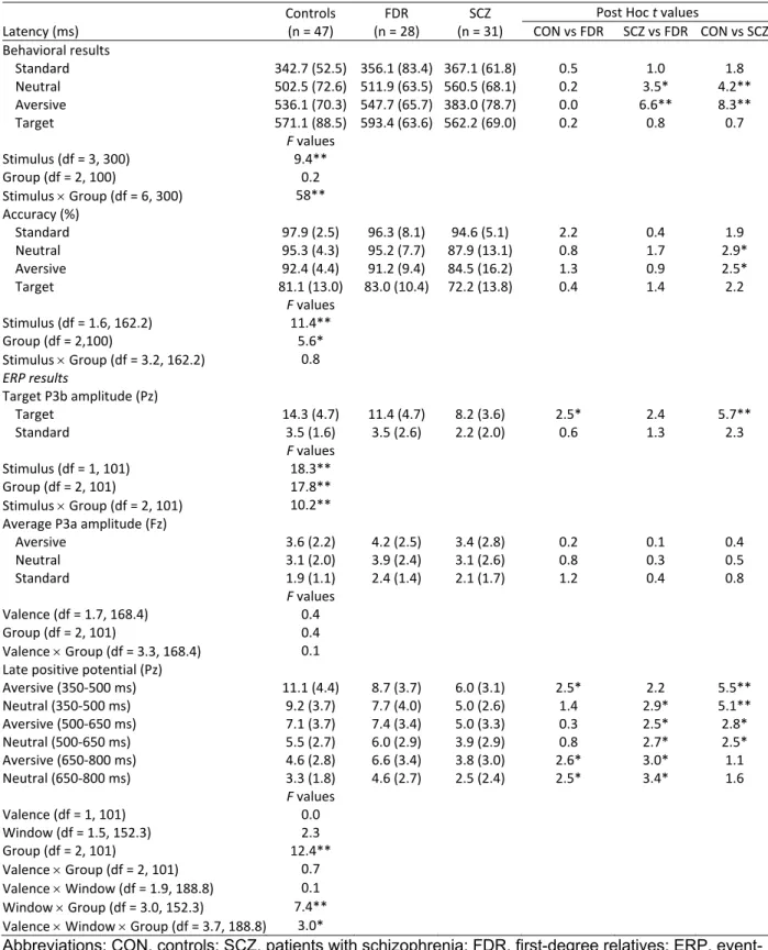
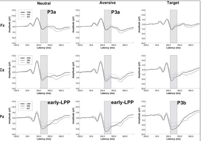
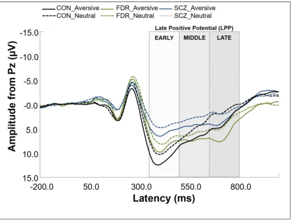
An oddball detection ERP paradigm, combining a target detection task with infrequent emotional distractor stimuli, is useful for distinguishing between attentional processing domains, such as directed attention to task-relevant events (P3b), novelty detection (P3a), motivated attention to salient stimuli (late positive potential [LPP]), and sustained attentional processing (continued late positivity). The P3a
al., 2009; van der Stelt et al., 2004), and have been related to enhanced positive symptom severity (Fisher et al., 2010; Turetsky et al., 2009). A parietally distributed P3b is elicited in response to task-relevant stimuli, indexing directed attention to “target” events (Falkenstein et al., 1999; Johnson, 1984; Katayama and Polich, 1998; Polich, 2007; Snyder and Hillyard, 1976; Squires et al., 1975). Diminished P3b amplitudes in auditory (Duncan, 1988; Kidogami et al., 1991; Mathalon, 2000; Roth and Cannon, 1972) and visual paradigms (van der Stelt et al., 2004) are among the most consistent findings in SCZ research (Duncan, 1988; Ford, 1999; Kidogami et al., 1991; Mathalon, 2000; Pritchard, 1986; Roth and Cannon, 1972; van der Stelt et al., 2004), and have been associated with abnormalities in directed attention, response selection, inhibition, and in aberrant salience attribution to task-relevant stimuli (Hajcak et al., 2010; Luck, 2005; Polich, 2007; Squires et al., 1975; van der Stelt and Belger, 2007). Reduced P3b (Kidogami et al., 1991; Price et al., 2006; van der Stelt et al., 2005) and auditory P3a amplitudes (Jahshan et al., 2012) have also been observed for individuals at clinical and familial high risk, suggesting that P3 components may be useful as endophenotypes and clinical state markers, as they can track fluctuations in clinical symptom severity (Mathalon et al., 2000). To our knowledge, P3a responses elicited in the visual modality have not been studied in individuals at familial high risk. Despite consistent findings of reduced P3 amplitudes in SCZ, similar effects have also been found in other neuropsychiatric disorders and may represent a trait marker for a central deficit in higher order processing of salient stimuli shared across psychopathologies (Bestelmeyer et al., 2009; Johannesen et al., 2012).