Influence of Beta Blockers on the Hemodynamic Response to
Inotropes in Acute Decompensated Heart Failure
By
Sarah M. Jagielski
Honors Thesis
UNC Eshelman School of Pharmacy University of North Carolina at Chapel Hill
February 15, 2019
Approved:
_____________________________________ Jo Ellen Rodgers, Faculty Mentor
Abstract:
Background: Previous studies have demonstrated altered hemodynamic response to
intravenous inotrope in chronic, stable heart failure patients receiving beta blocker (BB). The purpose of this study is to assess the impact of BB on hemodynamic response to intravenous inotrope in patients with acute decompensated heart failure (ADHF).
Methods: This prospective, single‐center, observational study assessed patients with ADHF deemed to require intravenous inotrope with pulmonary artery catheter‐based hemodynamic monitoring. Per institutional practice, patients received stepwise dosing of dobutamine (DOB) 1 to 6 mcg/kg/min followed by milrinone (MIL) 0.1 to 0.4 mcg/kg/min for dose‐finding of optimal single inotropic therapy. Patients were stratified by concomitant administration of BB
[carvedilol (CAR) or metoprolol succinate (MET) as prescribed prior to admission] or no BB. Hemodynamic parameters were measured at baseline and at each inotrope dose titration. The primary endpoint was change in Fick cardiac index (CI) from baseline to maximum, tolerated inotrope dose. Data is presented as median with interquartile range (IQR).
Results: Of the fifty patients enrolled, 78% received BB at baseline, with median doses of CAR 12.5 mg/d (n=21) and MET 50 mg/d (n=18). Forty‐nine patients received DOB and 44 received MIL; 43 received both DOB and MIL, but not concurrently. Baseline characteristics were similar among BB groups including left ventricular ejection fraction 18% (13‐23) and baseline Fick CI 1.8 L/min/m2 (1.5‐2.2). On MIL, Fick CI differed significantly between BB groups (p=0.049). Fick CI
significantly increased in the no BB group (+0.73, 0.35‐1.46), but did not significantly increase in the MET (+0.31, 0.23‐1.10) or CAR (+0.31, ‐0.24‐0.86) groups. Similar changes occurred with SVO2 (p=0.025) with significant increase with the no BB group (+16, 7‐24) but not the MET (+12,
‐1‐18) and CAR (+7, ‐7‐11) groups. In contrast, with DOB administration change in Fick CI and SVO2 did not differ among BB groups. Changes in all other parameters were similar with both
MIL and DOB administration regardless of BB therapy.
Conclusions: Both MET and CAR reduced hemodynamic response to MIL, but not DOB. Further
investigation is needed to confirm if BB should be withheld during concomitant inotrope therapy.
Abbreviation List:
Beta blocker; BB
Acute decompensated heart failure; ADHF Dobutamine; DOB
Milrinone; MIL Metoprolol; MET Carvedilol; CAR Cardiac Output; CO Cardiac Index; CI
INTRODUCTION:
In 2014, it was estimated that 6.5 million Americans over 20 years of age had heart failure with a projected increase to over 8 million by 2030.1 Heart failure is the leading cause of
hospitalization in those over 65 years of age and hospitalization is the primary cost driver of heart failure management. 2,3 The total cost of managing heart failure is anticipated to increase
from $30 billion in 2012 to $70 billion in 2030.4
When heart failure patients present with a sudden worsening of heart failure signs and symptoms including fluid overload and hypoperfusion to vital organs, they often require hospitalization which may include intravenous inotropic support.3 These patients are classified
as having acute decompensated heart failure (ADHF) and have a poorer prognosis than patients not requiring hospitalization.4 Per the 2013 ACC/AHA/HFSA guidelines, intravenous inotropes
may be reasonable in those hospitalized patients presenting with documented severe systolic dysfunction who present with low blood pressure and significantly depressed cardiac output to maintain systemic perfusion and preserve end‐organ performance.3 The rationale for using
intravenous inotropes in such patients is that they increase the contractility of the heart and improve perfusion to organs. This is measured by cardiac index (CI) which is equal to cardiac output (CO) divided by body surface area. Both in clinical practice and in research, CI is the gold standard for measuring heart function in the setting of ADHF.5 However, it is unknown how
select guideline directed medical therapies for heart failure, specifically beta blockers (BBs), impact the influence of inotropic therapy on CI and other hemodynamic parameters in ADHF patients.
Milrinone (MIL) and dobutamine (DOB) are the two intravenous inotropes available for use in the United States.6 MIL is a phosphodiesterase (PDE) inhibitor and DOB is a beta adrenergic
receptor agonist. Since DOB directly stimulates beta‐receptors, and MIL does not, it would be a logical assumption to conclude DOB would be less beneficial (have less effect on CI) than MIL in the presence of a BB. However, MIL is a known potent vasodilator and thus could lower blood pressure and thus may not be as well tolerated by patients with low blood pressure, a common finding in ADHF. In addition, both DOB and MIL increase heart rate and have been associated with the development of arrhythmias; however, it is currently unknown if one agent is
associated with a greater risk of this adverse effect. To date only two studies have assessed the influence of the presence of beta blocker on response to intravenous inotropic therapy.7,8
Metra et al assessed the effects of inotrope infusions with DOB and enoximone, a PDE inhibitor similar to MIL, in 29 chronic, stable heart failure patients receiving metoprolol tartrate or carvedilol.7 DOB’s effects on CI were significantly less with concomitant carvedilol compared to
no BB, but were not statistically different with metoprolol. In contrast, effects on CI with
enoximone were not significantly different with concomitant carvedilol therapy compared to no BB, but its effects on CI were significantly increased in the presence of metoprolol.
directed medical therapy for heart failure).
In another study, Bollano et al observed the effects of DOB in 10 patients with chronic, stable heart failure receiving BB therapy.8 With metoprolol succinate, CO increased with all doses of
DOB and this was attributed to an increase in heart rate. Of note, CI was not reported in this study. With carvedilol, CO was significantly increased with DOB 5 mcg/kg/min; however, CO significantly decreased once DOB doses were increased to 15 mcg/kg/min. The authors attributed the decline in CO to significant increase peripheral resistance, a vasopressor like effect. Unfortunately, the findings of this also have limited application to contemporary clinical practice given the high doses of DOB utilized and the inclusion of patients with chronic, stable heart failure rather than ADHF.
Importantly, in addition to those limitations previously listed, both studies utilize BB doses nearing, or even above, target doses recommended by guidelines.3 This factor could be
considered yet another limitation given these studies enrolled patients with chronic, stable heart failure. Unlike patients with chronic, stable heart failure, many patients who progress to advanced HF and experience frequent episodes of ADHF do not tolerate target BB doses, and therefore the inhibitory effects of lower BB doses on inotropic response could be less
pronounced. Additionally, in current practice the high doses of inotropic therapy utilized in these studies are rarely administered. Finally, both studies were of small sample size and at a single study center.
This study aimed to assess the hemodynamic effects of both DOB and MIL in the setting of concomitant BB or no BB therapy. These outcomes were assessed in a contemporary practice utilizing lower intravenous inotrope doses and two guideline‐directed BBs for heart failure, carvedilol and metoprolol succinate. The primary endpoint was change in the hemodynamic parameter, Fick CI, from baseline to maximally received inotrope dose. An secondary aim was to assess adverse events leading to discontinuation of DOB and MIL.
METHODS:
Study Participants:
This was a single center, observational study that enrolled patients who were hospitalized with ADHF. Patients were eligible for enrollment if ≥ 18 years of age, English speaking and admitted to the General Cardiology or Heart Failure Services at the University of North Carolina Hospitals. Only patients in whom the medical team were deemed to require intravenous inotrope
therapy, specifically a trial of DOB followed by MIL each at increasing doses per institutional practice (Table 1), and hemodynamic monitoring with a Swan‐Ganz catheter were included. At the discretion of the medical team, patients could be continued on all prior to admission
medications, including BB. However, patients must have had at least three consecutive doses of BB therapy (carvedilol, metoprolol succinate or metoprolol tartrate) to be analyzed as BB recipients. Patients who had missed at least three doses of BB therapy or who received no BB therapy were analyzed as a patient with no BB therapy.
blockers, nondihydropyridine calcium channel blockers or anti‐arrhythmic agents except for chronic stable doses of amiodarone, dofetilide or mexiletine. Additionally, use of intravenous inotropes or other intravenous vasoactive agents within the last 7 days, hemodynamically unstable arrhythmias (systolic blood pressure < 80 mmHg or heart rate > 110 bpm), uncorrected primary valvular disease or current mechanical support (left ventricular assist device, impella devices and intra‐aortic balloon pumps) were cause for exclusion.
This study was approved by the University of North Carolina Institutional Review Board and participants provided informed consent prior to enrollment.
Study Design:
Inotrope dose titration was based on an algorithm that is standard of care at our institution (Table 1). Inotrope titration including dose selection and frequency of titration was at the discretion of the medical team based upon clinical response and tolerability. In addition, the medical team could deem it medically inappropriate to administer DOB or MIL (i.e., only administer a single inotrope). Finally, BB dose down titration or discontinuation was allowed at the discretion of the medical team.
Baseline characteristics at time of admission were collected. Hemodynamic response was assessed by hemodynamic parameters obtained by Swan‐Ganz catheter and included central venous pressure, pulmonary artery systolic pressure, pulmonary artery diastolic pressure, mean pulmonary artery pressure, pulmonary capillary wedge pressure, CO, CI, systemic vascular resistance (SVR) and mixed venous oxygen saturation. The following parameters were assessed by both continuous/thermodilution method and Fick calculation: CO, CI and SVR. Vital signs were also obtained at these same time points and included systolic and diastolic blood
pressure, mean arterial pressure and heart rate. Hemodynamic parameters and vital signs were documented at baseline, then every 4 hours or at the end of each inotrope dose titration, whichever came first, as well as between DOB and MIL administration, if a washout was deemed allowable by the medical team. These parameters were only collected with each dose titration if the inotrope infusion was considered to have reached steady state per the
institutional titration algorithm. For patients who were deemed appropriate for an inotrope washout period between DOB and MIL, repeat baseline hemodynamic parameters and vital signs were collected.
Incidence of adverse effects requiring inotrope dose down titration or discontinuation (e.g., hypotension, arrhythmias) was obtained from daily progress notes. Duration of hospital stay was collected along with vital status upon discharge (e.g., home inotrope on discharge, discharge to palliative care, discharge status post left ventricular assist device or heart transplant). There was no required patient follow‐up after hospital discharge.
Statistical Analysis:
Based upon prior literature,7,8 power calculations were performed to assess our primary aim of
a change in Fick CI of 0.5 0.7 (90% power, two‐sided alpha 0.05). For patients receiving CAR, 34 patients were needed to detect a change in Fick CI of 0.3 0.6 (90% power, two‐sided alpha 0.05). Finally, for patients not receiving a BB, only 2 patients were needed to detect a change in Fick CI of 1.5 0.3 (90% power, two‐sided alpha 0.05); however, this arm was increased to 10 patients to provide a more meaningful sample size.
Data analysis consisted of a variety of descriptive and inferential statistics. First, all baseline characteristics and hemodynamic parameters were tested for normality. Since the data was not normally distributed, baseline characteristics and change in hemodynamic parameters including the primary endpoint, change in Fick CI, were assessed across the three BB groups (CAR, MET, no BB) by Kruskal‐Wallis for continuous data and chi‐square for categorical data. When a difference existed among the three groups, a Wilcoxon‐Rank Sum test was performed to assess for differences between individual groups (e.g. comparing change in Fick CI between CAR vs MET and CAR vs no BB). For patients who were deemed appropriate for an inotrope washout period between DOB and MIL, baseline hemodynamic parameters were repeated; however, this only occurred for 17 patients. The baseline hemodynamic Fick CI was compared to the washout Fick CI within each BB strata and no statistical difference was observed (p=0.743). Thus, baseline hemodynamics were used to calculate all changes in hemodynamic parameters.
Results:
Patient Population:
From December 2011 to September 2017, a total of 53 patients were enrolled. Three patients were excluded after enrollment due to concomitant sotalol (n=2) and having received only one dose of a single inotrope (n=1). The final analysis includes 50 patients, 49 patients received DOB and 44 patients received MIL. Overall, 43 patients received both inotropes. At the discretion of the medical team, six patients received only DOB and one patient received only MIL due to concerns regarding potential for intolerance to the alternative inotrope. The majority of patients were able to receive the maximum inotrope dose per the algorithm, 80% (39/49 patients) received DOB 5‐6 mcg/kg/min and 55% (24/44 patients) received MIL 0.375‐0.4 mcg/kg/min.
Prior to hospitalization, 18 patients were receiving MET (median dose 50 mg/d; IQR 25‐100) and 21 patients were receiving CAR (median dose 12.5 mg/d; IQR 12.25‐25). Beta blocker dose at time of DOB and MIL administration are described in Table 2. Eleven patients were either prescribed no BB as an outpatient, had BB held at admission or missed three consecutive doses prior to DOB initiation. Five patients did not remain on BB for the full inotrope algorithm. The BB was discontinued prior to DOB initiation in three patients receiving CAR and one patient receiving MET. The BB was restarted prior to the MIL infusion in one CAR and one MET patient. In one patient receiving only DOB, their MET was held prior to DOB infusion. Lastly, two
patients were switched from CAR to MET, one before DOB initiation and one before MIL
initiation. In all cases, three doses were missed and steady state was achieved prior to initiating the given inotrope.
Overall, patients were a median of 59 years of age, 80% male, and 44% African American (Table 2). Ischemic heart disease occurred in 42% and hypertension in 56%. The median left
ventricular ejection fraction was 17.5%. In addition to the low ejection fraction, several additional characteristics suggested a high severity of illness including low systolic blood pressure and elevated blood urea nitrogen, serum creatinine and N‐terminal pro‐natriuretic peptide level. Patients were well managed with standard guideline‐directed therapies for heart failure prior to admission including angiotensin converting enzyme inhibitor or receptor blocker (78%), beta blocker (78%) and aldosterone antagonist (72%). There were no significant
differences between the three BB groups in baseline characteristics, including hemodynamic parameters, with the exception of the CAR group having a significantly lower N‐terminal pro‐ natriuretic peptide on admission and the no BB group being significantly more likely to have atrial fibrillation and having a significantly lower diastolic blood pressure at baseline.
Hemodynamics Effects:
For the primary endpoint, following DOB there was no difference in change in Fick CI from baseline to maximum received inotrope dose among the BB groups, (CAR 0.35 L/min/m2; MET
0.43 L/min/m2; no BB 0.44 L/min/m2; p=0.53) (Table 3a). In addition, there was no difference in
change in any other hemodynamic parameter with DOB. In contrast, following MIL there was a significant difference in change in Fick CI among the BB groups (p=0.049) (Table 3b). This difference was due to a significant increase in Fick CI with no BB compared to CAR (no BB 0.73 L/min/m2, CAR 0.31 L/min/m2; p=0.02) (Figure 1). Similar to Fick CI, SVO
2 was significantly
increased with MIL with no BB compared to CAR (no BB 16%, CAR 7%; p=0.01) (Table 3b). There was no difference in any other hemodynamic parameters among the BB groups with MIL.
Inotrope Tolerability:
A higher rate of discontinuation due to any adverse drug event was observed with MIL (39%) compared to DOB (16%) (Table 4). While discontinuation due to arrhythmia occurred more commonly with DOB (12%) versus MIL (2%), discontinuation due to hypotension/dizziness occurred more commonly with MIL (20%) versus DOB (0%). Small event numbers precluded statistical analysis; however, no obvious trends were noted between BB groups beyond a higher rate of overall events in patients receiving CAR and MIL. We hypothesize this may be due to additive vasodilatory effects.
Length of Stay and Vital Status on Discharge:
Patient status varied at discharge (Table 5). The average length of stay was 18 15.2 days. Most patients were discharged on inotropic therapy (60%). Of those patients, more were discharged on MIL (34%) then DOB (26%). Additionally, 14% of patients received either a left ventricular assist device or a heart transplant, while 8% of patients were discharged on palliative care or were deceased before discharge.
DISCUSSION:
hemodynamic response observed with intravenous inotrope doses used in contemporary practice. These doses are lower than those previously studied.7,8 Our results suggest the change
in Fick CI with DOB is the same regardless of BB group. In contrast, our results suggest the change in Fick CI with MIL is more pronounced with no BB compared to CAR, but not MET. Neither of these findings are consistent with data presented in previous studies.7,8
Similar to Bollano et al, our study demonstrated an increase in Fick CI with CAR and a lower DOB dose, 5 mcg/kg/min. The rise in CO observed in the Bollano study with a lower DOB dose was 0.8 L/min (equals CI 0.36 L/min/m2 assuming a body surface area of 2.2 m2) is similar to the
change in Fick CI observed in our study, 0.34 L/min/m2. Importantly, our study retained the
inotropic effects with the DOB dose despite our patients being hospitalized with ADHF and receiving a lower CAR doses compared to patients in Bollano’s study who had chronic, stable heart failure and received higher CAR doses. Similar to Bollano, our study also demonstrated no increase in heart rate with CAR and DOB. Bollano hypothesized that, unlike with MET, beta receptors are not upregulated with CAR and this difference in receptor sensitivity might explain the lack of heart rate response. These authors further hypothesized that the increase in heart rate is partly a beta‐2 effect, which is more profoundly blocked by CAR. The vasopressor‐like effect observed with CAR and higher DOB dose observed in the Bollano study was not reproduced in this study and is likely due to our study not utilizing higher DOB doses.
Unlike our study which demonstrated a similar increase in Fick CI with MET and a lower DOB dose (0.43 L/min/m2) compared to that observed with CAR and DOB, the Bollano study
demonstrated a smaller increase in CO (equals CI 0.22 L/min/m2 assuming body surface area
2.2 m2) with MET and lower DOB dose. In the Bollano study, a significant increase in CO was not
observed with MET until a higher DOB dose was utilized and this was attributed to a significant decline in peripheral resistance and an increase in heart rate. This increase in CO was almost two‐fold what was observed in our study, 1.7 L/min (equals 0.77 L/min/m2 assuming body
surface area 2.2 m2). As stated above, these authors hypothesized that this change in CO was
associated with MET mediated beta receptor upregulation. It is unknown if our study patients would have observed as robust a response given the presence of ADHF and the lower MET dose. Perhaps more importantly, in contemporary practice many clinicians discontinue MET (a selective beta blocker) in the presence of DOB (a beta agonist) due to the theoretical inhibition of the beta agonist by the beta blocker; however, our study suggests an inotropic effect is retained similar to that observed with no BB, at least with lower DOB doses.
While a similar increase in CI was observed with MIL and both CAR and MET, the most
substantial increase in CI observed in our study occurred with MIL and no BB (0.73 L/min/m2).
While the study by Metra et al also reported similar change in Fick CI with CAR and MET treated patients receiving enoximone (in lieu of MIL), both responses were enhanced compared to prior BB treatment, ranging from 0.5 and up to 1.5 L/min/m2 with increasing enoximone doses. While
of changes in any other hemodynamic parameters, it is difficult to postulate a rationale for enhanced response to MIL with no BB other than those patients appearing to be more severely ill.
Overall, despite lower inotrope doses being utilized in contemporary practice, the incidence of adverse effects requiring inotrope discontinuation was high. As anticipated, hypotension was common with MIL and arrhythmias was common with DOB. This was also consistent with the previous literature, although limited to MIL.9 However, this previous study was not in the
presence of BB therapy. Unfortunately, there did not appear to be a reduced incidence of arrhythmias in patients treated with no BB; however, this may be due to the low BB dose not providing adequate cardioprotection. Despite the lack of formal statistical analysis, the incidence of these adverse effects may guide future selection of MIL and DOB. For example, one may consider avoiding MIL in the setting of low baseline blood pressure and avoiding DOB in patients with preexisting rhythm disturbances.
There are several limitations to this study. This was a small, single center, observational study. There were varying degrees of volume overload at inotrope initiation and its correction could contribute to improving hemodynamic parameters, especially Fick CI. Additionally, the study allowed for physician discretion in order to properly care for each patient. Given such, the algorithm was not always strictly followed and thus hemodynamic parameters were not obtained with all dose titrations, not every patient received both inotropes and MIL was
initiated first in two patients. Furthermore, less than half the patients were deemed by medical staff able to tolerate a washout period and thus baseline hemodynamic parameters were not obtained between DOB and MIL administration in all patients. Finally, due to the small sample size it is possible that the study is slightly underpowered for both MET and CAR groups. In order to account for these limitations, it is important to continue research within this field.
Overall, our study findings contribute to the existing literature and provide direction for
inotrope selection in patients with ADHF receiving BB therapy. Our study confirmed an increase in Fick CI with both DOB and MIL despite using lower doses than those previously studied. Our study also eliminates concern for a pharmacodynamic interaction between BBs and lower dose DOB precluding their concomitant use. In addition, the frequency of hypotension and
arrhythmias with MIL and DOB, respectively, will guide selection of these therapies in high risk patients already predisposed to these adverse effects.
REFERENCES:
1. Benjamin EJ, Blaha MJ, Chiuve SE, Cushman M, Das SR, Deo R, et al. Heart Disease and Stroke Statistics—2017 Update: A Report From the American Heart Association. Circulation. 2017 Mar 7;135(10):e146–603.
2. Allman KC, Shaw LJ, Hachamovitch R, Udelson JE. Myocardial viability testing and impact of revascularization on prognosis in patients with coronary artery disease and left ventricular dysfunction: a meta‐analysis. J Am Coll Cardiol. 2002 Apr;39(7):1151–8.
3. WRITING COMMITTEE MEMBERS, Yancy CW, Jessup M, Bozkurt B, Butler J, Casey DE, et al. 2013 ACCF/AHA Guideline for the Management of Heart Failure: A Report of the American College of Cardiology Foundation/American Heart Association Task Force on Practice Guidelines. Circulation. 2013 Oct 15;128(16):e240–327.
4. Nohria A, Mielniczuk LM, Warner Stevenson L. Evaluation and Monitoring of Patients with Acute Heart Failure Syndromes. Am J Cardiol. 2005 Sep;96(6):32–40.
5. Rodgers JE, Reed BN. Acute Decompensated Heart Failure. In: DiPiro JT, Talbert RL, Yee GC, Matzke GR, Wells BG, Posey LM, editors. Pharmacotherapy: A Pathophysiologic Approach, 10e [Internet]. New York, NY: McGraw‐Hill Education; 2017 [cited 2018 Nov 19]. Available from:
accesspharmacy.mhmedical.com/content.aspx?aid=1145177519
6. Aranda JM, Schofield RS, Pauly DF, Cleeton TS, Walker TC, Monroe VS, et al. Comparison of dobutamine versus milrinone therapy in hospitalized patients awaiting cardiac transplantation: A prospective, randomized trial. Am Heart J. 2003 Feb 1;145(2):324–9.
7. Metra M, Nodari S, D’Aloia A, Muneretto C, Robertson AD, Bristow MR, et al. Beta‐blocker therapy influences the hemodynamic response to inotropic agents in patients with heart failure. J Am Coll Cardiol. 2002 Oct;40(7):1248–58.
8. Bollano E, Täng MS, Hjalmarson A, Waagstein F, Andersson B. Different responses to dobutamine in the presence of carvedilol or metoprolol in patients with chronic heart failure. Heart Br Card Soc. 2003 Jun;89(6):621–4.
9. Felker GM, Benza RL, Chandler AB, Leimberger JD, Cuffe MS, Califf RM, et al. Heart failure etiology and response to milrinone in decompensated heart failure. J Am Coll Cardiol. 2003 Mar;41(6):997– 1003.
TABLES AND FIGURES:
Table 1 – Dobutamine and Milrinone Titration* Algorithms
Dobutamine Titration Titration Schedule
Step 1 1‐2 mcg/kg/min, titrate at 2 hours Step 2 3‐4 mcg/kg/min, titrate at 2 hours Step 3 5‐6 mcg/kg/min, titrate at 2 hours
Milrinone Titration Titration Schedule Based on Creatinine Clearance**
Dose > 60 mL/min 40 – 60 mL/min 20 – 40 mL/min < 20 mL/min Step 1 0.1 mcg/kg/min
4 + 1 hours 12 + 2 hours 18 + 3 hours 24 + 4 hours Step 2 0.3 mcg/kg/min
Step 3 0.375‐0.4 mcg/kg/min
*While all inotrope dose titrations (dose selection and titration frequency) were to the discretion of the
attending physician, the above titration schedule was the minimum time required to reach steady state prior to up titration.
**Creatinine clearance calculated by Cockcroft‐Gault, utilizing adjusted body weight if overweight
Table 2 ‐ Baseline Characteristics Characteristics Overall (n=50) CAR (n = 21) MET (n = 18) No BB (n =11) p value
Age (years) 58.5 (49.5‐68.25)) 56 (50‐68.5) 55.5 (44.25‐66.25) 68 (52‐71) 0.41
Gender (female) 10 (20%) 3 (14.3%) 5 (27.8%) 2 (18.2%) 0.57
Ethnicity (African American) 22 (44%) 9 (42.9%) 9 (50%) 4 (36.4%) 0.75
Medical History Ischemic Heart Disease Hypertension Atrial Fibrillation Diabetes 21 (42%) 28 (56%) 18 (36%) 21 (42%) 9 (42.9%) 15 (71.4%) 4 (19%) 8 (38.1%) 8 (44.4%) 9 (50%) 7 (38.9%) 9 (50%) 4 (36.4%) 4 (36.4%) 7 (63.6%) † 4 (36.4%) 0.91 0.13 0.04 0.69
Left Ventricular Ejection Fraction (%) 17.5 (12.5‐22.5) 18.55 (12.5‐22.5) 15 (14.4‐18.1) 20 (15‐22.5) 0.28 Systolic Blood Pressure (mmHg) 104 (92.75‐119) 107 (96‐120) 100.5 (93‐110) 103 (85‐120) 0.47
Heart Rate (bpm) 80 (72‐91.25) 80 (71‐85.5) 85.5 (72‐101) 76 (71‐93) 0.33
Blood Urea Nitrogen (mg/dL) 29.5 (21‐40.75) 23 (19.5‐32) 30.5 (21‐46) 34 (29‐61) 0.07 Serum Creatinine (mg/dL) 1.4 (1.08‐1.93) 1.3 (1‐1.6) 1.6 (1.1‐2.1) 1.5 (1.4‐2.0) 0.14 Serum Sodium (mg/dL) 138 (135‐140) 138 (134‐139.5) 137 (134.8‐141) 138 (135‐140) 0.94
NT‐proBNP (pg/mL) 7300
(3095‐12800) 3650 (1450‐6920) * 9375 (5952.5‐12825) 11550 (5610‐20625) 0.02 Medical Therapy and Devices Loop Diuretics ACE inhibitors or ARBs Sacubitril/Valsartan Aldosterone antagonist Digoxin Hydralazine Isosorbide dinitrate Amiodarone Mexelitine Implantable Cardioverter Defibrillator Cardiac Resynchronization Therapy 47 (94%) 39 (78%) 1 (2%) 36 (72%) 16 (32%) 7 (14%) 6 (12%) 15 (30%) 3 (6%) 45 (90%) 13 (26%) 21 (100%) 17 (81%) 1 (7.7%) 13 (62%) 3 (14.3%) 3 (14.3%) 3 (14.3%) 6 (28.6%) 1 (6.7%) 18 (85.7%) 4 (19%) 15 (83.3%) 13 (72.2%) 0 (0%) 14 (77.8%) 9 (50%) 2 (11.1%) 2 (11.1%) 4 (22.2%) 1 (8.3%) 16 (89%) 5 (27.8%) 11 (100%) 9 (81.8%) 0 (0%) 9 (81.8%) 4 (36.4%) 2 (18.2%) 1 (9.1%) 5 (45.5%) 1 (14.3%) 11 (100%) 4 (36.4%) 0.06 0.76 0.64 0.39 0.06 0.87 0.90 0.41 0.84 0.43 0.56 Hemodynamic Parameters MAP (mmHg) HR (bpm) PAD (mmHg)
Thermodilution CI (L/min/m2)
Fick CI (L/min/m2)
Thermodilution SVR (dyn∙s∙cm‐5)
SvO2 (%)
78 (71.5‐92) 80 (70‐85) 27 (22‐31.5) 1.75 (1.4‐2.1) 2 (1.55‐2.355) 1581 (1075‐2243.5) 60 (48.5‐67.2) 78.5 (69.5‐87.5) 89.5 (72‐101.75) 33.5 (27.75‐37) 1.5 (1.2‐2.175) 1.775 (1.5‐1.9) 1546.5 (1063‐1772) 52.4 (45.5‐58) 75 (62‐79) 76 (71‐93) 30 (23‐36) 1.9 (1.3‐2) 1.7 (1.3‐2.36) 1495 (1120‐2291) 49 (44‐58) 0.20 0.20 0.16 0.90 0.21 0.82 0.09 Beta Blocker Dose (mg)
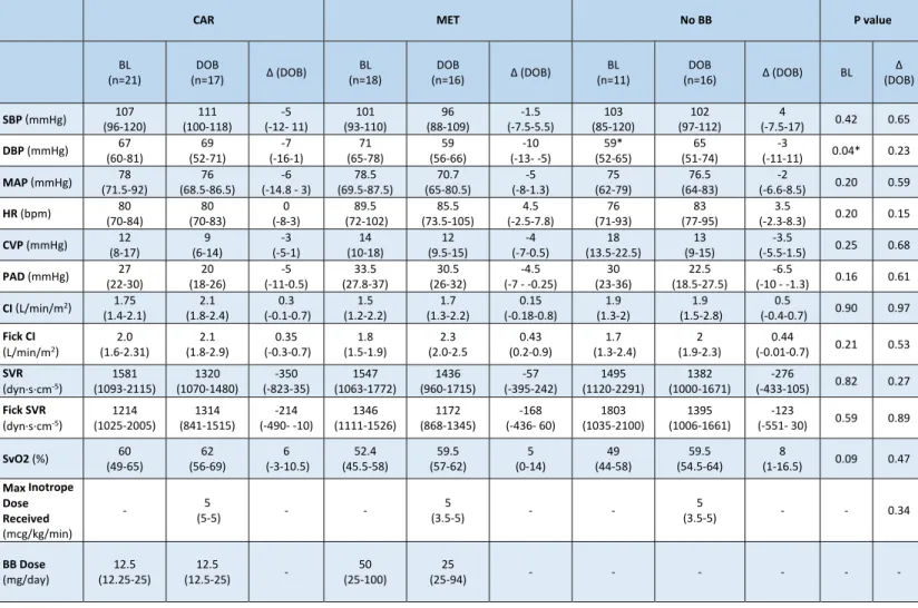
Table 3a – Hemodynamic Parameters during DOB Infusion
*When no BB was compared to CAR (p=0.0288) and MET (p=0.0133) DBP was significantly lower at baseline.
CAR MET No BB P value
BL (n=21) DOB (n=17) ∆ (DOB) BL (n=18) DOB (n=16) ∆ (DOB) BL (n=11) DOB
(n=16) ∆ (DOB) BL ∆ (DOB)
SBP (mmHg) 107 (96‐120) 111 (100‐118) ‐5 (‐12‐ 11) 101 (93‐110) 96 (88‐109) ‐1.5 (‐7.5‐5.5) 103 (85‐120) 102 (97‐112) 4
(‐7.5‐17) 0.42 0.65
DBP (mmHg) 67 (60‐81) 69 (52‐71) ‐7 (‐16‐1) 71 (65‐78) 59 (56‐66) ‐10 (‐13‐ ‐5) 59* (52‐65) 65 (51‐74) ‐3
(‐11‐11) 0.04* 0.23
MAP (mmHg) (71.5‐92) 78 (68.5‐86.5) 76 (‐14.8 ‐ 3) ‐6 (69.5‐87.5) 78.5 (65‐80.5) 70.7 (‐8‐1.3) ‐5 (62‐79) 75 (64‐83) 76.5 (‐6.6‐8.5) ‐2 0.20 0.59
HR (bpm) 80 (70‐84) 80 (70‐83) 0 (‐8‐3) 89.5 (72‐102) 85.5 (73.5‐105) 4.5 (‐2.5‐7.8) 76 (71‐93) 83 (77‐95) 3.5
(‐2.3‐8.3) 0.20 0.15
CVP (mmHg) (8‐17) 12 (6‐14) 9 (‐5‐1) ‐3 (10‐18) 14 (9.5‐15) 12 (‐7‐0.5) ‐4 (13.5‐22.5) 18 (9‐15) 13 (‐5.5‐1.5) ‐3.5 0.25 0.68
PAD (mmHg) 27 (22‐30) 20 (18‐26) ‐5 (‐11‐0.5) 33.5 (27.8‐37) 30.5 (26‐32) ‐4.5 (‐7 ‐ ‐0.25) 30 (23‐36) 22.5 (18.5‐27.5) ‐6.5
(‐10 ‐ ‐1.3) 0.16 0.61
CI (L/min/m2) 1.75
(1.4‐2.1) 2.1 (1.8‐2.4) 0.3 (‐0.1‐0.7) 1.5 (1.2‐2.2) 1.7 (1.3‐2.2) 0.15 (‐0.18‐0.8) 1.9 (1.3‐2) 1.9 (1.5‐2.8) 0.5
(‐0.4‐0.7) 0.90 0.97 Fick CI
(L/min/m2)
2.0 (1.6‐2.31) 2.1 (1.8‐2.9) 0.35 (‐0.3‐0.7) 1.8 (1.5‐1.9) 2.3 (2.0‐2.5 0.43 (0.2‐0.9) 1.7 (1.3‐2.4) 2 (1.9‐2.3) 0.44
(‐0.01‐0.7) 0.21 0.53 SVR
(dyn∙s∙cm‐5)
1581 (1093‐2115) 1320 (1070‐1480) ‐350 (‐823‐35) 1547 (1063‐1772) 1436 (960‐1715) ‐57 (‐395‐242) 1495 (1120‐2291) 1382 (1000‐1671) ‐276
(‐433‐105) 0.82 0.27 Fick SVR
(dyn∙s∙cm‐5)
1214 (1025‐2005) 1314 (841‐1515) ‐214 (‐490‐ ‐10) 1346 (1111‐1526) 1172 (868‐1345) ‐168 (‐436‐ 60) 1803 (1035‐2100) 1395 (1006‐1661) ‐123
(‐551‐ 30) 0.59 0.89
SvO2 (%) 60 (49‐65) 62 (56‐69) 6 (‐3‐10.5) 52.4 (45.5‐58) 59.5 (57‐62) 5 (0‐14) 49 (44‐58) 59.5 (54.5‐64) 8
(1‐16.5) 0.09 0.47 Max Inotrope
Dose Received (mcg/kg/min)
‐ (5‐5) 5 ‐ ‐ (3.5‐5) 5 ‐ ‐ (3.5‐5) 5 ‐ ‐ 0.34
BB Dose (mg/day) 12.5 (12.25‐25) 12.5 (12.5‐25) ‐ 50 (25‐100) 25
Table 3b‐ Hemodynamic Parameters during MIL Infusion
*When no BB was compared to CAR (p=0.0288) and MET (p=0.0133) DBP was significantly lower at baseline. †No BB was significantly higher when compared to CAR in Fick CI (p=0.0192) and SVO2 (p=0.0090).
CAR MET No BB P value
BL (n=21) MIL (n=15) ∆ (MIL) BL (n=18) MIL (n=17) ∆ (MIL) BL (n=11) MIL
(n=12) ∆ (MIL) BL ∆ (MIL)
SBP (mmHg) (96‐120) 107 (95‐116) 102 (‐22‐2) ‐8 (93‐110) 100.5 (90‐107) 98 (‐17‐4) ‐2 (85‐120) 103 (92‐114) 99 (‐4‐18) 8.5 0.42 0.13
DBP (mmHg) 67 (60‐81) 67 (52‐76) ‐14 (‐23‐ ‐14) 71 (65‐78) 58 (52‐70) ‐12 (‐21‐ ‐1) 59* (52‐65) 57 (47‐64) ‐5.5
(‐11‐2.5) 0.04* 0.22
MAP (mmHg) 78 (71.5‐92) 80 (68‐91) ‐7 (‐15‐14) 78.5 (69.5‐87.5) 68 (61‐73) ‐13 (‐36‐4) 75 (62‐79) 67 (59‐76) ‐5.5
(‐37‐7.5) 0.20 0.42
HR (bpm) 80 (70‐84) 84 (77‐91) 3 (‐5‐7) 90 (72‐101) 86 (81‐93) 4 (‐1‐6) 76 (71‐93) 90 (77‐103) 7
(1‐16) 0.20 0.28
CVP (mmHg) 12 (8‐17) 9 (7‐14) ‐2 (‐6‐1) 14 (12‐20) 11 (8‐14) ‐5 (‐11‐ ‐1) 18 (12‐21) 10 (9‐14) ‐4
(‐8‐ ‐2.5) 0.22 0.25
PAD (mmHg) 27 (22‐30) 20 (15‐27) ‐6 (‐11‐0) 33.5 (28‐37) 26 (21‐30) ‐7 (‐10‐0) 30 (23‐36) 25.5 (22.5‐31) ‐3.5
(‐8‐1) 0.16 0.63
CI (L/min/m2) 1.75
(1.4‐2.1) 2.4 (1.5‐2.7) 0.65 (‐0.3‐1) 1.5 (1.2‐2.1) 1.9 (1.6‐2.53) 0.4 (0‐0.8) 1.9 (1.3‐2.0) 2.15 (1.7‐2.65) 0.6
(0.15‐1.3) 0.90 0.71 Fick CI
(L/min/m2)
2.0 (1.6‐2.31) 2.28 (1.8‐2.92) 0.31 (‐0.24‐0.86) 1.8 (1.5‐1.9) 2.5 (2.11‐2.74) 0.31 (0.23‐1.10) 1.7 (1.3‐2.36) 2.59 (2.05‐3.36) 0.73 (0.35‐1.46) 0.21 0.049 † SVR
(dyn∙s∙cm‐5)
1581 (1093‐2115) 1151 (926‐1771) ‐461 (‐797‐ ‐36) 1547 (1067‐1728) 996 (840‐1379) ‐276 (‐625‐ ‐6) 1495 (1120‐2291) 1237 (1004‐1541) ‐483
(‐799‐ ‐214) 0.82 0.55 Fick SVR
(dyn∙s∙cm‐5)
1214 (1025‐2005) 1156 (839‐1510) ‐78 (‐977‐130) 1346 (1111‐1526) 902 (725‐1325) ‐417 (‐674‐ ‐196) 1803 (1035‐2100) 1045 (741‐1352) ‐381
(‐816‐ ‐173) 0.59 0.28
SvO2 (%) 60 (49‐65) 61 (50‐70) 7 (‐7‐11) 52 (46‐58) 61 (54‐68) 12 (‐1‐18) 49 (44‐58) 66 (56‐72) 16
(7‐24) 0.09 0.03† Max Inotrope
Dose Received (mcg/kg/min)
‐ 0.3
(0.2‐0.375) ‐ ‐
0.3 (0.25‐ 0.375)
‐ ‐ 0.375
(0.2‐0.4) ‐ ‐ 0.25
BB Dose (mg/day) 12.5 (12.25‐25) 12.5 (12.5‐25) ‐ 50 (25‐100) 25
Figure 1 – FICK CI, Change from Baseline to Maximum Tolerated Dose of Inotrope Therapy
Data presented as median. MET = metoprolol, CAR = carvedilol, no BB = no beta blocker, BL = baseline, WO =
washout, DOB = dobutamine, MIL = milrinone. *MIL + no BB group compared to MIL + CAR (p=0.019).
Table 4 – Tolerability of Inotrope Therapy
Adverse Event*
MIL DOB
CAR (n = 15)
MET (n = 17)
No BB (n = 12)
Total (n = 44)
CAR (n = 17)
MET (n = 16)
No BB (n = 16)
Total (n = 49) Inotrope
Discontinuation due to Adverse Event
7 (47%) 6 (35%) 4 (33%) 17 (39%) 4 (24%) 2 (13%) 2 (13%) 8 (16%)
Hypotension/ Dizziness
3 (20%) 4 (24%) 2 (17%) 9 (20%) 0 (0%) 0 (0%) 0 (0%) 0 (0%)
Arrhythmia 0 (0%) 0 (0%) 1 (8%) 1 (2%) 2 (12%) 2 (13%) 2 (13%) 6 (12%)
Other 4 (27%) 3 (18%) 2 (17%) 9 (20%) 2 (12%) 0 (0%) 0 (0%) 2 (4%)
Data is presented as number (percentage).
*Patients may have reported more than one adverse drug event leading to inotrope discontinuation.
Other includes headache (n=3), nausea (n=3), chest pain (n = 2), decreased renal function (n = 2), poor exercise tolerance (n = 2), hypertension (n = 2) and altered mental status (n = 1).
Table 5 – Length of Stay and Vital Status at Discharge
Total
(n = 50)
CAR (n = 21)
MET (n=18)
No BB (n =11)
Length of Stay (days)
18 + 15.2 16.5 + 16.1 21.1 + 15.9 16.7 + 10.8
Home Inotrope 17 MIL, 13 DOB 30 (60%) 5 MIL, 6 DOB 11 (52.4%) 6 MIL, 4 DOB 10 (55.6%) 6 MIL, 3 DOB 9 (82%) Left Ventricular
Assist Device 6 (12%) 2 (9.5%) 4 (22.2%) 0 (0%)
Heart Transplant 1 (2%) 1 (4.8%) 0 (0%) 0 (0%)
Palliative Care 1 (2%) 0 (0%) 0 (0%) 1 (9%)
Deceased 3 (6%) 1 (4.8%) 1 (5.6%) 1 (9%)
Data is listed as mean + standard deviation or number (%).
ADDEDUM:
Acknowledgements:
Nan Wang – Contributed to data analysis
Brent N. Reed – Contributed to patient recruitment Elizabeth A. Blair – Contributed to patient recruitment Ahmed Aldemerdash – Contributed to informed consent Khalid A. Alburikan – Contributed to data collection
Funding Support:
The authors have no funding support to disclose.
Conflicts of Interest: