ABSTRACT
MICHAEL N. BOUCHER. Comparative Hutagenicity of Urinary Metabolites of
Nitropolycylic Aromatic Hydrocarbons 1-Nitropyrene, 2-Nitrofluoranthene,
and 3-Nitrofluoranthene. (Under the Direction of Dr. LOUISE M. BALL)
This study compared the mutagenicity of urinary metabolites of
prevalent air pollutants 1-nitropyrene (1-NP), 2-nitrofluoranthene
(2-NFA), and 3-nitrofluoranthene (3-NFA). Rats were injected intra¬
peritoneal ly with specific doses and their urine was analyzed for
mutagenicity by an Ames plate incorporation assay using Salmonella
Typhimurium. Resulting data yielded revertant excretion rate plots used
for comparative analysis. To achieve maximum mutagenicity, 1-NP's
urinary metabolites required exogenous activation by S9, 2-NFA's did not
vary significantly with S9 activation, while 3-NFA's was decreased by S9
metabolism. 2-NFA's mutagenic metabolites' revertant excretion rate was
a quarter of the rates of 3-NFA's and 1-NP's mutagenic metabolites. The
strongest mutagenicity of urinary metabolites during the 48 hrs after
injection was from 1-NP which was double the amounts from 3-NFA and
2-NFA. However, the estimated total urinary mutagenicity showed 1-NP and
2-NFA potentially created equal amounts while 3-NFA 's amount was only
TABLE OF CONTENTS
I. INTRODUCTION...1
II. LITERATURE REVIEW...3
II. A. Background ...3
II. A. 1. Formation of Nitro-PAHs ...3
II. A. 2. Biological Reactivity of Nitro-PAHs ...4
II. A. 3. Analytical Techniques ...5
11. A. 3. (a). In Kivo Metabolism ...5
II. A. 3. (b). Ames Salmonella Assay ...7
II. A. 3. (c). Toxicokinetic Analysis ...8
II. B. Characterization of 1-Nitropyrene... 11
II. C. Characterization of 2- & 3-Nitrofluoranthene ...12
II. D. Metabolism Kinetics of 1-NP, 2-NFA, and 3-NFA...14
III. MATERIALS AND METHODS ...16
III. A. Materials ...16
III. A. 1. In Wvo Metabolism Study ...16
III. A. 2. In Vitro Ames Assay ...16
III. B. Methods ...17
III. B. 1. In Fivo Metabolism Study ...17
III. B. 2. In Kitro Ames Assay ...18
III. B. 3. Quality Assurance of Samples ...21
IV. RESULTS ...23
IV. A. Quality Assurance of Urine Samples...23
IV. B. Mutagenicity of Urine Samples ...23
IV. B. 1. 1-NP's Urinary Metabolite Mutagenicity ...2A IV. B. 2. 2-NFA's Urinary Metabolite Mutagenicity ...24
IV. B. 3. 3-NFA's Urinary Metabolite Mutagenicity ...25
IV. C. Toxicokinetic Analysis of Revertant Excretion Data ...25
IV. C. 1. 1-NP's Toxicokinetic Analysis ...25
IV. C. 2. 2-NFA's Toxicokinetic Analysis ...26
IV. C. 3. 3-NFA's Toxicokinetic Analysis ...26
V. DISCUSSION...___...46
V. A. Urinary Mutagenicity...46
V. B. Kinetic Relationship to Mutagenicity ...47
V. C. Dose Relationship to Mutagenicity ...51
V. D. Conclusions ...52
V. E. Recommendation For Future Research...53
VI. BIBLIOGRAPHY ...55
VII. APPENDICES...61
VII. A. Appendix A: Figures and Graphs Used in Data Analysis ...61
ill
LIST OF FIGURES
Page
Single Compartment Model Schematic...9
Excretion Rate Plot Schematic...10
Sigma Minus Plot Schematic...10
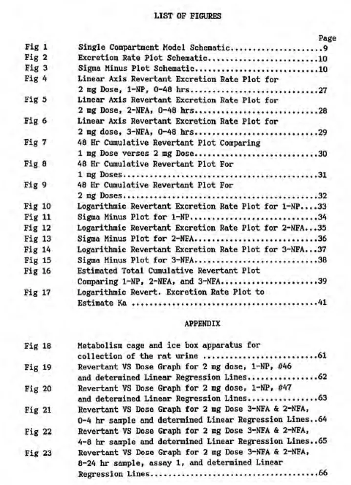
Linear Axis Revertant Excretion Rate Plot for
2 mg Dose, 1-NP, 0-48 hrs...27
Linear Axis Revertant Excretion Rate Plot for
2 mg Dose, 2-NFA, 0-48 hrs...28 Linear Axis Revertant Excretion Rate Plot for
2 mg dose, 3-NFA, 0-48 hrs...29 48 Hr Cimulative Revertant Plot Comparing
1 mg Dose verses 2 mg Dose... 30
48 Hr Cumulative Revertant Plot For
1 mg Doses...31
48 Hr Cumulative Revertant Plot For
2 mg Doses...32 Logarithmic Revertant Excretion Rate Plot for 1-NP....33 Sigma Minus Plot for 1-NP...34 Logarithmic Revertant Excretion Rate Plot for 2-NFA...35 Sigma Minus Plot for 2-NFA...36 Logarithmic Revertant Excretion Rate Plot for 3-NFA...37 Sigma Minus Plot for 3-NFA...38 Estimated Total Cumulative Revertant Plot
Comparing 1-NP, 2-NFA, and 3-NFA...39
Fig 17 Logarithmic Revert. Excretion Rate Plot to
Estimate Ka ...41
APPENDIX
Fig 18 Metabolism cage and ice box apparatus for
collection of the rat urine ...61
Fig 19 Revertant VS Dose Graph for 2 mg dose, 1-NP, M6
and determined Linear Regression Lines...62
Fig 20 Revertant VS Dose Graph for 2 mg dose, 1-NP, #47
and determined Linear Regression Lines...63
Fig 21 Revertant VS Dose Graph for 2 mg Dose 3-NFA & 2-NFA,
0-4 hr sample and determined Linear Regression Lines..64
Fig 22 Revertant VS Dose Graph for 2 mg Dose 3-NFA & 2-NFA,
4-8 hr sample and determined Linear Regression Lines..65
Fig 23 Revertant VS Dose Graph for 2 mg Dose 3-NFA & 2-NFA,
8-24 hr sample, assay 1, and determined Linear
Fig 24 Fig 25 • Fig 26 Fig 27 Fig 28
LIST OF FIGURES Continued
APPENDIX
Page
Revertant VS Dose Graph for 2 mg Dose 3-NFA & 2-NFA, 8-24 hr sample, assay 2, and determined Linear
Regression Lines...67
Revertant VS Dose Graph for 2 mg Dose 3-NFA & 2-NFA,
24-36 hr sample and determined Linear Regression
Lines...68 Revertant VS Dose Graph for 2 mg Dose 3-NFA & 2-NFA, 36-48 hr sample and determined Linear Regression
Lines...69
Revertant VS Dose Graph for 2 mg Dose, Controls (no S9), 0-48 hr samples and determined Linear
Regression Lines...70
Revertant VS Dose Graph for 2 mg Dose, Controls (+S9), 0-48 hr samples and determined Linear
Regression Lines...71 Fig 29 Revertant VS Dose Graph for 1 mg dose, 1-NP, #6,
0-48 hr sample and determined Linear Regression
Lines...72
Fig 30 Revertant VS Dose Graph for 1 mg dose, 1-NP, #7,
0-48 hr sample and determined Linear Regression
Lines...73
Fig 31 Revertant VS Dose Graph for 1 mg dose, 2-NFA & 3-NFA, 0-8 hr sample and determined Linear Regression Lines...74
Fig 32 Revertant VS Dose Graph for 1 mg dose, 2-NFA & 3-NFA,
8-24 hr sample, assay 1, and determined Linear
Regression Lines...75 Fig 33 Revertant VS Dose Graph for 1 mg dose, 2-NFA & 3-NFA,
8-24 hr sample, assay 2, and determined Linear
Regression Lines...76
Fig 34 Revertant VS Dose Graph for 1 mg dose, 2-NFA & 3-NFA,
24-36 hr sample and determined Linear Regression
Lines...77 Fig 35 Revertant VS Dose Graph for 1 mg Dose, Controls
(+S9), 0-36 hr samples and determined Linear
Regression Lines...78
Fig 36 Revertant VS Dose Graph for 1 mg Dose, Controls
(no S9), 0-36 hr samples and determined Linear
Regression Lines...79
Fig 37 Revertant VS Dose Graph for 2 mg Dose, Controls
(no S9), 0-48 hr samples including contaminated
assay CI, 8-24 hr sample and determined Linear
LIST OF TABLES
Page
Table 1 Cum. Revertant Values and Error For Figure 7 ....40 Table 2 Cum. Revertant Values and Error For Figure 8___40 Table 3 C\im. Revertant Values and Error For Figure 9 ....40 Table 4 Quality Assurance Results of all samples...42 Table 5 Linear Regression Values for all
Dose VS Revertant Plots...43
Table 6 Linear Regression Coefficients for all
1-Nitropyrene (1-NP) and nitrofluoranthenes (NFA) are prevalent air pollutants contributing significantly to mutagenic activity of
ambient organic particles in both cities and rural areas (Aray, et al
88; Ramdahl, et al 86; Tokiwa, et al 87). The everyday operation of
combustion engines exhausting into the atmosphere built these
pollutants' concentrations high enough to initiate scientific concern for human health. Assessment of 1-NP's mutagenicity is fairly complete implicating 1-NP as a potent mutagen and furthering concern about the carcinogenicity of Nitro-PAHs. The task continues to correlate current results to human risk and better categorize other nitroarenes and air pollutants according to their potential mechanisms of action.
This study provides a preliminary investigation into the mutagenicity of actual in vivo metabolites of 2-NFA and 3-NFA.
Characterization of 1-NP's in vivo fate and in vitro mutagenicity is established while only in vitro studies for 3-NFA and 2-NFA are available. This study uses the Ames bacterial assay to examine the
mutagenicity of urinary metabolites of 2-NFA and 3-NFA. Sprague Dawley
and 3-NFA dissolved in acetone. Developed Excretion Rate Plots
illustrate each compounds' mutagenicity and kinetics. Considering the past in vitro research results of the NFAs and using this study's in vivo results for all three compounds, preliminary characterizations of the urinary mutagenic metabolites of 2-NFA and 3-NFA are established
II. A. Background
II. A. 1. Fomiation of Mitro-PAHs
1-NP and 3-NFA are immediately formed by-products of combustion
due to the interactions of nitrogen oxides (NOx), nitric acid, and
fluoranthene molecules produced by incomplete combustion of fossil fuels (Pitts, et al 85; Ramdahl, et al 86). 1-NP, 3-NFA, and 2-NFA can be
formed any time after combustion during atmospheric chemical
interactions between the by products fluoranthene and dinitrogen
pentoxide (N2O5) catalyzed by a OH radical with NOx (Pitts, et al 85;
Ramdahl, et al 86; Zielinska, et al 87). Atmospheric conditions of
radiant energy, constituents' concentration and degradation rates,
organic particle interactions, and other forms of natural conditions
determine the rate of nitro-PAH formation. 2-NFA has been found in the
most abundance of the nitro-PAHs on ambient organic particles, although not found directly in diesel emissions as are 1-NP and 3-NFA (Aray, et
al 87; Ramdahl, et al 86). Nitro-PAH formation even occurs in the
charring process of some foods (Kinouchi, et al 86). All three
compounds are prevalent air pollutants due to their organic particle
II. A. 2. Biological Reactivity of Nitro-PMts
Nitro-PftHs can be absorbed via inhalation and/or ingestion during
normal everyday exposure. (King et al, 83; Bond & Sun 86; Bond, et al
86). Research has shown Nitro-PAHs require biological activation and
many are considered potent mutagens and subsequently, potential carcinogens (Ball, et al 84; Ball and King 85; Ball and Lewtas 86; Console, et al 89; Hirayama, et al 88). Indeed, Howard (et al 83) and
Dietrich (et al 88) showed 1-NP and 3-NFA metabolites can form a DNA
adduct. DNA adducts are suggestive of a chemical's carcinogenic
potential because they are proof of the chemical's ability to directly
interact with DNA.
In vivo metabolism of Nitro-PAHs involves many factors due to the
enterohepatic circulation. The compound may undergo oxidation, acetylation, and conjugation in the liver where the conjugated
metabolite may then be transported to the intestines. Intestinal flora
may reduce the metabolite while B-Glucuronidase- and
Sulfatase-containing bacteria may deconjugate the metabolite allowing absorption
into the enterohepatic circulation. Ball (et al 84) demonstrated the
importance of gut flora in the metabolism reduction of nitro-PAHs
followed by N-acetylation to the mutagenic acetylated metabolites.
Oxidation of Nitro-PAHs via the liver's mixed function oxidase results
in ring epoxidation and ring hydroxylation (King, et al 87; Howard,
et al 85; Ball, et al 84; King et al 84). Oxidative and reductive
nitro-PAH metabolism in vivo. Time dependence of specific metabolite
production signified the activity of the enterohepatic circulation in
nitro-PAH metabolism (Ball & King, 85). Several bacterial strains are
capable of these pathways, but unfortunately multiple strains can not
easily be used simultaneously. The use of urinary metabolites provides
a non-invasive look at in vivo metabolism. Bacterial strains recreate a
pseudo metabolism model to view the resulting body's exposure to
reabsorbed potentially mutagenic metabolites via the enterohepatic
circulation.
Characterizing nitro-PAHs via animal metabolism is limited in
scope since human P-450 metabolism of nitro-arenes may differ greatly
from rodents', questioning the rodent model's reliability as predictors of human metabolism (Howard, et al 90). Consequently, no direct human
health risks can be inferred, but we can develop a preliminary appraisal
of nitro-PAHs' in vivo metabolism, mutagenicity, and potential
carcinogenicity.
II. A. 3. Analytical Techniques
II. A. 3. (a). In Vivo Metabolism Study
Six week old, male, Sprague Dawley rats were selected for this in
vivo metabolism study. These rats are fairly competent nitroarene
Sprague Dawley rat liver microsomes (Belisario, et al 90). Although the
extent of P-450 epoxidation of nitroarenes differ between species (Howard, et al 88), a majority of previous research has accepted the Sprague Dawley rat as the standard model (Ball & King, 85; Belisario, et
al 90; Howard, et al 85; Howard, et al 88). To minimize confounding
between rat samples, all rats were treated identically. Acclimatization
time, food, water, cage type, injection and collection times, age, and
gender were consistent for all rats.
In vitro studies have shown similar metabolism rates for both 1-NP
and NFAs so in vivo rates could be proportionally similar. 1-NP's past
metabolism research shows a majority of the dose is excreted in the first 24 hrs, approximately 15% in urine and 40% in feces (Ball, et al
84; Ball & King, 85; Kinouchi, et al 90). Considering Stocking's (89)
findings of 2-NFA's slow in vitro metabolism, an extended collection period of 48 hrs ensured the majority of sample was collected. Only urinary metabolites were collected as the fecal metabolites should not
significantly elevate mutagenicity levels above background in Salmonella
Typhimurium strains as evidenced by 1-NP (Ball, et al 84).
Initial excretion of 1-NP metabolites before 8 hrs primarily
consisted of the oxidative products, diols and hydroxy metabolites, which are not potent mutagens. After 8 hrs, the enterohepatic
circulation enabled ring oxidation, nitroreduction, and N-acetylation
with peak mutagenicity levels attained around 12 hrs (Ball & King, 85;
Howard, et al 85; Kinouchi, et al 86). Shorter collection periods were
required to better characterize the initial portion of excretion. DMSO
proved to be an inefficient transport for nitro-PAHs into the systemic
system (Ball, et al 85) while acetone provided an increased delivery
efficiency. Less than 2% of metabolites excreted degrade by bacteria or
enzymes between collection and analysis (Ball, et al 84). Immediately
freezing samples upon collection and storage in a dark freezer ensured
samples authenticity until use in the ftroes assay.
II. A. 3. (b). Ames Salaonella Assay
The Ames Salmonella Typhimurium assay is widely accepted for
chemical and urinary mutagenicity research and specifically, the TA98
strain is frequently used for, and most responsive to nitro-PAH (Ball et
al 84; Ball et al 85; Howard, et al 87; Zeiger, et al 87; Zielinska, et
aJ 87). Salmonella Typhimurium strain TA98 is modified with a -1
frameshift disabling its histidine production capability. Lacking a
repair mechanism, a frameshift mutation is required to reset the gene
frame for proper function. Nitroreductase is a key metabolic activity
of TA98 as nonreductase deficient strains decrease mutation rate of
1-NP, a known nitroreductase dependent mutagen (Consolo, et aJ 89). The
bacteria is grown in a histidine deficient medium with the chemical of
histidine is evidence of a frameshift mutation of the genome. The assumption is that any chemical able to effect a mutation of the genome
is capable of other unidentifiable mutations of the genome presenting it as a potential carcinogen. Many mutagens recpiire exogenous activation
by S9, a mammalian system which provides a mixed function oxidase dependent on a NADPH, glucose-6-phosphate electron generating system.
S9 is from Aroclor 1254 induced animals which provides a wide range of
P-450 mixed function oxidases (Maron & Ames, 83). The Ames bacterial
assay has an overall positive predictive value of only 62% (Tennant, et
al 87), however, is over 90% efficient in correlating mutagenic nitroarenes as carcinogenic in rodents (Zeiger, 87).
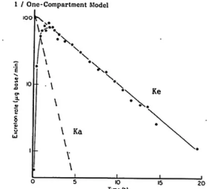
II. A. 3. (c). Toxicokinetic Analysis
Toxicokinetics has progressed rapidly over the past two decades becoming an extremely useful tool for invasive and non-invasive in vivo
studies. Particularly of interest to this study is the analysis of urinary excretion data. The urinary excretion rate of a compound is
identical to the plasma concentration level over time (Shargel & Yu,
85). Excretion rate data provide a profile of internal workings of the
animal. Excretion rate models consider the whole animal as one
kinetic model for first-order drug absorption and first-order elimination.
(Reprinted from Shargel and Yu, 85)
The rate of uptake and excretion of a substance is a function of
the rate constants, Ka, and Ke, which can both be estimated from
excretion rate data. The standardized method using an Excretion Rate
Plot is shown in Figure 2 (Shargel & Yu, 85). The slope of the terminal
end of the profile estimates the excretion rate constant, Ke, while Ka,
is the residue slope created by excretion line's data points subtracted
from the initial positive slope's data points. If Ka >>Ke, then the Y
intercept is the total cumulative amount of substance excreted which
also equals the total area xinder the curve (AUC). Also if Ka>>Ke then
the substance's excretion rate is formation rate limited (Shargel and
Yu, 85). The AUC can also be determined by using the linear excretion
rate to estimate the X intercept and calculating the AUC via the
trapezoidal rule. Once the AUC is known, a Sigma Minus Plot, Figure 3
(Shargel and Yu, 85), can plot the amount left to be excreted over time.
The Sigma Minus Plot's slope better estimates the excretion rate
constant Ke since all excretion rate data are used rather than just the
10
/ One-Compartment Model
lOO
ͣ
X.
\. \
10
ͣ
v
\
\
Ke
\
V Ka \
v
10
Time (h)
20
Fig. 2 Linear Axis Excretion Rate Plot to detenine Ke and Ka
(Reprinted froa Gibaldi and Perrier, 82)
1 / One-Compartment Model
100
o
<
a: 20
-o
<
Time (h)
II. B. l-Nitropyrene Mutagenicity Characterization
In vitro experiments initially charted each of 1-NP's metabolites
which were eventually found via in vivo research. The mutagenicity of
1-NP in vitro metabolites is decreased by S9 while curiously, its
urinary metabolites are activated by S9 (Ball, et al 84). 1-NP's in vivo pathway significantly differed from the in vitro route due to the
in vivo metabolism process, however, both paths essentially achieved
similar active mutagenic metabolites.
The majority of the dose after injection of rats was excreted in
bile as glutathione, cysteinylglycine, and cysteine conjugates (Bond, et
al 85; Howard, et al 85; Kinouchi, et al 90). Intestinal flora in the
intestines play significant roles in reducing 1-NP and metabolizing its
conjugates to reactivated metabolites enabling reabsorption via the enterohepatic circulation (EHC) (Ball & King, 85; Howard, et al 85; Kinouchi, et al 86; Kinouchi, et al 90). These reabsorbed metabolites
could be hydrolyzed in intestine and reduced by bacterial nitroreductase to reactive n-hydroxyl arylamine (NAA) derivatives (Ball & King, 85; Howard, et al 85; Kinouchi, et al 86; Kinouchi, et al 90). This
transformation to NAA may be common for all nitroarenes, but to what
extent is unknown (Ball & King, 85; Kinouchi, et al 90). The metabolite
6-hydroxy-NAAP accounted for the highest portion of urine mutagenicity excreted at 12 hours after dosing and required S9 for maximum bacterial
t^^j'/'i--*'!^f^'?i^-^^'fmw^m^m7r^'
12
metabolize the 6-OH-NSAP metabolite by esterification eventually
generating a nitrenium ion. Similarly, the parent, 1-NP undergoes in vitro nitroreduction to a hydroxylamine or nitrenium ion (Ball, et al 84). Howard (et aJ 83) showed the nitrenium ion formed by 1-NP can form a dG:C8 adduct N-(deoxyguanosin-8-yl)-l-ainino-fluoranthene. However,
1-NP metabolites reduce easier to DNA adducts than the parent 1-1-NP
(Djuric, et al 86). The excretion pathways of 1-NP appear to be
independent of exposure concentration where fecal remains the major
route accounting for twice as much as the urine route (Ball & King 85;
Bond, et al 85). The fecal does not contain significant mutagenic metabolites while very little parent 1-NP remained unmetabolized in the
urine (Bond, et al 85; Howard, et al 85; Ball and King, 85).
II. C. Characterizations of Kutagenicity of 2- & 3-Nitrofluoranthene
3-NFA is strongly mutagenic in bacterial strains containing nitro¬ reductase (Ball, et al 86), however 2-NFA is considered a weaker mutagen more closely related to 1-NP's levels. Nitroreduction aind
0-esterification have both been identified as slightly preferential
metabolic activators for 2-NFA and 3-NFA respectively (Console, et al
89). 2-NFA (Zielinska, et al 87; Belisario, et al 90) and 3-NFA
(Console, et al 89; Belisario, et al 90; Howard, et al 88; Stocking, 89)
likf, 1-NP (Ball & King, 85) were shown to be dependent on
esterification (McCoy, et al 83; Console, et al 89). Both NFAs are less dependent than 1-NP on nitroreductase in vitro, however, a reductase
lacking bacterial strain still decreased 3-NFA activity by 49-69%
(Consolo, et al 89). Moreover, 3-NFA in vitro metabolism pathway is
through epoxide intermediates (Howard, et al 88) and the resulting phenolic metabolites' mutagenicity remain unexplained (Consolo, et al
89). 2-NFA is less active and more dependent on nitroreductase than 3-NFA, but less affected by S9's mixed function oxidase (Zielinska, et al
88). Like 1-NP, 2-NFA's mutagenicity was decreased slightly by S9 as
was its in vitro metabolites 9-hydroxy-2NFA and 8-hydroxy-2NFA
(Zielinska, et al 87). 3-NFA's in vitro mutagenicity like 1-NP's, is decreased by S9 in the bacterial strain TA98 but unlike 1-NP's in vivo metabolites, 3-NFA does not produce in vitro metabolites more dependent on S9 (Consolo, et al 89). Specifically, the 3-NFA-8-ol metabolite was
as mutagenic as its parent 3-NFA, but other 3-NFA metabolites were 10
fold less mutagenic (Consolo, et al 89). This implies 3-NFA metabolites
further metabolism may result in detoxification indicating not all
nitro-PAHs' metabolism mechanisms are alike (Ball & Williams, 86).
Stocking (89) demonstrated in vitro reduction of 3-NFA efficiently
produced only the 3-AFA metabolite while reduction of NFA produced
2-AFA which itself underwent acetylation to form 2-NA2-AFA, but at a
considerably slower rate. Indeed 1-NP and 2-NFA metabolites have been
primary in vitro metabolites were largely dependent on 0-esterification (Console, et al 89). Unlike the hepatic metabolism of 1-NP shown to
involve both ring oxidation and nitroreduction (Ball, et al 84; Howard, et al 85), NFAs were shown to only undergo ring hydroxylation in aerobic and only reduction in anaerobic conditions (Belisario, et al 90).
Metabolism of these nitro-PAHs seems to be very dependent on their
environment suggesting their metabolism may depend on their in vivo kinetics. Metabolism may also vary with the in vivo model used as P-450 metabolism of 3-NFA and 1-NP vary greatly between species (Howard, et al
88).
II. D. Metabolis* Kinetics of 1-NP, 2-NFA, and 3-NFA
1-NP metabolism in vivo has been well characterized. Initial
metabolites excreted are phenols and dihydrodiols which rapidly give way in the 8-12 hr period to the acetylated 6-OH-NAAP constituting the
majority of the 1-NP dose excreted. Since very little actual parent compound remains unmetabolized, the terminal clearance rate is truly the metabolites' excretion rate. Expanding the comparative picture to
estimate NFAs' response, metabolism kinetics provides key evidence as to
metabolism opportunities, although NFAs' metabolism rate remains highly
varied. 3-NFA in bacterial strains undergoes 90X metabolism to 2-AFA in
6 hrs (Stocking, 89). 2-NFA metabolism is slower with 10% of the parent
15
to 2-AFA and 15% of the parent is metabolized to 2-NAAFA. 2-NFA's
nonlinear metabolism rate currently is attributed to a secondary
nitroreductase or an oxygen sensitive reductase which is activated
(Stocking, 89). 2-NFA and two metabolites, 8 and 9-hydroxy 2-NFA depend on nitroreduction and 0-acetylation in vitro (Zielinska, et al 87), no
in vivo metabolic pathways have yet been described. Comparing exposvire dose to urinary metabolite mutagenicity over time, 1-NP metabolites
become more mutagenic as the enterohepatic circulation provides
opportunity for acetylation for mutagenic metabolite production (Ball,
et al 84; Ball & King, 85). The combination of a fast metabolism rate
predicted by Stocking (89) and the detoxifying action of metabolism implies most 3-NFA urinary metabolites may be direct acting mutagens
III. MATERIALS AND METHODS
III. A. Materials
III. A. 1. In Vivo HetabolisB Study Materials
Six week old Sprague Dawely rats whose weights ranged from 200-230 grams were purchased from Charles River while the plastic metabolism
cages (Nalgene) and animal isolation booth were provided by Department
of Laboratory Animal Medicine (DLM), University of North Carolina,
Chapel Hill. Granular rat chow was supplied by DLM and ground to powder form. Nitro-PAHs injected: 1-nitropyrene (99.7% purity) was purchased
from Midwest Research Institute (Kansas City WO); 3-nitrofluoranthene purchased from Chemsyn Inc. (Lenexa, Ks); and 2-nitrofluoranthene was
synthesized and purified by Dr Louise Ball (Dept. of Environmental Science and Engineering, UNC) according to procedures outlined by Kloetzel (et al 1955). HPLC grade acetone was supplied by Fisher Scientific and sterile single use syringes were supplied by Sector Dickerson & Co. (Rutherford, N.J.). Sterile 0.22 and O.AO micron
filters were purchased from MSI (Westboro, MA).
III. A. 2. In Vitro Ames Assay Materials
Agar-Agar was purchased from U.S. Biochemical Corp (Cleveland, OH).
Sodium Azide, 2-nitrofluoranthene, ampicillin trihydrate, and crystal
NADP, and Glucose-6-Phosphate were supplied by Boehringer Mannheim (West Germany). Dextrose was purchased from EM Science (Cherry Hill, NJ). L-histidine, biotin, magnesium chloride, potassium chloride for the
Vogel-Brunner medium and S9 salt solution, along with molecular biology grade
Dimethyl Sulfoxide were acquired from Fisher Scientific (Fairlawn, NJ). 2-Anthramine was purchased from Sigma Chemical Co (St Louis, Mo). Oxide
broth was supplied by Oxoid Ltd (Basingstoke, Hants, England). S9 from
Aroclor 1254 treated male rats purchased from Maltox Molecular
Toxicology, Inc. (College Park, MD). Phosphate Buffer, PH=7.5, was
obtained from the Linberger Cancer Research Center's Tissue Culture Facility (UNC,Chapel Hill). Salmonella Typhimurium strain TA98 was obtained from Dr. Bruce Ames, University of California at Berkeley.
III. B. Methods
III. B. 1. In Vivo MetabolisB Study
After a one week acclimatization period, Sprague Dawley rats were injected intra-peritoneally through a 25g needle with varying doses of 1-NP, 2-NFA, and 3-NFA of 2 mg, 1 mg, and 0.5 mg in 0.2 ml and 0.1 ml
acetone solutions. Three rats were used for each chemical and one
control animal per dose received a pure acetone injection. Immediately after the injection, rats were placed in plastic metabolism cages in a controlled, pathogen free environment located in the DLM facility,
"18
the cage collected and immediately froze urine samples during the 48 hr collection period. A picture of this apparatus is shown in Fig. 18,
Appendix A. Urine was collected at intervals 0-4 hrs, 4-8 hrs, 8-24
hrs, 24-36 hrs, and 36-48 hrs and sterilized by filtration with a 0.4
micron pore size filter and stored at -80 C in darkness until
mutagenicity analysis. Rats had continuous access to drinking water and powdered rat chow.
After a brief visual inspection of the injection site for
unabsorbed chemical and general appearance of intra-peritoneal cavity,
the carcass was incinerated by DLM, UNC. Metabolism cages were cleaned
with 95% ethanol solution, then washed with a soap solution and rinsed
after each animal's use. All contaminated syringes and materials were
doubled bagged, labeled, and disposed of as trace carcinogenic waste
through the UNC's hazardous material collection department. Vinyl lab
gloves, surgical face mask, and lab coat were worn during animal handling, urine collection, and cage work.
III. B. 2. In Vitro Anes Assay
Mutagenicity analysis was via a Salmonella Typhimurium plate
incorporation test using TA98 strain with and with out S-9 fraction as outlined by Ames' 82 publication. Quality assurance of the strain was
performed on the master plates made from frozen permanents stored at
characteristics of histidine dependency, rfa, crystal violet, and UV
light sensitivity, and ampicillin resistance. A colony was picked off the master plate and incubated in 25 mis of nutrient broth in a dry,
dark, shaker at 37 C for 16 hrs to develope an approximate
9
concentration of 1.5 x 10 cell/ml. Acceptable urine samples (see Results' paragraph IV. A.) were resterilized through a 0.22 micron
filter and divided into three undiluted volumes ranging from 75 vL to 250 uL for 2 mg dosed rat samples and two undiluted volumes ranging from
100 uL to 250 \iL for 1 mg dosed rat samples. Two control 0 mg dosed
rats' samples were evaluated for backgroiind urine mutagenicity for each
assay. B-glucuronidase, 100 units/plate, was included in the top
overlay to account for glucuronide conjugate metabolite formation. One
half ml of lOOX activity Aroclor induced S9 (40 ug/100 uL) was added to half the plates for each sample. The top agar mixture was vortexed at low speed for 3-4 seconds and poured evenly on to the agar plate. Plates were allowed to solidify on a level surface for approximately 30
minutes then placed inverted in a lOOX humidity, 37° C dark incubator
for 24 hours. Samples with insufficient amoxints of urine to complete an assay were combined with other samples from the same time period and
chemical if necessary. The 2 mg dose of 1-NP's 0-4 hr sample and the 4-8 hr sample were combined by time period. All 1 mg doses required
combination of the 0-4 and 4-8 hr samples for each rat maintaining
20
using 2-nitrofluorene (50ug) evaluated the responsiveness of the strain
while duplicate control plates with 2-anthramine (23 ug) evaluated S9's
backgroxind activity. Duplicate spontaneous reversion plates verified
TA98's background activity and provided a zero dose revertant value for future analysis. Only plates producing twice the spontaneous reversion
count were considered evidence of mutation. Since a limited number of
samples can be run at any one time, the Ames assay is capable of only a finite number of samples per assay. Samples were stratified by
collection time and dose for the assays. This stratification
compensated for individual assay variances allowing each compound to receive equal assay bias reducing error in their future comparisons. The mutagenic activity of each sample was determined by least
squares linear regression analysis using the spontaneous revertant rate as the zero dose point. Assay results were recorded as revertant/dose and converted to a revertant/hr rate for each individual sample. When
more than one Revert./Dose linear regression line was able to be
determined, an average value was used in calculating the revertant rate and the standard deviation identified as error. The background
reversion rates of the control rats were subtracted from the PAH dosed
animals' rates to produce linear scale and logarithmic scale Revertant
Excretion Rate Plots. These plots were used to calculate revertant
excretion rates which in turn were used to estimate the total cumulative
III. B. 3. Quality Assurance of Sanples
Before, diiring, and after the in vivo metabolism study, the rats'
health, physical condition, eating and drinking habits, and urination
volumes were monitored and recorded. Persistence of an unusual
observation when confirmed by the visual autopsy caused exclusion of the rat. The brief autopsy examined the injection site and intestines to
confirm the compound was properly delivered to the rat.
III. B. 4. Toxicokinetic Analysis
Revertants are an indirect measure of the mutagenic species being excreted. No quantitative results concerning the physical number of mutagens can be inferred from the information provided since the number of revertants produced per mutagenic metabolite is unknown. However,
this information can provide an overall view of the mutagen excretion
for comparative analysis.
The Logarithmic Revertant Excretion Rate Plot's profile for each
compound indicated the animal system appeared to be a one compartment
model with first order kinetics concerning mutagen excretion. Applying product excretion kinetic analysis to the revertant data, the combined pseudo absorption and formation rate constant, Ka, and the revertant
excretion rate constant, Ke, were estimated. Identifying the highest
22
compartment model. The lack of data points immediately after injection
required the area under the curve, (AUC), be calculated to estimate the
resulting range of total cumulative revertants. A Sigma Minus plot
using all five values of the Excretion Rate Plot was developed using the
AUC values to provide a more accurate estimation of the excretion rate
constant. Lastly, the combined absorption and formation rate constant,
Ka, was estimated accounting for error using the total c\imulative
revertant value, AUC, as the Y intercept for the Logarithmic Revertant
IV. A. Quality Assurance of Urine Samples
Eighteen of the forty eight rats used provided acceptable samples
for use in this study. Table 4 outlines the reasons for exclusion of
any rat or selective sample. Notably, at least two rats were used for analysis of each injected compound. Both the 1 mg and 2 mg doses were analyzed whereas the 0.5 mg dose provided an insufficient revertant rate
beyond the 8th hour of collection.
Analysis of Ames assay results revealed an 18 September assay of the 8 - 24 hr period samples was unusable for compounds 2-NFA and 3-NFA due to an unusually high revertant rate for the control urine shown in
Fig. 37 in the appendix. Fortunately, two other acceptable assays were
completed for this sample period.
IV. B. Kutagenicity of Urine Samples
Least squares linear regression analysis of each Ames Assays'
Revertants vs Dose plots are presented in Figures 19 through 36 in
Appendix A and their numerical slope data listed in Table 5. All future results were based on these linear regression lines whose regression coefficients, Table 6, averaged 0.91 + 0.07, indicating the strong
,^ 24
between samples. Linear axis revertant excretion rate plots for the 2
mg doses Figures 4, 5, and 6 compare rats' mutagenicity excretion rates.
These figures also illustrate the significant difference in urination
rates between rats during the first 8 hour period of the in vivo study.
The extremely low revertant rates of the control rats confirm the
treated rats* revertant rates are direct results of the injected
nitro-PAHs.
IV. B. 1. 1-NP's Urinary Mutagenicity
Maximum mutagenicity required exogenous activation by S9 and was
excreted between 714 hours after injection of the 2 mg dose and 7
-20 hrs for the 1 mg dose extrapolated from excretion rate plot, Fig. 7.
The direct acting mutagenicity behaved similarly but only provided 30^ +
105t of the activity, shown in Excretion Rate Plots, Figures 8 and 9. The 1 mg dose provided approximately 33% + 13% of the total
activity achieved by the 2 mg dose for S9 activated mutagenicity.
Moreover, the direct acting mutagenicity in the 2 mg dose was 15% + 5%
higher than the 1 mg dose.
IV. B. 2. 2-NFA's Urinary Mutagenicity
2-NFA's maximiun mutagenicity occurred between 7-24 hrs period for
both S9 activated and direct acting mutagens for both 1 mg and 2 mg
equaled the direct acting mutagenicity for the 1 mg dose. Doubling of
the dose resulted in S9 activated mutagenicity increasing ^0% + 30i
while direct acting mutagenicity rose 27% +3%.
IV. B. 3. 3-NFA's Urinary Mutagenicity
Maximxim mutagenicity was direct acting and excreted between 4-10
hrs for the 2 mg and similarly was excreted between 0-15 hrs for the 1
mg dose shown in Fig. 7. Exogenous activation by S9 decreased this mutagenicity by 45% + 30% for the 2 mg dose, Fig. 9, and 70% + 25% for the 1 mg dose, Fig. 8. Doubling the dose raised direct acting
mutagenicity 40% + 27% and indirect acting mutagenicity 20% + 15%.
IV. C. Toxicokinetlc Analysis of Revertant Excretion Data
IV. C. 1. 1-NP's Toxicokinetic Analysis
Extrapolation of the logarithmic Excretion Rate Plot's, Fig. 10,
terminal end yielded a range of 4,850 + 2,530 Revert/(48 hr to
infinity). Extrapolation of this terminal rate estimates the total
mutagenicity produced was 41,478 + 3,700 revertants. The Sigma Minus
Plot, Fig. 11, estimated Ke, the Excretion Rate Constant's range was -0.061 + 0.015 (1/hr). The combined absorption/formation rate constant Ka was estimated at 1.14 (1/hr) as shown in Fig. 17. The revertant
excretion rate constant Ke is 18 times smaller than Ka, thus mutagen
26
IV. C. 2. 2-NFA's Toxicokinetic Analysis
Extrapolation of the Logarithmic Revertant Excretion Rate Plot's,
Fig. 12, terminal end showed the upper bound of the revertant excretion
rate was actually 0 rev/hr accounting for error. Therefore the highest
excretion rate and the "best fit" excretion rate were used. The
terminal end's cumulative revertant rate was 11,680 + 8,500 Revert/(48 hr to infinity). Extrapolation of the terminal rate indicates the total
mutagenicity was 25,448 + 14,170 revertants. The Sigma Minus Plot, Fig.
13, estimated the revertant excretion rate constant, Ke, was - 0.016 + 0.058 (1/hr). The combined absorption/formation rate constant Ka was
estimated at 1.14 (1/hr) as shown in Fig. 17. The revertant excretion
rate constant Ke is 40 times smaller than Ka, thus mutagen excretion is
formation rate limited.
IV. C. 3. 3-NFA's Toxicokinetic Analysis
Extrapolation of the Logarithmic Revertant Excretion Rate Plot's,
Fig. 14, terminal end yielded a revertant rate of 1,944 + 1,497
Revrt/hr. This value estimated the total revertants excreted was 23,240
+ 4,000 revertants. The Sigma Minus Plot, Fig. 15, estimated the
revertant excretion rate constant, Ke, was - 0.079 + 0.0043 (1/hr). The
combined absorption/formation rate constant Ka was estimated at -1.14
(1/hr) as shown in Fig. 17. The revertant excretion rate constant Ke is 15 times smaller than Ka, thus mutagen excretion is formation rate
2,500
For 2 mg Dose, 1»NP, 0-48 hrs
m 2,000
+ͣͣ*
c
rt t!
(1) 1,500 >
m
a
1,000
E
a
a 500
0
M
i
0-4 hrs 4-8 hrs 8-24 hrs 24-36 hrs
Time (Hrs)
#46-S9
ͤ
#46+39 H #47-39 HI #47+39 [1 AVG1NP-S9
F"igure 4
36-48 hrs
800
600
m
ͣ
+-ͣ»
c
as
>
DC
400
200
Linear Axis Revertant Excretion Rate Plot
For 2 mg Dose, 2rNFA, 0-48 lirs
0
0-4 hrs 4-8hrs 8-24 hrs
Hi #26-39
#26 -I-S924 24-36 hrs 36-48 hrs
Time (Hrs)
#27-S9 H #27+39 ^ 2-NFAAvg-S9 EU 2-NFAAvg+S9
Figure 5
48
2,500
2,000
B 1,500
C r:
0) 1,000
a:
500
0
For 2 rmg Dose, 3-NFA, 0-48 hrs
#3
0-4 hrs 4-8hrs
Hii
§A
;ii3 #14-39 #14+89
8-24 hrs 24 24-36 hrs 36-48 hrs 48
Time (Hrs)
#15-39
ͣ
#15+39 [2] Avg 3-NFA-S9
ͤ
Avg 3-NFA+39
0
48 Hr Cymulative Ftevertaot Plot
1 mg Dose verses 2 mg Dose
30,000
> 20,000
,r
10,000
20 30
Time (Hrs)
40 50
1-NP(1mg)+S9 1-NP(2mg)+S9 2-NFA(1mg)+S9 2-NFA(2mg)+S9 3-NFA(1mg)-S9 3-NFA(2mg)-S9
--- ---
ȋ
---U---
---H---O---#---Figure 7
COB
> CD
cr
E
14,000
12,000
10,000
1 mg Doses of 1-NP, 2-NFA, & 3-NFA
8,000
6,000
4,000
2,000
20
Time (Hrs)
30
1-NP-39 1-NP+39 2~NFA-S9 2-NFA+39 3-NFA-39 3-NFA+39
ͤ
m O # A A
Figure 8
40
32 it* O a E O m ec >
i
o 00 o O o^4-8
o o o o o o o o o" CM o o o o CO CO + z I CO to CO -—V 0) COo X + CO 0) <•
E
1-NFA-S9 2-NF
Oo i
CM
o
NP-S9 1-NP+S9 2
o^
W B
H
3,000
1,000
300
r
B 100
c
t
^ 30
0)
oc
10
3
--[I] ͣ Q..._ .. r ͣͣͣ '\... \ •v \
\
. ͣͣ>-,^
...:..ͣͣͣͣ">,
•ͣͣͣͣ... <r ͣ ^,.:^... 1 \ \ ...\ ... \ \ 1 1 "~'\^_
„, 1,.
20 'W 60
Time (Hrs)
1-NP +S9
ͤ
Figure 10
80 100 120
34 (D C/5 O Q E CM O Q. (A E (55
in
-\ 5 :
ͣ §
/
ͣ
/ : / ͣ: /
/ : -•-ͣ / / :y' - ͣ
: i r?:
ͣ
,>^
'X : ..-' 1
1 € :
^
-/ ͣ „'-' <D ͣ
/ a--"i &i
,'<'' 9 .
-ͣͣͣ
,--
rW-^-- i |m, 1 , 1 i 1 i 1 i 1 \ 1
o
o o
o in
J. It)
o o
o 00
I I.
Q)o in
o o o o o o o o O
o o o o o o o o O
o o o o o o in <M
o lO
o Cxi
o in OJ T—
1.000
500
200
100 X
0)
50
c
CO
t
CD > 20 CD
DC
10 5
2
-f-igure 12
100 120
Time (Hrs)
2NFA +S9
iJ
Revertants
Sigma Minus Plot For 2-NFA
2 mg Dose
20,000
10,000
5,000
2,000
1,000
500
200
100
ͣ
"th3.. "TEk.
\--0.075 (1/hi)
Avg Ke -- -0.016 I 0.058 (I.^lw)
-0.016(1.'%)
80
100 200
Time (Hrs)
(Xu-Xu)
Figure 13
300 330 400
3,000
1,000
300
^ 100
C
^ 30
Of tr 10 15 "-R -.^,__ ͣͣͣ
-..,ͤ"ͣ
^'"ͣͣͣ.,^^
r- ...X...ci-> Nr-i -<.,:; tU '-^^^ ͣ ^—
-..„:...X,- ... ...r^"..^
""'-y
^'..,
^ , ^"^v..
; ...ͣ...X
ͣ
•ͣ
-
~^-~^-.
.-1 1 1 \, 1 1 ""'"^-.. "--^ 30 60 Time (Hrs) 75 3-NF--S9 ͣ I
Figure 14
Revertants 50,000
Sigma Minus Plot For 3-NFA
2 mg Dose
20,000
ro,ooo
5,000
2,000
1,000
500
200
100 Ci-..
a _...
__ ... v.
D
Avg Ke = -0.079 + 0.033 (1/lir)
[;:
...Q--..
-0.046 (1/fir)
\,-.
-0.112 (1/hr)
I_________ ± ± _L
20 40 50 60 80
Time.(Hrs) (Xu'-Xu)
Figure 15
100 115120 140
Cum. Revertants 50,000
40,000
30,000
20,000
10,000
0
0
Figure 16
2 mg Dose, 1-NP, 2-NFA, & 3-NFA
r
1
1 .j/f^
ͣr /
-ͣ
ͣ
'; . /
ͣ
___________-—---
ͣ
---"
1 / / £^\^
1 JT/l „ ><c_'i
^^
1 (/-""' "t^
P^ 1 1
ͣ
1 1
100
1-NP+S9
n
200
Time (Hrs)
2-NFA+S9 3-NFA-S9
A O
300 400
CO
48 Hr Cumulative Rovertant Plot's Values
Fig T, 1 nig DcMM vi»ri« 2 mg Ooae
Table 1
48 Hr Cumulative Revertant Plot's Values
Pig a, 1 mg t3oiM» of 1 -NP, 2-NFA, fc 3-NFA
•
1-NP(1mg)+S9 1-NP(2mg)+S9 2-NFA(1mg)+S9 2-NFA(2mg)+S9 3-NFA(1mg)-S9
3-NFA(2mg)-S9 |
4 Hours 2,216
20B + 200 1,872 -t- 900
8 Hours 2.ͣ436 + 1.600 10.492 1,080 + 680
1.768 + 1.500 2,O0O + SCO 8,992 + 4.500
1 24 Hours 9.8SO + 4.100 3a,428 + 450 3,066 + 1,390
9,160 + 4.600 4.936 + 3.500 1 9.088 + 7,220 ] 1 36 Hours 11,194 + 4,300 34,168 + 450 3,608 + 1,390
1 0,948 + SOOO 4,936 + 3,500 1 9.988 + 7,460 |
1 48 Hours 36.628 + 1,200 13,768 + 5610 21.320 + 7,540 1
1 -NP -S9 1-NP +S9 2-NFA -S9 2-NFA +S9 3-NFA -S9
3-NFA +S9 1
8 Hours 784 + 350 2,436 + 1,600 1,224 + 9001,080 + 680 2,000 + 800 804 + 600
24 Hours 1,464 + 502 9,880 +4,100 2,948 + 1,400 3,056 + 1,390 4,936 + 3,500
1,397 + 1,200
36 Hours 1,608 + 502 11,164 + 4,300 3,224 + 1,400 3,608 + 1.390
4,936 + 3,500 1,397 + 1,200
Table 2
48 Hr Cumulative Revertant Plot for 2 mg Doses
Fig e, Values and Errom
1-NP-S9 1-NP +S9 2-NFA +S9 2-NFA -S9 3-NFA -S9
3-NFA +S9 1
4 Hours 480 2,216
208 + 200 316 + 3O0 1,872 + 900 1.708
8 Hours 3.116 10,492 1,768 + 1,500 560 + 300 8.992 + 4.500 4,232 1
1 24 Hours 13,436 + 2.B80 32,428 + 448 9.1 60 + 4,5O0 3,680 + 2,700 19.088 + 7.220 9,144 + 2,500 1
1 36 Hours 14,132 -t- 2.8SO 34,168 + 4SO 10,948 + 5,000 4.268 + 2.900 19,998 + 7,460 9,408 + 2.500 1
1 48 Hours 1 4,744 + 2.880 36,628 + 1,200 13,768 + 5,670 21,320 + 7,540 9,684 + 2,500
Table 3
Ln(Reverants/Hr) Y(Ke) -Y(Excr. Ftete Curve)
12 C]
ͣ
10 4>'
8
6
4
2
0
0
All Ka are approx. = -1.14 (1/hr)
D.. O
-/.A
3 6
1-NP
Time (hrs)
2"NFA
Figure 17
3~NFA
ͣ
- (T-). . . J^
-ij=^js«wr---TABLE 4
QUALITY ASSUBANCE OF SAMPLES
42
Compound Amount
Injected (mg)
# Acceptable Unacceptable Rats and
Rats Reasons
1-NP 2 mg 2 died
2 poor physical health
very ill
1 inadequate injection
1-NP 1 mg 2
2-NFA 2 mg 2
1 mg 2
3-NFA 2 mg 2
1 mg 3
Controls 2 mg
1 mg
k
5
1 inadequate injection 1 poor physical health
Obstructed/bloated intestine 1 poor physical health
Abnormal looking intestine 1 poor physical health
Internal fluid in cavity
2 died 1 died
LINEAR REGRESSION VALUES FOR REVERTANT VS DOSE PLOTS
2 MG DOSE
COW>OUND SAhPLE
1-NP 1-NP 1-NP 1-NP 1-NP 1-NP 2-NF 2-NF 2-NF 2-NF 3-WF 3-NF 3-NF 3-NF CNTRL CNTRL 46-S9 46+S9 47-S9 47+89 Avg -S9 Avg +S9 26-S9 26+S9 27-S9 27+59 14-S9 14+S9 15-S9 15+S9 -39 +S9 0-4 HRS Rev/uL Sample 2-14 9-11 0.27 0-19 1-15 0.59 0.53 0.11 0.12
4-8 HRS 8-24 HRS 8-24 HRS 24-36 HRS36-48 HRS Rev/uL Rev/uL Rev/uL Rev/uL Rev/uL
Sample Sample 1 Sample 2 Sample Sample
3-57 11-18 0.17 0.41 1-76 1.07 0.62 3.01 0.04 0.09 3 8.00 3.65 6.47 0-45 1.09 1-65 1.67 1.98 0-84 2.03 0.80 0.115 0-116 1.67 1.11 3.74 0-83 0.34 1-29 1-11 0-086 0.14 0.74 1.92 1.30 0.94 1.61 1-11 0-25 0.15 0.02 0.07 0-57 1-90 0-17 0.94 0-22 1-30 0.28 0.85 0.81 0.44 0.12 0-04 0.11
1 MS DOSE
CO^f>OUND SAff^LE
1-NP 1-NP 1-NP 1-NP 2-NF 2-NF 2-NF 2-NF 3-NF 3-NF 3-NF 3-NF CNTRL CNTRL 6-S9 6+S9 7-S9 7+S9 29-S9 29+59 31-S9 31+59 17-59 17+59 19-59 19+59 -S9 +59
0-8 HRS 8-24 HRS 8-24 HRS 24-36 HRS Rev-/uL Rev-/uL Rev/uL Rev./uL Sample Sample 1 Sample 2 Sample
TABLE 6 44
Linear Regression Coefficients
For
Revertant/Dose Plots' Regression Lines
Compound 0-4 4-8 8-24 24-36 36-48 Avg L.R.coeff
hrs hrs hrs hrs hrs
2 Me DOSE
1NP 46 -S9
1NP 46 +S9 0.98 0.99 0.80 1.00 0.98 0.95
1NP 47 -S9 1.00 0.98 0.86 0.95
1NP 47 +S9 0-97 0.99 0.89 0.79 0-66 0.86
2hF 26 -S9 0.96 0.97 0.94 0.96
2NF 26 +S9 0.97 0.96 0.94 0.97 0.96
2NF 27 -S9 0.92 0-98 0.73 0.88
2NF 27 +S9 0-68 0.98 0.99 0.99 0-99 0.93
3NF 14 -S9 0.92 0.98 0.98 0-96 0.87 0.94
3NF 14 +S9 0-93 0.96 0.99 &.91 0.95
3NF 15 -S9 0.53 0.96 0.87 0.93 0.98 0.85
3NF 15 +S9 0.88 0.66 0.77
CTRL -S9 0.63 0.96 0.74 0.16 0.50 0.60
CTRL +S9 0.64 0.64 0.40 0.84 0.63
Avg Coeff- 0.85 0.98 0.94 0.90 0.89
1 M6 DOSE 0-8
HRS 8-24 HRS 24-36 HRS Avg L.R.coeff
INP 6 -S9 0.95 0.81 0.88
INP 6 +S9 0.95 0.92 0.94
IMP 7 -S9 0.99 0.85 0.81 0.88
INP 7 +S9 0.98 0.93 0.92 0.94
2NF 29 -S9 0.99 0.96 0.60 0.85
2NF 29 +S9 0.99 0-91 0.92 0.94
2NF 31 -S9 0.98 0.95 0.97
2NF 31 +S9 0.98 0-95 0.97
3NF 17 -S9 0.96 0-92 0.94
3NF 17 +S9 0.97 0.95 0.96
3NF 19 -S9 0.93 0.90 0.26 0.70
3NF 19 +S9 0.98 0.86 0.65 0.83
CNTRL -S9 0.15 0.25 0.33 0.24
CNTRL +S9 0.74 0-80 0.33 0.62
Avg L.R.Coefficient 0.97 0.92 0.74
REVERTANTS/HOUR RATE FOR ALL SAii^LES
2 M6 nnfiE
1-NP Hrs 46-S9 1-NP 46+S9 1-NP 47-S9 1-NP 47+S9 1-NP C0MB-S9 1-NP
C0r>B+S9 CNTRL-S9 CNTRL+S9
0 4 8 24 36 48 0 515-63 0 1351.08 187.08 0 774.72 31.25 0 1391-39 146.46 223.75 0 120.5 658.88 645.17 58-23 51.25 0 554-38 2069.25 1371.23 145.21 205.42 0 13.25 10,5 20.73 3.44 18.33 0 15 27 26-4 16-04 50-42 Hrs 2-NFA 26-S9 2-NFA 26+S9 2-NFA 27-S9 2-NFA 27+S9 3-NFA 14-S9 3-NFA 14+S9 3-NFA 15-S9 15+S9 0 4 8 24 36 48 0 155.5 61.75 88.78 35.67 0 103.75 147.25 318.6 179.79 274.58 0 302.71 63.33 0 633 607.58 118.13 197.5 0 849-25 1126-38 506.15 89.06 116.67 0 427.5 631-75 194-1 22.92 0 86.13 2435.13 757-29 59-81 107-33 0 421-26 21.46
1 MG DOSE 1-NP Hrs 6-S9
V. DISOTSSI<»
Investigating in vivo metabolism via a small scale study is
inherently prone to individual animal variations. Averaging only two values is insufficient data to do statistical analysis, thus a graphical
range method was used to provide the most reliable and understandable presentation. Animals do not consistently urinate which generates large
standard deviations when averaging single collection periods although over the entire collection period individual rat's results are similar. Two rats injected with 2 mg of 3-NFA produced revertant excretion
amounts within 25% of each other, but during specific collection periods they varied up to 50%. The outcome is an overall error range of 50%, double the actual difference. Accordingly more value may be placed on
the best fit lines than normally expected.
V. A. Urinary Hutagenicity
Tin the first 48 hours after injection, 1-NP injected rats'
cumulative urinary mutagenicity was twice 3-NFA's direct mutagenicity,
fivejtimes 3-NFA's indirect mutagenicity, and triple 2-NFA's indirect
I
mutagenicity. Estimated total revertant amounts excreted by 1-NP
6 . - .. ͣ
injected rats remained the highest although matched by the upper range
of 2-NrA injected rat's total amount while 3-NFA injected rat's amount
remained equal to 2-NFA rat's median mutagenicity amount, as shown in
3-NFA injected rat's maximum urinary mutagenicity excretion rate
occurred in the initial phases 4-10 hrs following injection, while 1-NP injected rat's occurred slightly after during the 7-12 hrs following injection, and 2-NFA injected rats had the slowest and most consistent rate lasting a full day. This coincides with previous research results showing peak urinary mutagenicity from 1NP metabolism occurs in the 8
-12 hr period after injection and is dependent on S9 activation (Ball, et al 84; Ball & King, 85). Representing the middle of the road, 1-NP's
kinetics are bordered above by 3-NFA's fast kinetic rates and below by
2-NFA's slow kinetic rates. All three compounds had similar Ka
constants suggesting the Ka measured may have been also representative of the acetone carrier rather than just the compounds' structure.
Stocking (89) suggested 2-NFA to be less mutagenic than 3-NFA due
to its slower and inefficient bacterial metabolism. Indeed, 2-NFA's
slower metabolism generates a slower mutagenic production rate, however,
the duration of active mutagenic urinary metabolites being excreted is
much longer than for 1-NP. The result being a total revertant output
potentially as large as 1-NP and at least as large as 3-NFA's amount.
V. B. Kinetic Relationship to Hutagenicity
The resulting mutagenicity of each compound may be directly
related to the reciprocal dependency between the metabolism process and
48
as relying strongly on nitroreduction and enterohepatic circulation for
acetylation in the liver. Our study's results show urinary mutagenicity
from 1-NP metabolism behaved as estaJalish by several preceding studies
(Ball, et al 84; Ball and King, 85; Bond, et al 85; Howard, et al 85) and thus underwent predicted pathways and kinetics.
The revertant excretion rate plot profile and this study's
analysis strongly suggest the overall excretion of 2-NFA's mutagenic metabolites is significantly slower than 1-NP's and apparently slower
than 3-NFA's. In vitro research has shown 3-NFA is metabolized in 6 hrs
while 2-NFA takes up to 24 hrs (Stocking, 89). 2-NFA's slower
metabolism may be counterbalanced by its longer biological half life and subsequent increased probability of metabolism. The longer half life
may result from slow partitioning of 2-NFA and its metabolites in vivo
possibly due to structural differences discussed later. Stocking (89)
found 3-NFA to efficiently produce only 3-AFA via nitroreduction, while 2-NFA was reduced to 2-AFA which could undergo acetylation to 2-NAAFA.
2-NFA's longer biological half life and greater opportunity for metabolism may increase its ability to form both 2-AFA and its
metabolite 2-NAAFA. 2-NAAFA may cause the indirect acting mutagenicity
found from NFA metabolism similar to 1-NAAP production from 1-NP.
2-AFA, like its isomer 3-2-AFA, may be responsible for the direct acting
Moreover, 3-NFA had the fastest revertant excretion rate averaging
40% greater than 1-NP's rate and 400% faster than 2-NFA's rate. This
high revertant excretion rate for 3-HFA results may be indicative of two
situations. There may have been insufficient enterohepatic circulation of the compound for acetylation metabolism to produce the secondary mutagen species exhibited by 1-NP and 2-NFA. The second and more
probable situation is acetylation of 3-NFA may simply not produce an
active mutagenic species. Ball (et al 85; et al 86) did show further
metabolism of 3-NFA's metabolites in vitro detoxifies them, as shown
even in this study where the exogenous metabolism by S9 halved their total mutagenicity. 3-NFA's high mutagenicity excretion rate during the first 8 hrs was comparable to 1-NP's rate. 1-NP is known to produce
phenolic and dihydrodiol metabolites during initial hours of excretion
(Ball et al 84; Ball et al 85). 3-NFA mutagenic species' immediate excretion and direct acting mutagenicity correlates with in vitro results showing 3-NFA relies heavily on 0-esterification producing
epoxide intermediates (Zielinska, et al 87; Console, et al 89). This
may be evidence the initial and majority of its mutagenic urinary
metabolites are phenols and dihydrodiols. 3-NFA mutagenic metabolites'
fast excretion rate with inactivation of mutagenicity by further
metabolism, as seen with S9, also complies with in vitro research
(Howard, et al 88; Console, et al 89) discussed earlier in the
50
3-NFA metabolites may be excreted at a rate similar to 2-NrA, but detoxification by further metabolism allows the revertant excretion rate to provide a misconception of fast excretion kinetics. Indeed in vitro results suggested 3-NFA to be more mutagenic than both 2-NFA and 1-NP
(Stocking, 89; Console, et al 89). Considering that 3-NFA's cumulative
mutagenicity could be only half of 2-NFA's amount, it is probable the
revertant excretion rate actually represents the detoxifying metabolism
rate of 3-NFA's mutagenic metabolites as they enter the enterohepatic
circulation and undergo additional metabolism. Without the ability to
account for the total dose excreted this theory can not be sufficiently
evaluated.
The differing excretion kinetics may signify differing substrate binding affinities of 2-NFA and 3-NFA. The binding may be stronger for 3-NFA promoting metabolism or be stronger for 2-NFA delaying metabolism and lengthening the biological half life of mutagens or their
precursors. This complements Stocking's (89) second implication that,
the NFAs' nitro-groups may structurally affect nitroreductase's
availability to the active site according to Vance and Levin's (84)
research. Whether the mechanism is to catalyze nitroreduction, catalyze
esterification, or act as a competitor remains obscured. This
structural theory may also be relevant to the metabolism mechanism if
acetylation of 3-NFA truly does not produce active mutagenic metabolites
V. C. Dose Relationship to Ilutagenicity
All three compounds appear to exhibit a nonproportional rise
between indirect acting mutagens (S9 activated) and direct acting
mutagens with increasing dose. The general observation is the
predominant mutagenic metabolites of each specific compound received a
disproportionate increase with increasing dose. This observation is not
caused by assay bias but appears to be truly dose dependent since the
nonproportional increase occurred to 3-NFA's direct acting mutagenic
metabolites and 1-NP's and 2-NFA's indirect acting mutagens. Doubling
the 2-NFA dose seems to increase the indirect acting mutagens more. Stocking's (89) suggested a sattirable process existed in the bacterial strains metabolism of 2-NFA which may or may not be related to this
apparent event in vivo. A second possibility is the higher dose allows
a higher probability of enterohepatic circulation of products. This would account for 2-NFA's equal quantities of indirect and direct mutagens initially with the low dose but, then with increased dose an
increased percentage of compound undergoing reduction and acetylation
seemingly produces more indirect mutagenicity.
I suggest 2-NFA and 3-NFA are truly similar in their metabolism
pathways and mass excretion rates. Their main difference seems to be in
the metabolism mechanism and kinetics which allows production of the
potent NAAF metabolites by nitroreduction and acetylation. 3-NFA's
52
metabolism while 2-NFA, like 1-NP, produces a variety of mutagenic
species by additional metabolism introduced by the enterohepatic
circulation. Contrary to in vitro research, 2-NFA appears to present a higher mutagenicity dose in vivo similar to 1-NP while 3-NFA provides a
comparatively small in vivo mutagen dose due to the in vivo metabolism
system's detoxifying action.
V. D. Conclusions
This preliminary comparison of urinary mutagenicity of 1-NP,
2-NFA, and 3-NFA provides strong evidence:
1. Not all nitro-PAHs are metabolized to similar mutagenic
species
2. Revertant excretion rates of 3-NFA > 1-NP >> 2-NFA
3. Ctimulative urinary mutagenicity from 1-NP > 2-NFA > 3-NFA 4. 3-NFA urinary mutagenicity is direct acting and decreased by
additional metabolism.
5. 1-NP, 2-NFA, and 3-NFA urinary mutagen excretion is formation
V. E. RecoBHKndations For Future Research
The mutagenicity dependence on kinetics seen in this study by
2-NFA and 3-2-NFA has been difficult to analyze completely. Elucidation of
these kinetics is needed to better understand not only how mutagenic is a compound but, its actual in vivo mutagen dose. Future experiments
should be geared to thoroughly study and compare structural, in vitro,
and any other in vivo studies to fully grasp any relationship related to
their kinetic mechanisms. Researchers continuing the risk assessment of nitro-PAHs may next identify the nitro-PAH characteristics controlling the metabolism pathways and resulting biological exposure to mutagens.
I recommend repeating this study using radiolabled compounds at 1 mg to 3 mg range doses, to track and identify the urinary metabolites and their relationship in metabolism/excretion kinetics. Better
metabolite excretion rate plots are necessary requiring extension of the collection period out to three days with at least four animals per dose
per chemical. Collection of urine during the initial hrs after
injection at three 4 hour intervals or two 6 hour intervals will better
characterize the absorption/formation part of the excretion rate plot.
54
along with mutagenicity to better characterize potency. The ptreent&fsi of the initial dose of parent compound excreted over time should allow
investigation into the relationship between metabolism and resulting mutagenicity and the associated kinetics. The primary metabolites of
3-NFA and 2-3-NFA responsible for the direct and indirect mutagenicity need
BIBLIOGRAPHY
Ames, B., J. McCann, E. Yamasaki, 1975, "Methods for detecting
carcinogens and mutagens with the salmonella/mammalian microsome mutagenicity test", Mutation Research Vol. 31: 347-364
Arey, J., B. Zielinska, W.P. Harger, and R. Atinkinson, 1988, "The
contribution of nitrofluoranthenes and nitropyrenes to the
mutagenic activity of ambient particulate organic matter collected in southern California". Mutational Research, Vol. 207(2): 45-51 Ball, L. M., M.J. Kohan, L.D. Claxton, and J. Lewtas, 1984, "Metabolism
of l-nitro( )pyrene in vivo in the rate and mutagenicity of
urinary metabolites". Carcinogenesis, Vol. 5: 1557-1564.
Ball, L. M., and L.C. King, 1985, "Metabolism, Mutagenicity, and
activation of 1-Nitropyrene in vivo and in vitro". Environment
International Vol. 11: 335-362.
Ball, L. M., K. Williams, M.J. Kohan, and J. Lewtas, 1986, "S9-Dependent
Activation of l-nitropyrene and 3-nitrofluoranthene in bacterial mutagenicity assays".. Biological Reactive Intermediates III
(Plenum Publishing Corp. 1986)
Belisario, M.A., L. Carrano, A. de-Giulio, and V. Buonocore, 1986, "Role
of rat liver inducible enzymes in in vitro metabolic
transformation of l-nitropyrene". Toxicol-Lett. Jul-Aug;
Vol. 32(1-2): 89-96
Belisario, M.A., R. Pecce, R.D. Morte, and A.R. Arena, 1990,
"Characterization of oxidative and reductive metabolism in vitro
of nitrofluoranthenes by rat liver enzymes", Carcinogenesis,
Vol. 11(2): 213-218
Bond, J.A., J.D. Sun, M.A. Medinsky, R.K. Jones, and HC. Yeh, 1985,
"Deposition, metabolism, and excretion of l-[14C]nitropyrene and l-[14C]nitropyrene coated on diesel exhaust particles as
influenced by exposure concentration". Toxicol-Appl-Pharmacol.
Aug; Vol. 85(1): 102-17
Bond, J.A., M.A. Medinsky, and J.D. Sun, 1986, "Disposition and
metabolism of free and particle-associated nitropyrenes after
56
Consolo, M.C., M. Anders, and P.C. Howard, 1989, "Mutagenicity of the phenolic microsomal metabolites of 3-nitrofluoranthene and 1-nitropyrene in strains of Salmonella typhimurium"., Mutation
Research, Vol. 210(2): 263-9
Dietrich, A.M., C.R. Guenat, K.B. Tumor,and L.M. Ball, 1988
"Identification and characterization of the major DNA adduct
formed chemically and in vitro fonm the environmental genotoxin
3-nitrofluoranthene". Carcinogenesis Vol. 9(11): 2113-2119. Djuric, Z., D.W. Potter, R.H. Heflich, and F.A. Beland, 1986, "Aerobic
and anaerobic reduction of nitrated pyrenes in vitro". Chem-Biol-Interact. Oct., Vol. 59(3): 309-24
Djuric, Z., E.K. Fifer, P.C. Howard, and F.A. Beland, 1986, "Oxidative microsomal metabolism of 1-nitropyrene and DNA-binding of oxidized
metabolites following nitroreduction". Carcinogenesis. Jul;
Vol. 7(7): 1073-9
Djuric, Z., B. Coles, E.K. Fifer, B. Ketterer, and F.A. Beland, 1967, "In vivo and in vitro formation of glutathione conjugates from the K-region epoxides of 1-nitropyrene". Carcinogenesis. Dec; Vol.
8(12): 1781-1786
Durston, W.E., and B.N. Ames, 1973, "A simple method for the detection
of mutagens in urine: Studies with the carcinogen
2-acetylaminofluorene". National Academy of Science Vol. 3: 737-741 Edwards, M.J., S. Batmanghelich, S. Edwards, J.M. Parry, and K. Smith,
1986, "The induction of DNA adducts in mammalian cells exposed to
1-nitropyrene and its nitro-reduced derivatives". Mutagenesis.
Sep; Vol. 1(5): 347-52
Gibaldi, Milo, and D. Perrier, Pharmacokinetics, 2nd ed.. Marcel Dekker
Inc., N.Y. 1982
Hirayama, T., T. Watanabe, M. Akita, S. Shimomura, Y. Fujioka, S. Ozasa,
and S. Fukui, 1988, "Relationships between structure of nitrated
arenes and their mutagenicity in Salmonella typhimurium; 2- and
2,7-nitro substituted fluorene, phenanthrene and pyrene".
Mutat-Res. Sep-Oct; Vol. 209(1-2): 67-74
HoUoway, M.P., M.C. Biaglow, E.G. McCoy, M. Anders, H.S. Rosenkranz,
and P.C. Howard, 1987, "Photochemical instability of