R E V I E W A R T I C L E
Implant applications for children
Marzieh Mohamadi Moghadam
Department of Periodontics, Faculty of Dentistry, Mashhad University of Medical Sciences, Mashhad, Iran
Abstract
Background: Trauma and hypodontia are two most prevalent congenital dental abnormalities in children. A common treatment for them is the use of removable prosthesis, but this method may increase the residual alveolar bone resorption, periodontal problems, and secondary caries. Aim: The aim of the present article is to review the skeleton and dental growth trend and its effect on the application of implant and present guidelines based on related articles and books for prescription and nonprescription of implant. This paper is carried out by reviewing the data in Google Scholar, Pubmed, Embase, Blackwell Synergy, and Science Direct. Conclusion: Implants could protect the residual bone, increase retention, enhance the stability, beauty, and chewing power and improve the life quality in general. Clinical Significance: Due to bone integration and undesirable effects on maxillary and mandibular skeletal growth and subsequent tooth eruption, use of implant in growing patients arouses some concerns.
Keywords: Anodontia, children dental implant, hypodontia, skeleton and dental growth
Introduction
The growing patients are mostly in need of prosthetic rehabilitation in edentulous areas.[1] Trauma and hypodontia are the most prevalent abnormalities that cause congenital edentulous in children. A typical treatment, before full skeleton and dental growth is removable prostheses. However, the residual alveolar bone resorption, periodontal problems, and secondary caries will be increased by using a removable prosthesis.[2] Currently, in most of the cases implant is known as the best treatment plan for the edentulous area, in that it provides less dental preparation process in partial edentulous, more retention and stability in total edentulous increases beauty and chewing power, and improves the life quality. Moreover, its application is growing increasingly as a result of its successful prostheses (83% in maxilla and 94% in mandible).[3-5] Within a meta-analysis, the survival rate of implant, 6 months after the load bearing, is 93.6 and 97.5% for fixed partial dentures and single crowns, respectively.[6]
Dental and skeleton growth is the greatest concern for placing implant in children and teenagers. The growth effect on implant position, implant impact, and its prostheses and on dental and skeleton growth will bring about some limitations in typical application of implant in growing patients.[3] High demands for using implant, due to more awareness of parents of its positive psychological effects, as well as more inclination toward a more stable treatment and better physiological bone maintenance in congenital edentulous or traumatic areas, compared with
removable prosthesis,[7] have made the dentists to evaluate and be aware of the growth trend and mutual effects of implant positioning, more precisely.
Despite early application advantages for some patients with certain conditions (syndromes) who suffer from diastema, late positioning is more satisfactory until full dental and skeleton growth is reached and during this period alternative treatments, involving removable/fixed prosthesis and orthodontic treatment are more appropriate.[8] This is due to the fact, that implant is placed in a dynamic setting, does not work like a tooth, will remain fixed during teeth growth and replacement, and this would cause abnormalities and tooth row disarrangement.[1,9] On the other hand, various and unpredictable individual differences, especially before full development, have made any predictions impossible, as well. Therefore, placing an implant during mixed detention stage and even puberty has a limited prognosis, and in case the dentist is compelled to early positioning, longer and numerous edentulous areas (due to ankylotic nature and inhibition of vertical growth of implant along with the adjacent teeth) are privileged to single edentulous.[10,11]
This paper is conducted by reviewing the dental and skeleton growth trend and its effects on implant positioning and provides guidelines for the prescription or nonprescription of the implant by considering growth rate, number, and the place and type of edentulous based on the jaws.
Correspondence
Marzieh Mohamadi Moghadam, Department of Periodontics, Faculty of Dentistry, Mashhad University of Medical Sciences, Mashhad, Iran. Tel: +98-51-38067224, Fax: +98-51-38829500. E-mail: [email protected]
Received 18 January 2017 Accepted 28 February 2017
doi: 10.15713/ins.ijcdmr.114
How to cite the article:
Materials and Methods
The present review article was carried out by reviewing the data in Scholar Google, Pubmed, Embase, Blackwell Synergy, and Science Direct. Keywords sought were dental implant in children, dental and skeletal development, implant for children, hypodontia, and anodontia. Data related to various aspects of dental and skeletal growth trend, methods of selecting a patient and its contributing factors, the application of dental implant in children and its impact during the facial growth were gathered from available articles and books.
Implant and the Pattern of Dental and Skeleton Growth
In total, high rate growth accelerates during infancy, slows down during childhood, increases partially in adolescence, and finally reaches to a stable reduction during lifetime.[11] Limited reports are available of use of implant in children.[12-16] However, osseointegration is undoubtedly occurred in child’s jaw and dental implants, due to the existence of periodontal ligament and union to the bone, have the role of ankylosed primary teeth. Hence, such teeth are appropriate models for the evaluation of implant behavior in a growing child.[1,8,11] Among other complications of these teeth, we could refer to the defects of adaptive processes during dental and skeletal growth, burying, secondary severe abnormal occlusion, crooked teeth, and the difference between skeletal growth and adjacent dental area.[8,11]
Bjork argued that the overall development of the middle part of the face is in low and front directions and is related to the skull base that is mostly inactive up to the age of seven. However, during 7-15 the main factor for anterior displacement of nazo magazalari complex is the growth of maxilla and nose joints.
The lateral growth of the maxilla is controlled by the middle joint of the palate, which is an important growth area and its growth rate in posterior is 3 times higher than the anterior. This is the first dimension, the growth of which is equal to that of the adults [Figure 1].
The vertical growth of maxilla is also affected by the growth of facial joints and alveolar bone deposition. The resorption of internal nose, especially in frontal section and bone deposition at palatal level may cause the compensation of rotary motion at posterior maxilla.[17-19] Cronin and Oesterle introduced the vertical dimension of the maxillary growth as the most disturbing implant-positioning factor and predicted long-term destructive effects on the beauty and performance of upcoming tooth restoration.[1]
Moyers et al. suggested that the transformation of the primary teeth system to permanent as well as the lateral rotation of the maxilla is the main factors of dental arch length and perimeter reduction.[20] These changes may bring about the formation of smaller dental arch in 18 months than that of the 4 months.[20,21] In longitudinal dimension of dental growth, in addition to such dental motions, total dental set of the maxilla would be located at a more anterior position than the base of the upper jaw.[1]
Long-term studies indicated that the permanent detention superseded the primary ones and this displacement results in considerable incremental alterations in the lateral arch of the permanent detention.[4]
In boys (due to puberty), the size of the arch and its alterations takes a longer process, so it has a longer growth and higher rate, which is evaluated to be between 0.5 and 3 mm.[22]
Jones and Meredith illustrated 3-4 mm of maxilla height increase during primary teeth eruption, and exfoliation process and this value goes even further by the eruption of the permanent teeth.[23] The actual increase of dental height (about 5-6 mm) is occurred between the age of 5 and 15.[1,8] The anterior maxillary height has shown numerous individual differences, and the correlation coefficient between tooth size in the age of 15 and 4 is trivial.[23] Therefore, the mutual effects of implant use and dental alterations are unpredictable during childhood.
Iseri and Solow revealed that the eruption of upper jaw teeth might have a strong effect on implant treatment, such that the average annual eruption rate during development ages is 1.2-1.5 mm and the compensation of such alterations using the implant asks for 9-10 tough years in girls and is impossible in boys.[24] Any changes in palate height were also assessed by some Scientists as they calculated the distance between occlusal section and palate section about 2.5-6 mm. This amount includes the increase of alveolar height and vertical height of permanent detention.[20] The remarkable growth of bone height, especially at posterior palate, might cause the implant to be buried more than one centimeter at posterior maxilla and even the apical level to be appeared at the base of nasal cavity.[1]
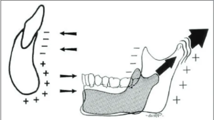
In contrast to the maxilla, the mandibular growth is not related to the development of skull bones and has a low and front direction. Bjork by overlaying the sequential cephalograms indicated that the bone deposition under the symphysis and angle is the main factor for nonlinear growth of the mandible and forward rotation.[17-19] Such growth process varies markedly individually and the direction of condylar growth generate the center of rotation, such that the vertical growth of condylar along with the center of rotation in incisors leads to the flattening of
the marginal mandibular, more labial eruption of lower incisors, and the decrease of gonial angle. While the anterior-posterior growth of condylar is at the center of molar rotation, it creates a steeped margin, a lingual tilt in incisors, and their vertical growth and no reduction in the gonial size. Thus, the direction and range of mandibular rotation has a significant effect on the final position of the tooth.[17] The amount of mandibular rotation (both internal and external rotation) is more than that of the maxilla, but bone resorption and deposition of the marginal mandibular hides this factor. Implant positioning at posterior mandible, due to the growth of condylar and burying of implant at various levels in children, especially in cases where there are extreme internal and external mandibular rotations, is divergent and unpredictable.[9] Implant at anterior mandible is also unable to perform the required angle changes to compensate the mandibular rotations like teeth with periodontal fibers, so it affects the beauty and performance of adjacent and opposite teeth. The longitudinal growth of mandible in ramus area is taken place with resorption in anterior margin and bone deposition at its posterior margin as well as the condylar area [Figure 2].[1] Meredith revealed that the ramus height and trunk length have the annual growth of 1-2 and 2-3 mm, respectively. Cronin declared that the cause of prominent chin in adulthood is the bone resorption in the previously mentioned dental area and the probability of implant protrusion, especially in earlier ages.[9]
A scientist suggested that the range of mandibular transverse growth is less than the maxillary and the simultaneous increase of length and width, especially in mandibular posterior arch is due to the V shape nature of the mandible [Figure 3].[25]
Due to early cessation of growth in the symphysis, the transverse growth of posterior mandible will reach to its maximum and remain stable before the eruption of deciduous teeth, and only a limited depositional growth will maintain.
During growth, mandibular incisors and canines will grow vertically. This process is fixed in dental height and alveolar bone and is more in men, such that it will continue up to the age 13-14 in girls and age of 15 in boys.[9]
Rilo specified that the height increase of anterior mandible in the age of 6-16 in girls and boys is about 6 and up to 10 mm, respectively and these values have strong effect on implant positioning.[26] The eruption of mandibular incisors could cause 2 (in women) to 3 (in men) millimeters of intercanine width increase, due to labial direction of permanent incisors, their greater size, and pushing primary canines to the primate space. These values will not change by the eruption of lateral incisors and canine and instead show a slight decrease in intercanine width in the age of 13-25.
Up to 4 years ago, Moyers et al. attributed the highest cross changes to premolars and indicated that the arch width will increase 2 mm by the eruption of premolars, 2-3 mm by the first molar, and 0-2 mm by the second molar.[20]
Such cross changes is vary significantly in different people.[27] The results of Holcomb illustrated that the prediction of arch width at the age of 15 through dental arch at the age of 4-5 is not reliable.[28] Therefore, although the minimum of changes is taken place at the mandible transverse dimension, various vertical,
and rotational changes result in the alteration of the complete mandibular system. The longitudinal changes of mandible arch are different from that of the maxillary and the eruption of permanent incisors is closer to their final arch position.[1-9] Moorrees and Reed showed that the loss of deciduous molars, eruption of premolars, and mesial drift of the first molar could cause 2 mm of arch length decrease and these change will increase by the mandibular growth.[29] Accordingly, the implant is unable to be adopted with the natural dental changes. Like maxillary, the whole mandibular system will replace seamlessly, but due to slighter limitations in joint growth and early closing of the symphysis, the extent of such development is trivial.[4]
Regarding such limited changes, adjusting the angle and vertical eruption of teeth (especially in incisors) in response to mandibular rotation, as well as more than one centimeter of increase of alveolar height must be regulated in implant positioning.[1]
Implant and Diversity of Growth Factors
The range of growth is specific to individual and various factors, involving genetic, nutrition, systematic diseases, and
Figure 2: A typical pattern of mandibular midline and ramus transformation
psychological problems contribute to the issue.[30] Major differences were also seen in skeleton jaws and direction of dental eruption. In general, the dental system compensate the skeleton diversity through change of direction and range of growth, the examples of which are long-faced people with longer and more vertical growth of incisors and prognathic people with labial steep of upper incisors and lingual steep lower incisors.[1] Sex differences are also among the main growth variables. In total, the average age of tooth eruption is earlier in girls (2 years).[31] With the onset of menstruation (around 15), the development process is almost complete in girls, whereas boys continue their growth up to 20. Such differences bring about longer skeletal and mandibular growth than maxillary.[1]
Development Assessment
Chronological height and age (due to diversity of confounders such as, gender, race, and individual differences in the range, and direction of facial development) are not appropriate determinants for evaluating growth.[1] Although there are no accurate criteria for the evaluation of growth cessation, skeleton age has relatively a better relationship with the growth of jaws than any other factors. Longitudinal studies illustrated that lifetime development will continue at much lower pace in the same direction as it occurred during adolescence, such that the facial growth is taken place following the development of long bones (dolichocephaly).[32]
The supply and superimpose of various cephalograms, individually, twice a year, and observing no changes in the growth of jaws during a year could be among the determinants of cessation.[1] The wrist X-ray and comparing that with natural atlases and the range of ossification of cervical vertebrae in cephalograms are other methods of evaluation growth threshold and bone age.[33,34]
Implant in Various Areas Anterior maxilla
According to Lederman et al., this area is important due to high traumatic capacity and various inherent dental absences.[35] Among the factors that might cause a halt in implant positioning in this area we could refer to high and divergent inherent growth of anterior, posterior, and vertical dimensions of this area, which result in overall developmental increase. Moreover, a need for permanent beauty of gingival gum restoration, the existence of mid-palatal joint, where placing implant next to that could cause growth cessation or uncoordinated width growth are other contributing factors. Materials should be taken into consideration, as well. Therefore, placing implant must be delayed in anterior maxilla up to the age of 15 in girls and 17 in boys. The evaluation of skeletal age, preparing consent form, and probable prescription of implant in future should be considered during the treatment process.[4]
Posterior maxilla
Enormous anterior-posterior and vertical growth diversity, the growth of width three times more than anterior, which leads to rotational development and mesial motion of molars, the activity of lowering joints of maxilla, resorption activity of nasal base, deposition of alveolar bone, and being concerned of increase of load on the implant due to more chewing power are factors of implant placement delay in this area.[3] Cronin introduced the detrimental effects of hard transpalatal prostheses or two-way support on transverse growth. Placing implant in this area is better to be postponed up to the age of 15 and 17 in girls and boys, respectively. Especial attention to the type of prosthesis and the amount of load is essential during treatment process.[4]
Anterior mandible
This area is the best dental space for implant positioning before the complete growth. Due to early closing of symphysis joint in the first 2 years of life and continuance of growth as bone deformities (deposition in facial area and resorption in dental area), implant placement, even in a 5-year child has shown positive results.[9-13] However, in case the implants are closer to the anterior, their prosthesis could be modified for 5-6 mm of increase in dental and bone height, and more incisors superseded the single teeth prognosis is more appropriate. The secondary burying to alveolar bone deposition and unfavorable rotations of mandible is probable.[9]
Posterior mandible
Growth in anterior-posterior and transverse dimension (the V shape nature of mandible), reduction of vertical height, high bone mass in the canal (bone resorption due to implant positioning), appositional growth of posterior alveolar along with various rotational potential of condyle, resorption procedure in lingual area, especially in more distal implants are the primary concerns of implant positioning in this area. Furthermore, the loss of secondary bone by removing the implant in a misplaced position, secondary bone resorption due to overload, the changing potentiality of crown to root proportion due to bone resorption and rotation of mandible are factors that postpone the implant positioning until the late growing period.[9]
Implant and Number of Edentulous Children with anodontia
Children with hypodontia
This group could be a part of a syndrome (ectodermal dysplasia) or non-syndrome. The most complicated decision-making is for this group and the number of absent teeth and the length of edentulous area is of great importance.[3] Before any action, tooth location and edentulous space are fixed using orthodontic, and if required piece prostheses. Placing implant until the growth cessation (1 year of cephalograms with no change) is safer, but sometimes some psychological factors like, need to beauty, performance, and more stability would lead to early implant positioning. It is vital to be informed of growth completion schedule for any corrective surgery or prosthesis placement. Vargervik believed that all of the abovementioned procedures for curing skeletal defects and cosmetic gum should be carried out after the growth completion.[3]
Children with full adentia
Most of children in this group are suffered from ectodermal dysplasia. Kearns et al. declared that the only concern in these children is the downward and frontal mandibular growth of and subsequent changes at jaw size.[36] The rationale for early implant positioning in this group is no dentoalveolar growth of edentulous arch and improvement of mental, social, and physical state. However, implant placement is better to be postponed until full dental hygiene care at 7.[3] Edman indicated that implant positioning in maxillary and anterior chin dimple (not placing the implant on posterior canine) could yield better results.[37] After growth completion, orthodontic surgery is performed to improve mandibular additional growth (fixing prognathism profile) and the presence of early implant-based prosthesis could facilitate the surgery.[3]
Implant and other Functionalities
Numerous studies substantiated the implants as invaluable tools in orthodontic treatments, called anchorage, the reason of which is their immobility during employing force, especially in cases that there is no appropriate dental anchorage, for example because of several dental absences.[38-42] Use of implant in children is also satisfactory by vast skeletal removal in maxillary and mandibular tumoral areas and long-term bone restoration.[3] Use of implant in bonded alveolar joint in children with palatal cleft and one/two-sided lip was satisfactory, as well.[36] Use of implant in congenital syndromes, such as ectodermal dysplasia, Williams-Beuren syndrome, dental agenesis, hemifacial microsomia is also a right choice.[43-47] Implant is also applicable for distraction osteogenesis (e.g. for maxillary protection or increase of mandibular length in mandibular hypoplasia).[1]
Discussion and Conclusion
The advantages of using implant in growing children are as much as its concerns about the early placement, which make it a challenging phenomenon. Since only few studies have been
conducted on this topic, the precise diagnosis and treatment of dentists for each individual case is the matter of the utmost importance.[2] Most studies alerted the early application of implant in growing children[48,49] and most dentists prefer to perform the implant treatment after the sexual maturity.[2] However, Smith
et al. reported the possibility of implant positioning in children aged even <5 in anterior mandible due to early stability of skeletal joint in the symphysis.[50] However, MacDonald suggested that implant positioning in anterior maxilla, with the most single hereditary and acquired edentulous, should be postponed due to existence of various joints and bone stability delay until the completed growth before puberty.[11] According to some studies, implant positioning should be avoided in children aged <16-18; otherwise, the tooth will erupt with infraocclusion shape in proportion to the alveolar bone and will remain at the opposite position. According to Bergendal et al., only in full aplasia case, like what is seen in ectodermal dysplasia, early implant positioning is applicable, as well.[51-54] However, by emphasizing on no affiliation with any implant manufacturer, Erosy despite vertical and transverse growth of adjacent bone to the implant, evaluated the developmental difference of these two areas at correctable and improvable level and declared that early implant placement could affect the maintenance and growth of bone.[53] Evaluating the effect of implant on factors including manner of dental and skeletal development, place and extent of edentulous, edentulous reasons (hereditary, along with a especial syndrome, trauma, tumor), range of remaining growth, application as an orthodontic support and the material and time of usage could affect implant positioning.[1,3,8,9,11] Providing a comprehensible suggestion for time and manner of implant placement in growing children due to difference in the range and direction of development in each edentulous area, edentulous differences, and specialized individual treatment is a complex process. Most dentists are opposed to temporary alternative treatments, involving fixed and removable prostheses. In contrast, due to some advantages like, higher bone protection, better positional blood supply, cellular immunity with the infiltration of most T lymphocyte cells in inflamed areas and plain bone restoration, beauty, and performance improvement some parents ask for the use of implant without considering the risks of early implant positioning. Various studies have suggested that use of implant, due to developmental changes of jaws and teeth in children, should be performed with high precision and systematic evaluation and, if possible, be postponed up to the age 15 for girls and 18 for boys, and be within a long-term process.[8] On the other hand, the evaluation of some factors, including causes of anodontia, patient’s sex, range of skeletal growth, prosthesis design, the range and quality of residual bone ridge, keeping hygiene and satisfying parents and patients’ demands should be taken into consideration for final decision-making on implant positioning.
References
2. Percinoto C, Vieira AE, Barbieri CM, Melhado FL, Moreira KS. Use of dental implants in children: A literature review. Quintessence Int 2001;32:381-3.
3. Sharma AB, Vargervik K. Using implants for the growing child. J Calif Dent Assoc 2006;34:719-24.
4. Oesterle LJ, Cronin RJ Jr, Ranly DM. Maxillary implants and the growing patient. Int J Oral Maxillofac Implants 1993;8:377-87. 5. Jemt T, Lekholm U, Adell R. Osseointegrated implants in the
treatment of partially edentulous patients: A preliminary study on 876 consecutively placed fixtures. Int J Oral Maxillofac Implants 1989;4:211-7.
6. Lindh T, Gunne J, Tillberg A, Molin M. A meta-analysis of implants in partial edentulism. Clin Oral Implants Res 1998;9:80-90.
7. Mehrali MC, Baraoidan M, Cranin AN. Use of endosseous implants in treatment of adolescent trauma patients. N Y State Dent J 1994;60:25-9.
8. Brahim JS. Dental implants in children. Oral Maxillofac Surg Clin North Am 2005;17:375-81.
9. Cronin RJ Jr, Oesterle LJ, Ranly DM. Mandibular implants and the growing patient. Int J Oral Maxillofac Implants 1994;9:55-62. 10. Tichler HM, Abraham JE. Management of a congenitally
missing maxillary central incisor. A case study. N Y State Dent J 2007;73:20-2.
11. Mcdonald RE, Avery DR. Dentistry for the Child and Adoleseent. 8th ed. St. Louis: Mosby; 2004. p. 516-20.
12. Guckes AD, Brahim JS, McCarthy GR, Rudy SF, Cooper LF. Using endosseous dental implants for patients with ectodermal dysplasia. J Am Dent Assoc 1991;122:59-62.
13. Guckes AD, Scurria MS, King TS, McCarthy GR, Brahim JS. Prospective clinical trial of dental implants in persons with ectodermal dysplasia. J Prosthet Dent 2002;88:21-5.
14. Kearns G, Sharma A, Perrott D, Schmidt B, Kaban L, Vargervik K. Placement of endosseous implants in children and adolescents with hereditary ectodermal dysplasia. Oral Surg Oral Med Oral Pathol Oral Radiol Endod 1999;88:5-10.
15. Thilander B, Odman J, Gröndahl K, Friberg B. Osseointegrated implants in adolescents. An alternative in replacing missing teeth? Eur J Orthod 1994;16:84-95.
16. Westwood RM, Duncan JM. Implants in adolescents: A literature review and case reports. Int J Oral Maxillofac Implants 1996;11:750-5.
17. Bjork A. Variations in the growth pattern of the human mandible: Longitudinal radiographic study by the implant method. J Dent Res 1963;42:400-11.
18. Björk A, Skieller V. Growth of the maxilla in three dimensions as revealed radiographically by the implant method. Br J Orthod 1977;4:53-64.
19. Bjork A. Cranial base development a follow up x-ray study of the individual variation in growth occurring between the ages of 12 and 20 years and its relation to main case and face development. AM J Orthod 1955;41:198-225.
20. Moyers RE, Van der Linden FP, Riolo ML, Mcnamara JA. Standards of Human Occlusal Development. Monograph 5. Craniofacial Growth Series. Ann Arbor: University of Michigan Press; 1976.
21. Moorrees CF, Gron AM, Lebret LM, Yen PK, Fröhlich FJ. Growth studies of the dentition: A review. Am J Orthod 1969;55:600-16. 22. Knott VB. Longitudinal study of dental arch widths at four
stages of dentition. Angle Orthod 1972;42:387-94.
23. Jones BH, Meredith HV. Vertical change in osseous and odontic portions of human face height between the ages of 5 and 15 years. Am J Orthod 1966;52:902-21.
24. Iseri H, Solow B. Continued eruption of maxillary incisors and first molars in girls from 9 to 25 years, studied by the implant method. Eur J Orthod 1996;18:245-56.
25. Enlow DH. Facial Growth. 4th ed. Philadelphia, PA: W.B
Saunders Co.; 1990. p. 87.
26. Rilo ML, Moyers RE, McNamara JA. An Atlas of Craniofacial Growth, Monograph 2, Craniofacial Growth Series. Ann Arbor, MT: University of Michigan Press; 1979.
27. Hopp WM, Meredith HV. A longitudinal study of dental arch width at the deciduous second molars on children 4 to 8 years of age. J Dent Res 1956;35:879-89.
28. Holcomb AE, Meredith HV. Width of the dental arches at the deciduous canines in white children 4 to 8 years of age. Growth 1956;20:159-77.
29. Moorrees CF, Reed RB. Changes in dental arch dimensions expressed on the basis of tooth eruption as a measure of biologic age. J Dent Res 1965;44:129-41.
30. Van Wieringen JC. Secular growth changes. In: Falkner F, Tanner JM, editors. Human Growth. 2nd ed. New York: Plenum
Press; 1986.
31. Marshall WA, Tanner JM. Puberty. In: Falkner F, Tanner JM, editors. Human Growth. 2nd ed. New York: Plenum Press; 1986.
32. Behrents RG. A Treatise on the Continuum of Growth in the Aging Craniofacial Skeleton. Ann Arbor: University of Michigan, Center for human Growth and Development; 1985. 33. Pyles SI. A Radiographic Standard of Reference for Growing
Hand and Wrist. Cleveland, OH: Press of Case Western Reserve University; 1971.
34. Proffit WR, Fields HW. Contemporary Orthodontics. 4th ed. St.
Louis: The C.V. Mosby Co.; 2007. p. 103-5.
35. Ledermann PD, Hassell TM, Hefti AF. Osseointegrated dental implants as alternative therapy to bridge construction or orthodontics in young patients: Seven years of clinical experience. Pediatr Dent 1993;15:327-33.
36. Kearns G, Perrott DH, Sharma A, Kaban LB, Vargervik K. Placement of endosseous implants in grafted alveolar clefts. Cleft Palate Craniofac J 1997;34:520-5.
37. Odman J, Gröndahl K, Lekholm U, Thilander B. The effect of osseointegrated implants on the dento-alveolar development. A clinical and radiographic study in growing pigs. Eur J Orthod 1991;13:279-86.
38. Higuchi KW, Slack JM. The use of titanium fixtures for intraoral anchorage to facilitate orthodontic tooth movement. Int J Oral Maxillofac Implants 1991;6:338-44.
39. Roberts WE, Helm FR, Marshall KJ, Gongloff RK. Rigid endosseous implants for orthodontic and orthopedic anchorage. Angle Orthod 1989;59:247-56.
40. Roberts WE, Marshall KJ, Mozsary PG. Rigid endosseous implant utilized as anchorage to protract molars and close an atrophic extraction site. Angle Orthod 1990;60:135-52. 41. Kokich VG. Managing complex orthodontic problems: The use
of implants for anchorage. Semin Orthod 1996;2:153-60. 42. Roberts WE, Nelson CL, Goodacre CJ. Rigid implant anchorage
to close a mandibular first molar extraction site. J Clin Orthod 1994;28:693-704.
dysplasia syndromes. Int J Prosthodont 2008;21:195-200. 44. Mass E, Oelgiesser D, Tal H. Transitional implants in a patient
with Williams-Beuren syndrome: A four-year follow-up. Spec Care Dentist 2007;27:112-6.
45. Bergendal B. When should we extract deciduous teeth and place implants in young individuals with tooth agenesis? J Oral Rehabil 2008;35 Suppl 1:55-63.
46. Sarnäs KV, Rune B, Aberg M. Maxillary and mandibular displacement in hemifacial microsomia: A longitudinal Roentgen stereometric study of 21 patients with the aid of metallic implants. Cleft Palate Craniofac J 2004;41:290-303. 47. Iseri H, Kisnisci R, Altug-Ataç AT. Ten-year follow-up of a
patient with hemifacial microsomia treated with distraction osteogenesis and orthodontics: An implant analysis. Am J Orthod Dentofacial Orthop 2008;134:296-304.
48. Heij DG, Opdebeeck H, van Steenberghe D, Kokich VG, Belser U, Quirynen M. Facial development, continuous tooth eruption, and mesial drift as compromising factors for implant placement. Int J Oral Maxillofac Implants 2006;21:867-78.
49. Bergendal B, Ekman A, Nilsson P. Implant failure in young children with ectodermal dysplasia: A retrospective evaluation of use and outcome of dental implant treatment in children in Sweden. Int J Oral Maxillofac Implants 2008;23:520-4.
50. Smith RA, Vargervik K, Kearns G, Bosch C, Koumjian J. Placement of an endosseous implant in a growing child with ectodermal dysplasia. Oral Surg Oral Med Oral Pathol 1993;75:669-73. 51. Dietschi D, Schatz JP. Current restorative modalities for
young patients with missing anterior teeth. Quintessence Int 1997;28:231-40.
52. Mackie IC, Quayle AA. Implants in children: A case report. Endod Dent Traumatol 1993;9:124-6.
53. Bergendal B, Bergendal T, Hallonsten AL, Koch G, Kurol J, Kvint S. A multidisciplinary approach to oral rehabilitation with osseointegrated implants in children and adolescents with multiple aplasia. Eur J Orthod 1996;18:119-29.
54. Ersoy AE, Ellialti DB, Dogan N. Implant-supported prosthetic applications upon development of children and adolescents: A pilot study in pigs. Implant Dent 2006;15:412-9.