Tiago Branco
, Markus A. Stahlberg
, Camin Dean
, Claudio O. Fernández
, Ira Milosevic
and Tiago F. Outeiro
1,5,6*Abstract
Alpha-synuclein (aSyn) plays a crucial role in Parkinson’s disease (PD) and other synucleinopathies, since it misfolds and accumulates in typical proteinaceous inclusions. While the function of aSyn is thought to be related to vesicle binding and trafficking, the precise molecular mechanisms linking aSyn with synucleinopathies are still obscure. aSyn can spread in a prion-like manner between interconnected neurons, contributing to the propagation of the pathology and to the progressive nature of synucleinopathies. Here, we investigated the interaction of aSyn with membranes and trafficking machinery pathways using cellular models of PD that are amenable to detailed
molecular analyses. We found that different species of aSyn can enter cells and form high molecular weight species, and that membrane binding properties are important for the internalization of aSyn. Once internalized, aSyn accumulates in intracellular inclusions. Interestingly, we found that internalization is blocked in the presence of dynamin inhibitors (blocked membrane scission), suggesting the involvement of the endocytic pathway in the internalization of aSyn. By screening a pool of small Rab-GTPase proteins (Rabs) which regulate membrane
trafficking, we found that internalized aSyn partially colocalized with Rab5A and Rab7. Initially, aSyn accumulated in Rab4A-labelled vesicles and, at later stages, it reached the autophagy-lysosomal pathway (ALP) where it gets degraded. In total, our study emphasizes the importance of membrane binding, not only as part of the normal function but also as an important step in the internalization and subsequent accumulation of aSyn. Importantly, we identified a fundamental role for Rab proteins in the modulation of aSyn processing, clearance and spreading, suggesting that targeting Rab proteins may hold important therapeutic value in PD and other synucleinopathies.
Keywords:Alpha-synuclein, Parkinson’s disease, Uptake, Spreading, Rab proteins
Introduction
The aggregation and accumulation of proteins in the brain is a common feature among several neurodegenerative disorders such as Parkinson’s disease (PD) and dementia with Lewy bodies (DLB) [24]. These diseases are part of a group known as synucleinopathies, characterized by the accumulation of proteinaceous inclusions enriched in alpha-synuclein (aSyn) [55, 56, 59, 60, 64], an abundant protein in the brain that is found in presynaptic terminals and also in other subcellular compartments. The precise
physiological function of aSyn remains elusive, but it is thought to be involved in synaptic vesicle trafficking and biology [47]. aSyn can be divided into three distinct re-gions based on the amino acid composition: the N-terminal region (residues 1–60) adopts amphipathic α-helical structure when associated with membranes ([13,
15]; the central region (residues 61–95) is highly
hydro-phobic and essential for aggregation ([23]; and the C-terminal region (residues 96–140) is enriched in acidic residues and is involved in several protein-protein interac-tions [26], conferring to the protein a chaperone-like func-tion [31,43,54]. aSyn is an intrinsically disordered protein (IDP), characterized by the lack of defined secondary structure under physiological conditions. However, aSyn adopts helical structure in the first 100 residues upon interaction with membranes [21,28,51].
* Correspondence:[email protected]
1Department of Experimental Neurodegeneration, Center for Biostructural Imaging of Neurodegeneration, Center for Nanoscale Microscopy and Molecular Physiology of the Brain, University Medical Center Goettingen, 37073 Göttingen, Germany
5Max Planck Institute for Experimental Medicine, Göttingen, Germany Full list of author information is available at the end of the article
The precise physiological form of aSyn is still a matter of debate. Initially, the protein was thought to be monomeric, existing in a balanced equilibrium between a cytosolic, free state, and a state bound to the plasma membrane and vesi-cles. Recently, multimeric forms of aSyn, mainly tetrameric, have been isolated from various cell types [3,9,65], initiat-ing a debate about its natural state.
Several mutations in theSNCAgene have been identi-fied in familial forms of PD (A53T [45], A30P [32], E46K [67], H50Q [2], G51D [35] and A53E [44]). In addition, overexpression of wild-type aSyn (aSyn WT) due to duplication [16] or triplication [49] of theSNCA
gene are also associated with autosomal dominant forms of PD.
Intense efforts have focused on the study of the mo-lecular mechanisms underlying aSyn misfolding and ag-gregation. Recently, cell-to-cell spreading of aSyn has become an attractive model to explain the progressive nature of these diseases and the typical patterns of path-ology deposition in neuroanatomically connected regions of the diseased brain. Multiple studies demonstrated that aSyn oligomers and pre-formed fibrils (PFFs) enter cul-tured cells and accumulate in the cytoplasm [37,38,63]. However, it is still unclear how aSyn enters cells and where aggregation starts. The hypothesis that aSyn mul-timerizes upon interacting with lipid membranes [9] raised the question of whether α-helical aSyn multimers directly transition into β-strand-rich cytotoxic forms, or whether it is the unstructured, monomeric form that transitions to aggregates inside cells, during the process-ing and compartmentalization in different organelles and the interaction with effector proteins.
We have previously shown that small Ras-like GTPases (Rabs) proteins, key mediators of the membrane traf-ficking and vesicle recycling, can also modulate aSyn oligomerization and aggregation [5, 17, 25]. Rabs act as molecular“switches”that alternate between two conform-ational states: the GTP-bound ‘on’ form, and the GDP-bound ‘off’form [57]. Notably, mutations in RAB genes (e.g.RAB7L1 and RAB39B) and in their regulators or ef-fectors have been implicated in several neurological and neurodevelopmental disorders, suggesting that impair-ment in the function of these proteins might be linked to familial forms of PD [36,48,66].
Here, we investigated the internalization of aSyn and the role of Rab proteins in mediating this pro-cess. We found that membrane interactions by aSyn enable internalization and that Rab proteins mediate the intracellular distribution of the protein. In total, our study provides novel insight into the molecular mechanisms associated with aSyn internalization and aggregation, paving the way for future intervention strategies aimed at interfering with the spreading of aSyn pathology.
Materials and methods
aSyn purification
aSyn WT, aSyn A30P and aSyn A11P/V70P were ob-tained by transformingE.coliBL21-DE3 competent cells with plasmids encoding corresponding cDNA sequences (pET21-aSyn, pET21-A30P, pET21-A11P/V70P).
Purification was performed as previously reported [26] with minor modifications. Briefly, BL21-DE3 cells were grown in LB medium in the presence of ampicillin (100μg/ml). Protein expression was induced with 1 mM IPTG for 4 h at 37 °C. Afterwards, cultures were har-vested and the cell pellet was resuspended in Lysis Buf-fer (50 mM Tris HCL, 150 mM NaCl, 1 mM EDTA and Inhibitor Protease cocktail) at pH 8.0. Cells were recov-ered, sonicated on ice, boiled for 20 min at 95 °C, and cell debris were discarded by centrifugation. Subsequent precipitation first with streptomycin sulphate (10 mg/ ml) and later with ammonium sulphate (361 mg/ml) was used to obtain aSyn-enriched precipitate.
Anion exchange high-performance liquid-chromatography (AEC) was carried out on an Äkta-HPLC Purifier (GE Healthcare). The pellet was resuspended then in 25 mM Tris-HCl (pH 7.7), and loaded onto a Mono Q column or bounded to a Hi-Trap column (GE Healthcare). The mono-meric proteins were eluted at∼300 mM NaCl with a linear salt gradient of elution buffer from 0 mM to 1 M NaCl. The pure proteins (judged by PAGE) were dialyzed overnight against the appropriate buffer and further size exclusion chromatography (SEC) purification step using a Superdex 75 column (GE Healthcare) was performed.
Protein concentration was estimated from the absorb-ance at 274 nm using an extinction coefficient of 5600 M−1
cm−1. The protein stocks were frozen in sin-gle aliquots at−80 °C.
Fibril formation
Three aliquots of 300 μL of aSyn WT were prepared
from the protein stocks, and diluted in phosphate saline buffer (PBS) to reach a final concentration of 60 μM. Samples were incubated in an Eppendorf Thermomixer Comfort (Eppendorf, USA) with 0.02% sodium azide at 600 rpm and 37 °C.
The transition of aSyn from initial soluble monomeric form to aggregated state was determined by measuring light scattering in a Jasco FP-8200 spectrofluorometer (Jasco Inc., MD, USA) with an excitation wavelength of 330 nm and emission range from 320 to 340 nm at 25 ° C. Solutions without protein were used as negative con-trols. All experiments were carried out in triplicates.
Cell line cultures and treatments with aSyn
Human neuroglioma H4 cells were maintained at 37 °C
cells or bound to the dish (for a maximum time of 30 s), incubated again with medium (in order to stop the tryp-sinization) and then washed one last time with PBS.
Transfections were performed with calcium phosphate following the procedure from www.flemingtonlab.com. Shortly, 3 h prior to transfection, fresh cell medium was added to the cells. DNA was diluted in 1× HBS buffer with 25 mM 4-(2-hydroxyethyl)-1-piperazi-neethanesulfonic acid, 140 mM NaCl, 5 mM KCl,
0.75 mM Na2HPO4 ·2H2O, 6 mM Dextrose, pH 7.1.
After mixing, 2.5 M CaCl2 was added dropwise and
vigorously mixed. Followed 20 min of incubation, the mixture was added dropwise to the cells. In the next morning cells were fed with fresh medium.
Immunoblot analysis
Cells were solubilised with RIPA buffer (50 mM Tris pH 8.0, 150 mM NaCl, 0.1% Sodium-Dodecyl-Sulphate (SDS), 1% Nonidet P40, 0.5% Sodium-Deoxycholate, protease inhibi-tors) and protein quantification was done using the Bradford assay (BioRad). All samples were measured in triplicate.
Cell lysates were separated by SDS-PAGE under redu-cing conditions in 12% separating gels with 7% stacking gels. After electrophoresis, proteins were transferred onto 0.45 μm nitrocellulose membranes for 20 min per membrane at constant 25 mA in a semi-dry transfer
chamber Trans-Blot® Turbo™ Transfer Solution from
Bio-Rad (Bio-Rad Laboratories, Inc., Hercules, CA, USA). Free binding sites were blocked with 10% (w/v) skim milk dissolved in TBS-T (50 mM Tris (hydroxy-methyl)-aminomethane (TRIS) supplemented with 0.05% (v/v) Tween-20) for 1 h at Room Temperature (RT).
For detection, the primary antibodies were dissolved in TBS and incubated over night at 4 °C. Detection of proteins on immunoblots was performed to detect aSyn, (aSyn C-20, 1:1000, Santa-Cruz Biotechnology) β-actin (Actin beta, 1:10.000, Sigma Aldrich), tubulin (tubulin β-III, 1:10.000, Santa-Cruz Biotechnology), transferrin receptor (Tfr Receptor, 1:1000, Invitrogen-Life Technologies) and Rab GTPases fused to GFP (GFP Ab, 1:1000, Roche). After washing with TBS-T, secondary HRP-conjugated antibodies (GE Healthcare) were diluted 10,000-fold in TBS-T and in-cubated with the membrane for 2 h at RT.
Membranes were visualized using Fusion Fx (Vilber Lourmat, Marne-la-Vallée, France) with Immobilon
size Protean nitrocellulose membrane (Schleicher & Schuell Bioscience GmbH, Dassel, Germany). The mem-brane was subsequently blocked with 5% skim milk in TBS (to prevent unspecific staining) for 1 h.
Membranes were incubated with primary antibody (aSyn BD transduction, 1:2000; BD Biosciences) diluted in 1% skim milk in TBS, or 5% bovine serum albumin (BSA) in TBS, overnight at 4 °C. At the end of the incubation, mem-branes were washed three times with TBS-T for 10 min.
Subsequently, membranes were incubated with
HRP-conjugated secondary antibody (GE Healthcare) di-luted 1:10,000 in TBS. Afterwards, membranes were visu-alized using Fusion Fx (Vilber Lourmat, Marne-la-Vallée, France) with Immobilon Western Chemiluminescent HRP Substrate (Merck Millipore, Billerica, MA, USA).
Triton X-100 fractionation assay
Cells were plated and treated as described above. At the end of the treatment, cells were lysed in Lysis Buffer I (25 mM Tris pH 7.5, 150 mM NaCl, 1 mM EDTA and cocktail of protease inhibitors) and centrifuged at 100.000 g for 30 min at 4 °C. Supernatants were col-lected (soluble fraction) and the pellets (insoluble frac-tion) were washed with cold PBS and transferred to new tubes. Samples were centrifuged once again 14.000 rpm for 10 min at 4 °C and the resulting pellet, correspond-ing to the insoluble fraction, was subsequently resus-pended in Lysis Buffer II (75 mM Tris, pH 6.8, 3% SDS, 15% Glycerin, 3.75 mM EDTA pH 7.4 and a cocktail of protease inhibitors). Finally, samples were sonicated (10 pulse/second) and immunoblotting analysis were per-formed as described above.
Membrane biotinylation assay
The day before the experiment, cells were plated in 100 mm Petri dishes at a density of 4*106 cells, and grown until 60–70% confluence. Thereafter, cells were treated with 1μM of aSyn recombinant monomers or fi-brils of different aSyn variants (aSyn WT, aSyn A30P or aSyn A11P/V70P) for 24 h.
The non-bound biotin was removed by incubating cells with 100 mM solution of Glycine for 15 min at 4 °C.
To remove the excess of glycine, cells were briefly washed with PBS and thereafter cell lysate was prepared in PBS containing Protease Inhibitor α-complete (La Roche, Basel, Switzerland), 0.1% SDS and 1% Triton X-100. The lysates were sonicated for 30 s and centri-fuged for 5 min at 17000 × g. The supernatant was
fur-ther incubated with 100 μL of NeutrAvidin Agarose
Resin (ThermoFisher) for 2 h, in a rotatory shaker with gentle agitation, at 4 °C. After the incubation with the resin, the supernatant (corresponding to the Cytoplasmic cell lysate fraction) was collected, and a Bradford assay was performed to evaluate the amount of total protein concentration in each of the samples.
Biotinylated proteins were then washed 3 times with PBS and then eluted with 2× Laemmli Buffer by boiling the samples at 95 °C for 5 min.
Samples were then processed by western blotting. Transferrin receptor was used as a positive control of the biotinylated fraction, whereas tubulin was used as a positive control for the cytoplasmic cell lysate fraction.
Immunocytochemistry (ICC)
For ICC analysis, cells were plated in multi-well plates with different formats, previously coated with coverslips. 24 or 48 h after transfection, H4 cells were washed with PBS and fixed with 4% paraformaldehyde (PFA) for 10 min at RT, followed by a permeabilisation step with 0.5% Triton X-100 (Sigma Aldrich, St. Louis, MO, USA) for 10 min at RT.
After blocking in 10% normal goat serum (PAA, Cölbe, Germany)/DPBS for 1 h, cells were incubated with primary antibody. Primary antibodies used were: rabbit anti-aSyn (Human aSyn (C-20): sc-7011-R, 1:1000, Santa Cruz Biotechnology, Dallas, USA) or mouse anti-aSyn (Human aSyn, 610,787, 1:2000, BD Transduction), for 3 h or over-night and secondary antibody (Alexa Fluor 555 donkey anti-mouse IgG and/or Alexa Fluor 555 goat anti rabbit IgG, (Life Technologies- Invitrogen, Carlsbad, CA, USA) for 2 h at RT. In some of the experiments cells were incu-bated with Phalloidin (Phalloidin 488, A12379, Phalloidin 594, A12381, 1:50 in PBS, ThermoFisher Scientific, Massachusetts, USA), in order to stain acting filaments. Phalloidin was added to the samples after the secondary antibody, for 1–2 h at RT. Finally, cells were stained with Hoechst 33,258 (Life Technologies- Invitrogen, Carlsbad, CA,USA) (1:5000 in DPBS) for 5 min, washed again and then fixed with Mowiol (Sigma Aldrich, St. Louis, MO, USA) for epifluorescence and confocal microscopy.
Microscopy and imaging
Images in Fig. 1 and in Fig. 5 were acquired using a Leica Inverted Microscope DMI 6000 B (Leica, Wetzlar,
Germany), using a 40× objective (HCX Pl Fluotar) or a 63× objective (HCX Pl Fluotar).
Total internal reflection (TIRF) microscopy was con-ducted on a Zeiss Axio Observer.Z1 microscope equipped with 405, 488, 561 and 639 nm lasers and an Evolve 512 EMCCD camera (Photometrics) and Zen Blue software, using 63× EC Plan-NEOFLUAR and 100×
αPlan-APOCHROMAT objectives.
For the screening of Rab proteins, images were taken using an Olympus IX81-ZDC microscope system, with a 40× objective (FN 26,5). Sixty four images were ran-domly taken using the Olympus Scan^R Image Analysis Software in three independent experiments. Analysis of the aggregation patterns were performed qualitatively, by the experimenters, and the percentage of cells with inclusions was determined.
All the other images were acquired using a Airyscan Confocal Zeiss LSM800 with 40× or 63× oil immersion objectives.
For the colocalization analysis in Fig. 3, the Pearson’s
R value was calculated via the use of Coloc2 plugin from ImageJ.
For the mean fluorescence intensity values in Figs. 8
was used Fiji (ImageJ) Software and GraphPad Prism for the statistical analyses and graph generation.
NMR experiments
All NMR spectra were recorded on Bruker 600 MHz Avance III spectrometer, equipped with a cryogenically cooled triple resonance 1H(13C/15N) TCI probe. Experi-ments were recorded at 15 °C using protein samples dis-solved in Buffer B supplemented with 10% D2O. For the 1
H-15N HSQC experiments we used 16 scans, 1024
complex points (sweep-width of 16 ppm in the 1H di-mension) and 256 complex points (sweep-width of 26 ppm in the15N dimension). Sequence-specific assign-ments for the backbone of aSyn WT and aSyn A11P/ V70P were transferred from previously published studies [39, 42]. Only unambiguously assigned, well resolved peaks were included in the analysis. The I/I0ratios
ob-tained for aSyn WT and aSyn A11P/V70P, in absence and presence of SUVs were plotted as a function of the protein sequence to obtain the intensity perturbation profiles [33]. Mean weighted chemical shifts
displace-ments (MWΔCS) for 1
H-15N were calculated as [(Δδ1
H)2+ (Δδ15N/10)2]1/2.
Acquisition and processing of NMR spectra were per-formed using TOPSPIN 3.2 (Bruker Biospin). 2D spectra analyses were performed with CCPN.
SUV preparation
SUVs were prepared from a molar ratio of 1:1 of Coagu-lation Reagent I containing DOPE:DOPS:DOPC (5:3:2
dissolved in chloroform yielding a final molar ratio of DOPE:DOPS:DOPC (5:3:12 w/w). The lipid solution formed a thin film under evaporation of the solvent with nitrogen gas and was further dried by lyophi-lization under vacuum. The dried phospholipids were dissolved in MES buffer (20 mM MES, 100 mM NaCl, pH 6.5) and underwent several cycles of freeze-thawing and water bath sonication until the so-lution became clear. The size distribution was also
checked by DLS. For the NMR experiments a SUV stock solution of 85 mM (6.6% w/v) in respect to the monomers was used.
Software and statistical analyses
Colocalization in ICC samples was measured by using ImageJ software and Pierson’s Coefficient was calculated and detailed colocalization analysis were performed with
C
E
F
D
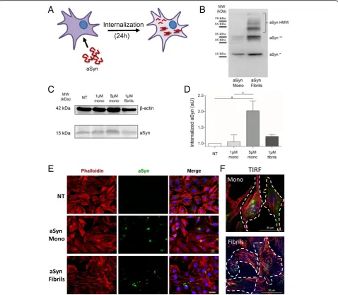
Fig. 1Recombinant aSyn monomers and fibrils are internalized by H4 cells.aRecombinant aSyn monomers (aSyn Mono) or fibrils (aSyn Fibrils) were added to the cell culture medium and incubated for 24 h.bSDS-PAGE and immunoblot analysis of the recombinant monomeric or fibrillar species of aSyn used in the experiments. The monomers show also the presence of a small fraction of dimers, as displayed by the faint band at 35 kDa. In the fibril preparation, one can observe the presence of higher molecular weight (HMW) species that are stable even on an SDS-PAGE.c
the use of Coloc2 Plugin from Fiji (ImageJ software). Figures were composed with CorelDRAW X8 (Corel Corporation, Ottawa, Canada) or with Microsoft Power Point (Microsoft Corporation).
Statistical analysis was performed using Microsoft Excel (Microsoft Corporation) and GraphPad PRISM 5 (GraphPad Software, San Diego, CA, USA). Images were processed with ImageJ V1.41, NIH, USA and/or Corel-DRAW X8 (Corel Corporation, Ottawa, Canada).
Statistical tests performed were Student’s-two-tailed t-test, one-way-Analysis of Variance (ANOVA) and repeated-measures ANOVA for grouped analysis, followed by Tukey’s post-hoc tests for multiple comparison.
Data were expressed as mean ± SEM and a 0.5% gen-eral significance level was defined, with significance levels as follows: *:p< 0.05; **:p< 0.01; ***:p< 0.001.
Results
aSyn is internalized and forms intracellular inclusions In order to investigate the molecular determinants of aSyn internalization, we compared the behaviour of two distinct forms of recombinant aSyn (monomers or fibril-lar aggregates, hereafter named fibrils) (Fig. 1a). aSyn monomers and fibrils were generated as described in Methods, and the species were characterized by
Trans-mission Electron Microscopy (TEM) [62] and by
SDS-PAGE (Fig.1b). The preparation of aSyn monomers showed also the presence of a smaller amount of dimers, as illustrated by the band at ~ 35 kDa. Due to contrast-ing studies reportcontrast-ing the ability of monomeric aSyn to passively enter cells, we tested two different concentra-tions of this form (1μM and 5μM), and 1μM of fibrils of aSyn (calculated based on the initial concentration of monomeric aSyn). The internalization of aSyn was ana-lysed by immunoblotting and by immunocytochemistry (ICC) after 24 h of incubation with cells. Immunoblot analysis revealed that both aSyn monomers and fibrils were internalized, given the increase in the levels of aSyn in treated cells (Fig.1c and d). Furthermore, microscopy analysis (confocal and TIRF microscopy) demonstrated that, in cells exposed to monomeric aSyn, the protein accumulated in distinct perinuclear puncta, whereas in cells exposed to fibrils aSyn accumulated in larger cyto-solic inclusions (Fig.1e-f).
aSyn interacts with the plasma membrane and accumulates as high molecular weight species
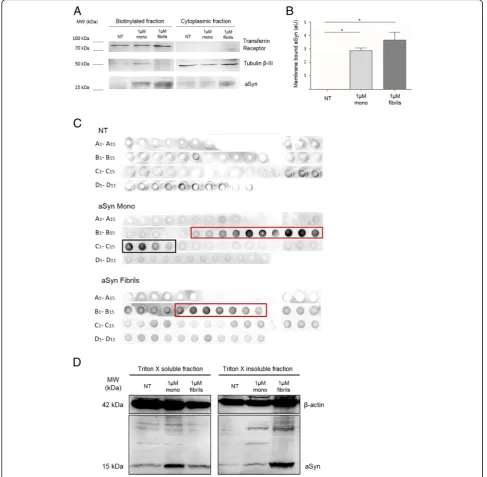
Based on previously reported aSyn binding to membranes, we hypothesized that the internalization of aSyn might in-volve an interaction with the plasma membrane. In order to test this, we performed a cell surface biotinylation assay in cells treated with aSyn. aSyn was found in the biotinyl-ated fraction trebiotinyl-ated with either monomers or fibrils, indi-cating that extracellular aSyn interacted with the plasma
membrane (Fig. 2aand b), and then accumulated within the cell in punctae (Fig.1e-f).
To further investigate the biochemical nature of the intracellular aSyn, we performed size exclusion chroma-tography (SEC) of cell lysates, and then dot blot analysis of the various fractions collected. Recombinant aSyn monomers were assessed by SEC in parallel, in order to establish the elution profile of this form of aSyn (Additional file 1: Figure S1A and S1B).
As expected, dot blot analysis of non-treated cells (NT) showed low signal, given the lower levels of aSyn (Fig.2c). In cells treated with aSyn monomers, we detected the pro-tein in fractions C1 to C4 (black box, Fig.2c) as reported in the chromatogram (Additional file 1: Figure S1B). We also detected aSyn in the final B fractions (from B6 to B15, red box), indicating the presence of higher molecular weight aSyn species as well (Fig. 2c). Such species were also found in cells treated with aSyn fibrils (fractions B5 to B11, red box), but not monomeric species of aSyn (Fig. 2c), suggesting that the species accumulating in the cells were biochemically distinct depending on the species of aSyn added to the cells.
To further confirm the biochemical differences ob-served, we performed differential fractionation of the cell lysates using Triton X-100. Immunoblot analysis showed higher levels of Triton X-100-soluble aSyn in cells treated with monomers, and higher levels of Triton X-100-insoluble aSyn in cells treated with fibrils, consist-ent with the results of the SEC analysis (Fig. 2d, left side). Interestingly, we also detected the formation of high molecular weight aSyn species in cells treated with monomeric aSyn, suggesting that, upon internalization, aSyn monomers start to aggregate (Fig.2d, right side).
Taken together, these results suggest that both mono-meric and fibrillar aSyn enter cells and accumulate in aggregated, high molecular weight species.
aSyn partially colocalizes with Rab5A and Rab7
Next, we performed a microscopy-based screen of mam-malian Rab proteins in order to identify the interplay be-tween aSyn and the trafficking pathways. The experiment consisted of treating cells overexpressing each individual mammalian Rab protein (fused to EGFP: Rab-GFP) with aSyn monomers or fibrils in order to assess (i) the effect of aSyn on the subcellular distribution of the Rab proteins, and (ii) the effect of each Rab protein on the subcellular distribution of aSyn. From the screen, we selected a set of Rab proteins whose localization was altered, or that colo-calized with aSyn (Additional file 2: Table S1). Of these, we selected Rab4A, Rab5A and Rab7 since they resulted in the strongest phenotype. Interestingly, the Rab proteins identified in the screen are compatible with a hypothesis that aSyn is internalized via an active endocytic
compartments, such as endosomes and lysosomes [57]. Therefore, we next focused our study on the these Rab proteins.
First, we assessed the degree of colocalization of aSyn and Rab5A-GFP, or Rab7-GFP, in cells treated with aSyn monomers or fibrils (Fig. 3a). The colocalization was quantified using the Coloc2 plugin of ImageJ
Software (Fig. 3b). In cells treated with aSyn mono-mers, we observed a strong colocalization between aSyn (in red) and Rab5A-GFP vesicles (in green) (Fig.
3a, left column, central panel), as well as a partial, al-though weaker, colocalization with Rab7-GFP (Fig. 3a, right column, central panel). Interestingly, the colocali-zation was not observed when cells were treated with
aSyn fibrils. This supports the idea that the internaliza-tion and sorting of aSyn monomers and fibrils is
differ-ent, as one might expect given their distinct
biochemical properties.
aSyn form inclusions in Rab4A-positive compartments Next, we examined the interplay between Rab4A and aSyn. We found no effect on the distribution of Rab4A-GFP in cells treated with aSyn fibrils. Likewise, we also found no
colocalization between Rab4A-GFP and aSyn in those cells (Fig. 4, right panel). In contrast, when Rab4A-GFP-expressing cells were treated with aSyn monomers, we observed a prominent increase in the size of endosomes, as well as a massive internalization of aSyn that accumu-lated in compartments surrounded by large, abnormal rings of Rab4A (Fig.4, central panel on the top and lower panels). This change in the size of early endosomes suggested that exposure to aSyn monomers altered the
A
B
normal biology of Rab4A and, therefore, the endosome-related trafficking processes.
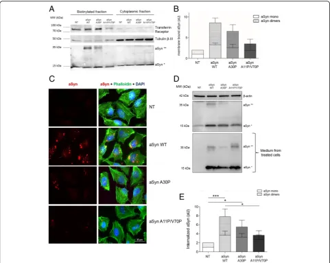
Membrane binding properties are essential for the internalization of aSyn
Based on the stronger effects of aSyn monomers, we de-cided to focus on the effects of monomeric aSyn. To in-vestigate whether intrinsic aSyn properties affected the internalization of the protein, we took advantage of dif-ferent aSyn mutants that have difdif-ferent membrane bind-ing abilities. Specifically, we used WT aSyn, the aSyn A30P familial mutant, known to display weaker binding to membranes [4, 12, 29, 30, 61], and the artificial mu-tant (A11P/V70P) designed to severely impair mem-brane binding [8, 10]. First, we performed membrane biotinylation assays with the different mutants, and de-tected a clear trend in the amount of protein present in the biotinylated fractions that reflected the different membrane binding properties of the aSyn mutants (aSyn A30P and aSyn A11P/V70P) (Fig.5a and b). In particu-lar, we detected a consistent trend in the levels of aSyn dimeric species in the biotinylated fraction, suggesting that membrane binding is important for dimerization and aggregation of aSyn.
Next, we tested the ability of the two aSyn mutants to enter cells and accumulate in intracellular inclusions, via immunocytochemistry (ICC) approach. We observed a significant reduction in the accumulation of aSyn in in-clusions in cells treated with A30P or A11P/V70P aSyn mutants when compared with cells treated with WT aSyn (Fig.5c), consistent with a difference in the intern-alization of the mutants. We further quantified the in-ternalization of the different variants of aSyn using immunoblot analysis. We confirmed a strong difference in the internalization of the various mutants (Fig.5dand e),
although the amount of aSyn present in the medium was identical (Fig.5d).
Taken together, these results suggest that membrane binding is essential for the internalization and, therefore, for the formation of intracellular aSyn inclusions.
aSyn A11P/V70P is unable to bind membranes
To test the contribution of different aSyn regions to-wards aggregation and membrane binding, several artifi-cial mutants, such as the aSyn A11P/V70P have been designed [9, 11]. First, we validated the effect of the A11P/V70P mutation on the biding of aSyn to small unilamellar vesicles (SUVs) (5:3:12 mixture of DOPE: DOPS:DOPC) using NMR spectroscopy (Additional file3: Figure S2). While WT aSyn interacted with DOPE: DOPS:DOPC SUVS, as previously described [22, 34] (Additional file 4: Figure S3A, panel on the left), the aSyn A11P/V70P mutant displayed a drastic reduction (more than 80%) of the signal broadening starting from residue 11 (where the first Pro mutation is located) until residue 140 (Additional file 4: Figure S3A, panel on the right), confirming the impairment in membrane binding.
Next, we tested the effect of aSyn A11P/V70P in the cellular model, in comparison to WT aSyn, and also in the presence or absence of Rab4A-GFP, as we had ob-served increased internalization of aSyn in H4 cells. Using immunoblot analysis and ICC, we found that the internalization A11P/V70P aSyn was negligible when compared to that of WT aSyn (Additional file 4: Figure S3B-D). Importantly, we did not detect internalization when we increased the concentration of A11P/V70P aSyn or when we overexpressed Rab4A-GFP, conditions which significantly increased the levels of intracellular WT aSyn (Additional file4: Figure S3B-D).
The endocytic pathway is involved in the internalization of aSyn
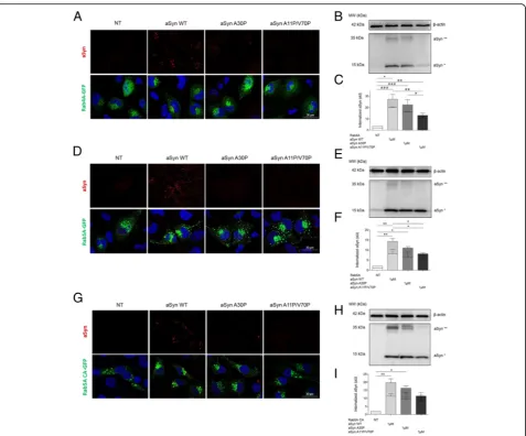
In order to investigate whether mutants with different membrane binding properties altered the internalization and intracellular fate of internalized aSyn, we used cells overexpressing Rab4A-GFP, Rab5A-GFP, or a constitu-tively active (CA) mutant of Rab5A-GFP. As described above, WT aSyn was readily internalized and accumulated in Rab4A-GFP-positive vesicles. In contrast, the internal-ization of the artificial A11P/V70P aSyn mutant was strongly impaired. Curiously, the PD-associated mutant A30P displayed an intermediate phenotype (Fig.6a-c).
Interestingly, the levels of internalized aSyn (monomers and dimers) were higher in cells expressing these Rab pro-teins than in naïve cells (shown above in Fig.5d-e), sug-gesting that increased levels of Rab4A altered the dynamics of internalization and dimerization of aSyn. The same trend was observed in in cells overexpressing Rab5A-GFP indicating, once again, that stimulation of the early steps of endosome formation increased the internal-ization of aSyn, as long as the membrane binding proper-ties of the protein are preserved (Fig. 6d-f). In cells overexpressing Rab5A, we also observed an increase in the levels of aSyn dimeric species.
Finally, to confirm the functional involvement of Rab5A on the internalization of aSyn, we used a mutant in which the GTPase activity is deregulated, resulting in permanent activation constitutively active mutant -Rab5ACA-GFP (Fig. 6g-i). In cells expressing this mu-tant Rab5A, we found overall higher levels of aSyn
in-ternalization, further confirming the role of the
endocytic pathway in the internalization of aSyn.
Rab7 sorts aSyn for degradation and reduces its intracellular accumulation
Next, we investigated the intracellular fate of internal-ized aSyn along the endocytic pathway by using
Rab7-GFP as a marker. Cells expressing Rab7-GFP were treated with WT, A30P, or A11P/V70P aSyn mutants, and analysed by ICC and immunoblotting, as described above. Surprisingly, we found that the internalization of aSyn, and the formation of dimers, was significantly re-duced in cells overexpressing Rab7, and that there were no differences in internalization between WT aSyn or the two mutants. (Fig.7a-c).
We hypothesized that this effect could be due to the sorting of aSyn for degradation in the lysosome, due to increased stimulation of the lysosomal pathway by over-expression of Rab7. To test this, we repeated the experi-ment in cells expressing a dominant negative (DN)
Fig. 6The A30P and A11P/V70P aSyn mutants are less internalized than WT aSyn.aICC andbImmunoblotting of cells transfected with Rab4A– GFP and treated as in experiments shown in Fig.5.dandeICC and Immunoblotting of cells transfected with Rab5A-GFP and treated as above.g
andhICC and Immunoblotting of cells transfected with Rab5ACA-GFP (constitutively active) and treated as above.c,fandiQuantifications of the immunoblots in panelsb,eandh. Dotted bars refer to the band corresponding to aSyn dimers (aSyn**), and clear bars refer to aSyn monomers (aSyn*). Statistical tests were performed using one-way ANOVA with repeated-measures for grouped analysis, followed by Tukey’s post-hoc tests. Data are expressed as mean ± SEM and a 0.5% general significance level was defined, with significance levels as follows: *:p< 0.05; **:p< 0.01; ***:p< 0.001. Statistical significance is indicated with the symbol“#”for the monomers,“+”for the dimers, and“*”for the
mutant of Rab7 (Rab7DN-GFP) that impairs its activity. Interestingly, we found that the internalization and dimerization of aSyn was restored to the initial levels, suggesting that the Rab7DN mutant blocked the sorting of aSyn to the lysosome (Fig.7d-f).
The internalization of aSyn is mediated by dynamin Next, we investigated the mechanism involved in the in-ternalization of aSyn by using two well established chemical blockers of endocytosis: PitStop2 (PitStop) and Dyngo 4A (Dyngo).
Pitstop is a selective inhibitor of clathrin-mediated endocytosis (CME) [50, 53], while Dyngo blocks all dynamin-dependent endocytic mechanisms [40].
Naïve cells or cells overexpressing Rab4A-GFP were treated with each of the two compounds for 30 min prior to the treatment with aSyn monomers, and were then incubated together with aSyn for 24 h and proc-essed for ICC or immunoblotting, as described above.
In naive cells, Dyngo efficiently blocked the internal-ization of aSyn (Additional file 5: Figure S4A). Yet, the opposite effect was observed using PitStop, which increased the accumulation of intracellular aSyn (Additional file 5: Figure S4B-D).
Similarly, in cells overexpressing Rab 4A, Dyngo pre-vented the internalization and accumulation of aSyn in Rab4A-surrounded vesicles. In contrast, PitStop failed to produce a significant effect (Fig.8a-b).
Internalized aSyn is degraded by lysosomes
To investigate the fate of internalized aSyn, we tested whether blocking lysosomal and autophagic function would affect the levels of internalized aSyn. We treated cells expressing Rab7-GFP with bafilomycin A1 or chloro-quine for 30 min, and then added monomeric aSyn.
Cells were then incubated for 24 h, and then processed for ICC analysis. We confirmed that blocking lysosomal acidification and consequently autophagy inhibited the degradation of internalized aSyn and led to its accumula-tion, possibly in late endosomes and lysosomes (Fig.8c-d). Identical results were obtained in naïve cells (Additional file 5: Figure S4, A-D). Interestingly, treatment with chloroquine resulted in stronger accumulation of aSyn than that observed with Bafilomycin A1, consistent with chloroquine being more potent inhibitor then bafilomycin A1.
Taken together, our results suggest that aSyn is inter-nalized via dynamin-mediated endocytosis and trafficked
via the endocytic pathway and then sorted and targeted for degradation via the autophagy-lysosomal system.
Discussion
A key premise for the prion-like spreading hypothesis of aSyn pathology in PD and other synucleinopathies is that aSyn assemblies are taken up by a recipient cell, are sorted, and then seed the aggregation of endogenous aSyn, thereby propagating pathology [6, 7]. Nevertheless, the precise mo-lecular mechanisms involved are still elusive. Most studies have focused on the understanding of the mechanisms through which aSyn can be released from cells. However, present understanding of the uptake and fate of internalized aSyn are even more limited. Here, we focused on the initial steps of this complex process, and investigated how mem-brane binding properties might affect the internalization and processing of aSyn in the cell. Using a simple cell-based paradigm, we established that both monomeric and fibrillar aSyn proteins can interact with the plasma membrane and be internalized, accumulating in high molecular weight species.
We also investigated the role of aSyn membrane binding on internalization by using mutants that are known to affect the membrane binding properties of aSyn. The PD-associated mutant A30P displays reduced membrane interactions and reduced internalization, when compared to WT aSyn. Structurally, the substitution of the alanine
by the proline disrupts the α-helical domains formed in the N-terminal and central regions of aSyn, thereby affect-ing the ability of the protein to interact and bind to mem-branes. Consistently, we found that the artificial mutant A11P/V70P, designed to disrupt membrane binding [9,
11], indeed disrupts the N-terminal region and compro-mises membrane binding, thereby impairing internaliza-tion of aSyn. Interestingly, it has been widely suggested that the aSyn A30P mutant may cause PD by affecting at least partially different cellular pathways that those af-fected by WT aSyn. Our study is consistent with this idea, and forms now the basis for in vivo investigations of the prion-like spreading ability of this mutant, which has not been documented, since the studies performed thus far fo-cused on WT aSyn [57–60].
Importantly, our findings suggest that one cannot dismiss that, even monomeric aSyn might be sufficient to initiate the process of aggregation and therefore, the spreading of aSyn pathology. Only two studies have reported the intern-alization of aSyn monomers and the involvement of the endocytic pathway, although the purpose and the readouts used in those studies were different [1,58]. Interestingly, a comparable study reported consistent results regarding tau, suggesting that extracellular monomeric tau is sufficient to initiate the spreading of tau pathology [41]. However, one caveat of our study is the concentration of aSyn used, which is higher than that used in several other studies [30, 61].
B
D
Nevertheless, our study establishes proof of concept that, perhaps a local and even just temporary increase in the concentration of aSyn, caused for example by dying cells in the brain, might lead to the release of monomeric aSyn that could be internalized by neighbouring cells, thereby initiat-ing the spreadinitiat-ing of pathology.
Using selective inhibitors of the endocytic pathways, such as Dyngo and PitStop, we demonstrate that aSyn is internalized in a dynamin-dependent process, but not through CME. It is possible that when clathrin-mediated mechanisms are blocked (PitStop-related effects), the clathrin-independent processes (blocked by Dyngo) which are presumably involved in the internalization of aSyn are exacerbated. In this way, a larger amount of aSyn can be internalized in naïve cells (Additional file 5: Figure S4). Additional studies are required to further dissect the mo-lecular mechanisms involved in aSyn endocytosis.
In an unbiased screen of Rab proteins, we found that, once internalized, aSyn partially colocalizes with Rab5A and Rab7, suggesting that the endosomal pathway is
involved in the sorting and processing of aSyn. Further-more, we demonstrated that Rab4A modulates the internal-ization of aSyn, and surrounds the internalized protein. Both Rab4A and Rab5A are localized in the early endo-somes, contributing to the recycling/transport of proteins to the plasma membrane, respectively. Rab7 is localized in the late endosomes, lysosomes and phagosomes, contribut-ing to the fusion of late endosome with lysosome [27]. Re-cent studies suggest that, even when different Rabs localize in the same compartment, they can occupy distinct mem-brane microdomains with different functions, called Rab domains. These are dynamic structures but, interestingly, do not mix significantly over time [52]. One of the possible explanations for this segregation mechanism is that it could be, at least in part, mediated by effector proteins and their association with additional molecules in the organelle membranes [52]. Consistently, a recent study showed that Rab4A is essential for the recruitment of adaptor proteins to the tubular subdomain of the early endosomal mem-brane, orchestrating a signalling cascade that initiates the
gation may be favoured by the low pH - that is reported to vary between 5.0 and 6.0 - in endosomes [14], as well as by molecular crowding due to the presence of acidic endosomal proteins. It is also possible that aSyn aggregation occurs due to problems in the late endosome-lysosomal compartments, which we found to be involved in the degradation of aSyn. Thus, impaired autophagic degradation of aSyn could lead to the accumulation of aggregation-competent species [19].
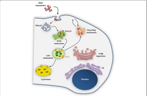
Our data enable us to propose a model for the internal-ization of aSyn, based on the interaction with trafficking machinery components (Fig.9). After interacting with the membrane, aSyn monomers are internalized through the endocytic pathway, where Rab4A plays an important role on the protein sorting and on the transport from/to the plasma membrane. aSyn is thereafter sorted to the early endosome (in colocalization with Rab5A). It is possible that during the progress from early to late endosome (where it colocalizes with Rab7), due to pH acidification, aSyn monomers start to oligomerize, and to assemble in high molecular weight species, that can then escape to the cytoplasm, leading to its accumulation and facilitating the spreading of aSyn pathology.
Conclusions
In total, our study emphasizes the importance of mem-brane binding for the internalization of aSyn and highlights the fundamental role of Rab proteins in the internalization, sorting, and processing of aSyn, suggesting that targeting specific Rab proteins and/or specific intracellular trafficking components might prove to be valuable targets for modu-lating the spreading of aSyn pathology and, consequently, disease progression in PD and other synucleinopathies.
Additional files
Additional file 1:Figure S1.Characterization of recombinant aSyn monomers. (A) Fractions collected upon protein separation on Superose 6 10/300 size exclusion column. (B) Chromatogram of recombinant aSyn monomers showing the fractions in which monomeric aSyn was recovered. (PDF 190 kb)
Additional file 2:Table S1.Results of the Rab protein screen. Rab-GTPase family members selected in a screen where we assessed alterations in the subcellular distribution of the Rab protein or the colocalization with aSyn in cells treated with aSyn monomers or fibrils. In the column“morphology”, a
“11% more Rab-vesicles”statement means that in the 11% of the cells ana-lysed, the localization of Rabs is more vesicular (suggesting an increase of 11% in the active, GTP-bound Rab protein) compared to the localization pattern
and clear bars refer to aSyn monomers (aSyn*). Statistical tests were performed using one-way-analysis of variance (ANOVA) with repeated-measures for grouped analysis, followed by Tukey’s post-hoc tests. Data were expressed as mean ± SEM and a 0.5% general significance level was defined, with significance levels as follows: *:p< 0.05; **:p< 0.01; ***:p< 0.001. Significance is shown with the symbol“#”for the monomers, with the symbol“+”for the dimers and with the symbol“*”for the sum between monomers and dimers. (D) ICC of H4 cells transfected with Rab 4A-GFP and treated with 1μM or 5μM of aSyn wild type and aSyn A11P/V70P. Scale bar: 30μm. (PDF 3696 kb)
Additional file 4:Figure S3.Dyngo blocks whereas PitStop enhances the internalization of aSyn. (A) ICC of H4 cells treated with 1μM aSyn monomers and with vehicle, PitStop 30μM or Dyngo 50μM. Both PitStop and Dyngo are inhibitors of the endocytic processes. (B) Quantification of the aSyn mean fluorescence intensity in the three conditions. Scale bar: 30μm. (C) Immunoblotting of H4 cells treated with different concentrations of PitStop and Dyngo. (D) Quantification of the immunoblot. Dotted bars refer to the band corresponding to aSyn dimers (aSyn**), and clear bars refer to aSyn monomers (aSyn*). Statistical tests were performed using one-way-analysis of variance (ANOVA), with repeated-measures for grouped analysis, followed by Tukey’s post-hoc tests. Data were expressed as mean ± SEM and a 0.5% general significance level was defined, with significance levels as follows: *:p< 0.05; **:p< 0.01; ***:p< 0.001. Significance is shown with the symbol“#”for the monomers, with the symbol“+”for the dimers and with the symbol“*”for the sum between monomers and dimers. Scale bar: 30μm (PDF 1051 kb)
Additional file 5:Figure S4.Blocking of autophagy inhibits the degradation of aSyn. (A) ICC of H4 cells treated with 1μM aSyn monomers and with vehicle, Bafilomycin 100 nM (Baf 100 nM), or with Chloroquine 50μM (Chlq 50μM). Bafilomycin and chloroquine are inhibitors of the ALP. (B) Quantification of the aSyn mean fluorescence intensity in the three conditions. Scale bar: 30μm. (C) Immunoblotting of H4 cells treated with 1μM aSyn WT and incubated with Bafilomycin 100 nM or Chloroquine 50μM. (D) quantification of the immunoblot in panel C. Dotted bars refer to the band corresponding to aSyn dimers (aSyn**), and clear bars refer to aSyn monomers (aSyn*). Statistical tests were performed using one-way-analysis of variance (ANOVA), with repeated-measures for grouped one-way-analysis, followed by Tukey’s post-hoc tests. Data is expressed as mean ± SEM and a 0.5% general significance level was defined, with significance levels as follows: *:p< 0.05; **:p< 0.01; ***:p< 0.001. Significance is shown with the symbol“#”for the monomers, with the symbol“+”for the dimers and with the symbol“*”for the sum between monomers and dimers. Scale bar: 30μm. (PDF 1301 kb)
Acknowledgements
We thank Dr. Andrés Binolfi for technical assistance with NMR and fruitful discussions. TFO is supported by the DFG Center for Nanoscale Microscopy and Physiology of the Brain (CNMPB) and by SFB1286. TFO is also supported by a grant from La Marató de TV3, Spain. IM is supported by Schram-Stiftung T287/25457 and Deutsche Forschungsgemeinschaft (Emmy Noether Young Investigator Award MI-1702/1 and SFB1190/P2).
Funding
T287/25457 and Deutsche Forschungsgemeinschaft (Emmy Noether Young Investigator Award MI-1702/1 and SFB1190/P2).
Availability of data and materials
All data is available and original.
Authors’contributions
CM conducted experiments, analysed data, and wrote the manuscript. MH, EG, TLF, AVP, MAS, CD and TB conducted experiments. CD, COF and IM designed experiments and revised the manuscript. TFO designed experiments, supervised the project, and wrote the manuscript. All authors read and approved the final manuscript.
Ethics approval and consent to participate
Not applicable.
Consent for publication
All authors agreed with the content and consent publication.
Competing interests
The authors declare that they have no competing interests.
Publisher’s Note
Springer Nature remains neutral with regard to jurisdictional claims in published maps and institutional affiliations.
Author details
1Department of Experimental Neurodegeneration, Center for Biostructural Imaging of Neurodegeneration, Center for Nanoscale Microscopy and Molecular Physiology of the Brain, University Medical Center Goettingen, 37073 Göttingen, Germany.2Max Planck Laboratory for Structural Biology, Chemistry and Molecular Biophysics of Rosario (MPLbioR, UNR-MPIbpC) and Instituto de Investigaciones para el Descubrimiento de Fármacos de Rosario (IIDEFAR, UNR-CONICET), Universidad Nacional de Rosario, Ocampo y Esmeralda, S2002LRK Rosario, Argentina.3European Neuroscience Institute, University Medical Center Göttingen, Göttingen, Germany.4Trans-synaptic Signaling Group, European Neuroscience Institute, Grisebachstrasse 5, 37077 Goettingen, Germany.5Max Planck Institute for Experimental Medicine, Göttingen, Germany.6Institute of Neuroscience, The Medical School, Newcastle University, Framlington Place, Newcastle Upon Tyne NE2 4HH, UK.
Received: 3 July 2018 Accepted: 28 July 2018
References
1. Ahn KJ, Paik SR, Chung KC, Kim J (2006) Amino acid sequence motifs and mechanistic features of the membrane translocation of a-synuclein. J Neurochem 97:265–279
2. Appel-Cresswell S, Vilarino-Guell C, Encarnacion M, Sherman H, Yu I, Shah B et al (2013) Alpha-synuclein p.H50Q, a novel pathogenic mutation for Parkinson’s disease. Mov. Disord 28:811–813
3. Bartels T, Choi JG, Selkoe DJ, Hospital W (2012) a-Synuclein occurs physiologically as a helically folded tetramer that resists aggregation. Nature. 2011;477(7362):107-110.https://doi.org/10.1038/nature10324
4. Bertoncini CW, Fernández CO, Griesinger C, Jovin TM, Zweckstetter M (2005) Familial mutants ofα-synuclein with increased neurotoxicity have a destabilized conformation. J Biol Chem 280:30649–30652
5. Breda C, Nugent ML, Estranero JG, Kyriacou CP, Outeiro TF, Steinert JR et al (2015) Rab11 modulatesα-synuclein-mediated defects in synaptic transmission and behaviour. Hum Mol Genet 24:1077–1091
6. Brundin P, Melki R (2017) Prying into the prion hypothesis for Parkinson’s disease. J Neurosci 37:9808–9818
7. Brundin P, Melki R, Kopito R (2010) Prion-like transmission of protein aggregates in neurodegenerative disorders. Nat. Rev. Mol. Cell Biol. 11:301–307 8. Burré J, Sharma M, Südhof TC (2012) Systematic mutagenesis of aSyn
reveals distinct sequence requirements for physiological and pathological activities. J Neurochem 15227:15242
9. Burré J, Sharma M, Südhof TC (2014) a-synuclein assembles into higher-order multimers upon membrane binding to promote SNARE [Internet]. Proc. Natl. Acad. Sci. U. S. A 111:E4274–E4283. Available from:http://www.
pubmedcentral.nih.gov/articlerender.fcgi?artid=4210039&tool= pmcentrez&rendertype=abstract
10. Burré J, Sharma M, Südhof TC (2015) Definition of a molecular pathway mediating a-Synuclein. J Neurosci 35:5221–5232
11. Burré J, Sharma M, Tsetsenis T, Buchman V, Südhof TC (2010)α-Synuclein Promotes SNARE-Complex Assembly in vivo and in vitro. Science (80-.) 329: 1663–1667
12. Bussell R, Eliezer D (2001) Residual structure and dynamics in Parkinson’s disease-associated mutants of a-Synuclein. J Biol Chem 276:45996–46003 13. Bussell RJ, Eliezer D (2003) A structural and functional role for 11-mer
repeats in a -Synuclein and other exchangeable lipid binding proteins. J Mol Biol 2836:763–778
14. Casey JR, Grinstein S, Orlowski J (2010) Sensors and regulators of intracellular pH. Nat. Rev. Mol. Cell Biol. 11:50–61
15. Chandra S, Chen X, Rizo J, Jahn R, Su TC (2003) A broken a-helix in folded a-Synuclein. J Biol Chem 278:15313–15318
16. Chartier-Harlin M-C, Kachergus J, Roumier C, Mouroux V, Douay X, Lincoln S et al (2004)α-Synuclein locus duplication as a cause of familial Parkinson’s disease. Lancet 364:1169–1171
17. Chutna O, Gonçalves S, Villar-Piqué A, Guerreiro P, Marijanovic Z, Mendes T et al (2014) The small GTPase Rab11 co-localizes withα -synuclein in intracellular inclusions and modulates its aggregation, secretion and toxicity. Hum Mol Genet 23:6732–6745
18. D’Souza RS, Semus R, Billings EA, Meyer CB, Conger K, Casanova JE (2014) Rab4 orchestrates a small GTPase cascade for recruitment of adaptor proteins to early endosomes. Curr Biol 24:1187–1198
19. Danzer KM, Kranich LR, Ruf WP, Cagsal-Getkin O, Winslow AR, Zhu L et al (2012) Exosomal cell-to-cell transmission of alpha synuclein oligomers. Mol Neurodegener 7:42
20. Elia G (2008) Biotinylation reagents for the study of cell surface proteins. Proteomics 8:4012–4024
21. Ferreon ACM, Gambin Y, Lemke EA, Deniz AA (2009) Interplay of a-synuclein binding and conformational switching probed by single-molecule fluorescence. Proc Natl Acad Sci 106:5645–5650
22. Fonseca-Ornelas L, Eisbach SE, Paulat M, Giller K, Fernández CO, Outeiro TF et al (2014) Small molecule-mediated stabilization of vesicle-associated helicalα-synuclein inhibits pathogenic misfolding and aggregation. [Internet]. Nat. Commun 5:5857. Available from:http://www.ncbi.nlm.nih. gov/pubmed/25524885
23. Giasson BI, Murray IVJ, Trojanowski JQ, Lee VM (2001) A hydrophobic stretch of 12 amino acid residues in the middle of a-Synuclein is essential for filament assembly. J Biol Chem 276:2380–2386
24. Golde TE, Borchelt DR, Giasson BI, Lewis J (2013) Thinking laterally about neurodegenerative proteinopathies. J Clin Invest 123:1847–1855
25. Gonçalves SA, Macedo D, Raquel H, Simões PD, Giorgini F, Ramalho JS et al (2016) shRNA-based screen identifies endocytic recycling pathway components that act as genetic modifiers of alpha-Synuclein aggregation, secretion and toxicity. PLoS Genet 12:1–26
26. Hoyer W, Cherny D, Subramaniam V, Jovin TM (2004) Impact of the Acidic C-Terminal Region Comprising Amino Acids 109–140 on a-Synuclein Aggregation in Vitro. Biochemistry:16233–16242
27. Hutagalung AA, Novick PJ (2011) Role of Rab GTPases in membrane traffic and cell physiology. Physiol Rev 91:119–149
28. Jao CC, Hegde BG, Chen J, Haworth IS, Langen R (2008) Structure of membrane-bound alpha-synuclein from site-directed spin labeling and computational refinement. Proc Natl Acad Sci U S A 105:19666–19671 29. Jensen PH, Nielsen MS, Jakes R, Dotti G, Goedert M (1998) Binding of
alpha-synuclein to brain vesicles is abolished by familial parkinsons-disease mutation. J Biol Chem 273:26292–26294
30. Jo E, Fuller N, Rand RP, St George-Hyslop P, Fraser PE (2002) Defective membrane interactions of familial Parkinson’s disease mutant A30P
α-synuclein 1 1Edited by I. B Holland J Mol Biol 315:799–807
31. Kim TD, Paik SR, Yang C-H (2002) Structural and functional implications of C-terminal regions of alpha-synuclein. Biochemistry 41:13782–13790 32. Krüger R, Kuhn W, Müller T, Woitalla D, Graeber M, Epplen JT et al (1998)
Ala30Pro mutation in the gene encoding a-synuclein in Parkinson’s. Nat Genet 18:231–236
38. Luk KC, Song C, O’Brien P, Stieber A, Branch JR, Brunden KR et al (2009) Exogenous alpha-synuclein fibrils seed the formation of Lewy body-like intracellular inclusions in cultured cells. Proc Natl Acad Sci U S A 106:20051– 20056
39. Maltsev AS, Ying J, Bax A (2012) Impact of N-terminal acetylation of a-synuclein on its random coil and lipid binding properties. Biochemistry 51:5004–5013 40. Mccluskey A, Daniel JA, Hadzic G, Chau N, Clayton EL, Mariana A et al (2013)
Building a better dynasore: the dyngo compounds potently inhibit dynamin and endocytosis. Traffic 14:1272–1289
41. Michel CH, Kumar S, Pinotsi D, Tunnacliffe A, George-Hyslop PS, Mandelkow E et al (2014) Extracellular monomeric tau protein is sufficient to initiate the spread of tau protein pathology. J Biol Chem 289:956–967
42. Miotto MC, Valiente-Gabioud AA, Rossetti G, Zweckstetter M, Carloni P, Selenko P et al (2015) Copper binding to the N-terminally acetylated, naturally occurring form of alpha-Synuclein induces local helical folding. J Am Chem Soc 137:6444–6447
43. Park SM, Jung HY, Kim TD, Park JH, Yang C, Kim J. Distinct Roles of the N-terminal-binding Domain and the C-terminal-solubilizing Domain of A-Synuclein , a Molecular Chaperone. J Biol Chem 2002; 277: 28512–28520 44. Pasanen P, Myllykangas L, Siitonen M, Raunio A, Kaakkola S, Lyytinen J et al (2014)
A novelα-synuclein mutation A53E associated with atypical multiple system atrophy and Parkinson’s disease-type pathology. Neurobiol Aging 35:1–5 45. Polymeropoulos MH (1997) Mutation in the -synuclein gene identified in
families with Parkinson’s disease. Science (80-. ) 276:2045–2047
46. Scheurer SB, Rybak JN, Roesli C, Brunisholz RA, Potthast F, Schlapbach R et al (2005) Identification and relative quantification of membrane proteins by surface biotinylation and two-dimensional peptide mapping. Proteomics 5: 2718–2728
47. Scott DA, Tabarean I, Tang Y, Cartier A, Masliah E (2010) A pathologic cascade leading to synaptic dysfunction in a-synuclein induced neurodegeneration. J Neurosci 30:8083–8095
48. Shi M, Shi C, Xu Y (2017) Rab GTPases: the key players in the molecular pathway of Parkinson’s disease. Front Cell Neurosci 11:1–8
49. Singleton AB, Farrer M, Johnson J, Singleton A, Hague S, Kachergus J et al (2003) a-Synuclein locus triplication causes Parkinson’s Disease. Science (80-. ) 302:841
50. Smith CM, Haucke V, McCluskey A, Robinson PJ, Chircop M (2013) Inhibition of clathrin by pitstop 2 activates the spindle assembly checkpoint and induces cell death in dividing HeLa cancer cells. Mol Cancer 12:1–15 51. Snead D, Eliezer D (2014) Alpha-Synuclein function and dysfunction on
cellular membranes. Exp Neurobiol 23:292
52. Sönnichsen B, De Renzis S, Nielsen E, Rietdorf J, Zerial M (2000) Distinct membrane domains on endosomes in the recycling pathway visualized by multicolor imaging of Rab4, Rab5, and Rab11. J Cell Biol 149:901–913 53. Soohoo AL, Puthenveedu Ma (2013) Divergent modes for cargo-mediated
control of clathrin-coated pit dynamics. Mol Biol Cell 24:1725–1734 S1-12 54. Souza MY, Giasson BI, Lee VM, HI Y (2000) Chaperone-like activity of
synucleins 474:116–119
55. Spillantini MG, Crowther RA, Jakes R, Hasegawa M, Goedert M (1998) alpha-Synuclein in filamentous inclusions of Lewy bodies from Parkinson’s disease and dementia with lewy bodies. Proc Natl Acad Sci U S A 95:6469–6473 56. Spillantini MG, Schmidt ML, Lee VM-Y, Trojanowski JQ, Goedert M (1997)
a-Synuclein in Lewy bodies. Nature. 1997;388(6645):839–840
57. Stenmark H (2009) Rab GTPases as coordinators of vesicle traffic. Nat Rev Mol Cell Biol 10:513–525
58. Sung JY, Kim J, Paik SR, Park JH, Ahn YS, Chung KC (2001) Induction of neuronal cell death by Rab5A-dependent endocytosis of a-Synuclein. J Biol Chem 276:27441–27448
–
27708160,http://www.pubmedcentral.nih.gov/articlerender.fcgi?artid= PMC5081623
63. Volpicelli-daley L a, Luk KC, Patel TP, Tanik SA, Dawn M, Stieber A et al (2011) Exogenous aSynuclein fibrils induce Lewy body pathology leading to synaptic dysfunction and neuron death. Neuron 72:57–71
64. Wakabayashi K, Yoshimoto M, Tsuji S, Takahashi H (1998) A-Synuclein immunoreactivity in glial cytoplasmic inclusions in multiple system atrophy. Neurosci. Lett 249:180–182
65. Wang W, Perovic I, Chittuluru J, Kaganovich A, Nguyen LTT, Liao J et al (2011) A soluble a-synuclein construct forms a dynamic tetramer. Proc Natl Acad Sci 108:17797–17802
66. Wilson GR, Sim JCH, McLean C, Giannandrea M, Galea CA, Riseley JR et al (2014) Mutations in RAB39B cause X-linked intellectual disability and early-onset parkinson disease withα-synuclein pathology. Am J Hum Genet 95: 729–735