ONLINE FIRST
This is a provisional PDF only. Copyedited and fully formatted version will be made available soon.
ISSN: 0015-5659
e-ISSN: 1644-3284
Anatomical variations of the abductor pollicis longus: a pilot
study
Authors: Piotr Karauda, Łukasz Olewnik, Michał Podgórski, Michał Polguj, Kacper Ruzik, Bartłomiej Szewczyk, Mirosław Topol
DOI: 10.5603/FM.a2019.0134
Article type: ORIGINAL ARTICLES
Submitted: 2019-10-12
Accepted: 2019-11-03
Published online: 2019-12-04
This article has been peer reviewed and published immediately upon acceptance.
It is an open access article, which means that it can be downloaded, printed, and distributed freely, provided the work is properly cited.
Articles in "Folia Morphologica" are listed in PubMed.
Anatomical variations of the abductor pollicis longus: a pilot study
Variants of the abductor policis longus
Piotr Karauda1, Łukasz Olewnik1, Michał Podgórski2, Michał Polguj1, Kacper Ruzik1,
Bartłomiej Szewczyk3, Mirosław Topol1
1Department of Normal and Clinical Anatomy, Interfaculty Chair of Anatomy and Histology,
Medical University of Lodz, Poland
2Polish Mother’s Memorial Hospital Research Institute, Department of Diagnostic Imaging
Lodz, Poland
3Department of Clinical Morphology, Medical University of Lodz, Poland
Address for correspondence: Piotr Karauda, Department of Normal and Clinical Anatomy,
Interfaculty Chair of Anatomy and Histology, Medical University of Lodz, ul. Kościuszki 4,
90-419 Łódź, Poland, Poland, e-mail: [email protected]
ABSTRACT
Background: The abductor pollicis longus (APL) originates from the lateral part of the dorsal
surface of the body of the ulna below the insertion of the anconeus muscle, from the
interosseous membrane, and from the middle third of the dorsal surface of the body of the
radius. However, the number of its accessory bands and their insertion vary considerably.
Material and methods: Fifty upper limbs (2 paired, 31 male, 19 female) were obtained from
adult Caucasian cadavers, and fixed in 10 % formalin solution before examination
Results: The APL muscle was present in all specimens. The muscles were divided into three
main categories, with Type II and III being dived into subtypes. Type I was characterized by a
single distal attachment, with the tendon inserting to the base of the I metacarpal bone. Type
II was characterized by a bifurcated distal attachment, with the main tendon inserting to the
base of the first metacarpal bone; this type was divided into three subtypes (A-C). Type III
was characterized by the main tendons inserting to the base of the first metacarpal bone, while
the accessory band was characterized by mergers (fusion) with other tendons. This type was
divided into two subtypes (A-B).
Key words:abductor policis longus, abductor pollicis longus tendon, abductor pollicis
longus tendon, tendon graft, anatomical variations
INTRODUCTION
The forearm bears three muscles that move the thumb, these being the extensor
pollicis brevis (EPB) and extensor pollicis longus (EPL), known as the deep extensors, and
the abductor pollicis longus muscle (APL). The APL lies immediately below the supinator,
and is sometimes united with it. It originates from the lateral part of the dorsal surface of the
body of the ulna below the insertion of the anconeus muscle, from the interosseous
membrane, and from the middle third of the dorsal surface of the body of the radius [1]. The
muscle belly of the APL passes obliquely downward and lateralward, becoming a tendon
which, together with the tendon of the EPB, runs through a groove on the lateral side of the
distal part of the radius and inserts at the radial side of the base of the first metacarpal bone
[1]. The APL itself is innervated by the posterior interosseous nerve, a branch of the radial
nerve [1]. Its primary function is to abduct the thumb at the carpometacarpal joint, thereby
moving the thumb anteriorly, and to assist in extending and rotating the thumb.
The dorsal surface of the hand typically includes a number of muscles divided between
six compartments: the EPB and the APL are in the first compartment, the extensor carpi
radialis longus and extensor carpi radialis brevis in the second, the EPL in the third, the
extensor digitorum and extensor indicis in the fourth, the extensor digiti minimi muscle in the
fifth, and the extensor carpi ulnaris in the sixth [1]. Both the first and the third compartment
serve as the limits of the “anatomical snuffbox”.
The APL demonstrates a high degree of variability with regard to the number of its
accessory bands and their insertion [2–10]. The presence of multiple tendons originating from
the APL is clinically relevant, as it can lead to the development of de Quervain’s syndrome,
which is caused by stenosing tenosynovitis. In addition, these accessory bands may also serve
as a potential source of transplant material, especially for tendon reconstruction [8].
The primary aim of the study is to characterize the morphology of the APL and its accessory
bands; however, it is also intended to prepare a classification for distinguishing different types
of APL when planning surgical procedures in the area.
Fifty upper limbs (31 male, 19 female) were obtained from adult Caucasian cadavers,
and fixed in 10 % formalin solution before examination. The upper limbs were stored in a
pool of preservative liquid for three to eight years. The cadavers were the property of the
Department following donation to the university anatomy program. The inclusion criteria
comprised sufficient specimen quality and the lack of evidence of surgical intervention in the
examined area; this is needed to allow for complete identification of the tendon insertion. The
dissection of the forearm and hand areas was performed according to the standard technique
[11, 12]:
Dissection started with the removal of the skin, subcutaneous tissue and superficial
fascia. Following this, starting proximal to the retinaculum, the tendon and muscle belly of the
APL was exposed and precisely dissected to the bony attachment. Following this, the entire
tendon was separated to the distal attachment.
Upon dissection, the following morphological features of the APL were assessed:
— The type of APL tendon insertion (location, shape of tendon insertion). — Morphometric measurements of the APL.
— An electronic digital calliper was used for all measurements (Mitutoyo Corporation,
Kawasaki-shi, Kanagawa, Japan). Each measurement was carried out twice with an
accuracy of up to 0.1 mm. The size of each band was measured. Consent was obtained
from the Local Bioethical Commission to perform the anatomical stage
(RNN/09/19/KE).
Statistical analysis
Statistical analysis was performed with Statistica 12 software (StatSoft Polska,
Cracow, Poland). The Chi2 test with contingency tables were used to evaluate differences
between the distribution of APL types according to gender and body side. The normality of
the data distribution was checked with the Shapiro-Wilk test. As the data was not normally
distributed, the morphological measurements of different APL types were compared using the
Mann-Whitney U-test (two groups) or the Kruskal-Wallis test by ranks with a dedicated post
hoc test (more than two groups).
A p-value lower than 0.05 was considered significant. The results are presented as
mean and standard deviation unless otherwise stated.
The APL was present in all specimens. Based on its morphology, three main types
were distinguished:
Type I – This type possesses a single distal attachment. The tendon inserts to the base of the I
metacarpal bone. This type was observed in 5 upper limbs – Fig. 1.
Type II – This type is characterised by a bifurcated distal attachment. The main tendon
inserts to the base of the first metatarsal bone. This type was present in 6 cases. Two subtypes
were determined based on the place of attachment.
Subtype a – the accessory band inserts to the shaft of the first metacarpal bone. This
subtype was found in 3 upper limbs. – Fig. 2.
Subtype b – the accessory band inserts to the trapezius bone. This subtype occurred in
3 cases – Fig. 3.
Subtype c – the accessory band was divided into two: the first band connected with
the abductor pollicis brevis, while the second connected with the opponens pollicis
muscle. This subtype was present in 12 cases – Fig. 4.
Type III – This type is also characterised by a double tendon; however, the main band inserts
to the base of the first metacarpal bone, while the accessory band fuses with other tendons. It
was further divided into two subtypes:
Subtype a –wherethe accessory band fuses with the APB. This subtype was present in 25 cases – Fig. 5.
Subtype b –where the accessory band fuses with the EPB, and inserts to the trapezius
bone. This type was present in 2 cases – Fig. 6.
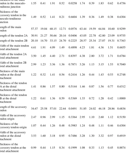
The morphological characteristics of the tendons and muscle bellies according to body side,
gender and each APL type are presented in tables 1 and 2.
Table 1. Parameter Gender P value Body side P value Female Male Right Left
Mean SD Mean SD Mean SD Mean SD
Forearm Length 25.19 1.61 27.03 1.35 0.0005 26.07 1.61 26.71 1.76 0.1948
Width of the main tendon in the musculo-tendinous junction
5.11 1.66 5.83 1.67 0.1418 5.40 1.73 5.75 1.64 0.3532
Width of the accessory
tendinous junction
Thickness of the main tendon in the musculo-tendinous junction
1.55 0.41 1.91 0.52 0.0258 1.74 0.39 1.83 0.62 0.4756
Thickness of the accessory tendon in the musculo-tendinous junction
1.49 0.52 1.41 0.21 0.6604 1.39 0.36 1.49 0.38 0.6304
Length of the main
tendon 57.37 19.04 68.15 12.71 0.0576 63.44 19.59 64.84 10.60 0.9299
Length of the tendon 2A 30.91 21.27 50.66 20.16 0.0406 43.05 22.78 42.80 23.09 0.9755
Length of the tendon 2B 20.10 14.70 33.15 24.70 0.2225 28.57 25.34 27.07 19.31 0.7363
Width of the main tendon
distal attachment 4.61 1.91 4.09 1.49 0.4898 4.23 1.81 4.36 1.51 0.6655
Width of the tendon 2A
distal attachment 3.50 1.45 4.40 2.71 0.8397 4.38 2.80 3.72 1.71 0.6784
Width of the tendon 2B
distal attachment 2.99 1.23 3.36 1.36 0.7871 3.26 1.13 3.15 1.53 0.7040
Thickness of the main tendon at the distal attachment
1.22 0.32 1.41 0.56 0.2416 1.26 0.44 1.43 0.53 0.2748
Thickness of the tendon 2A at the distal
attachment attachment
1.41 0.86 1.57 0.80 0.5144 1.46 0.87 1.56 0.77 0.6312
Thickness of the tendon 2B at the distal
attachment
1.22 0.61 1.36 0.59 0.5369 1.33 0.72 1.28 0.42 1.0000
Length of the accessory
tendon 34.67 25.58 57.01 22.64 0.0491 51.85 24.02 46.18 28.86 0.6836
Width of the accessory
tendon origin 2.47 0.96 2.99 1.15 0.3364 2.95 1.10 2.60 1.12 0.5150
Thickness of the
accessory tendon origin 1.07 0.44 1.26 0.48 0.3983 1.24 0.48 1.11 0.46 0.6304
Width of the accessory tendon at the distal attachment
3.53 1.60 3.18 0.95 0.7486 3.28 1.30 3.32 0.97 0.6919
Thickness of the accessory tendon at the distal attachment
0.99 0.44 1.15 0.34 0.1999 1.08 0.33 1.13 0.45 0.8874
Table 2.
Parameter
APL type
P value I II III
Mean SD Mean SD Mean SD
Forearm Length 25.60 1.82 26.47 1.61 26.43 1.75 0.5863
Width of the main tendon in the
Width of the accessory tendon in the
musculo-tendinous junction 3.49 1.13 -
Thickness of the main tendon in the
musculo-tendinous junction 1.92 0.37 1.68 0.54 1.82 0.51 0.3167
Thickness of the accessory tendon in the
musculo-tendinous junction 1.44 0.36 -
Length of the main tendon 65.03 10.20 69.73 13.09 60.10 17.98 0.0972
Length of the tendon 2A 42.60 25.03 43.68 16.52 0.9202
Length of the tendon 2B 33.04 24.41 18.12 11.54 0.2090
Width of the main tendon at the distal
attachment 5.12 1.45 4.15 1.76 4.21 1.64 0.2261
Width of the tendon 2A at the distal attachment 2.99 1.12 5.56 2.78 0.0037
Width of the tendon 2B at the distal attachment 2.47 0.86 4.33 0.98 0.0014
Thickness of the main tendon at the distal
attachment 1.52 0.36 1.16 0.45 1.44 0.51 0.1666
Thickness of the tendon 2A at the distal
attachment 1.25 0.72 1.88 0.82 0.0189
Thickness of the tendon 2B at the distal
attachment 0.96 0.36 1.83 0.46 0.0014
Length of the accessory tendon 40.63 31.52 50.93 25.20 0.5524
Width of the accessory tendon origin 2.82 1.10 -
Thickness of the accessory tendon origin 1.20 0.46 -
Width of the accessory tendon at the distal
attachment 3.29 1.17 -
Thickness of the accessory tendon at the distal
attachment 1.10 0.37 -
DISCUSSION
Our work provides a systematic classification of the accessory bands of the APL and
their type of insertion. The proposed classification is the first to systematize the relevant APL
insertion variants into three main types (I-III), with type II and III being divided into two or
three subtypes.
Different types of accessory band insertions have been described in the literature [2–
10, 13], and such great morphological variability is believed to have an embryological basis.
The APL and EPB differentiate from a common muscle mass and are characterised by
different anatomies in other primates [14], with a continuum of differentiation of common
muscle mass observed between species: the APL inserts to the radial side of the shaft of the
first metacarpal bone in chimpanzees, but inserts to the trapezium bone in gorillas.
Phylogenetically, the process is still visible in infancy and therefore it is not surprising that
Anatomical variations of APL tendons were first described in detail by Anson [13],
who reported seven different types, the most common being one characterized by insertion
into both the first metacarpal and the greater multangular bones (45.8 %) [13]. In the second
most common type, the APL inserts into the first metacarpal and, additionally to the abductor
pollicis brevis and the greater multangular (14.1 %) [13]. The third most frequent insertion
was into the first metacarpal bone and the APB (12.8 %). The other four types were
characterized by a variable distal attachment, such as fusions with the opponens pollicis and
insertion to the styloid process of the radii and the volar carpal ligament; however, these
variations were much more rarely observed and were not found among our specimens [13].
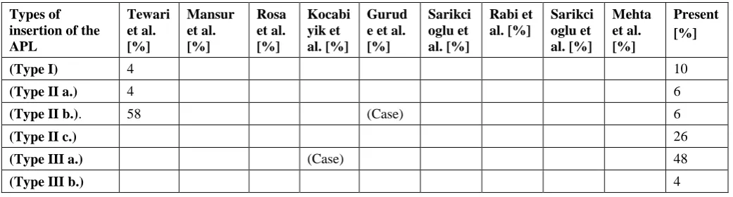
Differences between variations described by other authors and these found in our work are
presented in Table 3.
Table 3. The differences between the classifications given in the present study and those of other authors.
Types of insertion of the APL Tewari et al. [%] Mansur et al. [%] Rosa et al. [%] Kocabi yik et al. [%] Gurud e et al. [%] Sarikci oglu et al. [%] Rabi et al. [%] Sarikci oglu et al. [%] Mehta et al. [%] Present [%]
(Type I) 4 10
(Type II a.) 4 6
(Type II b.). 58 (Case) 6
(Type II c.) 26
(Type III a.) (Case) 48
(Type III b.) 4
Our prosed classification is intended to assist clinicians while preparing for surgery or
during diagnosis. In this case, a classification based on ten, eight or even seven types is too
unwieldy; a simpler and more practical approach would be to divide the types based on single,
bifurcated (with subtypes) and trifurcated types (with subtypes), as in the proposed
classification.
A good knowledge of the variations of the APL is important from the clinical point of
view. For example, de Quervain’s tenosynovitis often develops due to repetitive and
continued strain of the APL and EPB tendons as they pass under a thickened and swollen
extensor retinaculum [15]. Symptoms include pain at the radial side of the wrist, spasms,
tenderness, an occasional burning sensation in the hand and swelling over the thumb side of
the wrist, and difficulty gripping with the affected side of the hand. The onset is often gradual
the forearm [15]. The presence of additional APL bands running in separate sheaths may
influence the choice of treatment, as they can reduce the penetration of steroids used in
conservative approaches and prevent full surgical release, thus resulting in failure. Similarly,
abnormal insertion into the phalanx or metacarpal can cause trapezio-metacarpal subluxation
or arthritis. In addition, ABL is also commonly used as material for tendon grafts; hence, the
presence of additional bands might affect the grafting procedure, the quality of the graft and
the remaining function of the thumb [8, 15].
The present study has some limitations: no sample size calculation was performed and
a relatively low number of dissected limbs were examined. Nevertheless, based on available
material, we were able to propose classification that can cover all variations of the APL and
can serve as a basis for further studies, and as a foundation for communication with surgeons
in clinical practice. One final limitation is that the study explores only variations of the APL
and not the EPB, despite the fact that it might also present variants with a significant influence
important on the aetiology and treatment of de Quervain’s tenosynovitis.
CONCLUSIONS
The APL is characterized by high morphological variability associated with the
presence of additional bands and fusion with neighbouring muscles. Our proposed
classification is simple and serves as a foundation for further development with rare variants.
Acknowledgements
The authors wish to express their gratitude to all those who donated their bodies to medical
science.
Ethical approval and consent to participate
The anatomical protocol of the study was accepted by Bioethics Committee of Medical
University of Lodz (resolution RNN/09/19/KE).
The Local Bioethics Commission gave consent for the study (agreement no. RNN/09/19/KE).
The cadavers belong to the Department of Normal and Clinical Anatomy of the Medical
University of Lodz.
This article does not contain any studies with human participants or animals performed by any
References
1. Moore K, Arthur F, Dalley I, et al. Clinically Oriented Anatomy. Lippincott Williams & Wilkins, 2013.
2. Tripathy S, Tewari J, Mishra P. Anatomical variation of abductor pollicis longus in Indian population A
cadaveric study. Indian J. Orthop. 2015; 49 (5): 549.
3. Coleman SS, McAfee DK, Anson BJ. The insertion of the abductor pollicis longus muscle; an
anatomical study of 175 specimens. Q. Bull. Northwest. Univ. Med. Sch. 1953; 27 (2): 117–122.
4. El-Beshbishy RA, Abdel-Hamid GA.Variations of the abductor pollicis longus tendon: An anatomic
study. Folia Morphol. 2013; 72 (2): 161–166.
5. Thwin SS, Zaini F, Than M. Multiple variations of the tendons of the anatomical snuffbox. Singapore
Med. J. 2014; 55 (1): 37–40.
6. Fabrizio P, Clemente FR. A Variation in the Organization of Abductor Pollicis Longus. Clin. Anat.
1996; 375 (9): 371–375.
7. Opreanu RC, Wechter J, Tabbaa H, et al. Anatomic variations of the first extensor compartment and
abductor pollicis longus tendon in trapeziometacarpal arthritis. Hand. 2010; 5 (2): 184–189.
8. Bravo E, Barco R, Bullón A. Anatomic study of the abductor pollicis longus : A source for grafting
material of the hand. Clin. Orthop. Relat. Res. 2010; 468 (5): 1305–1309.
9. Mahakkanukrauh P, Mahakkanukrauh C. Incidence of a septum in the first dorsal compartment and its
effects on therapy of de Quervain’s disease. Clin. Anat. 2000; 13 (3): 195–198.
10. Bharambe V, Patel D, Rao Manvikar P, et al. A study of extensor pollicis longus and brevis and abductor
pollicis longus from the perspective of evolution. J. Med. Res. 2017; 3 (3): 146–150.
11. Olewnik Ł, Wysiadecki G, Polguj M, et al. Anatomical variations of the palmaris longus muscle
including its relation to the median nerve: A proposal for a new classification. BMC Musculoskelet.
Disord. 2017; 18 (1): 1–9.
12. Olewnik Ł, Podgórski M, Polguj M, et al. Anatomical variations of the pronator teres muscle in a Central
European population and its clinical significance. Anat. Sci. Int. 2018; 93 (2): 299–306.
13. Anson B. An Atlas of Human Anatomy. W.B. Saunders Company, Philadelphia, 1963.
14. Giles KW. Anatomical variations affecting the surgery of de Quervain’s disease. J. Bone Jt. Surg. 1960;
42 (B): 352–5.
15. Goel R, Abzug JM. de Quervain’s tenosynovitis: a review of the rehabilitative options. Hand. 2015; 10
(1): 1–5.
Figure 1. Type I of abductor pollicis longus insertion. APL abductor pollis longus EPL
extensor pollicis longus EPB extensor pollicis brevis RA radial artery
Figure 2. Type II a of abductor pollis longus insertion. APL abductor pollis longus EPL
Figure 3. Type II b of abductor pollis longus insertion. APL abductor pollis longus EPL
extensor pollicis longus EPB extensor pollicis brevis
Figure 4. Type II c of abductor pollis longus insertion. APL abductor pollis longus EPL
extensor pollicis longus EPB extensor pollicis brevis
Figure 5. Type III a of abductor pollis longus insertion. APL abductor pollis longus EPL
extensor pollicis longus EPB extensor pollicis brevis
Figure 6. Type III b of abductor pollis longus insertion. APL abductor pollis longus EPL