Published online 2015 August 29. Research Article
The Effect of Different Training Modes on Serum Apelin and Pain Threshold
in Morphine-Dependent Rats
Ebrahim Zarrinkalam,
1and Ali Heidarianpour
1,*1Department of Exercise Physiology, Faculty of Physical Education and Sport Science, Bu Ali Sina University, Hamadan, IR Iran
*Corresponding author: Ali Heidarianpour, Department of Exercise Physiology, Faculty of Physical Education and Sport Science, Bu Ali Sina University, Hamadan, IR Iran. Tel: +98-9188187984, E-mail: [email protected]
Received 2015 June 1; Revised 2015 July 15; Accepted 2015 August 15.
Abstract
Background: Apelin has recently been identified as an analgesic agent and a novel neuropeptide. On the other hand, it has been shown that exercise can lead to reduced pain in morphine-dependent patients.
Objectives: Therefore, the aim of this study was to evaluate apelin and pain threshold changes in healthy and morphine-dependent rats in response to two exercise paradigms.
Materials and Methods: In this study, 30 healthy and 30 morphine-dependent rats were used. Morphine-dependent and healthy rats were divided into six groups: 1, a control (healthy) group; 2, a healthy endurance group; 3, a healthy strength-training group; 4, an addicted control group; 5, an addicted endurance group; 6, an addicted strength-training group. Then, the training groups performed aerobic and strength training for eight weeks. After the training program, the tail flick and formalin tests were used to assess pain. Apelin was also measured by ELISA.
Results: Regardless of the type of exercise, exercise significantly increased the apelin serum levels in healthy rats. The apelin levels significantly increased in the morphine-dependent rats compared with the healthy control group. Endurance, unlike strength training, significantly increased apelin in the serum compared to the addicted control group. The training led to pain relief in the morphine-dependent rats and returned it to the healthy control group level. The Pearson correlation showed a reverse significant correlation between the serum apelin level and the tail flick test in the morphine-dependent rats.
Conclusions: The results showed that endurance training reduced pain by increasing apelin in morphine-dependent rats. Therefore, it is suggested that this type of training be considered for the morphine-dependent patients for pain relief.
Keywords: Exercise Training, Analgesics, Pain Threshold, Morphine, Apelin
Copyright © 2015, Hamadan University of Medical Sciences. This is an open-access article distributed under the terms of the Creative Commons Attribution-Non-Commercial 4.0 International License (http://creativecommons.org/licenses/by-nc/4.0/) which permits copy and redistribute the material just in noncommercial usages, provided the original work is properly cited.
1. Background
Drug abuse is one of the most important health, social, and cultural issues. Over 90% of people have expressed seri-ous concerns about drug abuse as a worldwide problem. Ac-cording to the latest data from the united nations office on drugs and crime (UNODC), drug abuse is rising at a gentle slope; in 2012 – 2013, an average of 226 million (5%) of people between the ages of 15 and 64 years old had used drugs at least once per year (1). Addiction is now being introduced as a disease associated with molecular and physiological changes in which various factors are involved, including genetic, environmental, and neurobiological factors; there-fore, therapy methods are very different and complicated and no satisfactory results have yet been achieved (2). Exer-cise has been reported to be effective in the treatment and even prevention of many disorders, including depression, memory impairment, Alzheimer disease, and addiction (3, 4). It has been shown that the release of several neurotrans-mitters like dopamine, glutamate, acetylcholine, sero-tonin, and androgen opioids in the brain can be altered by exercise (5). Exercise can also compensate for the reduced
with the descending pain transmission system, such as the amygdala, hypothalamus, and dorsal raphe nucleus (9). Xu et al. (2009) showed that apelin can demonstrate its anal-gesic effect through the hair and receptors in the hippo-campus, which these APJ play a key role in drug withdrawal syndrome by increasing the levels of opioids. They showed that the apelin levels are very high at the areas of brain in which the highest level of opioids can be found, indicating interrelationships between apelin and analgesic effects (7).
2. Objectives
Most studies regarding the reduction of pain are focused on aerobic activity; however, the effect of strength training is not clear. This study aimed to investigate the relation-ship between the serum apelin levels and analgesic effects in morphine-dependent rats following two types of exer-cise (aerobic and strength-training) for the first time.
3. Materials and Methods
In this experimental study, male Wistar rats aged 6 – 8 weeks weighing 180 – 200 g (Razi Pasteur institute, Iran) were used. Animals were kept in the rodent’s standard laboratory (12 hours light-dark cycle at 2 ± 22 C°). They had free access to food and water.
3.1. Dependency Induction Method
In this study, morphine dependency was induced orally with morphine at continuous concentrations of 1.0, 2.0, and 3.0 mg/mL for 48 hours, then 4.0 mg/mL in the next 15 days was poured in the drinking water. Sucrose sulfate (3%) was added to the drinking water due to the bitter taste of morphine. The water and morphine were covered by thin aluminum sheets to prevent the degradation of morphine by light. The rats were addicted to morphine. Naloxone (3 mg/kg body weight) was injected intraperi-toneally to one or two rats in each group randomly to examine their morphine dependency. After confirming rats’ addiction to morphine, morphine-dependent and healthy rats were divided into the following six groups: 1, a control (healthy) group; 2, a healthy endurance group; 3, a healthy strength-training group; 4, an addicted con-trol group; 5, an addicted endurance group; and 6, an ad-dicted strength-training group.
3.2. The Training Protocols
3.2.1. Endurance Training
The rats were trained for 8 weeks, 5 days per week. The training period was divided to 2 stages: overload and load intensity stabilization stages. During the first week, the rats ran every day for 10 minutes at a speed of 17 m/min on a treadmill. Gradually, from the second to the fifth week (overload stage), the training length was increased to 55 minutes per session. From the sixth to the eighth week (the length stabilization stage), the rats ran on treadmill
for 55 minutes at 30 m/min (75% VO2max), (Table 1) (10). The incline of the treadmill was 0° in all stages.
3.2.2. Strength Training
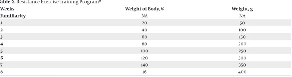
The rats were placed 3 days per week for 15 minutes on a 36-step ladder (Iran) with no weights for adaptation and reducing their stress. Then, the regular exercise protocol was performed 5 days per week in three sets, which in-cluded 4 trainings with 3-minute intervals between the sets and 15 seconds between iterations, for 8 weeks. The rats were trained in the first 3 weeks with weights weigh-ing 20, 40, and 60% of their body weight; with weights weighing 80, 100, and 120% of their body weight in the following 3 weeks; and finally with weights weighing of 140 and 160% of their body weight in the final 2 weeks. The ladder was located perpendicular to the ground next to the wall (Table 2). All the rats had 2 days of rest.
3.3. Tail Flick Test
After training, a tail flick test was used to measure the pain threshold in the studied groups. The tail flick apparatus consisted of two parts: a restrainer and a control system. The rats were placed in the restrainer while their tail was out and the beginning of the tail was on a light-sensitive sensor. In this study, the light intensity was adjusted to achieve a ba-sic average responsiveness time of 4 to 5 seconds to obtain approximately 50°C, which was appropriate for testing; 12 seconds was considered the cut-off time to the middle of the tail. For adaption with the restrainer, the animals were trained 3 days before the experiment, 1 hour per day to re-duce the stress caused by the apparatus immobilization. After putting a rat in the restrainer, the tail was fixed and the start button was pressed for radiation. The mean with-drawal time was measured 3 times in 1-minute intervals. To prevent tissue damage, the rats were removed from the light when there was no reaction for 30 seconds (11).
3.4. Formalin Test
3.5. Apelin Measurement Method
The blood samples (3 cc) were taken from the rats’ eyes 48 hours after the last training session. They were centri-fuged at 10,000 rpm for 10 minutes. The serum samples were stored at -80°C before the measurements. Apelin was measured using ELISA kits (Apelin ELISA kit, RAB0018, Sigma Aldrich USA) and the ELISA method according to the manufacturer’s instructions by Elisa reader (ELX800, USA).
3.6. Statistical Analysis
Data were analyzed using SPSS 18. The normality of the data was confirmed using the Shapiro-Wilk test. The one-way ANOVA and Tukey’s post-hoc tests were used to evalu-ate the differences between the groups. Data are presented as mean ± SEM, and α < 0.05 was considered significant.
4. Results
Apelin: The one-way ANOVA results showed that the ape-lin levels were significantly different between groups (P = 0.0001, F = 34.59). Tukey’s post-hoc test results showed a significant difference between the control group and the other addicted groups. There was a significant difference between the healthy groups and the addicted strength-training group. Tukey’s test results showed a significant difference between the endurance group and the control and the healthy strength-training groups. There was sig-nificant difference between strength-training group and control (healthy) group (Figure 1).
4.1. The Pain Threshold
The tail flick test results showed that there is a significant dif-ference between the groups’ pain threshold (P = 0.02, F = 7.04). Tukey’s post-hoc test results showed that the pain threshold in the addicted control group was significantly higher than the other groups. The results showed no significant difference between the trained groups and the control (healthy) group. According to Tukey’s post-hoc test, there were no significant differences between the trained groups (Figure 2).
The one-way ANOVA results showed a significant differ-ence between the groups in the pain threshold in the for-malin test (P = 0.04, F = 5.73). Tukey’s post-hoc test results showed that the pain threshold in the addicted control group is significantly higher than other groups. Moreover, the results showed no significant difference between the trained groups and the healthy control group. According to Tukey’s post-hoc test, there was no significant difference between the trained groups as well (Figure 3).
4.2. Pearson Correlations Between Parameters
There was no significant correlation between apelin and the tail flick test in healthy rats (α = 0.072, R = -0.34) (Figure 4). There was also no significant correlation be-tween apelin and the formalin test in the healthy rats (α = 0.68, R = -0.01) (Figure 5).
There was a significant inverse correlation between ape-lin and the tail flick test in morphine-dependent rats (α = 0.0001, R = -0.79) (Figure 6). However, there was no signifi-cant correlation between apelin and the formalin test in the morphine-dependent rats (α = 0.47, R = -0.14) (Figure 7).
Table 1. Endurance Exercise Training Programa
Weeks Duration, min Speed, m/min Intensity (VO2max), %
0 15 5 45
1 20 10 50
2 25 15 60
3 30 20 65
4 35 25 70
5 40 30 75
6 45 30 75
7 50 30 75
8 55 30 75
aThe speed and duration were gradually increased to 30 m/min and 55 min.VO
Table 2. Resistance Exercise Training Programa
Weeks Weight of Body, % Weight, g
Familiarity NA NA
1 20 50
2 40 100
3 60 150
4 80 200
5 100 250
6 120 300
7 140 350
8 16 400
Abbreviation: not available.
Figure 1. Effect of Different Exercise Training Modes on Serum Apelin in all Experimental Groups
HC HE HS AC AE AS Groups
Apelin pg/ml
1600 1400 1200 1000 800 600 400 200 0
P,C,D,E,F
A,E A,E A,E A,E A,B,C,D,F
Each column and bar represents mean ± S.E.M: A, significant difference from the HC group at the level of P < 0.05; B, significant difference from the HE group at the level of P < 0.05; C, significant difference from the HS group at the level of P < 0.05; D, significant difference from the AC group at the level of P < 0.05; E, significant difference from the HE group at the level of P < 0.05; F, significant difference from the HE group at the level of P < 0.05.
Figure 2. Effect of Different Exercise Training Modes on Tail flick test in all Experimental Groups
HC HE HS AC AE AS Groups
6
5
4
3
2
1
0
Tail Dliek T
est ,
s
Each column and bar represents mean ± S.E.M significant difference from the other experimental groups at the level of P < 0.05.
Figure 3. Effect of Different Exercise Training Modes on Formalin Test in all Experimental Groups
HC HE HS AC AE AS Groups
2.5
2
1.5
1
0.5
0
Formalin T
est (Score)
Values are mean ± SEM; Significant difference from the other experimen-tal groups at the level of P < 0.05.
Figure 4. There are No Correlation Between Apelin and Tail Flick Test in Healthy Rats
Apelin pg/ml
Tailflick
3.40 3.50 3.60 3.70 3.00 1400.00
1200.00
1000.00
800.00
600.00
400.00
r = -034, alpha = 0.072
R Sq Linear = 0.119
r = -0.34; P = 0.072.
Figure 5. There are no Correlation Between Apelin and Formalin Test in Healthy Rats
Apelin pg/ml
Formalin
2.00 2.10 2.20 2.30 2.40 2.50 1400.00
1200.00
1000.00
800.00
600.00
400.00
r = -01, alpha = 0.68
R Sq Linear = 0.01
r = -0.1; P = 0.68.
Figure 6. Significant Inverse Correlation Between Serum Apelin and Tail Flick Test in Morphine-Dependent Rats
Apelin pg/ml
Tailflick
3.30 4.00 4.20 4.40 4.60 4.80 5.00 1400.00
1200.00
1000.00
800.00
r = 0.79, alpha = 0.0001
R Sq Linear = 0.635
Figure 7. There are No Correlation Between Apelin and Formalin Test in Morphine-Dependent Rats
Apelin pg/ml
Formalin
0.80 1.00 1.20 1.40 1.60 1.80 1400.00
1200.00
1000.00
800.00
r = 0.14, alpha = 0.47
R Sq Linear = 0.022
r = -0.14; P = 0.47.
5. Discussion
Apelin is a multi-functional neuropeptide that plays an important role in energy homeostasis regulation, immune system function, gastrointestinal function, and pain relief regulation (7, 8). Morphine is used ex-tensively to relieve acute pain, but morphine injections to relieve chronic pain are associated with side effects, including addiction (2). This study was the first to show that strength training can provide relief in morphine-dependent rats by increasing the serum apelin neuro-peptide. Regardless of the type of exercise, morphine led to a significant increase in the serum apelin levels in healthy rats. Moreover, no significant differences were observed between the trained groups and the healthy subjects. On the other hand, the serum apelin levels sig-nificantly increased in the addicted groups compared with the control (healthy) group. The results showed that strength training unlike endurance training sig-nificantly increased the serum apelin levels compared with the addicted control group. On the other hand, endurance training can lead to a reduced pain thresh-old in morphine-dependent rats and change it to the control (health) group’s level. The Pearson correlation results showed a reverse correlation between the serum apelin levels and the tail flick test in morphine-depen-dent rats. This may be due to the wide distribution of the apelin receptors in the pain control centers like the spinal cord, hypothalamus, and medulla oblongata (12-14). The distribution of apelin receptors in the brain pain control centers can imply the physiological func-tion of apelin in pain relief. The results of this study are consistent with those of Yang et al., indicating that 0.3 to 3 g intraventricular injections of the apelin-13 using water immersion tests in healthy rats reduced pain by activating apelin-specific receptors and hair receptors. They also showed that these changes do not mean that apelin is able to reduce all types of pain (15). The results demonstrated that there is a significant correlation
Footnote
Authors’ Contribution:Ali Heidarianpour wrote the manuscript; Ebrahim Zarrinkalam executed the study protocols.
References
1. van Boekel LC, Brouwers EP, van Weeghel J, Garretsen HF. Public opinion on imposing restrictions to people with an alcohol- or drug addiction: a cross-sectional survey. Soc Psychiatry Psychiatr Epidemiol. 2013;48(12):2007–16. doi: 10.1007/s00127-013-0704-0. [PubMed: 23657876]
2. Alaei H, Huotari M, Piepponen PT, Ahtee L, Hanninen O, Mannisto PT. Morphine rele ases glutamate through ampa receptors in the ventral tegmental area: A microdialysis study in conscious rats.
MJIRI. 2003;17(3):225–31.
3. McGovern MK. The effects of exercise on the brain. Serendip.
2005;31(6):125–34.
4. Shokraviyan M, Miladi-Gorji H, Vaezi GH. Voluntary and forced exercises prevent the development of tolerance to analgesic ef-fects of morphine in rats. Iran J Basic Med Sci. 2014;17(4):271–7. [PubMed: 24904720]
5. Marghmaleki VS, Alaei HA, Malekabadi HA, Pilehvarian A. Effect of physical activity on symptoms of morphine addiction in rats, after and before of lesion of the mpfc area. Iranian journal of basic medical sciences. 2013;16(10):1091.
6. Lv S, Yang YJ, Hong S, Wang NB, Qin YJ, Li W, et al. Intrathecal apelin-13 produced different actions in formalin test and tail-flick test in mice. Protein & Peptide Letters. 2013;20(8):926–31. doi: 10.2174/0929866511320080010.
7. Xu N, Wang H, Fan L, Chen Q. Supraspinal administration of ape-lin-13 induces antinociception via the opioid receptor in mice.
Peptides. 2009;30(6):1153–7. doi: 10.1016/j.peptides.2009.02.011. [PubMed: 19463749]
8. Lv SY, Qin YJ, Wang NB, Yang YJ, Chen Q. Supraspinal antinocicep-tive effect of apelin-13 in a mouse visceral pain model. Peptides.
2012;37(1):165–70. doi: 10.1016/j.peptides.2012.06.007. [PubMed: 22732665]
9. Véras-Silva AS, Mattos KC, Gava NS, Brum P, Negrão CE, Krieger EM. Low-intensity exercise training decreases cardiac out-put and hypertension in spontaneously hypertensive rats.
American Journal of Physiology-Heart and Circulatory Physiology.
1997;273(6):H2627–31.
10. Le Bars D, Gozariu M, Cadden SW. Animal models of nociception.
Pharmacol Rev. 2001;53(4):597–652. [PubMed: 11734620]
11. O’Carroll AM, Selby TL, Palkovits M, Lolait SJ. Distribution of mRNA encoding B78/apj, the rat homologue of the human APJ re-ceptor, and its endogenous ligand apelin in brain and peripheral
tissues. Biochimica et Biophysica Acta (BBA) - Gene Structure and Ex-pression. 2000;1492(1):72–80. doi: 10.1016/s0167-4781(00)00072-5. 12. Reaux A, De Mota N, Skultetyova I, Lenkei Z, El Messari S, Gallatz
K, et al. Physiological role of a novel neuropeptide, apelin, and its receptor in the rat brain. J Neurochem. 2001;77(4):1085–96. [PubMed: 11359874]
13. Hosoya M, Kawamata Y, Fukusumi S, Fujii R, Habata Y, Hinuma S, et al. Molecular and functional characteristics of APJ. Tissue distribution of mRNA and interaction with the endogenous li-gand apelin. J Biol Chem. 2000;275(28):21061–7. doi: 10.1074/jbc. M908417199. [PubMed: 10777510]
14. Lv SY, Yang YJ, Qin YJ, Xiong W, Chen Q. Effect of centrally ad-ministered apelin-13 on gastric emptying and gastrointestinal transit in mice. Peptides. 2011;32(5):978–82. doi: 10.1016/j.pep-tides.2011.01.023. [PubMed: 21291936]
15. Yang YJ, Lv SY, Xiu MH, Xu N, Chen Q. Intracerebroventricular ad-ministration of apelin-13 inhibits distal colonic transit in mice.
Peptides. 2010;31(12):2241–6. doi: 10.1016/j.peptides.2010.09.006. [PubMed: 20849897]
16. Befort K, Filliol D, Darcq E, Ghate A, Matifas A, Lardenois A, et al. Gene expression is altered in the lateral hypothalamus upon acti-vation of the mu opioid receptor. Ann N Y Acad Sci. 2008;1129:175– 84. doi: 10.1196/annals.1417.028. [PubMed: 18591478]
17. Fujie S, Sato K, Miyamoto-Mikami E, Hasegawa N, Fujita S, Sanada K. Reduction of arterial stiffness by exercise training is associat-ed with increasing plasma apelin level in middle-agassociat-ed and older adults. PloS one. 2014;9(4). doi: 10.1371/journal.pone.0093545. 18. Kadoglou NP, Fotiadis G, Kapelouzou A, Kostakis A, Liapis CD,
Vrabas IS. The differential anti-inflammatory effects of exercise modalities and their association with early carotid atheroscle-rosis progression in patients with type 2 diabetes. Diabet Med.
2013;30(2):e41–50. doi: 10.1111/dme.12055. [PubMed: 23078531] 19. Zhang J, Ren CX, Qi YF, Lou LX, Chen L, Zhang LK, et al. Exercise
training promotes expression of apelin and APJ of cardio-vascular tissues in spontaneously hypertensive rats. Life Sci.
2006;79(12):1153–9. doi: 10.1016/j.lfs.2006.03.040. [PubMed:
16674982]
20. Kleinz MJ, Skepper JN, Davenport AP. Immunocytochemical localisation of the apelin receptor, APJ, to human cardiomyo-cytes, vascular smooth muscle and endothelial cells. Regul Pept.
2005;126(3):233–40. doi: 10.1016/j.regpep.2004.10.019. [PubMed: 15664671]
21. Andersen CU, Hilberg O, Mellemkjaer S, Nielsen-Kudsk JE,
Si-monsen U. Apelin and pulmonary hypertension. Pulm Circ.
2011;1(3):334–46. doi: 10.4103/2045-8932.87299. [PubMed:
22140623]