R E S E A R C H
Open Access
SP-A2 contributes to miRNA-mediated sex
differences in response to oxidative stress:
pro-inflammatory, anti-apoptotic, and
anti-oxidant pathways are involved
George T. Noutsios
1†, Nithyananda Thorenoor
1†, Xuesheng Zhang
1, David S. Phelps
1, Todd M. Umstead
1,
Faryal Durrani
1and Joanna Floros
1,2*Abstract
Background:Human innate host defense molecules, surfactant protein A1 (SP-A1), and SP-A2 differentially affect the function and proteome of the alveolar macrophage (AM). We hypothesized that SP-A genes differentially regulate the AM miRNome.
Methods:Humanized transgenic mice expressing SP-A1 and SP-A2 were subjected to O3-induced oxidative stress (OxS) or filtered air (FA), AMs were isolated, and miRNA levels were measured.
Results:In SP-A2 males, we found significant changes in miRNome in terms of sex and sex-OxS effects, with 11 miRNAs differentially expressed under OxS. Their mRNA targets included BCL2, CAT, FOXO1, IL6, NF-kB, SOD2, and STAT3. We followed the expression of these transcripts as well as key cytokines, and we found that (a) the STAT3 mRNA significantly increased at 4 h post OxS and returned to baseline at 18 h post OxS. (b) The anti-oxidant protein SOD2 level significantly increased, but the CAT level did not change after 4 h post OxS compared to control. (c) The anti-apoptotic BCL2 mRNA increased significantly (18 h post OxS), but the levels of the other transcripts were decreased. The presence of the SP-A2 gene had a protective role in apoptosis of AMs under OxS compared to mice lacking SP-A (knockout, KO). (d) Pro-inflammatory cytokine IL-6 protein levels were significantly increased in SP-A2 mice compared to KO (4 and 18 h post OxS), which signifies the role of SP-A2 in
pro-inflammatory protein expression. (e) SOD2 and CAT mRNAs changed significantly in OxS indicating a plausible role of SP-A2 in the homeostasis of reactive oxygen species. (f) Gonadectomy of transgenic mice showed that sex hormones contribute to significant changes of the miRNome expression.
Conclusions:We conclude that SP-A2 influences the miRNA-mediated sex-specific differences in response to OxS. In males, these differences pertain to inflammatory, anti-apoptotic, and anti-oxidant pathways.
Keywords:Surfactant protein A, Ozone, Sex differences, STAT3, IL-6
* Correspondence:[email protected]
†Equal contributors
1Center for Host Defense, Inflammation, and Lung Disease (CHILD) Research,
Department of Pediatrics, The Pennsylvania State University College of Medicine, Hershey, PA 17033-0850, USA
2Department of Obstetrics and Gynecology, The Pennsylvania State
University College of Medicine, Hershey, PA 17033-0850, USA
Background
The major function of the lung is to enable the uptake of oxygen from ambient air and eliminate carbon diox-ide from the circulation. As the lung performs this life-sustaining function, it encounters many insults from the environment, such as ozone (O3), particulate matter,
al-lergens, bacteria, and viruses. Although healthy individ-uals bearing a well-regulated innate host defense system can neutralize these insults and maintain normal lung function, O3-induced oxidative stress (OxS) can have
dire effects on pulmonary innate host defense mecha-nisms and consequently on lung function [1].
In the alveolus, the alveolar macrophages (AMs), and to a lesser extent, the alveolar epithelial cells provide the primary innate defense against inhaled insults that may trigger a cascade of inflammatory responses after O3
ex-posure. The alveolus is lined by epithelial type I and type II cells and contains various epithelial cell metabolic products, including pulmonary surfactant as well as teins from the lung interstitial fluid. Both surfactant pro-teins and lipid constituents of surfactant influence the regulation and function of AM [2–9].
Surfactant protein A (SP-A) is an important host defense molecule and has both innate host defense func-tions [2, 10–12] and surfactant-related functions that in-fluence surfactant secretion and structure. We and others have highlighted the function of SP-A as innate immune molecule itself by showing that SP-A knockout (KO) mice are more prone to several infections com-pared to mice that express SP-A [13–17]. Previous stud-ies have described the regulatory effects of SP-A either by directly affecting AM function [2, 18], the proteomic expression profile of AM [9, 19], and the cell shape and activation state of AM [20] or indirectly via the produc-tion of cytokines and chemokines by AM [11, 21]. Ozone exposure induces production of reactive oxygen species (ROS) by bronchoalveolar cells [22]. ROS dam-age the lung epithelium [23] and oxidize SP-A, and in turn, this compromises its innate immune functions [24–29]. The model used in this study provides advan-tage over other types of OxS, such as hyperoxia (which occurs over days), as it produces effects in several hours, allowing us to examine primary endpoints rather than secondary downstream events.
Ozone exposure has resulted in significant sex differ-ences in survival, with females showing higher morbidity and mortality rates than males in several lung diseases including respiratory infections, COPD, asthma, and CF [30–34]. In our studies, we observed differences in sur-vival rates between males and females after bacterial in-fection. Females appeared to resolve the bacterial respiratory infection more readily than males, but when OxS was introduced prior to infection, the outcome was reversed, i.e., females showed lower survival rates than
males [13, 17, 35–38]. Moreover, these sex differences were further accentuated in A KO mice or when SP-A was oxidized, highlighting once again the important role of SP-A as a regulatory innate immune molecule [13, 17]. The observed sex differences are in part, if not entirely, attributable to circulating gonadal hormones, which are believed to influence the innate immune re-sponses [36], although the specific roles of these hor-mones and the underlying mechanisms of regulation remain unknown.
In the present study, we hypothesized that the two human SP-A1 and SP-A2 gene products differentially regulate the AM miRNome and this explains in part the previously observed differences in AM function and the AM proteome. We exposed male and female mice expressing SP-A1 or SP-A2 to O3 or FA (control), and
we measured the expression levels of 372 micro-RNAs (miRNAs). The miRNAs with significantly altered levels in response to OxS were identified and used for further analysis. The findings showed that the AM miRNome is regulated by O3 exposure and that the SP-A2 male
miRNome is associated with genes involved in inflam-mation pathways, regulation of reactive oxygen species, and apoptosis. Moreover, the miRNA differences in SP-A2 male and female mice after gonadectomy showed that sex hormones play an important role in the ob-served miRNA differences by affecting the regulation of miRNAs. We postulate that miRNAs contribute to mechanisms involved in the regulation of AM function and the AM proteome by the SP-A2 gene in males.
Methods Animals
We used humanized transgenic (hTG) C57BL/6 mice, expressing SP-A1 (6A2) or SP-A2 (1A0), or SP-A KO mice [39] at 12 weeks of age. Both males and syn-chronized females (regarding the estrous cycle) were used in this study. For synchronization, we used dirty bedding from a male’s cage to stimulate estrus in fe-males. Females were group-housed on male bedding for exactly 10 days before exposure to FA or O3. For
each experimental condition, there were 3 animals/ group (n= 36 for miRNA study and n= 60 for gene expression analysis, terminal deoxynucleotidyl
trans-ferase (TdT)-mediated dUTP Nick-End Labeling
Ozone exposure
Mice were exposed to 2 ppm ozone (O3) or to filtered
air (FA) (control) at room temperature and 50% humid-ity for 3 h, as described previously [40]. In all cases, we used at least 3 mice/group, i.e., 3 males, 3 females, 3 SP-A KO, 3 hTG SP-SP-A2, 3 hTG SP-SP-A1, 3 for O3 exposure
and 3 for filtered air, and 3 male and 3 female SP-A2 and KO gonadectomized mice (n= 96). Each sample was analyzed individually, and we did not pool any samples. All FA and O3 exposures were conducted in parallel
[41]. Mice were sacrificed 4 and 18 h after exposure.
Mouse alveolar macrophage purification
Mouse AMs were obtained by bronchoalveolar lavage (BAL) [9]. AMs were separated from the BAL by centri-fugation (150×g for 5 min). The BAL supernatant, which was cell-free (3.0 ml), was stored at−80 °C until further use. The cell pellets were washed with 1× PBS (Gibco, Waltham, MA), and cells were counted. A frac-tion of the cells was placed in a cytocentrifuge and used to prepare cytospins, cells were stained, and a differen-tial cell count was performed. AM purity was 95% as assessed by Papanikolaou staining. The remaining AM pellet was resuspended in 0.5 ml solution of 90% fetal bovine serum (Gibco) and 10% DMSO (Sigma-Aldrich, St. Louis, MO) and was cryopreserved in liquid nitro-gen until further use.
Isolation of miRNAs and qRT-PCR
Total RNA from the AM was isolated using QIAzol Lysis Reagent (#79306, Qiagen, Valencia, CA), and the miRNA-enriched fraction was isolated and purified with miRNeasy Micro Kit (#217084, Qiagen). cDNA was gen-erated with the miScript II RT Kit (#218161, Qiagen) and then used as a template for real-time qPCR with the miS-cript SYBR Green PCR Kit (#218075, Qiagen). The ex-pression profiles of the 372 most abundantly expressed and best-characterized miRNAs in miRBase were deter-mined by miRNA PCR Array (Array #MIMM-3001Z, Qiagen). The miRNA PCR array covers most of the mouse miRNA orthologs (~ 90%) of the human miRNA annotations. The variability across the 3 samples is assessed bypvalues (p< 0.05). The miRNAs with signifi-cantly changed levels were studied further (p< 0.05).
Data analysis
miRNAs (miR-21a-5p, miR-23a-3p, mmu-miR-2137, and mmu-let-7c-5p) were constant under our experimental conditions and therefore were selected for use in the normalization of all miRNA values. The arith-metic mean values of these miRNAs were used for normalization, and all statistical analyses were done after normalization.
The Ct cutoff was 35. The fold change (2−ΔΔCt) was calculated as the normalized individual miRNA expres-sion (2−ΔCt) in each individual experimental specimen divided by the same normalized miRNA expression 2 −ΔCt
in the corresponding KO (control). The fold regula-tion represented fold change results in a biologically meaningful way. Fold change values greater > 1 indicated a positive or an upregulation, and the fold regulation was equal to the fold change. Fold change values < 1 indicated a negative or downregulation, and the fold regulation was the negative inverse of the fold change. Thepvalues were calculated based on Student’sttest of the replicate 2−ΔCt values for each miRNA in the KO control group and treatment groups. All data can be found in Additional file 1. Data analysis was performed with manufacturer’s software (https://www.qiagen.com/ us/resources/geneglobe/).
Gonadectomy and ozone exposure
Male and female SP-A2 mice were gonadectomized (Gx) as described [36]. Two weeks after gonad removal, mice were exposed to O3(2 ppm) for 3 h and were sacrificed
4 h post OxS. The RNA from AM was isolated, and the differential expression of miRNAs was quantified as tag count data [42] by RNA sequencing at the Pennsylvania State University College of Medicine Genomic Core Facility, with default false discovery rate 0.1 (cutoff ). The miRNAs identified from Gx mice were selected based on the average tag count data (≥100) and with good correl-ation data count between mice (2 out of 3). We success-fully identified 63 miRNAs in Gx-SP-A2 and KO (male and female) and used these in the present analysis. First, the changes in miRNA expression in SP-A2 mice were calculated by normalizing to KO, i.e., the level of expres-sion of each individual experimental miRNA (i.e., in Gx-SP-A2) was divided by the same miRNA expression in the corresponding Gx-KO. Next, the differentially expressed miRNAs between SP-A2 males and females were determined by dividing a specific individual male miRNA by the corresponding specific female miRNA. Lastly, the fold changes of the identified miRNAs from Gx (SP-A2 male vs female) were compared to the corre-sponding miRNAs identified from non-gonadectomized (NGx) SP-A2 male vs female. Samples from 12 animals (6 males and 6 females for SP-A2 and KO) were individually analyzed.
Gene expression assays
AMs were lysed by the addition of QIAzol Lysis Reagent (Qiagen). Total RNA was purified with Direct-zol RNA Mini Prep kit (#R2052, Zymo Research, Irvine, CA), and RNA concentration and quality were confirmed by
Nanodrop and Bioanalyzer. RNA was
according to the manufacturer’s protocol. Real-time PCR was performed using RT2 SYBR Green ROX qPCR Mas-termix (#330520, Qiagen) on a QuantStudio 12K Flex Real-Time PCR System (Applied Biosystems, Waltham, MA) at the Pennsylvania State University College of Medi-cine Genomic Core Facility. The expression levels of GAPDH, SOD2, CAT, IL-6, STAT3, BCL2, FOXO1, FOXO3, BECN1, IKKβ, and NF-kB-p65 were detected by qRT-PCR with specific RT2 qPCR Primer Assays (Qiagen). Samples from 3 animals/treatment (FA and O3)
and 3 replicates/animal were individually analyzed and quantified relative to GAPDH mRNA expression. The relative expression levels of target genes were determined by the equation 2−ΔCT, and 2−ΔΔCT, in whichΔCTwas
cal-culated as follows: ΔCT=CTgene-of-interest−CT housekeeping gene. TheΔΔCTwas calculated by the difference between
ΔCTvalues of experimental (O3exposure) and control (FA
exposure) (ΔΔCT=ΔCTexperimental− ΔCTcontrol).
Western blot analysis and antibodies
Equal amounts of AM protein lysates for each set of sam-ples from control and O3 exposure mice were separated
by sodium dodecyl sulfate-polyacrylamide gel electrophor-esis (SDS-PAGE), transferred to PVDF membrane, and the expression levels of STAT3, NF-kB-p65, FOXO1, CAT, SOD2, and GAPDH were detected by Western blot-ting with specific antibodies. Samples from 3 animals/ treatment (control and O3) were individually analyzed and
quantified relative to GAPDH expression. The antibodies utilized were obtained from the following sources: NF-kB (#8242), FOXO1 (#2880), GAPDH (#2118), STAT3 (#12640), SOD2 (#13194), and catalase (#14097) (Cell Signaling Technology, Danvers, MA).
Measurement of IL-6, TNF-α, and IL-1β
Data from BAL from 3 animals/treatment (FA, O3) and 3
replicates/animal were used to assess levels of IL-6,
TNF-α, and IL-1β using a conventional enzyme-like immuno-sorbent assay (ELISA) as per vendor’s protocol (#SEM03015A, SEM03113A, and SEM03109A, SABios-ciences, Valencia, CA). Levels of IL-6, TNF-α, and IL-1β were measured using BAL samples. (a) BAL from SP-A2 mice after 4 h of ozone (O3) exposure was used to measure
IL-6 levels by diluting the BAL 1:2, 1:4, 1:8, and 1:16. (b) BAL from SP-A2 and KO mice after 18 h of O3exposure
was used to measure IL-6 levels by diluting 1:2, whereas the levels of TNF-α and IL-1β were measured without BAL dilution. The optical density of the end product was measured at 450 nm using Tecan Spectrafluor spectropho-tometer (Agilent Technologies, Santa Clara, CA).
TUNEL assay
The TdT-mediated dUTP Nick-End Labeling (TUNEL;
#4822-96-K, HT Titer TACS Assay, Trevigen,
Gaithersburg, MD) assay was used to detect the ability of ozone to induce apoptosis according to the vendor’s protocol. Briefly, AM cells (70,000 cells/well) after 18 h of O3 exposure from hTG SP-A2 and KO mice were
fixed in 3.7% formaldehyde in 1× PBS. The cells were in-cubated for 1 h at 37 °C in the presence of TdT. In positive control TACS-Nuclease was used, whereas no TdT was added in the unlabeled control (data not shown). The apoptotic cell absorbance was measured at 450 nm using Tecan Spectrafluor spectrophotometer (Agilent Technologies).
Statistical analysis
All experiments were done using 3 animals for each condition. Replicates were analyzed using Student’s t test; a pvalue of < 0.05 was considered statistically sig-nificant. Bonferroni correction of the p values was ap-plied to account for the sex, treatment, and gene variability in our miRNA arrays. Therefore, we consid-ered significant miRNA changes with p< 0.0166. More-over, to control false discovery rates (FDR) within our miRNA arrays, we also determined the FDR by Benja-mini and Hochberg approach [43]. We focused our ana-lysis only on miRNAs passing both the Bonferroni correction and an FDR-adjusted threshold of 0.05 (expressed asq< 0.05) (see Additional file 1).
To report sex differences, a two-way analysis of vari-ance (ANOVA) with factors of treatment and sex was performed to show a significant interaction between sex and treatment, meaning that the treatment effect dif-fered in the two sexes (see Additional file 1). Also, the effect of gonadal removal in O3 response was assessed
by one-way ANOVA (see Additional file 3).
Results
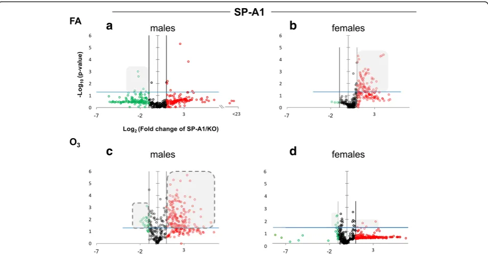
SP-A genes differentially regulate the AM miRNome The expression levels of the hTG SP-A1 and SP-A2 AM miRNomes were determined in males and females that were exposed to FA or O3 and compared to the
corre-sponding KO AM. The miRNome levels are presented as volcano plots to show the fold change regulation differ-ences between levels of miRNAs in hTG and KO mice, as well as their statistical significance (Figs. 1 and 2).
(of both magnitude and significance) in females com-pared to males. Following O3 exposure (panels C and
D), the SP-A1 mice show a very different picture. The female mice (panel D) exhibit fewer differences of lower magnitude than what is observed with the males (panel C). The pattern seen in OxS is almost the opposite of the basal conditions (compare shaded areas in Fig. 1).
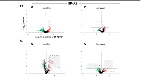
When we compare the SP-A2 hTG males and fe-males under basal conditions, we do not see any ro-bust differences as with the SP-A1 hTG (Fig. 2, panels a and b, respectively). However, under OxS, females have less pronounced differences than males (Fig. 2, panels c and d). Under OxS, the male miR-Nome in both SP-A1 and SP-A2 mice is more re-sponsive compared to females and exhibits a higher number of changing miRNAs that reach the Bonfer-roni corrected significance threshold p< 0.0166 (com-pare shaded areas). A two-way ANOVA test for the sex and treatment effects showed that the F stat for the SP-A2 mice regarding the sex effect is F= 26.34 with F crit = 3.84 and p= 3.23 × 10⁻⁷
. The F stat for the interaction between the two factors (sex and treat-ment) wasF= 25.25 withFcrit = 3.84 andp= 5.65 × 10⁻⁷.
The same analysis for the SP-A1 mice miRNome did not show that sex, treatment, or the combination of these two factors were significantly different (see Additional file 1). The ANOVA test for the SP-A1 mice to check the effect of treatment (FA or O3), sex (male, female), and the
interaction between the two variables (treatment and sex effects) showed that theFstat for SP-A1 mice wasF= 1.17 lower than the F crit = 3.85 and p value not signifi-cant p= 0.279. These data show that in the AM miR-Nome, there is a difference between sexes in response to O3 exposure with the presence of SP-A1 or SP-A2.
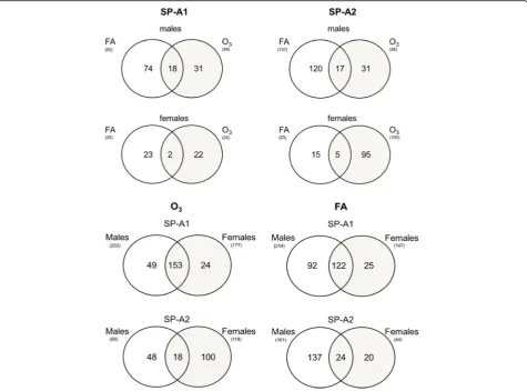
Regulation of AM miRNome in response to OxS
Next, we focused on the AM miRNAs whose expression is altered in response to OxS from SP-A1 and SP-A2 male and female mice. We compared the A1 and SP-A2 AM miRNome altered ≥2-fold between males and females under basal conditions and OxS (Fig. 3). We found that in SP-A1 mice, in males, 92 miRNAs were changed under control conditions and 49 under O3,
whereas in females, only 25 miRNAs were changed in FA and 24 in OxS. The same comparison in SP-A2 AM miRNome showed that for males, 137 miRNAs were
a
b
c
d
regulated in FA and 48 miRNAs in O3, while in females,
only 20 were regulated in FA and 100 by OxS (Fig. 3).
Sex- and gene-specific AM miRNAs that are regulated by OxS
To identify sex- and gene-specific AM miRNome changes in response to OxS, we compared via Venn dia-grams the SP-A1- and SP-A2-regulated miRNAs that were altered in the two conditions (FA and O3) (Fig. 3
and Additional file 2). The diagrams show the distribu-tion of the differentially expressed SP-A1 and SP-A2 miRNAs regulated ≥2-fold that are unique or common under basal conditions and under OxS in A1 or SP-A2 male and female mice.
We next assessed the miRNAs unique and common for male and female SP-A1 and SP-A2 mice after O3and
FA exposure. Out of the total 372 miRNAs of our array, we identified 49 miRNAs that are unique to SP-A1 male and 24 miRNAs unique to SP-A1 females in OxS, and 153 miRNAs were found to be in common in SP-A1 males and females after O3 exposures (Fig. 3). In the
case of SP-A2 mice, we identified 48 miRNAs unique to SP-A2 male and 100 miRNAs unique to SP-A2 females in OxS, and only 18 miRNAs were commonly identified after O3exposure. Similarly, under control conditions in
filtered air (FA), more miRNAs (n= 122) were in common in SP-A1 male and female mice than in SP-A2
(n= 24) (Fig. 3). These data indicate sex variability in the SP-A2 and SP-A1 miRNome.
Ingenuity Pathway Analysis of the AM miRNome and the genes that they affect in response to OxS in SP-A2 male mice To better understand and integrate the AM miRNome data, we performed Ingenuity Pathway Analysis (IPA) for the AM miRNAs whose expression was significantly altered by OxS. Only miRNAs that were shown to pass the corrected Bonferroni p< 0.0166 and an FDR-adjusted q< 0.05 were used to ensure that sex, treat-ment, gene, and array variability do not lead to false dis-coveries. IPA for the male SP-A2 miRNome (n= 31 miRNAs were male-specific) showed several mRNA transcripts, the expression of which could be affected by miRNAs that were changed by OxS. We found direct as-sociations between some of these miRNAs with IL-6, NF-kB, STAT3, BCL2, and FOXO1 mRNAs.
miR-191-5p, which binds IL-6 [44], is downregulated. The levels of microRNAs (miR-21-5p, miR-181a-5p, miR-1195) in response to O3are shown in Table 1; these
are known to interact directly with STAT3.
We found five miRNAs to be directly associated with the BCL2 gene. Both miR-21-5p and miR-16-5p were significantly downregulated in our study. miR-195a-5p that has the same seeding sequence with miR-16-5p and
is predicted to bind BCL2 mRNA was also
a
b
c
d
downregulated while miR-153-3p that was also found to bind BCL2 experimentally (by Western blot, qRT-PCR, and LUC) [45] was increased.
Two miRNAs (miR-9-5p and miR-183-5p) were regu-lated by O3, and these were shown to target the NF-kB
protein and mRNA experimentally (Western blot and qRT-PCR) [46] and by in silico analysis (TargetScan). miR-9-5p expression levels were downregulated. miR-183-5p, which was upregulated, probably has the opposite effect. FOXO1 is targeted by a multitude of miRNAs that are changed in our study miR-9-5p, miR-21-5p, miR-16-5p, miR-183-5p [47], miR-486b-5p, and miR-153-3p. The above six miRNAs were either up- or downregulated in our experiments (Table 1) and could potentially regulate the expression levels of FOXO1 in AM by OxS.
Only the male SP-A2 AM miRNome in response to O3
associates with genes involved in OxS
When an IPA like the one done with data of male SP-A2 hTG mice was performed, using the 95 miRNAs found in the SP-A2 female AM miRNome, the main associa-tions found were for cervical cancer and reproductive system disorders (data not shown). Although the SP-A2 females express the same miRNAs as the males, the changes resulting from O3 exposure are lower than
those observed in the males, leading to the identification of different pathways. For the SP-A1-regulated miRNAs in males that changed in response to O3 (31 miRNAs),
regulation of 4 miRNAs identified in our study, along with argonaute-2 protein (AGO2), which is involved in biogenesis of miRNAs (data not shown).
Effect of gonadectomy and OxS on expression of miRNAs in SP-A2 male and female mice
To identify the effect of sex hormones on the expression of miRNAs after OxS, we performed miRNA expression analysis in AM from gonadectomized (Gx) SP-A2 and KO male and female mice and compared it with that of non-gonadectomized (NGx) mice after O3exposure. For
this analysis, we used 63 miRNAs that were identified in both NGx (male and female) and Gx (male and female) groups. Of these, in the NGx (male vs female) group, only 37 miRNAs had their levels significantly changed (fold change ≥2) (Fig. 4, Additional file 3). In the Gx group (male vs female) compared to the corresponding NGx (male vs female) group, expression of 2 miRNAs (5.4%) was significantly increased (fold change≥2), and expression of 34 miRNAs (94.6%) was significantly de-creased (fold change ≤2). Of the 37 miRNAs differen-tially expressed in Gx male and female, 17 miRNAs (45.9%) are Gx female specific and 7 miRNAs (18.9%) are Gx male specific (Fig. 4 inset, Additional file 3). Moreover, a one-way ANOVA pertaining to the gonad-ectomy effect on the miRNA expression showed a sig-nificant difference with F stat = 11.98 with F crit = 3.92 and p= 0.00073 (see Additional file 3). These data indi-cate that sex hormones play an important role in the ob-served miRNA sex differences by affecting regulation of miRNAs, as most of the miRNAs that were increased in NGx were decreased in Gx.
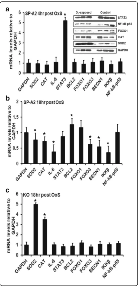
Expression of genes regulated by OxS in SP-A2 males We performed qRT-PCR to assess the expression levels of SOD2, CAT, IL-6, STAT3, BCL2, FOXO1,
FOXO3, BECN1, IKKβ, and NF-kB-p65 genes at
mRNA level in the male SP-A2 AM. We found that under OxS, the levels of STAT3 mRNA were in-creased significantly (p< 0.05) by 5-fold (Fig. 5a). No significant differences were observed for the other mRNAs studied.
We examined the protein expression levels of STAT3, NF-kB, FOXO1, CAT, and SOD2 in the male SP-A2 AM after 4 h OxS. The Western blot analysis indicated no significant difference in the protein levels of STAT3, NF-kB, FOXO1, and CAT at 4 h after OxS exposure, whereas for SOD2, a significant increase was observed compared to control (Fig. 5a, inset).
To account for half-life differences and study further the expression of these genes, we used AM from male SP-A2 and KO mice after 18 h of exposure to O3and found a
significant increase in the mRNA level of BCL2 (Fig. 5b). The levels of the other genes studied (SOD2, CAT, IL-6, FOXO3, BECN1, and IKKβ) were decreased significantly (Fig. 5b). In contrast, in KO, the expression of SOD2 and CAT was increased significantly, but no significant changes in the expression of the other genes were ob-served (IL-6, STAT3, BCL2, FOXO3, BECN1, IKKβ, and Table 1Levels and statistical significance of the male SP-A2 AM
miRNAs compared to SP-A KO in OxS and shown by IPA to be directly associated with genes IL-6, NF-kB, STAT3, BCL2, and FOXO1
Mature ID Fold regulation pvalue
miR-191-5p −2.1515 0.00196
miR-153-3p 4.2526 0.000167
miR-15b-5p −1.7279 0.01395
miR-16-5p −1.8298 0.000532
miR-181a-5p −1.5004 0.000539
miR-183-5p 2.0782 0.000372
miR-195a-5p −1.2763 0.030639
miR-21a-5p −2.4629 0.009501
miR-9-5p −3.0084 0.001602
miR-486b-5p 1.407 0.196574
miR-1195 2.1028 0.000323
−indicates downregulation
0 20 40 60 80 100 NGx Gx yll ai t n er ef fi d s A N Ri m f o e g at n e cr e
P expressed in SP-A
2 Gx and NGx Increased Decreased n=37 94.59% n=35 5.4% n=2
M vs F M vs F
NF-kB-p65 (Fig. 5c). A trend of increase in the level of FOXO1 was observed in SP-A2 and KO (Fig. 5b, c).
To investigate the role of IL-6 in the increased STAT3 levels, we measured IL-6 protein by ELISA in the BAL of male SP-A2 hTG mice after exposure to O3 and FA.
We found that IL-6 levels were increased significantly compared to control conditions (FA) at 4 h post OxS (Fig. 6a) and at 18 h post OxS (Fig. 6b). We also mea-sured the levels of IL-1βand TNF-αat 18 h post OxS in SP-A2 and KO. We observed a significant increase in their levels in SP-A2 compared to FA (Fig. 6b), whereas in the case of KO, the level of these proteins was signifi-cantly decreased after O3 exposure compared to FA
(Fig. 6b), indicating a role of SP-A2 in these processes.
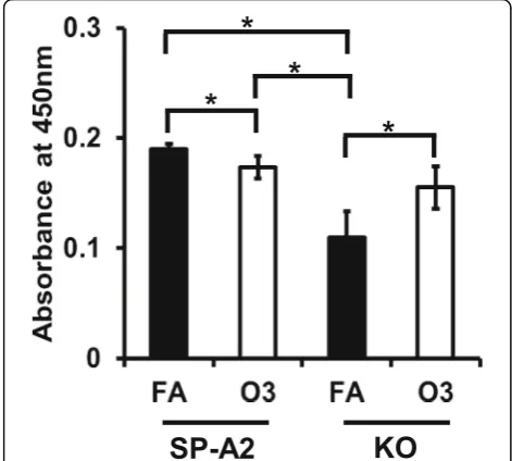
Apoptosis induced by OxS in male SP-A2 and KO mice We used a TUNEL assay to assess apoptosis. We ana-lyzed apoptotic cells from AM of SP-A2 and KO male mice at 18 h post OxS. In response to O3exposure, no
significant differences were observed between SP-A2 and KO. However, a small but significant decrease in apoptotic cells was observed in SP-A2 O3-exposed mice
compared to SP-A2 FA-exposed mice, whereas an in-crease in apoptotic cells was observed in O3-exposed
KO mice compared to FA-exposed KO (Fig. 7). Our results indicate that ozone differentially affects apoptosis in KO and SP-A2 AM, with SP-A2 providing perhaps some protection from the effect of OxS.
Discussion
SP-A is an important host defense molecule. In humans, there are two SP-A functional genes, SFTPA1 (SP-A1) and SFTPA2 (SP-A2), and their gene products have been shown to differ functionally from each other [39, 48–50]. Differences have been seen in the prote-omic expression of AM and the AM actin cytoskeleton after exposure to SP-A1 or SP-A2 variants [20, 48]. We
*
*
*
*
* *
*
*
*
*
a
b
c
have also previously shown sex differences in the KO AM proteome after treatment with SP-A1 or SP-A2 gene products where more changes occurred with SP-A1 and SP-A2 treatment in females and males, respect-ively [51]. The present study was done to further
understand mechanisms underlying the previously ob-served sex differences in the AM function and prote-ome in response to SP-A genes (SP-A1 and SP-A2).
We studied the AM miRNome in male and female mice that express human SP-A1 and SP-A2 after O3
ex-posure and compared it to that of KO mice that do not express SP-A. In both male and female miRNomes, changes were found after exposure to O3 that may
re-flect an attempt to mitigate the consequences of OxS. In the male miRNome of both transgenic mice (SP-A1 and SP-A2), there was an increase in the number of miRNAs that changed significantly after O3 exposure, whereas
in females, OxS did not change the miRNome substantially.
We found miR-191-5p, shown previously to regulate IL-6 [44], to be downregulated and this could increase the levels of IL-6, which is consistent with our experimental findings. IL-6 was found to be expressed in the BAL of males after O3exposure, but not in females [38]. IL-6 has
been shown to have both pro- and anti-inflammatory functions [52], and in mice, IL-6 overexpression has been shown to protect against hyperoxia-induced lung mitochondrial damage [53].
Pro-inflammatory cytokine IL-6 has been reported to activate STAT3 via phosphorylation [54–59] and OxS to trigger STAT3 phosphorylation that is responsible for transcriptional activation of cytokine-encoding genes that are involved in inflammation and cell injury [60].
miR-21-a
b
Fig. 6Effect of O3on IL-6, TNF-α, and IL-1βprotein levels in the bronchoalveolar lavage of male SP-A2 and KO mice compared to FA exposure.
aProtein levels were measured by ELISA 4 h post exposure. OxS significantly increased IL-6 levels in SP-A2 male mice compared to FA (control) SP-A2 mice (p< 0.05).bProtein levels were measured at 18 h post exposure in SP-A2 and KO. OxS significantly increased IL-6, IL-1β, and TNF-αin SP-A2 mice (p< 0.05) and significantly decreased in KO mice (p< 0.05). Data were generated using 3 animals/group,n= 18 and 3 replicate/animal
5p, miR-181a-5p, and miR-1195 shown in the present study to be altered significantly in response to O3 have
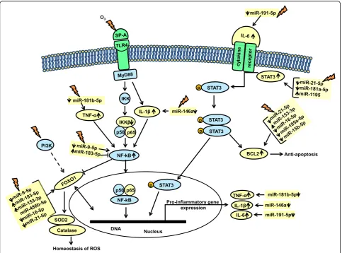
also been shown to directly interact with STAT3. Specific-ally, miR-21-5p is predicted to bind STAT3 mRNA (Tar-getScan). In our experimental conditions, the observed downregulation of this miRNA is associated with in-creased levels of STAT3. However, activation of STAT3 by IL-6 has been shown experimentally to increase expres-sion of miR-21-5p [61]. miR-1195 has been shown by in silico analysis to bind STAT3 [62], while miR-181a-5p has been shown with quantitative RT-PCR to be increased after activation of STAT3 by IL-6 [63]. In response to OxS, the expression levels of 21-5p, 1195, miR-181a-5p, and miR-191-5p (as shown in Fig. 8) were chan-ged significantly and this could result in the observed in-creased levels of STAT3. We postulate that an activated or dimerized STAT3 translocates to the nucleus where tran-scription initiation of pro-inflammatory genes takes place (Fig. 8). Moreover, the significantly increased STAT3 mRNA levels and the increased levels of TNF-α, IL-6, and
IL-1β after O3 exposure in the SP-A2 male AM (4 and
18 h post OxS) support this possibility. The IL-6 driven STAT3 phosphorylation and activation in OxS has been shown before [64] and has been linked with inflammation, cell injury, and cancer [65, 66], but the potential role of SP-A2 in this process is novel.
STAT3 activates other genes involved in cell cycle and upregulates anti-apoptotic genes such as BCL2 [67]. Of interest, BCL2 was found to be regulated by the male SP-A2 AM miRNome in OxS; BCL2 has been shown previously to be expressed in AM [68]. We found five SP-A2-regulated miRNAs that are directly associated with the BCL2 gene. miR-21-5p is predicted to bind both, BCL2 and STAT3 mRNAs (TargetScan), and miR-16-5p has been shown to bind BCL2 by sev-eral experimental approaches (Western blot, qRT-PCR, and luciferase reporter assays) [69, 70]. Both miR-21-5p and miR-16-5p were downregulated significantly in our study, and this is associated with increased mRNA levels of BCL2 (at 18 h post exposure, Fig. 5b) that may
contribute to inhibition of apoptosis and cell prolifera-tion in SP-A2 males, but not in KO males, where the expression of BCL2 did not change (Fig. 5c). miR-195a-5p that has the same seeding sequence with miR-16-miR-195a-5p, is predicted to bind BCL2 mRNA, and this was also downregulated in our study and may increase further the anti-apoptotic effects of miR-21-5p and miR-16-5p. miR-153-3p that binds BCL2 experimentally (by WB, qRT-PCR and LUC) [45] was increased in our study and may have the opposite effect, i.e., attenuating the anti-apoptotic effect of the other miRNAs. A further support for a role of BCL2 in the inhibition of apop-tosis and cell proliferation in response to O3is provided
by the TUNEL assay. Fewer apoptotic cells were found in SP-A2 compared to FA and an increase in apoptotic cells in KO compared to FA (Fig. 7). Of interest, in the absence of SP-A (i.e., in KO mice), higher inflammation and lower survival rates were observed in both FA- and ozone-exposed mice compared to wild type animals [35, 40]. These findings together indicate that ozone differen-tially affects apoptotic pathways in SP-A2 and KO mice and that the lungs from SP-A2 mice may have a protective anti-apoptotic role.
Another transcriptional factor that we found to be tar-geted by the male SP-A2 AM miRNome and is involved in immune responses is NF-kB [71]. Two miRNAs (miR-9-5p and miR-183-5p) were significantly changed by O3, and
these were shown to target the NF-kB mRNA experimen-tally [46] and by in silico analysis (TargetScan), respectively. miR-9-5p expression levels were downregulated signifi-cantly compared to control, and this may increase NF-kB mRNA levels that in turn may induce anti-proliferative ef-fects [72]. miR-183-5p that was upregulated may have the opposite effect on NF-kB. Our data showed a decrease in IKKβwithout any change in the NF-kB-p65 levels. It is well established that the IKKβ-dependent NF-kB signaling is a regulator of cell survival, immunity, and inflammation [73]. We know that SP-A activates NF-kB in THP-1 cells, a hu-man macrophage-like cell line [74]. The signal may be in-duced through MyD88, IL-1β, and IKKβkinase (IKK), with subsequent NF-kB translocation to the nucleus to facilitate transcription of pro-inflammatory genes, such as TNF-α and IL-1βas observed in the present study. We have ob-served that O3exposure decreases the ability of SP-A to
ac-tivate the NF-kB pathway as indicated by lack of increase of nuclear p65 and decrease of cytoplasmic IKBa [25]. How-ever, no studies up to now have addressed the impact of O3
on SP-A2-mediated NF-kB activation.
The two transcriptional factors identified here, NF-kB and STAT3, are believed not to act in isolation but to cross talk with each other in inflammatory conditions [75]. It has been documented in the literature that IL-6 increases levels of NF-kB [76] and siRNA inhibition of IL-6 results in reduced levels of NF-kB [77]. Along with
IL-6, TNF-α also increases the binding of NF-kB to STAT3 [78]. In our experimental conditions, 18 h post OxS, we observed an increase in TNF-αand IL-1βlevels and 4 h post OxS we observed a decrease in miR-181-5p and miR-146a; these miRNAs are known to target
TNF-α and IL-1β, respectively [79]. This may result in an in-creased level of TNF-αand IL-1β(Figs. 6b and 8). More-over, an increased level of TNF-αand IL-1βmay activate NF-kB, causing its translocation to the nucleus to facili-tate transcription of pro-inflammatory genes as dis-cussed above. Furthermore, an activated NF-kB has been shown to increase activation of BCL2 [80], whereas a mutant NF-kB decreased BCL2 [81]. The present data indicate that SP-A2 may play a role in these processes in the male AM miRNome in response to OxS.
Another transcript in our study that was targeted by several miRNAs is the FOXO1. This mRNA is targeted by miR-9-5p [47], miR-21-5p, miR-16-5p (TargetScan), miR-183-5p [47], miR-486b-5p [82], and miR-153-3p [47]. From the above six miRNAs, all but miR-486b-5p were changed significantly in our study and these poten-tially could regulate the expression levels of FOXO1 in AM under OxS (Fig. 8). FOXOs are transcription factors known to be involved in the homeostasis of ROS and can function as a negative feedback loop to control cel-lular reactive oxygen species [83]. FOXO1 regulates the expression of anti-oxidant genes such as CAT and SOD2, both of which are known to neutralize free radi-cals generated by ROS. Although no significant changes were observed in FOXO1 mRNA levels at 4 or 18 h post OxS in either SP-A2 or KO mice, the mRNA levels of SOD2 (superoxide dismutase) and CAT (catalase) sig-nificantly decreased at 18 h post OxS in SP-A2 males. However, for SOD2 (but not for CAT) at 4 h post OxS, the protein levels were increased. In contrast, in KO males, the mRNA levels of SOD2 and CAT were signifi-cantly increased. This striking difference between the KO and the hTG indicates that the SP-A2 gene may play a role in the homeostasis of ROS.
profile of SP-A2 mice was most similar to that of WT mice, particularly for proteins regulated by Nrf2, which were downregulated compared to KO, suggesting that these proteins were utilized to ameliorate the effect of oxidative stress in SP-A2 [9, 48], by OxS.
Conclusions
In summary, SP-A2 regulates miRNAs that play a role in pathways involved in inflammation, apoptosis, and ROS homeostasis. The observations made pertain only to the male SP-A2 AM miRNome in response to OxS exposure and not to females. The involvement of sex hormones in the observed miRNA sex-specific differ-ences was supported by the findings in gonadectomized mice where we observed that the regulation of the miR-Nome of the SP-A2 male mice compared to that of fe-male mice in response to OxS is significantly altered after gonadectomy. Collectively, our findings support and in part explain our previous observations of sex differences after OxS, where we observed females to be more susceptible in terms of survival to OxS than males. Here, we show that the male SP-A2 AM miR-Nome in response to OxS is associated with OxS-related inflammatory genes, regulation of ROS, and apoptosis. In females, although these miRNAs are expressed, their levels do not change significantly. The miRNAs identified here may correlate with the male survival advantage in OxS compared to females.
Additional files
Additional file 1:Original miRNA data used to generate Figs. 1 and 2. (XLSX 1320 kb)
Additional file 2:Original miRNA data used to generate Venn Diagrams (Fig. 3). (XLSX 75 kb)
Additional file 3:Original gonadectomy miRNA tag count data (Fig. 4). (XLSX 123 kb)
Abbreviations
AM:Alveolar macrophage; ANOVA: Analysis of variance; BCL2: B cell lymphoma 2; BECN1: Beclin-1; CAT: Catalase;Fcrit: Critical value inFdistribution;Fstat:F test or Fisher’s statistic; FA: Filter air; FDR: Benjamini and Hochberg false discovery rate; FOXO1: Forkhead box O1; FOXO3: Forkhead box O3; Gx: Gonadectomized; hTG: Humanized transgenic mice; IKKβ: Inhibitor of nuclear factor kappa-B kinase subunit beta; IL-1β: Interleukin 1 beta; IL-6: Interleukin 6; KO: Knockout; miRNA: Micro-RNA; NF-kB: Nuclear factor kappa-light-chain-enhancer of activated B cells; NGx: Non-gonadectomized; O3: Ozone; OxS: Oxidative stress; qRT-PCR: Quantitative real-time polymerase chain reaction; SFTP-A1: Gene encoding SP-A1; SFTP-A2: Gene encoding SP-A2;
SOD2: Superoxide dismutase 2; SP-A: Surfactant protein A; STAT3: Signal transducer and activator of transcription 3; TNF-α: Tumor necrosis factor alpha; TUNEL: TdT-mediated dUTP Nick-End Labeling
Acknowledgements
We would like to thank Dr. Rongling Wu for his consultation on the statistical analysis of miRNAs with multiple comparisons and Dr. Yuka Imamura from Pennsylvania State University College of Medicine Genomic Core Facility for the identification of miRNAs from Gx samples.
Funding
This work was supported by NIHR01HL34788 and CHILD fund, Department of Pediatrics, College of Medicine, at Pennsylvania State University.
Authors’contributions
GTN designed the study, performed the experiments, run the statistics, analyzed and synthesized the data, and contributed to the manuscript writing. NT performed the experiments, analyzed and synthesized the data, and contributed to the manuscript writing. XZ and TMU performed the maintenance and breeding of mouse lines, exposed the mice to
experimental conditions (FA and O3), and isolated the alveolar macrophages. FD performed all the gonadectomy experiments. DSP contributed to the data analysis and synthesis and manuscript writing. JF provided oversight to the entire project and involved in the data analysis, integration, and writing of the manuscript. All authors read and approved the final manuscript.
Ethics approval
All protocols used in this study were evaluated and approved by the Pennsylvania State University College of Medicine Institutional Animal Care and Use Committee and conformed to the guidelines of the National Institutes of Health on the care and use of laboratory animals.
Consent for publication
Not applicable
Competing interests
The authors declare that they have no competing interests.
Publisher’s Note
Springer Nature remains neutral with regard to jurisdictional claims in published maps and institutional affiliations.
Received: 22 June 2017 Accepted: 1 November 2017
References
1. Al-Hegelan M, Tighe RM, Castillo C, Hollingsworth JW. Ambient ozone and pulmonary innate immunity. Immunol Res. 2011;49:173–91.
2. Phelps DS. Surfactant regulation of host defense function in the lung: a question of balance. Pediatr Pathol Mol Med. 2001;20:269–92. 3. Huang W, Wang G, Phelps DS, Al-Mondhiry H, Floros J. Combined
SP-A-bleomycin effect on cytokines by THP-1 cells: impact of surfactant lipids on this effect. Am J Physiol Lung Cell Mol Physiol. 2002;283:L94–L102. 4. van Iwaarden JF, Claassen E, Jeurissen SH, Haagsman HP, Kraal G. Alveolar
macrophages, surfactant lipids, and surfactant protein B regulate the induction of immune responses via the airways. Am J Respir Cell Mol Biol. 2001;24:452–8.
5. Kremlev SG, Phelps DS. Effect of SP-A and surfactant lipids on expression of cell surface markers in the THP-1 monocytic cell line. Am J Phys. 1997;272: L1070–7.
6. Kremlev SG, Umstead TM, Phelps DS. Surfactant protein A regulates cytokine production in the monocytic cell line THP-1. Am J Phys. 1997;272:L996–1004. 7. Song M, Phelps DS. Comparison of SP-A and LPS effects on the THP-1
monocytic cell line. Am J Physiol Lung Cell Mol Physiol. 2000;279:L110–7. 8. Phelps DS, Umstead TM, Floros J. Sex differences in the response of the
alveolar macrophage proteome to treatment with exogenous surfactant protein-A. Proteome Sci. 2012;10:44.
9. Phelps DS, Umstead TM, Quintero OA, Yengo CM, Floros J. In vivo rescue of alveolar macrophages from SP-A knockout mice with exogenous SP-A nearly restores a wild type intracellular proteome; actin involvement. Proteome Sci. 2011;9:67.
10. Wright JR. Immunoregulatory functions of surfactant proteins. Nat Rev Immunol. 2005;5:58–68.
11. Crouch E, Wright JR. Surfactant proteins a and d and pulmonary host defense. Annu Rev Physiol. 2001;63:521–54.
12. Griese M. Pulmonary surfactant in health and human lung diseases: state of the art. Eur Respir J. 1999;13:1455–76.
14. LeVine AM, Bruno MD, Huelsman KM, Ross GF, Whitsett JA, Korfhagen TR. Surfactant protein A-deficient mice are susceptible to group B streptococcal infection. J Immunol. 1997;158:4336–40.
15. LeVine AM, Whitsett JA, Gwozdz JA, Richardson TR, Fisher JH, Burhans MS, Korfhagen TR. Distinct effects of surfactant protein A or D deficiency during bacterial infection on the lung. J Immunol. 2000;165:3934–40.
16. LeVine AM, Gwozdz J, Stark J, Bruno M, Whitsett J, Korfhagen T. Surfactant protein-A enhances respiratory syncytial virus clearance in vivo. J Clin Invest. 1999;103:1015–21.
17. Mikerov AN, Hu S, Durrani F, Gan X, Wang G, Umstead TM, Phelps DS, Floros J. Impact of sex and ozone exposure on the course of pneumonia in wild type and SP-A (−/−) mice. Microb Pathog. 2012;52:239–49.
18. Floros J, Wang G, Mikerov AN. Genetic complexity of the human innate host defense molecules, surfactant protein A1 (SP-A1) and SP-A2—impact on function. Crit Rev Eukaryot Gene Expr. 2009;19:125–37.
19. Phelps DS, Umstead TM, Floros J. Sex differences in the response of the alveolar macrophage proteome to treatment with exogenous surfactant protein-A. Proteome Sci. 2011;10:44.
20. Tsotakos N, Phelps DS, Yengo CM, Chinchilli VM, Floros J. Single-cell analysis reveals differential regulation of the alveolar macrophage actin cytoskeleton by surfactant proteins A1 and A2: implications of sex and aging. Biol Sex Differ. 2016;7:18.
21. Floros J, Phelps D. Pulmonary surfactant protein A; structure, expression, and its role in innate host defense. In: Nakos G, Lekka M, editors. Surfactant-update of intensive care medicine. Greece: University of Ioannina; 2002. p. 87–102. [Nakos G, ME L (Series Editor).
22. Voter KZ, Whitin JC, Torres A, Morrow PE, Cox C, Tsai Y, Utell MJ, Frampton MW. Ozone exposure and the production of reactive oxygen species by bronchoalveolar cells in humans. Inhal Toxicol. 2001;13:465–83.
23. Kehrl HR, Vincent LM, Kowalsky RJ, Horstman DH, O’Neil JJ, McCartney WH, Bromberg PA. Ozone exposure increases respiratory epithelial permeability in humans. Am Rev Respir Dis. 1987;135:1124–8.
24. Huang W, Wang G, Phelps DS, Al-Mondhiry H, Floros J. Human SP-A genetic variants and bleomycin-induced cytokine production by THP-1 cells: effect of ozone-induced SP-A oxidation. Am J Physiol Lung Cell Mol Physiol. 2004; 286:L546–53.
25. Janic B, Umstead TM, Phelps DS, Floros J. Modulatory effects of ozone on THP-1 cells in response to SP-A stimulation. Am J Physiol Lung Cell Mol Physiol. 2005;288:L317–25.
26. Wang G, Umstead T, Phelps D, Al-Mondhiry H, Floros J. The effect of ozone exposure on the ability of human surfactant protein a variants to stimulate cytokine production. Environ Health Perspect. 2002;110:79–84.
27. Mikerov AN, Umstead TM, Gan X, Huang W, Guo X, Wang G, Phelps DS, Floros J. Impact of ozone exposure on the phagocytic activity of human surfactant protein A (SP-A) and SP-A variants. Am J Physiol Lung Cell Mol Physiol. 2008;294:L121–30.
28. Connor AJ, Laskin JD, Laskin DL. Ozone-induced lung injury and sterile inflammation. Role of toll-like receptor 4. Exp Mol Pathol. 2012;92:229–35. 29. Cho HY, Morgan DL, Bauer AK, Kleeberger SR. Signal transduction pathways
of tumor necrosis factor—mediated lung injury induced by ozone in mice. Am J Respir Crit Care Med. 2007;175:829–39.
30. Sims EJ, Green MW, Mehta A. Decreased lung function in female but not male subjects with established cystic fibrosis-related diabetes. Diabetes Care. 2005;28:1581–7.
31. Mudway IS, Kelly FJ. An investigation of inhaled ozone dose and the magnitude of airway inflammation in healthy adults. Am J Respir Crit Care Med. 2004;169:1089–95.
32. Wong CM, Thach TQ, Chau PY, Chan EK, Chung RY, Ou CQ, Yang L, Peiris JS, Thomas GN, Lam TH, et al. Part 4. Interaction between air pollution and respiratory viruses: time-series study of daily mortality and hospital admissions in Hong Kong. Res Rep Health Eff Inst. 2010:283–362. 33. Caracta CF. Gender differences in pulmonary disease. Mt Sinai J Med. 2003;
70:215–24.
34. Tam A, Churg A, Wright JL, Zhou S, Kirby M, Coxson HO, Lam S, Man SF, Sin DD. Sex differences in airway remodeling in a mouse model of chronic obstructive pulmonary disease. Am J Respir Crit Care Med. 2016;193:825–34. 35. Mikerov AN, Gan X, Umstead TM, Miller L, Chinchilli VM, Phelps DS, Floros J. Sex differences in the impact of ozone on survival and alveolar macrophage function of mice after Klebsiella pneumoniae infection. Respir Res. 2008;9:24. 36. Durrani F, Phelps DS, Weisz J, Silveyra P, Hu S, Mikerov AN, Floros J. Gonadal
hormones and oxidative stress interaction differentially affects survival of
male and female mice after lung Klebsiella pneumoniae infection. Exp Lung Res. 2012;38:165–72.
37. Mikerov AN, Cooper TK, Wang G, Hu S, Umstead TM, Phelps DS, Floros J. Histopathologic evaluation of lung and extrapulmonary tissues show sex differences in Klebsiella pneumoniae-infected mice under different exposure conditions. Int J Physiol Pathophysiol Pharmacol. 2011;3:176–90.
38. Mikerov AN, Phelps DS, Gan X, Umstead TM, Haque R, Wang G, Floros J. Effect of ozone exposure and infection on bronchoalveolar lavage: sex differences in response patterns. Toxicol Lett. 2014;230:333–44. 39. Wang G, Guo X, Diangelo S, Thomas NJ, Floros J. Humanized SFTPA1 and
SFTPA2 transgenic mice reveal functional divergence of SP-A1 and SP-A2: formation of tubular myelin in vivo requires both gene products. J Biol Chem. 2010;285:11998–2010.
40. Haque R, Umstead TM, Ponnuru P, Guo X, Hawgood S, Phelps DS, Floros J. Role of surfactant protein-A (SP-A) in lung injury in response to acute ozone exposure of SP-A deficient mice. Toxicol Appl Pharmacol. 2007;220:72–82. 41. Umstead TM, Phelps DS, Wang G, Floros J, Tarkington BK. In vitro exposure
of proteins to ozone. Toxicol Mech Methods. 2002;12:1–16.
42. Sun J, Nishiyama T, Shimizu K, Kadota K. TCC: an R package for comparing tag count data with robust normalization strategies. BMC Bioinformatics. 2013;14:219. 43. Benjamini Y, Drai D, Elmer G, Kafkafi N, Golani I. Controlling the false
discovery rate in behavior genetics research. Behav Brain Res. 2001;125:279–84.
44. Tufekci KU, Oner MG, Genc S, Genc K. MicroRNAs and multiple sclerosis. Autoimmune Dis. 2011;2011:807426.
45. Xu J, Liao X, Wong C. Downregulations of B-cell lymphoma 2 and myeloid cell leukemia sequence 1 by microRNA 153 induce apoptosis in a glioblastoma cell line DBTRG-05MG. Int J Cancer. 2010;126:1029–35. 46. Guo LM, Pu Y, Han Z, Liu T, Li YX, Liu M, Li X, Tang H. MicroRNA-9 inhibits
ovarian cancer cell growth through regulation of NF-kappaB1. FEBS J. 2009; 276:5537–46.
47. Myatt SS, Wang J, Monteiro LJ, Christian M, Ho KK, Fusi L, Dina RE, Brosens JJ, Ghaem-Maghami S, Lam EW. Definition of microRNAs that repress expression of the tumor suppressor gene FOXO1 in endometrial cancer. Cancer Res. 2010;70:367–77.
48. Phelps DS, Umstead TM, Silveyra P, Hu S, Wang G, Floros J. Differences in the alveolar macrophage proteome in transgenic mice expressing human SP-A1 and SP-A2. J Proteom Genom Res. 2013;1:2–26.
49. Mikerov AN, Umstead TM, Huang W, Liu W, Phelps D, Floros J. SP-A1 and SP-A2 variants differentially enhance association ofPseudomonas aeruginosa with rat alveolar macrophages. Am J Physiol Lung Cell Mol Physiol. 2005; 288:L150–8.
50. Mikerov AN, Umstead TM, Huang W, Liu W, Phelps DS, Floros J. SP-A1 and SP-A2 variants differentially enhance association of Pseudomonas aeruginosa with rat alveolar macrophages. Am J Physiol Lung Cell Mol Physiol. 2005;288:L150–8.
51. Phelps DS, Umstead TM, Floros J. Sex differences in the acute in vivo effects of different human SP-A variants on the mouse alveolar macrophage proteome. J Proteome. 2014;108:427–44.
52. Scheller J, Chalaris A, Schmidt-Arras D, Rose-John S. The pro- and anti-inflammatory properties of the cytokine interleukin-6. Biochim Biophys Acta. 1813;2011:878–88.
53. Waxman AB, Kolliputi N. IL-6 protects against hyperoxia-induced mitochondrial damage via Bcl-2-induced Bak interactions with mitofusins. Am J Respir Cell Mol Biol. 2009;41:385–96.
54. Moore TC, Bush KL, Cody L, Brown DM, Petro TM. Control of early Theiler’s murine encephalomyelitis virus replication in macrophages by interleukin-6 occurs in conjunction with STAT1 activation and nitric oxide production. J Virol. 2012;86:10841–51.
55. Nguyen VA, Gao B. Cross-talk between alpha(1B)-adrenergic receptor (alpha(1B)AR) and interleukin-6 (IL-6) signaling pathways. Activation of alpha(1b)AR inhibits il-6-activated STAT3 in hepatic cells by a p42/44 mitogen-activated protein kinase-dependent mechanism. J Biol Chem. 1999;274:35492–8. 56. Miller AM, Wang H, Park O, Horiguchi N, Lafdil F, Mukhopadhyay P, Moh A, Fu XY,
Kunos G, Pacher P, Gao B. Anti-inflammatory and anti-apoptotic roles of endothelial cell STAT3 in alcoholic liver injury. Alcohol Clin Exp Res. 2010;34:719–25. 57. Myung SJ, Yoon JH, Yu SJ. STAT3 & cytochrome P450 2C9: a novel signaling
pathway in liver cancer stem cells. Biomed Pharmacother. 2012;66:612–6. 58. Piekorz RP, Nemetz C, Hocke GM. Members of the family of IL-6-type
59. Zhang Z, Jones S, Hagood JS, Fuentes NL, Fuller GM. STAT3 acts as a co-activator of glucocorticoid receptor signaling. J Biol Chem. 1997;272:30607–10. 60. Carballo M, Conde M, El Bekay R, Martin-Nieto J, Camacho MJ, Monteseirin
J, Conde J, Bedoya FJ, Sobrino F. Oxidative stress triggers STAT3 tyrosine phosphorylation and nuclear translocation in human lymphocytes. J Biol Chem. 1999;274:17580–6.
61. Li L, Zhang J, Diao W, Wang D, Wei Y, Zhang CY, Zen K. MicroRNA-155 and MicroRNA-21 promote the expansion of functional myeloid-derived suppressor cells. J Immunol. 2014;192:1034–43.
62. Hegde VL, Tomar S, Jackson A, Rao R, Yang X, Singh UP, Singh NP, Nagarkatti PS, Nagarkatti M. Distinct microRNA expression profile and targeted biological pathways in functional myeloid-derived suppressor cells induced by
Δ9-tetrahydrocannabinol in vivo: regulation of CCAAT/enhancer-binding proteinαby microRNA-690. J Biol Chem. 2013;288:36810–26.
63. Iliopoulos D, Jaeger SA, Hirsch HA, Bulyk ML, Struhl K. STAT3 activation of miR-21 and miR-181b-1 via PTEN and CYLD are part of the epigenetic switch linking inflammation to cancer. Mol Cell. 2010;39:493–506. 64. Ng IH, Yeap YY, Ong LS, Jans DA, Bogoyevitch MA. Oxidative stress impairs
multiple regulatory events to drive persistent cytokine-stimulated STAT3 phosphorylation. Biochim Biophys Acta. 1843;2014:483–94.
65. Levy DE, Darnell JE Jr. Stats: transcriptional control and biological impact. Nat Rev Mol Cell Biol. 2002;3:651–62.
66. Yu H, Pardoll D, Jove R. STATs in cancer inflammation and immunity: a leading role for STAT3. Nat Rev Cancer. 2009;9:798–809.
67. Barre B, Avril S, Coqueret O. Opposite regulation of myc and p21waf1 transcription by STAT3 proteins. J Biol Chem. 2003;278:2990–6. 68. Drakopanagiotakis F, Xifteri A, Tsiambas E, Karameris A, Tsakanika K,
Karagiannidis N, Mermigkis D, Polychronopoulos V, Bouros D. Decreased apoptotic rate of alveolar macrophages of patients with idiopathic pulmonary fibrosis. Pulm Med. 2012;2012:981730.
69. Cimmino A, Calin GA, Fabbri M, Iorio MV, Ferracin M, Shimizu M, Wojcik SE, Aqeilan RI, Zupo S, Dono M, et al. miR-15 and miR-16 induce apoptosis by targeting BCL2. Proc Natl Acad Sci U S A. 2005;102:13944–9.
70. Xia L, Zhang D, Du R, Pan Y, Zhao L, Sun S, Hong L, Liu J, Fan D. miR-15b and miR-16 modulate multidrug resistance by targeting BCL2 in human gastric cancer cells. Int J Cancer. 2008;123:372–9.
71. Hayden MS, West AP, Ghosh S. NF-kappaB and the immune response. Oncogene. 2006;25:6758–80.
72. Rath PC, Aggarwal BB. Antiproliferative effects of IFN-alpha correlate with the downregulation of nuclear factor-kappa B in human Burkitt lymphoma Daudi cells. J Interf Cytokine Res. 2001;21:523–8.
73. Luo JL, Kamata H, Karin M. IKK/NF-kappaB signaling: balancing life and death—a new approach to cancer therapy. J Clin Invest. 2005;115:2625–32. 74. Koptides M, Umstead TM, Floros J, Phelps DS. Surfactant protein A activates NF-kappa B in the THP-1 monocytic cell line. Am J Phys. 1997;273:L382–8. 75. He G, Karin M. NF-κB and STAT3—key players in liver inflammation and
cancer. Cell Res. 2011;21:159–68.
76. Haugen F, Drevon CA. Activation of nuclear factor-kappaB by high molecular weight and globular adiponectin. Endocrinology. 2007;148:5478–86. 77. Shah A, Verma AS, Patel KH, Noel R, Rivera-Amill V, Silverstein PS, Chaudhary
S, Bhat HK, Stamatatos L, Singh DP, et al. HIV-1 gp120 induces expression of IL-6 through a nuclear factor-kappa B-dependent mechanism: suppression by gp120 specific small interfering RNA. PLoS One. 2011;6:e21261. 78. Snyder M, Huang J, Huang XY, Zhang JJ. A signal transducer and activator of
transcription 3·nuclear factorκB (Stat3·NFκB) complex is necessary for the expression of fascin in metastatic breast cancer cells in response to interleukin (IL)-6 and tumor necrosis factor (TNF)-α. J Biol Chem. 2014;289:30082–9. 79. Zhang Y, Wang F, Lan Y, Zhou D, Ren X, Zhao L, Zhang Q. Roles of
microRNA-146a and microRNA-181b in regulating the secretion of tumor necrosis factor-alpha and interleukin-1beta in silicon dioxide-induced NR8383 rat macrophages. Mol Med Rep. 2015;12:5587–93.
80. Heckman CA, Mehew JW, Boxer LM. NF-kappaB activates Bcl-2 expression in t(14,18) lymphoma cells. Oncogene. 2002;21:3898–908.
81. Jacque E, Schweighoffer E, Visekruna A, Papoutsopoulou S, Janzen J, Zillwood R, Tarlinton DM, Tybulewicz VL, Ley SC. IKK-induced NF-κB1 p105 proteolysis is critical for B cell antibody responses to T cell-dependent antigen. J Exp Med. 2014;211:2085–101.
82. Wang LS, Li L, Chu S, Shiang KD, Li M, Sun HY, Xu J, Xiao FJ, Sun G, Rossi JJ, et al. MicroRNA-486 regulates normal erythropoiesis and enhances growth and modulates drug response in CML progenitors. Blood. 2015;125:1302–13. 83. Essers MA, Weijzen S, de Vries-Smits AM, Saarloos I, de Ruiter ND, Bos JL,
Burgering BM. FOXO transcription factor activation by oxidative stress mediated by the small GTPase Ral and JNK. EMBO J. 2004;23:4802–12.
• We accept pre-submission inquiries
• Our selector tool helps you to find the most relevant journal
• We provide round the clock customer support
• Convenient online submission
• Thorough peer review
• Inclusion in PubMed and all major indexing services
• Maximum visibility for your research
Submit your manuscript at www.biomedcentral.com/submit