CrossMark Published by DiscoverSys
ABSTRACT
Background and aim: Tumor-associated tissue eosinophilia (TATE) is commonly present in different neoplasms, yet its relationship with cancers has not been established. Squamous cell carcinoma (SCC) is one of the most common malignancies of the head and neck region. The aim of this study was to evaluate the relationship between TATE and histological grading of head and neck SCC (HNSCC) using Luna staining technique.
Materials and methods: In the present descriptive study, 67 samples consisting of 4-µm cross-sections of HNSCC tissue stained with hematoxylin and eosin (H&E) were re-evaluated for histological grading. The samples were divided into three categories of well differentiated, moderately differentiated, and poorly differentiated. Luna histochemical staining was used to evaluate infiltration of eosinophils in the tumor. Eosinophils were randomly counted in 10
microscopic fields and divided into 4 categories. Kruskal-Wallis test and Spearman’s correlation coefficient were used to evaluate the relationship between eosinophilic infiltration and other relevant factors.
Results: The frequency of tissue eosinophilia was zero in only one case (1.5%) and was positive in the rest 66 cases (98.5%). The mean count of eosinophils was 7.5±4.5 cells per high-power field (HPF). There was a significant relationship between histopathological grading and TATE (p = 0.04). However, there were no significant relationships between TATE and factors such as age, gender, and lesion location.
Conclusion: The tissue eosinophil counts in the group with poorly differentiated SCC of the head and neck region were less than those in the two other groups; regardless, no significant relationship was found with other clinicopathological factors.
Keywords:Eosinophils, squamous cell carcinoma, head and neck, Luna staining
Cite This Article: Khatibi, A.H., Sabzijate. M., Ghiasian. T., Rahrotaban. S., Rastegar, E., Eftekharian, S.H. 2018. Quantification analysis of tissue eosinophilia in squamous cell carcinoma of the head and neck region. Bali Medical Journal 7(1): 165-169. DOI:10.15562/bmj.v7i1.787
Quantification analysis of tissue eosinophilia
in squamous cell carcinoma of the head and
neck region
Khatibi AH,1 Sabzijate M,1* Ghiasian T,2 Rahrotaban S,3 Rastegar E,1 Eftekharian SH1
INTRODUCTION
Squamous cell carcinoma (SCC) is the most common malignancy of the head and neck region and the oral cavity, particularly originating from the stratified squamous epithelium of the mucosa. It accounts for more than 90% of malignancies of the oral cavity with multifactorial and multistage etio-pathogenesis.1 In the past 30 years, despite advances in diagnostic techniques and treatment modalities, the incidence and prevalence of oral SCC have been on the increase, mainly in young adults.2 The stage of SCC in the oral cavity at the time of diagnosis is the most important factor for its prognosis, which is determined using the TNM system. This system works by integrating the three basic clinical prop-erties of tumor size, metastasis to lymph nodes and distant metastasis. In this context, the smaller the size of the initial tumor and the absence of any evidence of regional lymph node involvement favor a better prognosis, and the bigger the size of the initial tumor and involvement of regional or distant lymph node indicates a worse prognosis.2,3
Previous studies on SCC have denoted relation-ships between several factors regarding the tumor
and the risk of its metastasis to lymph nodes along with the survival rate of the patients. The factors assessed including clinical and anatomical proper-ties of the tumor, such as the location and extent of the tumor, the stage and the degree of histological differentiation of the initial tumor, the thickness of the tumor and its invasion to vessels and nerves3,4 One of the factors that have been assessed in rela-tion to the risk of metastasis to cervical lymph nodes that affects the prognosis of oral squamous cell carcinoma (OSCC) is inflammatory cells, espe-cially eosinophils.4 The inflammatory infiltration in the tumor tissue matrix consists of primarily monocytic cells and low amounts of neutrophils and eosinophils.5,6 Eosinophils originate from the bone marrow and account for 1‒3% of the leuko-cytes in the peripheral blood flow.7 These cells are characterized by specific round and ovoid shaped cells containing cytotoxic granules including major basic protein (MBP), eosinophil-derived neuro-toxin (EDN), eosinophilic peroxidase (EPX) and eosinophil cationic protein (ECP). Those protein granules are found in the cell cytoplasm and their 1Orthodontics Resident, Shahed
University of Medical Sciences, Tehran, Iran
2Dentist, Private Practitioner, Tehran, Iran
3Assistant Professor of Oral and Maxillofacial Pathology Dentistry, Tehran University of Medical Sciences, Tehran, Iran
*Correspondence to: Sabzijate M, Orthodontics Resident, Shahed University of Medical Sciences, Tehran, Iran
Received: 2017-08-01 Accepted: 2017-10-23 Published: 2017-10-28
Volume No.: 7
Issue: 1
First page No.: 165
P-ISSN.2089-1180
E-ISSN.2302-2914
two-lobed nucleus. They are deeply stained with eosin and clearly visible under a light microscope.8 It should be pointed out that eosinophils have the potential to synthesize and secrete inflammatory mediators such as GM-CSF, IL-3, IL-5, TGF-β, TNF-α and TGF-α, which play a role in the lysis of cells including tumoral cells.9,10
Tumor-associated tissue eosinophilia (TATE) is defined as the infiltration of inflammatory cells with eosinophilic predominance around or within the tumor. Such inflammatory infiltration has been found to be present in many areas, for instance, the pharynx, larynx, lungs, stomach and the oral cavity.9,11‒13 A large number of studies have analyzed TATE status and its relationship with prognosis and the patients’ survival rate.12‒16 However, contradic-tory results have been reported on the relationship between eosinophilic infiltration and clinicopatho-logical factors as well as the prognosis of head and neck SCC (HNSCC).14‒19 Since the majority of stud-ies have reported TATE as a favorable prognostic indicator, theoretically, it might be possible in the future to use immunotherapy with the assistance of the anti-tumoral feature of eosinophils to treat cancers.20
Based on what has discussed above regarding the discrepancy between the results of previous studies, the present study was aimed to evaluate tissue eosinophilia in patients with HNSCC and its relationship with the histological differentiation of the tumor and other clinicopathological char-acteristics. Luna histochemical staining technique was used in this study to stain all the eosinophilic granules red, contrasting with the underlying blue background to promote better visibility under the microscope. This staining technique is specific for eosinophils and is commonly used to identify them in tissues.21,22
MATERIALS AND METHODS
In the present cross-sectional study, 107 files of patients with HNSCC (including oral cavity, pharynx, larynx, and lips) were evaluated and were sorted further based on the following criteria:
1. The lesion was treated with surgical technique only
2. The lesion was a primarily found lesion with no report of recurrence
3. The patient has no history of radiotherapy and chemotherapy
4. The sample is provided with complete demo-graphic data of the patient
Histological cross-sections were prepared from the selected samples, and the hematoxylin
and eosin (H&E) slides were categorized in terms of tumor differentiation grades based on Broder criteria.3 A nasal polyp sample was used as a posi-tive control for evaluation of eosinophil staining. A total of 67 final slides were evaluated to deter-mine eosinophil counts. The relevant paraffin blocks of the final samples were sectioned into 4 µm cross-sections using a microtome (Leitz, Germany), selected within the approximate tumoral tissue site and prepared for histochemical staining. The cross-sections slides were transferred to alcohol to remove any wrinkles and then transferred to lukewarm water (45-48°C). Appropriate cross-sec-tions were retrieved from the surface of the water with the use of lamellae coated with albumin glue. Cross-sections spread on lamellae were cautiously dehydrated and placed in an autoclave at 60°C for half an hour to remove paraffin so that the tissue would properly adhere to the lamellae. To remove the embedded paraffin completely, the samples were then immersed in xylol for 20 minutes. After the samples were deparaffinized, they are ready for Luna staining.
In the first step of Luna’s staining method, the samples were dyed with a mixture of hematoxylin and Biebrich scarlet for 5 minutes. Subsequently, they were rinsed with 1% acid alcohol solution (hydrochloric acid), followed by rinsing in water. In the next stage, the samples were rinsed with lithium carbonate solution and rinsed in running water for 2 minutes afterward.12 Finally, 95% and absolute alcohol were used for dehydration. In the final stage, the samples were covered with lamellae. In order to determine eosinophil counts in the connective tissue within the tumor, ten areas within the tumor were selected in a random manner under a light microscope (Olympus, Japan) and eosino-phil counts were determined in each area at 400x magnification, and the sums of these counts were categorized into 4 groups.23
0 eosinophils found: absence of eosinophils 1‒4 eosinophils found: poor infiltration 5‒19 eosinophils found: moderate infiltration ≥20 eosinophils found: severe infiltration Statistical analyses were carried out with SPSS 19, using non-parametric tests, Mann-Whitney, chi-square and Kruskal-Wallis tests and Spearman’s correlation coefficient. Statistical significance was set at p < 0.05.
RESULTS
The highest frequency was noted in the 45-59 years of age group with 32 (47.8%) cases. The oldest subject was 90 years old, and the youngest was 36 years old. The most frequent locations of the tumor
were on the tongue and the pharynx, and the least frequent location was on the larynx. All the samples were histologically graded in three groups: 37.3% samples were well differentiated (n = 25 cases); 44.8% samples were moderately differentiated (n = 30 cases); and 17.9% samples were poorly differen-tiated (n = 12 cases).
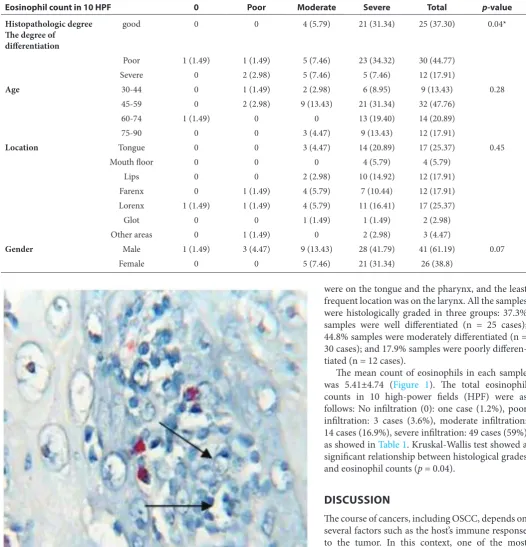
The mean count of eosinophils in each sample was 5.41±4.74 (Figure 1). The total eosinophil counts in 10 high-power fields (HPF) were as follows: No infiltration (0): one case (1.2%), poor infiltration: 3 cases (3.6%), moderate infiltration: 14 cases (16.9%), severe infiltration: 49 cases (59%) as showed in Table 1. Kruskal-Wallis test showed a significant relationship between histological grades and eosinophil counts (p = 0.04).
DISCUSSION
The course of cancers, including OSCC, depends on several factors such as the host’s immune response to the tumor. In this context, one of the most common cells in inflammatory infiltration against tumor cells is eosinophils. These cells secrete cyto-toxic proteins such as MBP and ECP and are capa-ble of activating the immune system through the release of some interleukins such as IL-2, IL-4, and IL-5, resulting in the reinforcement of the immune system and combat against the tumor; however, the release of IL-10 and TGF-β contributes to tumor Table 1 The frequencies of TATE in the head and neck SCC, in term of the relevant factors
Eosinophil count in 10 HPF 0 Poor Moderate Severe Total p-value
Histopathologic degree The degree of
differentiation
good 0 0 4 (5.79) 21 (31.34) 25 (37.30) 0.04*
Poor 1 (1.49) 1 (1.49) 5 (7.46) 23 (34.32) 30 (44.77) Severe 0 2 (2.98) 5 (7.46) 5 (7.46) 12 (17.91)
Age 30-44 0 1 (1.49) 2 (2.98) 6 (8.95) 9 (13.43) 0.28
45-59 0 2 (2.98) 9 (13.43) 21 (31.34) 32 (47.76)
60-74 1 (1.49) 0 0 13 (19.40) 14 (20.89)
75-90 0 0 3 (4.47) 9 (13.43) 12 (17.91)
Location Tongue 0 0 3 (4.47) 14 (20.89) 17 (25.37) 0.45
Mouth floor 0 0 0 4 (5.79) 4 (5.79)
Lips 0 0 2 (2.98) 10 (14.92) 12 (17.91)
Farenx 0 1 (1.49) 4 (5.79) 7 (10.44) 12 (17.91) Lorenx 1 (1.49) 1 (1.49) 4 (5.79) 11 (16.41) 17 (25.37)
Glot 0 0 1 (1.49) 1 (1.49) 2 (2.98)
Other areas 0 1 (1.49) 0 2 (2.98) 3 (4.47)
Gender Male 1 (1.49) 3 (4.47) 9 (13.43) 28 (41.79) 41 (61.19) 0.07
Female 0 0 5 (7.46) 21 (31.34) 26 (38.8)
progression.12,24 The result of the present study showed a significant relationship between histo-logical grading and TATE (p = 0.04), which means there was a decrease in TATE with higher histologi-cal grading and poorer differentiation of the tumor, confirming the anti-tumoral role of eosinophils.
On the other hand, some studies reported conflicting verdicts. For example, Leighton et al. evaluated TATE and its effect on nasopharyngeal carcinoma. In that study, TATE was categorized into several groups based on eosinophil counts: poor (<10), moderate (10‒100) and severe (>100); along with result indicating no relationships between TATE and factors involved in the tumor prognosis.25 In another study by Tadbir et al., the role of TATE was appraised as a factor to determine the prognosis in OSCC; the result showed no signif-icant relationship between TATE and tumor differ-entiation.26 In addition, in two separate studies, Goldsmith et al. (1987 and 1992) evaluated TATE and eosinophils in head and neck malignancies and deduced that eosinophils were associated with favorable prognosis. Despite that, the relationship between TATE and histological grading of the tumor was not evaluated in those studies.17,18 The discrepancies between the results of one study to another might be attributed to variation in the tech-niques used to determine eosinophil counts, the staining techniques used and the grading system used to specify the eosinophil counts.
In all of the studies discussed above, hema-toxylin and eosin (H&E) staining technique was used despite there are other staining methods with higher sensitivity. However, other techniques including auto fluorescence or non- histochemical techniques have been used in some studies to identify sound or degranulated eosinophils in the tumor. The use of those other staining methods is due to several reasons, such as the presence of unusual and uncommon morphology of granulo-cytes particularly eosinophils, examining fibrotic tissue or severely infiltrated samples, and difficulty identifying the cells on the microscopic plates.27 For example, in a study by Joshi et al., Congo red staining technique was used for identification of eosinophils, which was significantly more effective than H&E for identification of these cells.24 The study by Dorta et al. also showed no significant distinction in the mean counts of eosinophils in each millimeter-square surface area between H&E and immunohistochemical (IHC) staining tech-niques in OSCC. Nevertheless, it was reported that when a monoclonal antibody is used, CD125 of this antibody reacts with only one epitope on the mole-cule, resulting in the reaction of antibodies with a limited number of antigens; therefore, the number
of eosinophils identified by IHC is less than that by H&E staining.27 Other than that, a study of the effect of angiogenesis and immune cells on the SCC of the tongue with the use of carbol chromotrope by Alkhabuli showed no relationship between eosinophil concentration and histological grading of the tumor.12 Into the bargain, Luna histochemi-cal staining was used in the present study owing to its lower cost, relatively easily accessible compared to other techniques, and its ability to stain eosin-ophils selectively hence increasing the accuracy of the speed of the evaluation.21 As discussed previ-ously, no significant relationship was demonstrated between TATE and variables such as gender and tumor location, consistent with the results of the study by Oliveira et al., in which no significant relationship was found between TATE and gender, ethnicity, tumor location and tumor stage. In that study, tumors with higher and more severe TATE were significantly associated with a history of alco-hol use and smoking.28
Many of the studies mentioned above have reported inconsistent results in HNSCC, and there is still no definitive conclusion available relating to the presence of eosinophils in the HNSCC. The differences in the results might be ascribed to the reasons conferred above, including the tumor loca-tion and other factors. A new consideraloca-tion in the present study was the use of Luna staining tech-nique. Unfortunately, due to the lack of adequate access to patient data and sufficient information in the patients’ files, it was not possible to evaluate the rates of recurrences, survival, and metastasis.
CONCLUSION AND
RECOMMENDATIONS
It appears that tissue eosinophils decrease signifi-cantly with a decrease in the differentiation of tumor cells in the head and neck SCCs.
REFERENCES
1. Lo ML, Santarelli A, Panzarella V, Campisi G, Carella M, Ciavarella D, Di Cosola M, Giannone N, Bascones A. Oral squamous cell carcinoma and biological markers: an update on the molecules mainly involved in oral carcino-genesis. Minerva Stomatologica. 2007; 56(6): 341-7. 2. Feller L, Lemmer J. Oral Squamous Cell Carcinoma:
Epidemiology, Clinical Presentation and Treatment. Journal of Cancer Therapy 2012; 3: 263-268.
3. Neville BW, Damm DD, Allen CM, Bouquot JE. Oral & maxillofacial pathology.3nd ed. Philadelphia: Saunders; 2009. Chapter:10.
4. Amouian S, Tayebi Meybodi N, Ataranzadeh A, Montazer M, Rahimi M. Survey the Association between Tissue Eosinophil Counts and Prognostic Factors of Esophageal SCC 2010; 19 (73): 55-60.
a clinicopathologic scoring system for evaluating risk of cervical lymph node metastasis. Laryngoscope 1995; 105: 728- 733.
6. Davoine F, Sim A, Tang C, Fisher S, Ethier C, Puttaguna L, et al. Eosinophils in human oral squamous carcinoma; role of prostaglandin D2. J Inflamm (Lond) 2013;10 (1): 4. 7. Rothenberg E, Epthtein H. Mechanism of disease. The new
England Journal of Medicine. 1998; 328:1592-1600. 8. Waseem S, Ahmed B, Mazhar H, Aslam M. Eosinophils
and eosinophil products in asthma. J oral pathol Moo 2003; 32: 9-195.
9. Dorta RG1, Landman G, Kowalski LP, Lauris JR, Latorre MR, Oliveira DT. Tumor-associated tissue eosino-philia as a prognostic factor in oral squamous cell carcino-mas. Histopathology 2002; 41: 152-7.
10. Martinelli-Klay CP, Mendis BRRN, Lombardi T. Eosinophils and oral squamous cell carcinoma: a short review. J Oncol 2009; 3: 110-132.
11. Altemani AM,Guimaraes P, Metze K, Queiroz LS. Quantification analysis of modes of invasion and lymph node metastases in oral squamous cell carcinoma. Neoplasma 1999; 46: 323-8
12. Alkhabuli JO. Significance of neo-angiogenesis and immu-no-surveillance cells in squamous cell carcinoma of the tongue. Libyan J Med 2007; 2: 30-9.
13. Falconieri G, Luna MA, Pizzolitto S, DeMaglio G, Angione V, Rocco M. Eosinophil-rich squamous carci-noma of the oral cavity: A study of 13 cases and delineation of a possible new microscopic entity. Ann Diagn Pathol 2008; 12(5): 322-7.
14. Lowe D, Jorizzo J, Hutt MS. Tumor-associated eosino-philia: a review. J Clin Pathol 1981; 34(12): 1343-8. 15. Gao J, He Y, Wu L. The clinical analysis of eosinophil
infiltration in human squamous cell carcinoma of buccal mucosa. Hua Xi Kou Qiang Yi Xue Za Zhi 1997; 15(3): 228-,232
16. Deron P, Goossens A, Halama AR. Tumor-associated tissue eosinophilia in head and neck squamous cell car-cinoma. ORL J Otorhinolaryngol Rolat Spec 1996; 58(3): 167-70.
17. Goldsmith MM, Cresson DH, Askin FB. The prognostic significance of stromal eosinophilia in head and neck can-cer. Otolaryngol Head Neck Surg 1987; 96: 319-24 18. Goldsmith MM, Belchis DA, Cresson DH, Merritt WD,
Askin FB. The importance of eosinophil in head and neck cancer. Otolaryngol Head Neck Surg 1992; 106(1): 27-33.
19. Debta P, Mohan debta F, Chaudhary M, Dani A. Evaluation of infiltration of immunological cells (Tumor associated tissue eosinophils and mast cells) in oral squamous cell carcinoma by using special stains. British journal of medi-cine & medical research 2012; 2(1): 75-85.
20. Costello R, O’ Callaghan T, Sebahoun G. Eosinophils and antitumor response. Rev Med Interne2005; 26(6): 479-84. 21. Ono Y1, Fujii M, Kameyama K, Otani Y, Sakurai Y,
Kanzaki J. xpression of matrix metalloproteinase-1 mRNA related to eosinophilia and interleukin-5 gene expres-sion in head and neck tumor tissue. Virchows Arch 1997; 431(5): 305- 10.
22. Tomasi VH, Pe’res MA, Itoiz ME. Modification of Luna’s technique for staining eosinophils in the hamster cheek pouch. Biotech Histochem 2008; 83(3-4):147-51. 23. Said M, Wiseman S, Yang J, Alrawi S, Douglas W, Cheney R.
Tissue eosinophilia: a morphologic marker for assessing stromal invasion in laryngeal squamous neoplasms. BMC Clin Pathol 2005; 5(1): 1
24. Joshi PS, Kaijkar MS. A histochemical study of tissue eosinophilia in oral squamous cell carcinoma using congo red staining. Dent Res J (Isfahan) 2013; 10(6): 784-9. 25. Leighton SE, Teo JG, Leung SF, Cheung AY, Lee JC, Van
Hasselt CA. Prevalence and prognostic significance of tumor-associated tissue eosinophilia in nasopharyngeal carcinoma. Cancer 1996; 77(3): 436-40.
26. Tadbir AA, Ashraf MJ, Sardari Y. Prognostic significance of stromal eosinophilic infiltration in oral squamous cell carcinoma. J Craniofac Surg 2009; 20(2): 287-9
27. Lorena SC, Dorta RG, Landman G, Nonogaki S, Oliveira DT. Morphometric analysis of the tumor asso-ciated tissue eosinophilia in the oral squamous cell carcinoma using different staining techniques. Histol Histopathol 2003;18(3):709-13.
28. Oliveira DT, Biassi TP, Faustino SE, Carvalho AL, Landman G, Kowalski LP. Eosinophils may predict occult lymph node metastasis in early oral cancer. Clin Oral Investig 2012;16(6):1523-8.