Dr. Akash Yadav
IPS Academy, College of Pharmacy, Indore (M.P.) India E-mail: [email protected]
Address for correspondence
Access this article online www.japer.in
Formulation development and characterization of
nanocochleates for the improvement of permeability of drug
INTRODUCTION
Cochleates are elongated rolled microstructures that consist of a series of lipid bilayers formed as a result of condensation of small unilamellar negatively charged liposomes. These structures were first reported by Papahadjopoulos and Wilschut (Papahadjopoulos and Wilschut, 1979). These are solid particulates which are made of continuous lipid bi layer sheets rolled up into a spiral structure by interaction with multi cationic metal ion such as Ca2+ and have no internal aqueous space. Cochleate particles are stable phospholipid-cation precipitates composed of simple, naturally occurring phospholipids, such as dioleylphosphatidylserine (Zarif, 2002) and/or a mixture of lipids containing atleast phosphatidylserine, phosphatidylinositol, phosphatic acid, phosphatidyl glycerol upto 75% by
weight with phosphatidylcholine,
phosphatidylethanolamine and other less abundant phospholipids (Sankar and Reddy, 2010). Because of this unique structure, cochleates have the ability to encapsulate hydrophilic as well as hydrophobic moieties of any shape and size thus making them the most versatile carrier for delivery of a wide range of drug molecules, proteins and peptides. They also protect the entrapped molecule from harsh conditions of pH, temperature and lipase degradation.
Rifampicin is a semisynthetic antibiotic (bactericidal) produced from Streptomyces mediterranei. It is a broad antibacterial spectrum, having activity against several forms of Mycobacterium. It is the first choice drug in the treatment of tuberculosis but requires a high dose for a period of 4-6 months in Multi Drug Therapy (MDT) along with isoniazid, ethambutol and pyrazinamide. The BCS classification of rifampicin puts it under BCS class II (low solubility, high permeability) having an apparent permeability of about 5.79 ± 0.053 × 10⁻⁶ cm/s which is above the critical limit of 2 × 10⁻⁶ cm/s (Biganzoli et al., 2009), and it is expected to have a bioavailbility of over 90% (Artursson and Karlsson, 1991). But low rifampicin Rifampicin is the first choice drug for treatment of tuberculosis (TB), but it shows variable absorption in presence of other anti-TB drugs, so high dose is required. To overcome this problem nanocochleates of rifampicin were prepared through the interaction of calcium ions with the negatively charged phospholipids. It was done by two step process; first the liposomes of the drug were prepared by film hydration method using rotary evaporator, then dropwise calcium ions were added to the liposomes under constant stirring to form nanocochleates. Liposomes were evaluated for parameters of vesicle size and percentage entrapment, and the best formulations were selected for preparing nanocochleates. Cochleates thus prepared were subjected under particle size analysis, size distribution and SEM, which shows the elongated rod shaped sub micron size particles. Permeability studies were performed using the ex-vivo model, everted rat intestine was used to determine apparent permeability of rifampicin and comparing it to the apparent permeability shown by the rifampicin loaded nanocochleates. A significant increase in the apparent permeability of
rifampicin (3.56 × 10⁻⁶cm/sec) was shown when given as nanocochleates of
rifampicin (7.78 × 10⁻⁶cm/sec), thus this strategy can be used to improve the
absorption of rifampicin through small intestine.
Keywords: Rifampicin, nanocochleates, cochleates, apparent permeability, liposomes.
ABSTRACT Akash Yadav*1,
Shaily Chaudhary2,3
1IPS Academy, College of Pharmacy, Indore (M.P.) 2Faculty of Pharmacy, Pacific University, Udaipur (Rajasthan) 3Acropolis Institute of Pharmaceutical Education and Research, Indore (M.P.)
concentrations have been observed in tuberculosis (TB) patients. Also the apparent permeability coefficient of rifampicin is variable and changes throughout the intestinal mucosa. Concentrations of antimycobacterial drugs below than normally expected during tuberculosis (TB) therapy may cause appearance of resistant strains of Mycobacterium tuberculosis (Peloquin, 2002). Low antimycobacterial drug concentrations can be due to inadequate dosing and irregular drug intake. However, other factors that are reported to compromise the bioavailability of anti-TB drugs are malabsorption due to intestinal function impairment under treatment, and when they are consumed with food or antacids.
The determining factor for any drug absorption is the absorptive capacity of the intestinal mucosa, which may be altered in several clinical situations; eg- HIV infected TB patients have low serum concentrations of rifampicin and ethambutol. Nanocochleates loaded with rifampicin can be a good alternative solution for overcoming its variable absorption through instetinal mucosa. Reported strategies to improve drug absorption through cross membrane diffusion include drug delivery using a lipid based system. Nanocochleates can entrap the hydrophobic drug molecule effectively and the entrapped moiety remains unaffected by its surrounding environment thus providing a reasonable solution for the problem. Formulating rifampicin as nanocochleates helps in increasing its permeability through instestinal mucosa, lowering the dose of rifampicin and reduction of gastrointestinal tract side-effects such as abdominal cramps and diarrhoea.
2. MATERIALS AND METHODS
2.1 Materials
Rifampicin was obtained as a gift sample from Modern Laboratories, Indore, India. Soya lecithin was obtained as generous gift from Lipoids, Germany and used as vesicle forming phospholipid, cholesterol (Sigma-Alderich) provides flexibility to the formed liposomal vesicles thus maintaining its shape. Chloroform (Loba
Chem, India) was used as the organic solvent used to dissolve both soya lecithin and cholesterol. Bovine serum albumin (Sigma-Alderich) was used as an aggregation inhibitor while formation of nanocochleates to stop them from aggregating and forming clumps. Calcium chloride (Loba Chem, India) was used as the cochleation agent as the Ca2+ cations are required to roll up the lipid bilayer sheets into cochleate structures. All solvents used were of analytical grade.
2.2 Preparation of small unilamellar vesicles by lipid film hydration method
Negatively charged liposomes were prepared by lipid film hydration method. Constitutive lipids (Soya Lecithin: Cholesterol) were taken in different molar ratios (5:5, 6:4, 7:3, 8:2, 9:1) and were dissolved in minimum volume of choloroform. Known amount of drug rifampicin (100 mg) dissolved in organic phase (cholroform) was added to the lipid solution. Organic solvent was slowly removed using a rotary evaporator (Lab India) at temperature of about 37 ± 2oC, so that a thin film of dry lipid was deposited on the inner wall of the flask. This lipid film was left overnight to allow any remaining traces of organic solvent to evaporate. The thin lipid film was dispersed in phosphate buffer saline (PBS, pH 7.4) to give a final phospholipid concentration of 20 mg/ml by mechanical agitation by wrist shaking for 3 hours. Liposomes thus obtained were subjected to ultra sonication for 2 cycles of 10 min each to obtain small unilamellar vesicles.
2.3 Vesicle size and percent drug entrapment of small unilamellar vesicles
The small unilamellar vesicles thus formed were studied for vesicle size, percent entrapment and external morphology by photo microscopy. Nanotrac-150, USA was used to determine vesicle size. Nanotrac uses a newer, advanced power spectrum analysis of Doppler shifts to produce a full spectrum of vesicle sizes provided as a vesicle size distribution of volume (mass) percent and intensity. The liposomal
suspension is directly taken in a cell. The combined light passes through a fiber-optic cable to a single detector, and advanced electronics and software analyze the signals to calculate the Doppler shifts corresponding to particle size.
Percent drug entrapment was determined and expressed as the ratio of experimentally measured amount of drug in dispersion and initial amount used for entrapment. Vesicles were lysed by adding 1.0ml of 0.1% (v/v) Triton X-100, and were subjected to centrifugation at about 13000 rpm for 15 min at 4oC, and supernatant was taken for analysis of rifampicin using UV spectrophotometric method at a maximum wavelength of 334 nm.
2.3 Preparation of small cochleate cylinders.
Bovine serum albumin was added to the liposomal preparation as an aggregation inhibitor such that the final concentration of it remains to about 1% of the cochleate composition. Cochleate cylinders were then formed from liposomal preparations by trapping method, following addition of stock solution of calcium chloride in liposomal preparation while under constant stirring. The final Ca2+ concentration was 10 mM. The size of cochleate cylinders was controlled by subjecting the preparation to 2 cycles of ultra sonication of about 10 min each. After the evaporation of water solid form of cochleates were extracted.
2.4 Size measurement of cochleate particles
The vesicle size and size distribution of nanocochleates were measured by Malvern Zetasizer 2000 (Malvern, UK). Vesicular suspensions were mixed with the appropriate medium (PBS, pH 7.4) and the measurements were taken in a multimodal mode. All measurements were performed at 250C after 5 min of thermal equilibration. The Malvern Zetasizer 2000 (Malvern, UK) detects backscattering at an angle of 1730 for an improved sizing of especially larger particles at higher concentrations. Viscosity of the medium of 0.89 mPas and refractory index of 1.33 were assumed.
2.5 Scanning electron microscopy studies
Surface morphology of the specimens will be determined by using a scanning electron microscope (SEM), Model JSM 840A, JEOL, Japan. The samples are dried thoroughly in vaccum desicator before mounting on brass specimen studies. Gold-palladium alloy of 1200A was coated on the sample using sputter coating unit (Model E5 100 Polaron U.K.) in Argon at ambient of 8-10 Pascal with plasma voltage about 20MA. The sputtering was done for nearly 5 minutes to obtain uniform coating on the sample to enable good quality SEM images. The SEM was operated at low accelerating voltage of about 10KV with load current of about 80MA. The objective lens aperture has a diameter of 240 microns and the working distance (WD) of 39mm.
2.6 Permeability studies (ex-vivo evert gut sac model) Male Albino Wistar rats (150-200 g) were used for the present study. The rat was sacrificed humanely by cervical dislocation, the abdomen was opened by a midline incision, and the entire small intestine was then washed out with a solution of 0.9% (w/v) NaCl containing 0.3% (w/v) glucose. The whole of the small intestine was then removed by cutting across the upper end of the duodenum and the lower end of the ileum and manually stripping the mesentery from the intestine.
assembly (beaker and apparatus with tissue) was placed on a magnetic stirrer and a magnetic bead was allowed to rotate at 25 rpm in beaker and the temperature was maintained at 370C with adequate aeration. The samples were collected at every 5 min for 1 hour and analyzed by HPLC for estimation of permeability.
The apparent permeability (Papp) was calculated by the equation.
(Le Ferrec et al.) Where,
Papp: Apparent permeability co-efficient
dQ/dT: Cumulative amount of drug (Q) appearing in the acceptor compartment as a function of time, and was obtained from the slope of the linear portion of the amount transported-versus-time plot
A: Surface area of the intestine (cm2), taking 0.18 cm as radius (Shirasaka et al,. 2008)
C0: Initial concentration of drug in the donor compartment (μg/ml)
V: Volume of sample (ml)
3. RESULTS AND DISCUSSION
3.1 Characterization of vesicles formed
The vesicles formed were spherical in shape and unilamellar in nature. However, the vesicle size of the formed vesicle tends to vary as the molar ratio of the
structural lipids is changed. The vesicles also show variable entrapment efficiency with the maximum percent entrapment of 39.86 ± 0.38 %. The results are compiled under table 1.
Table 1: Characterization of small unilamellar vesicles
S.
No. Composition (molar ratio) Vesicle size (nm) Entrapment (%) 1. Cholesterol (5:5) Soya lecithin: 401 27.63 ± 0.15 2. Cholesterol (6:4) Soya lecithin: 435 34.73 ± 0.52 3. Cholesterol (7:3) Soya lecithin: 431 39.86 ± 0.38 4. Cholesterol (8:2) Soya lecithin: 451 26.95 ± 0.19 5. Cholesterol (9:1) Soya lecithin: 506 25.29 ± 0.44
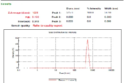
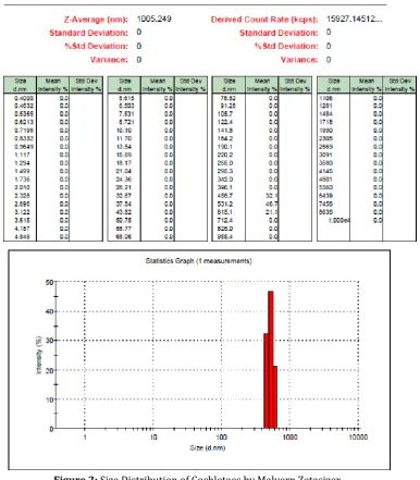
3.2 Size and size distribution of nanocochleates formed The nanocochleates were subjected to particle size analysis and particle size distribution and average diameter (Z- Average) was found out to be 1005 nm with peak obtained at 522.6 nm, following results were obtained:
Figure 1: Particle size of cochletaes by Malvern zetasizer
Figure 2: Size Distribution of Cochletaes by Malvern Zetasizer
3.3 Surface morphology of cochleate particles (SEM studies).
The images are shown in Figure 3. The micro size rods and clusters of such elongated rod shaped cochleates were observed.
3.4 Permeability estimation
The absorption rate as the cumulative concentration obtained at various time intervals across the intestinal segment is depicted in Fig. 4 for rifampicin and rifampicin loaded nanocochleates. The absorption of both was linear after initial lag phase and then had a plateau. The slope of the linear phase was used to
determine the dQ/dt and then the apparent permeability co-efficient. The Papp values were found to be 3.56 × 10⁻⁶ and 7.78 × 10⁻⁶ cm/sec respectively for rifampicin and rifampicin loaded nanocochleates respectively.
Table 2: Absorption kinetics of rifampicin and rifampicin loaded nanocochleates by everted rat instestine method
S. No. Time (sec) Cumulative Concentration (µg)
Rifampicin Rifampicin loaded nanocochleates
1. 300 36.075 ± 4.5 96.135 ± 7.5
2. 600 51.870 ± 5.2 238.485 ± 17.6
3. 900 70.590 ± 8.3 292.110 ± 21.3
4. 1200 109.394 ± 9.6 365.235 ± 23.3
5. 1500 187.590 ± 12.3 437.580 ± 24.6
6. 1800 243.490 ± 23.4 524.745 ± 26.7
7. 2100 300.300 ± 22.1 587.925 ± 21.2
8. 2400 302.835 ± 21.4 737.100 ± 20.2
9. 2700 327.210 ± 23.7 771.615 ± 27.8
10. 3000 369.525 ± 23.4 807.495 ± 21.9
11. 3300 376.155 ±24.4 828.750 ± 23.3
12. 3600 381.030 ± 21.2 842.945 ± 25.6
Absorption kinetics of rifampicin and rifampicin loaded nanocochleates using
everted instestine apparatus.
Time (sec)
Cu
m
u
la
ti
v
e
Co
n
c
e
n
tr
a
ti
o
n
(
µ
g
)
300 600 900 1200 1500 1800 2100 2400 2700 3000 3300 3600
0 200 400 600 800 1000
Rifampicin
Rifampicin loaded Nanocochleates
Figure 4: Absorption kinetics of rifampicin and rifampicin loaded nanocochleates using everted instestine apparatus
4. CONCLUSION
Formulation of nanocochleates of rifampicin is a promising and good strategy for improving its absorption through instestinal mucosa. It was observed that the cochleate formulation of rifampicin shows significant increase in apparent permeability.
Since the cochleate structure are formed from the interaction of divalent Ca2+ ion with small unilamellar vesicles of rifampicin, the formulation was started by selecting a suitable lipid, cholesterol composition for the preparation of SUV. Soya lecithin was selected as the structural phospholipid as it is easily available and
consists of three types of phospholipids; phosphatidylcholine (PC), phosphatidylethanolamine (PE) and phosphotidylinositol (PI) which can be used in formulation of negatively charged vesicles used for formulation of cochleates.
Initial experiments were conducted to optimize the liposomes formed by the process of film hydration method. The parameters studied included vesicle size and percent entrapment. It was observed that the vesicles formed by taking soya lecithin and cholesterol in the molar ratio of 7:3 shows maximum percent drug entrapment at about 39.86 ± 0.38 %. This can be attributed to the lipophilic nature of the drug since the entrapment is dependent upon lipid: aqueous phase ratio. While the vesicle size of various formulations varied marginally between 400-500 nm. The vesicle size of liposomes increases with the increase in the phospholipid content in the formulation. Furthermore, morphologically the vesicles were spherical in shape and unilamellar in nature.
Liposomal formulation showing the best percent drug entrapment of rifampicin was taken for formulation of cochleates. Initial experiments were carried out using variable concentration of Ca2+ ion, which suggested that for the better cochleation of liposomal bilayer into rod shaped structure and formation of small cochleate structures concentration of Ca2+ ion should be increased. Ca2+ concentration of 10 mM was maintained in the final formulation. On microscopic study of the formulation it was observed that the cochleate cylinders tend to aggregate to form large clumps which can affect the absorption kinetics, therefore bovine serum albumin (1% w/v of the total cochleate formulation) was used as an anti-aggregation agent, it was incorporated in the liposomal solution prior to the addition of Ca2+ ions. Characterization of nanocochleates on various parameters such as particle size and size distribution, scanning electron microscopy were performed. Average diameter (Z- Average) of cochleates was found out to be 1005 nm with peak obtained at 522.6 nm, this difference in the average diameter and peak
can be attributed to the elongated shaped structure of cochleate particle. Size distribution report of the formulation also shows maximum particle intensity in the range of 450-620 nm.
To further confirm the morphological structure of the cochleates, the particles were subjected to scanning electron microscopy (SEM) and elongated rod shaped structures of cochleates were observed. Permeability studies of the cochleate formulation of rifampicin shows more than two fold increase in the apparent permeability over normal rifampicin. The apparent permeability of rifampicin was found to be 3.56 × 10⁻⁶ cm/s. Formulation of drug as nanocochleates increased its absorption through the instestinal mucosa. This resulted in a significant increase in the apparent permeability to 7.78 ×10⁻⁶ cm/s. Hence the presented work proves a marked increase in permeability of rifampicin through small instestine by its formulation as nanocochleates, which is a good strategy to overcome the problem of malabsorption of rifampicin in presence of other anti-TB drugs as well as food and antacid, thus lowering the dose and side effects of the drug and requires further clinical studies to confirmation.
5. REFERENCES
1. Acocella G, Pagani V, Marchetti M, Barconi CG and Nicolis FB (1971). Kinetic studies on rifampicin. Chemotherapy, 16: 365-370.
2. Artursson P and Karlsson J (1991). Correlation between oral drug absorption in humans and apparent drug permeability coefficients in human intestinal epithelial (Caco-2) cells. Biochem. Biophys. Res. Commun., 175: 880–885.
3. Biganzoli E, Cavenaghi LA, Rossi R, Brunati MC and Nolli ML (1999). Use of a Caco-2 cell culture model for the characterization of intestinal absorption of antibiotics. Farmaco., 54: 594–599.
5. Fogerite SG, Kheiri MT, Zhang F, Wang Z, Scolpino AJ, Feketeova E, Canki M and Mannino RJ (1998). Targeting immune response induction with cochleates and liposome based medicines. Adv. D. Del. Rev., 32: 273–287.
6. Le Ferrec E, Chesne C, Artursson P, Brayden D, Fabre G, Gires P, Guillou F, Rousset M, Rubas W and Scarino ML (2001). In vitro models of the intestinal barrier. Atla, 29: 649-668.
7. Papahadjopoulos D and Wilschut J (1979). Ca2+ -induced fusion of phospholipid vesicles monitored by mixing of aqueous contents. Nature, 281: 690-692.
8. Peloquin CA (2002). Therapeutic drug monitoring in the treatment of tuberculosis. Drugs, 62: 2169-2183. 9. Perlin DS (2004). Amphotericin B cochleates: a
vehicle for oral delivery. Curr. Opin. Investig. Drugs, 5(2): 198-201.
10. Sankar V and Reddy Y (2010). Nanocochleate- A new approach in lipid drug delivery. Int. J. Pharm. Pharm. Sci., 2(4): 220-223.
11. Santangelo R, Paderu P, Delmas G, Chen ZW, Mannino R, Zarif L and Perlin DS (2000). Efficacy of oral cochleate-amphotericin B in a mouse model of systemic candidiasis. Antimicrob. Agents Chemother., 44: 2356–2360.
12. Syed UM, Woo AF, Plakogiannis F, Jin T and Zhu H (2008). Cochleates bridged by drug molecules. Int. J. Pharm., 363: 118-125.
13. Zarif L (2002). Elongated supramolecular assemblies in drug delivery. J. Control. Rel., 81: 7-23.
14. Zarif L, Graybill J, Perlin D and Mannino RJ (2000). Cochleates- New lipid based drug delivery system. J. Liposome Res., 10(4): 523-538.
How to cite this article: Bappa Sarkar, Payel Manna, Sanjib Bhattacharya, Moulisha Biswas; In vitro free radical scavenging effect of Butea monosperma leaf extracts; J. Adv. Pharm. Edu. & Res. 2016: 6(3): 14-21.
Source of Support: Nil, Conflict of Interest: Nil