T
ADEUSZŁ
UKIEŃCZUK1, 2, G
RAŻYNAD
MOCHOWSKA3, A
GNIESZKAK
RZYŻAŃSKA3,
K
RZYSZTOFK
ALISZEWSKI1, 2, P
AWEŁD
OMOSŁAWSKI1, S
TANISŁAWP
ÓŁTORAK4,
J
ACEKR
OGOLIŃSKI4, T
ADEUSZD
OBOSZ3“Loss of Microsatellite”, a New Type of Microsatellitic
Change in the Telomeric Region of Chromosomes,
as a Possible Characteristic Feature of Papillary
Thyroid Cancer – Preliminary Report
Loss of microsatellite
, nowy typ zmian mikrosatelitarnych
chromosomowych rejonów telomerowych jako charakterystyczna cecha
raka brodawkowatego tarczycy – doniesienie wstępne
1Department of General, Gastroenterological and Endocrinological Surgery, Silesian Piasts University
of Medicine in Wrocław, Poland
2Faculty of Public Health, Department of Clinical Nursing, Division of Clinical Procedures, Silesian Piasts
University of Medicine in Wrocław, Poland
3Department of Forensic Medicine, Molecular Technique Unit, Silesian Piasts University of Medicine in
Wrocław, Poland
4Center of Oncology, Maria Sklodowska−Curie Memorial Institute, Gliwice Branch, Poland
Adv Clin Exp Med 2008, 17, 1, 27–31 ISSN 1230−025X
ORIGINAL PAPERS
© Copyright by Silesian Piasts University of Medicine in Wrocław
Abstract
Background. Papillary thyroid cancer is the most common type of thyroid malignancy. The prognosis is favorable, but only in cases where the illness is detected and qualified for surgical treatment very early. Therefore, early and reliable preoperative diagnostic tools of the most common thyroid cancer are still demanded. In this study changes in the telomeric region of chromosomes were observed which can be detected in various kinds of thyroid tumors by PCR. A third type of microsatellitic change was identified, not described until now as a separate unit, where the subtelomeric microsatellite disappears. The name LOM (Loss of Microsatellite) is proposed for this phenomenon. Objectives. The aim of the study was to investigate LOM as a possible characteristic feature of papillary thyroid cancer.
Material and Methods. Genetic material from fifteen patients with three of the most common types of thyroid lesions (nodular goiter, follicular thyroid adenoma and papillary thyroid cancer) was investigated. DNA isolated from tumors tissues remaining after performing all necessary routine diagnostic tests was used. As a comparative material, DNA from the blood of the patients where tumor was removed surgically was used. PCR was applied. Results. LOM was observed in 7 (46.7%) patients with papillary thyroid carcinoma, 4 (30.76%) with follicular thy− roid adenoma, and 4 (30.76%) with nodular goiter.
Conclusions. LOM occurred in papillary thyroid cancer more frequently than in benign thyroid lesions (thyroid goiters and follicular thyroid adenomas). This is a preliminary report and the authors intend to examine LOM in a larger group of patients (Adv Clin Exp Med 2008, 17, 1, 27–31).
Key words: microsatellite, instability, papillary thyroid cancer.
Streszczenie
Thyroid nodules are a very frequent pathology and occur in 4–7% of the general population [1]. Some data suggest that thyroid nodules are pre− sented in 30–67% of ultrasonography examina− tions and almost 7% of them are malignant [1, 2]. Thyroid cancer is the most common endocrine malignant disease and its incidence is increasing [3]. Papillary thyroid cancer (PTC) is the most common type of thyroid malignancy and makes up about 50–80% of all thyroid cancers [4]. The prog− nosis in PTC is favorable, but only in cases where illness is detected and qualified for surgical treat− ment very early. Therefore, early and reliable pre− operative diagnostic tools of this most common thyroid cancer are still demanded.
Microsatellitic changes occurring in cancer tissue were described until now as microsatellite instability (MSI) or loss of heterozygosity (LOH) [5]. Microsatellite instability, identified with addi− tional “alleles” which are different from those occurring in healthy tissues of the same patient, occurs commonly (with a frequency to 2/3) in var− ious types of familial and sporadic cancers [6, 7]. Loss of heterozygosity (LOH) is identified with a total loss or significant decrease in the amount of DNA forming one of the alleles found in the can− cer cells (in relation to the healthy tissue of the examined patient).
In sporadic cancers, LOH is considered fun− damental for the initiation of cancerogenesis [8, 9]. Analysis of LOH is inseparably associated with the interpretation of the obtained results. Because fragments of surgically removed cancer tissue remaining after performing routine diag− nostic tests are commonly used for LOH testing, the selection of the fragment designated for analy− sis is crucial. Proper surgical techniques require the removal of the tumor with the proper margin of healthy tissue left surrounding the cancer. Isolation of DNA from such a large piece of mate− rial gives a mixture of cancer DNA and DNA from healthy tissue. This makes interpretation of
the results difficult. Attempts to solve this prob− lem include genetic analyzers and primers labeled with fluorochromes or by making densitometric diagrams of electrophoretic analysis and dividing the total area under particular DNA bands from healthy tissue cells by the total area under analo− gous bands of DNA from cancer cells. A quotient smaller than 0.6 allows one to accept the presence of LOH [10]. Results with the lowest quotients, according to the proposition of Berti et al. [11], may be called AIM (allelic imbalance) and are uninformative.
During the progress of malignancy, develop− ing tumors gradually accumulate chromosomal aberrations as a result of partial or complete loss of chromosomes. Loss of heterozygosity caused by microdeletion in subtelomeric regions of chromo− somes were also reported by other authors, for instance on 1q [7], 2q [8], 3p [9], 3q [8], 4p [10], 4q [8], 5q [8,11], 6p, 6q, 7p [8], 8p [12] and 9p [8,13], and on 11q, 12p, 14q, 15q, 17p, 17q, 20p,
obserwowanych zmian stwierdzono, że jest to trzeci typ zmian mikrosatelitarnych, które dotychczas nie były opi− sywane jako osobne zjawisko. Zjawisko to autorzy nazwali LOM(Loss of Microsatellite).
Cel pracy. Dokładna analiza zjawiska LOM jako potencjalnej cechy charakterystycznej komórek raka brodawko− watego tarczycy.
Materiał i metody. Do badania użyto DNA pobranego od pacjentów z trzema najczęstszymi patologiami gruczo− łu tarczowego (wole guzowate, gruczolak pęcherzykowy tarczycy, rak brodawkowaty tarczycy). DNA izolowano z tkanki guza pobranej podczas zabiegu chirurgicznego. Materiał porównawczy stanowiło DNA uzyskane z krwi obwodowej od tych samych chorych. Do badania wykorzystano technikę PCR.
Wyniki. Obserwowano zjawisko LOM u 7 (46,7%) chorych z rakiem brodawkowatym tarczycy, u 4 (30,76%) cho− rych z gruczolakiem pęcherzykowym tarczycy oraz także u 4 (30,76%) z wolem guzowatym.
Wnioski. Zjawisko LOM występuje częściej u chorych z rakiem brodawkowatym tarczycy w porównaniu z pa− cjentami ze zmianami łagodnymi (wole guzowate, gruczolak pęcherzykowy). Jest to doniesienie wstępne, dlatego autorzy chcą zbadać zjawisko LOM na większej liczbie chorych (Adv Clin Exp Med 2008, 17, 1, 27–31).
Słowa kluczowe: mikrosatelity, niestabilność, rak brodawkowaty tarczycy.
Type of tumors Number of Detected LOM (Rodzaj guza) cases inves− percentage
tigated (Odsetek
(Liczba wykrytych zbadanych LOM)
zmian)
Papillary thyroid 15 46.7
carcinoma
(Rak brodawkowaty tarczycy)
Follicular thyroid 13 30.76
adenoma
(Gruczolak pęcherzy− kowy tarczycy)
Nodular goiter 13 30.76
(Wole guzowate)
Table 1.Types of thyroid tumors investigated in the study
20q, 22q, and Xq [8]. Theoretically, a double LOH (on both chromosomes) is possible, so a vanishing of the microsatellite may occur on each chromo− some and at each place. This phenomenon, described in 2006 by Berti et al. [11] and named DEL (complete deletion), has been observed in 2% of lung cancer cases. Other researchers involved in the genetic studies of microsatellites have probab− ly also noticed this phenomenon but, according to our knowledge, have never published, especially as a separate type of the change. This report builds
on this phenomenon in more detail, limited to microsatellites from subtelomeric regions of dif− ferent chromosomes, especially from the p arm of chromosomes 3, 8, and 17, that seem to play a par− ticular role in pathogenesis, at least in head and neck cancers [14]. Some authors suggest [15] that position 17p13.3 is occupied by some tumor−sup− pressor gene.
The aim of this study was to investigate LOM as a possible characteristic feature of papillary thy− roid cancer.
System Location Primers
(Umiejscowienie) (Startery)
1QTEL19 1q GGA GTT AAG GTT GAA GAG CC
TTC ACG TAC AAC AGT ATC TC
2PTEL12 2p CTG TGC TTC TGC AGG TTA GA
TAC CTA GGT GAG AGT TAT CC
3PTEL01 3p AGA GTT CTC TAG AGG GAC AG
TTG CCT GCA GTG CTT CTG CC
3QTEL05 3q TCA CAG TGG CCA AGA TAT CA
TCC ATG TTG CTG CAA ATG AC
3QTEL06 3q TCA CAA GGG AAA TAA CTG TTT TAC
TTC CTG TAA CCC TCC AAA AT 4PTEL04 4p CTA GTC TTG ATT CTA TTG ACC
GGT CTA AAT CAA TGA CCT AAG C
5QTEL70 5q CTA TTT TTA TTT CAG TTG GCT GTT T
AAG GAA ACG TTC CTC TAA GTT ATT A
6QTEL54 6q CAG AAC AGA TTA AGA CTC AG
GCA TTT ATC AAC TTG TGT CC
9QTEL33 9q ATC TGT GTT GGA TTC TTG GC
ACT GAA CAC ACC TGT ACA GG
10PTEL35 10p CAG AGA CTG GCA TTC CCA A
GTC TTG AAA GTC ACC AGT CC
12QTEL82 12q GTT CCA AGG GAG AGTTTC AT
TAA AAT GAT AGT TTG CAC AAT AAT GG 13QTEL56 13q TTG CAG TGA GCT GAT ATC GC
TAA CAG GAT CTG TGT AAG CG
14QTEL23 14q GAT CAC GCC AAA TAG TAT GT
TGA GAT CTG TCT TGG AAA CC
17PTEL49 17p AGT AGG TTT CAG TTG CCT TTT C
AGA GAC ACA CAC AAT GAC AAT TAG
17QTEL13 17q CTG GCC ACT CAA ATA TAA AC
CAA AAT AAA AAC TGC AAG CAA TAT A 18QTEL11 18q CCT ATT TAA GTT TCT GTA AGG
ATG GTG TAG ACC CTG TGG AA 21QTEL14 21q CTA AGG ACA CAT GCC CAA TG
ACA GAG AAG GTG GGA GAT TG
22Q 22q TTG CAG ACA GCA GAC TAC AGG
TTC AGT CTG TGG CTG TCC AG
XYQ XYq GGC CTG AAT TCA TTT ATT CTA ATA G
GAA CAG GCA AAG ATG CCC ACT CTC
Table 2. Chromosomal localization of the tested microsatellite systems and primer sequences
Material and Methods
Genetic material from fifteen patients with the three most common types of thyroid lesions (nodular goiter, follicular thyroid adenoma, and papillary thyroid cancer) was investigated. These were obtained from the Institute of Oncology in Gliwice and from the Department of General, Gastrointestinal, and Endocrinological Surgery of Silesian Piasts University of Medicine in Wro− cław. In these studies, DNA isolated from tumors tissues remaining after performing all necessary routine diagnostic tests was used. DNA from the blood of patients where tumor was removed surgi− cally was used as a comparative material. The types of thyroid tumors are listed in Table 1 (see previous page).
The DNA used in this study, both from tumors and from blood, was isolated using a QIAamp DNA Mini Kit (Qiagen, Hilden, Germany) or Sherlock AX (DNA Gdańsk, Gdańsk, Poland). Very small amounts (ca. ¼ mm3) of the tested
material was used to minimize the danger of obtaining a mixture of DNA from cancer and healthy tissues. However, this requires from the surgeon the removal of very small pieces of tissue, definitely originating from the tumor itself.
The investigations were accomplished by the PCR method. The primer compositions are described in Table 2. The primer sequences were according to those used by Rosenberg et al. [16] and Colleaux et al. [12]. PCR was run using the Qiagen Multiplex PCR Kit and Hybaid OmniGene thermocycler. The amplification program consist− ed of an initial 2 min. denaturation at 94°C fol− lowed by 32 cycles of 94°C for 1 min., 60°C (or 63°C for 1QTEL19) for 45 s, and 72°C for 45 s. Upon completion of the 32 cycles the reaction was finished with 20 min. of elongation at 72°C.
Results
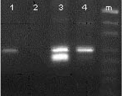
LOM was observed in 46.7% of the patients with papillary thyroid carcinoma, 30.7% with fol− licular thyroid adenoma, and 30.76% with nodular goiter. The results (LOH and LOM) are shown in Figure 1 and Table 1.
Discussion
This study suggests that a new type of microsatellitic change exists, distinct from LOH, and involving subtelomeric regions. It can have
considerable significance for the early diagnosis of cancer changes. It seems important that the diag− nosis of LOM may be done by inexpensive and simple agarosis minigel electrophoresis as well as using genetic analyzers (DNA sequencers).
The current explanation for the “immortality” of cancer cells is structural rearrangements in the chromosome, for instance by cryptic translocation [13, 17], in patients with myeloid disorders [16]. The present authors suppose that another mecha− nism is also possible: malignancy of a particular cancer depends significantly on its ability to reconstruct telomeres consumed during chromoso− mal division. Perhaps the “awakening” of fetal telomerase takes place in the cancer cell after con− sumption of the last telomeric unit. At this moment the normal cell would normally undergo apoptosis, but the “awakened” telomerase prevents this. The new, rebuilt telomeres do not terminate an intact chromosome, but a damaged one with shortened subtelomeric regions. These deletions could be a very early change characteristic in papillary thy− roid cancers, and such a feature has been sought for early diagnosis and prediction. Further investi− gations should clarify if subtelomeric LOM is real− ly a diagnostically useful early molecular change.
Fig. 1.Sample results: 1 – example of normal homozy− gote from blood, 2 – example of LOM in the same patient from tumor, 3 – example of normal heterozy− gote from blood, 4 – example of LOH in the same patient from tumor, m – size marker
References
[1] Ezzat S, Sarti DA, Cain DR:Thyroid incidentalomas: prevalence by palpation and ultrasonography. Arch Intern Med 1994, 154, 1838–1840.
[2] Pineda P, Rojas P, Liberman C, Moyano L, Goecke I:Detection of malignancy markers in thyroid nodules by reverse transcriptase polymerase chain reaction (RT−PCR). Rev Med Chil 2003, 131, 9, 965–972.
[3] Ross D:Nonpalpable Thyroid Nodules – Managing and Epidemic. J Clin Endocrinol Metab 2002, 87, 1938–1940. [4] Sherman S:Thyroid carcinoma. The Lancet 2003, 361, 501–511.
[5] Dobosz T, Łukieńczuk T, Sąsiadek M, Kuczyńska A, Jankowska E, Blin N:Microsatellite Instability in Thy− roid Papillary Carcinoma and Multinodular Hyperplasia. Oncology 2000, 58, 305–310.
[6] Boland CR, Thibodeau SN, Hamilton SR, Sidransky D, Eshleman JR, Burt RW, Meltzer SJ, Rodriguez−Bi− gas MA, Fodde R, Ranzani GN, Srivastava S:A National Cancer Institute Workshop on Microsatellite Instabi− lity for cancer detection and familial predisposition: development of international criteria for the determination of microsatellite instability in colorectal cancer. Cancer Res 1998, 58, 5248–5257.
[7] Kennedy D, Silver MM, Winsor EJT:Inverted duplication of the distal short arm of chromosome 3 associated with lobar holoprosencephaly and lumbosacral meningomyelocele. Am J Med Genet 2000, 91, 167–170. [8] Li X, Lee NK, Ye YW Waber PG, Schweitzer C, Cheng QC, Nisen PD:Allelic loss at chromosome 3p, 8p,
13q and 17p associated with poor prognosis in head and neck cancer. J Natl Cancer Inst 1994, 86, 1524–1529. [9] Florida G, Piantanida M, Minelli A:The same molecular mechanism at the maternal meiosis I produces mono−
and dicentric 8p duplications. Am J Hum Genet 1996, 58, 785–796.
[10] Fearon E:Tumor supressor Genes. In: The Genetic Basis of Human Cancer. Eds.: Vogelstein B, Kinzler K, Mc− Grew−Hill Health Professional Division, New York 1998, 229–240.
[11] Berti A, Barni F, Lago G:Tumor progression and microsatellite instability of forensic STR loci in human lung cancer. Promega Newsletter Identity 2006.
[12] Colleaux L, Rio M, Heuertz S, Moindrault C:A novel automated strategy for screening cryptic telomeric rear− rangements in children with idiopathic mental retardation. Eur J Hum Genet 2001, 9, 319–327.
[13] Tsezou A, Kitsiou S, Galla A, Petersen MB, Karadima G, Syrrou M, Sahlen S, Blennow E:Molecular cyto− genetic characterization and origin of two de novoduplication 9p cases. Am J Med Genet 2000, 91, 102–106. [14] White GRM, Stack M, Santibanez−Koref M, Liscia DS, Venesio T, Wang JC, Helms C, Donis−Keller H,
Betticher DC, Altermatt HJ, Hoban PR, Heighway J:High levels of loss at the 17p telomere suggest the clo− se proximity of a tumour suppressor. Br J Cancer 1996, 74, 863–870.
[15] Cave H, Guidal C, Elion J, Vilmer E, Grandchamp B:A low rate of loss of heterozygosity is found at many different loci in childhood B−lineage acute lymphocytic leukemia. Leukemia 1996, 10, 1486–1491.
[16] Rosenberg M, Hui L, Ma J, Nusbaum HC, Clark K, Robinson L, Dziadzio L, Swain PM, Keith T, Hudson TJ, Biesecker LG, Flint J:Characterization of Short Tandem Repeats from Thirty−One Human Telomeres. Genome Res 1997, 7, 917–923.
[17] Simpson AJ:The natural somatic mutation frequency and human carcinogenesis. Adv Cancer Res 1997, 71, 209–240.
Address for correspondence:
Tadeusz ŁukieńczukFaculty of Public Health
Department of Clinical Nursing, Division of Clinical Procedures Silesian Piasts University of Medicine
Bartla 5 51−618 Wrocław Poland
E−mail: [email protected]
Conflict of interest: None declared