R E S E A R C H
Open Access
Antigen-specific CD8+ memory stem T cells
generated from human peripheral blood
effectively eradicate allogeneic targets in
mice
Liping Guan, Xiaoyi Li, Jiali Wei, Zhihui Liang, Jing Yang, Xiufang Weng
*and Xiongwen Wu
*Abstract
Background:As the implantation and long-term existence of tumor-specific T cells in host are the prerequisite for adoptive immunotherapy, memory stem T cells (TSCM) with self-renewal and differentiation capacity show the greatest potential to implant and long-term exhibit function in vivo, compared with other T cells of differentiation stages. Hence, tumor-specific TSCMhave become potential candidate for adoptive T cell therapy of cancer. Here, we reported a protocol to generate allogeneic antigen-specific CD8+ TSCMcells from human PBLs.
Methods:To prepare allogeneic antigen-specific CD8+ TSCM, we used an LCL named E007 of defined HLA allotyping as simulator, a co-culture of E007 and allogeneic PBLs was carried out in the presence of differentiation inhibitor TWS119 for 7 days. Sorting of proliferation cells ensured the E007-specificity of the prepared TSCMcells. The sorted lymphocytes underwent further expansion by cytokines IL-7 and IL-15 for further 7 days, making the
E007-specific CD8 + TSCMexpanded in number. The stem cell and T memory cell properties of the prepared CD8+ TSCM
were observed in NOD-SCID mice.
Results:Our protocol began with 1 × 10⁷PBLs and resulted in 2 × 10⁷E007-specific CD8+ TSCMcells in 2 weeks of preparation. The prepared TSCMcells exhibited a proliferative history and rapid differentiation into effector cells upon the E007 re-stimulation. Importantly, the prepared TSCMcells were able to exist long and reconstitute other T cell subsets in vivo, eradicating the E007 cells effectively after transferred into the LCL burden mice.
Conclusions:This protocol was able to prepare allogeneic antigen-specific CD8+ TSCMcells from human PBLs. The prepared TSCMshowed the properties of stem cells and T memory cells. This study provided a reference method for generation of antigen-specific TSCMfor T cell adoptive immunotherapy.
Keywords:T memory stem cells, Allogeneic antigen specificity, Preparation in vitro, Adoptive immunotherapy
Background
Adaptive immunity is an effective strategy for cancer treatment. T cells are the main force to combat microbes and cancer cells in adaptive cells [1, 2]. However, T cell is heterogeneous, which exists in a continuum of differ-entiation states. According to differdiffer-entiation stages, T cells can be divided into naive T cells (TN), memory
stem cells (TSCM), central memory cells (TCM), effector
memory cells (TEM) and differentiated effectors (TEF) [3,
4]. Adoptive T cell therapy (ACT) for clinical application is normally based on the use of terminally differentiated TEFcells, which have short lifespan and inferior
engraft-ment capacities [5–7]. TSCMis defined as the earliest
de-velopmental stage of memory T cells, which possesses capacities of self-renewal and differentiation [5, 8]. Phenotypically, TSCM can be identified by the expression
of CD3+ CD45RA+ CD62L+ CD95+ CCR7+ CD28+ [5, 9–11]. TSCM has longer lifespan, as it is reported TSCM
is able to be tracked in vivo for 12 years after infusion of genetically modified lymphocytes [7]. In particular, TSCM
* Correspondence:[email protected];[email protected]
Department of Immunology, School of Basic Medicine, Tongji Medical College, Huazhong University of Science and Technology, 13 Hangkong Rd, Wuhan 430030, China
mediates longer and more robust efficiency of tumor re-jection in vivo compared to other memory and effector subsets [5, 6, 8, 12–14]. It is these characteristics that make TSCM a good candidate for immunological
cytotherapy of cancer.
TSCMconstitutes a small proportion of the T cell
sub-set, 2–4% of the total T cells in peripheral blood [5,8]. The premise of antigen-specific TSCM used for clinical
therapy is to accomplish in vitro TSCM preparation in a
large number. It is demonstrated that small molecule chemical inhibitor TWS119, a potent inhibitor of the serine–threonine glycogen synthase kinase 3β (GSK-3β) able to induce the Wnt-β-catenin signaling, helps the enrichment of TSCM through differentiation inhibition
both in mice and in human [15–17]. On the other hand, TSCM can be amplified with cytokines IL-7 and IL-15;
IL-21 is also reported to promote the generation of TSCMunder ex vivo culture conditions [6,18–26].
Allogeneic hematopoietic stem cell transplantation (allo-SCT) is an effective immunotherapeutic approach with curative potential in patients with malignancies. The therapeutic basis is mainly dependent on the donor T cell alloresponses against the recipient’s malignant cells named as graft versus leukemia (GVL) or graft ver-sus tumor (GVT) effect [27]. T cell responses to alloanti-gen are of peptide-MHC complex (pMHC) specificity in the same way as those to nominal antigen [28, 29]. Transfer of alloreactive T cells with defined specificity, such as for leukemia- or tumor-associated antigens, is proposed to separate the GVL or GVT effect from the deleterious graft versus host disease (GVHD) [30, 31]. As the implantation and long-term existence of tumor-specific T cells in host are the prerequisite for adoptive immunotherapy, it is of importance to prepare alloantigen-specific TSCMcells in vitro.
In this study, we explored a methodology of alloantigen-specific TSCM (allo-specific TSCM) preparing
in vitro. Although both CD4+ and CD8+ TSCMcells are
reported, we focused on CD8+ TSCM cells. The TSCM
cells were generated in an allogeneic co-culture, enriched by TWS119, then sorted with proliferation, and finally expanded by IL-7 and IL-15. Our protocol pre-pared 2 × 10⁷allo-specific TSCM cells from 1 × 10⁷PBLs.
An LCL burden mouse model was introduced in for the TSCM cell behavior in vivo. LCL cells were human B
lymphoblastoid cells immortalized by EB virus infection. Although this model would reflect lymphoproliferative disorders, the LCL cells in mice could act as allogeneic targets to measure eradication efficacy of T cells in vivo in our study. Importantly, the prepared TSCM cells
ex-hibited both stem cell and memory T cell properties, and were able to implant and effectively eradicate the targets after adoptive transfer into mice. This study pro-vided a practical method for generation of allo-specific
TSCM cells in vitro, which would be expected to be
re-ferred in preparation of allogeneic TSCM grafts with
de-fined antigen specificity for the purpose of adoptive immunotherapy.
Materials and methods
Peripheral blood lymphocyte isolation
Peripheral blood was obtained from healthy donors after informed consent under a protocol approved by the Eth-ics Committee of Tongji Medical College, Wuhan, China. Peripheral blood mononuclear cells (PBMCs) were isolated by centrifugation through a ficoll-hypaque gradient (Ficoll-Hypaquedensity 1.077 g/ml) and cul-tured in RPMI-1640 medium supplemented with 10% fetal bovine serum (FBS), then placed in dish for 2 h to remove the adherent cells. The non-adherent cells were collected as peripheral blood lymphocytes (PBLs) for the co-culture.
Cell lines and antibodies
The EB virus (EBV) transformed B lymphoblastoid cell lines (LCLs) E007 and E001 were established in our lab according to reported protocol [32]. HLA typing for LCLs was performed with PCR-SSP [33]. The HLA class I alleles of E007 and E001 are mismatched, E007 carried A*110x, 310x; B*510x, 550x; C*030x, 150x; and E001 A*020x, 240x; B*460x, 540x; C*010x, 080x.
Fluorescent antibodies for cells staining included hu-man CD3-APC-Cy7 (clone HIT3a), CD8-BV510 (clone SK1), CD3-Percp-Cy5.5 (clone HIT3a), CD62L-PE-Cy7 (clone DREG-56), CD45RA-APC (clone HIT100), CD45RA-BV421 (clone HI100), CD95-PE (clone DX2), CXCR7-PE (clone 10D1-J16), CD28-APC (clone CD28.2), IL-2-APC (clone MQ1-17H12), IFN-γ-PE (clone 4S.B3), TNF-α-PE (clone MAb11), the above anti-bodies were purchased from BioLegend, USA. Corre-sponding isotype for each antibody were used as isotype control. Cells were analyzed on a BD LSR flow cyt-ometer. T cell subsets were determined using fluores-cence minus one (FMO) controls for interesting antibody.
Allogeneic co-culture and allo-specific TSCMpreparation Allogeneic PBLs were co-cultured with the E007 for raising alloreactive TSCM cells. PBLs were stained with
proliferation in the co-culture, CFSEdim cells were sorted by FACS with BD FACS AriaII on day 7. Sorted cells were expanded by cytokines IL-7 and IL-15 (Pepro-tech, USA) of 25 ng/ml each for a further week.
T cell subsets in the co-culture bulks were identified by their surface markers, CD3+ CD8+ CD45RA+ CD62L+ CCR7+ CD95+ CD28+ for TSCM, CD3+ CD8+
CD45 RA- CD62L+ for TCM, CD3+ CD8+
CD45RA-CD62L- for TEM, and CD3+ CD8+ CD45 RA+
CD62L-for TEF.
Preparation of allo-specific TEMand TEFfor mouse transfer Allo-specific TEM and TEF cells were prepared by a
co-culture of allogeneic PBLs with E007 set up as above, but no TWS119 was added. CFSEdim cells were sorted on day 7. Sorted cells were further expanded by 300 U/ ml IL-2 (Peprotech, USA) instead of IL-7 and IL-15 for a further week.
In vitro TSCMcells differentiation assay and intracellular cytokine staining
The prepared TSCMcells were incubated with E007, E001
cells and Dynabeads Human T-Activator CD3/CD28 (α-CD3/CD28) (Gibco, USA), respectively, in a 5:1 ratio. Cells were collected at 6, 12, and 24 h, and labeled with corresponding fluorescent antibodies for differentiation assay. The TSCM cells incubated with the above
stimula-tions in the presence of 1 × BFA (eBioscience, USA) were collected for intracellular IFN-γ, IL-2, and TNF-αstaining after 4 h incubation. Samples were analyzed on BD Verse flow cytometer. The data analysis was performed with Flowjo software version 10.0 (Tree Star).
Quantitative real-time PCR
T cell subsets in the co-culture were sorted by BD Aria II flow cytometer based on the corresponding pheno-types on day 7. Total DNA of the T cells was isolated using a genomic Extraction kit (Takara, Japan), following manufacturer’s instructions. TRECs of T cell subsets were detected using fluorescently quantitative PCR kit (Takara, Japan) with TREC-specific primers [34] on Bio-Rad CFX Sequence detection system.
Animal experiments
6-week-old female NOD-SCID mice were purchased from Vital River Lab Animal Co, Ltd. (Beijing; a dis-tributor of the Jackson Laboratory). Animal experiments in this study were approved by the Ethical Committee of Tongji Medical College. The mice were irradiated with 2.0 Gy and randomized in five groups (n= 5), then intra-venous injection with 8 × 10⁶ E007 or E001 on day −3. The mice were injected with the 1 × 10⁷ allo-specific TSCMor allo-specific TEMand TEFon day 0. Blood
sam-ples were taken every week from mice after the T cell
transfer. The number and phenotype of human T cells in mouse peripheral blood were determined by flow cy-tometry. The signs of GVHD were monitored daily. Mice were euthanized on day 35 and bone marrow and spleen were removed to detect human T cells and re-sidual LCL cells. The LCL cells were detected by the la-tent membrane protein 1 (LMP1) of EB virus using antibodies LMP1-FITC (primary antibody: mouse EBV LMP1, clone CS1–4, Abcom; secondary anti-body: goat anti-mouse IgG-FITC, clone poly 4053, Bio-Legend) and flow cytometry.
Immunofluorescence analysis for mouse spleen
Formaldehyde-fixed spleen specimens were embedded in paraffin and cut into sections for immunofluorescent de-tection of human CD3+ T cells with antibodies CD3-488 (primary antibody: rabbit anti-human CD3, clone SP7, ThermoFisher; secondary antibody: goat anti-rabbit lgG H&L-Alexa Fluor 488, Abcom) and LMP1-CY3 (second-ary antibody: goat anti-mouse lgG H&L-Cy3, Abcom) respectively. Nuclei were stained with DAPI (BioLegend).
Statistical analysis
The statistical significance of differences between two groups was assessed with a 2-tailed paired or unpairedt test. Comparisons of more than two groups were per-formed by one-way ANOVA with multiple comparison tests. Data are shown as the mean ± standard deviation (SD). Difference were marked as NS, P> 0.05; *P< 0.05; **P< 0.01, and ***P< 0.001. All the data obtained from the study was analyzed using SPSS 22.0 (IBM, USA).
Results
Our in vitro protocol is able to prepare allo-specific CD8+ TSCMcells effectively
To prepare allo-specific TSCM, this study began with a
co-culture of a simulator cells and allogeneic PBLs on day 0. The stimulator was an LCL named as E007 with defined HLA allotyping. Due to the difference in HLA alleles among random donors, allo-specific TSCM cells
were generated from TN through proliferation during
the co-culturing (Fig. 1a). The TSCM were enriched in
the presence of differentiation inhibitor TWS119, of which the optimal concentration was 5μM in the allo-geneic co-culture (Additional file 1: Figure S1A B). On day 7, the TSCM cells in the co-culture bulks were
de-fined by the phenotype CD3+ CD8+ CD45RA+ CD62L+ CD95+ CCR7+ CD28+ (Fig.1b). The inhibition of differ-entiation in the allogeneic co-culture enriched the TSCM
sorting reached above 98% purity of the proliferative cells (Fig. 1d). It would be rational that the prepared TSCM cells were E007 specific. After sorting, the cells
were cultured in the presence of IL-7 and IL-15 (25 ng/
ml each) for the next 7 days. The allo-specific TSCM
in-creased by another 150 folds on day 14 (Fig. 1e). The lymphocyte distribution at this time not only showed the cultural bulks were mainly CD8+ TSCM (60.1 ± 11.2%),
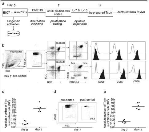
Fig. 1Allo-specific TSCMcells are effectively prepared in vitro.aProtocol for preparation of allo-specific TSCM. The method began with setting up an allo-reactive co-culture by mixing of 2 × 10⁶E007 cells and 1 × 10⁷allogeneic PBLs (PBLs) on day 0 (allogeneic activation), to generate allo-specific T cells. The presence of 5μM TWS119 enriched TSCMin the co-culture (differentiation inhibition). The CFSE diluted cells in the co-culture bulks were sorted by FACS on day 7 (proliferation sorting). The sorted cells were cultured with IL-7 and IL-15 (25 ng/ml each) to expand the TSCM for the next 7 days (cytokine expansion). Then, the cultural bulks were used as the prepared TSCMcells for the tests in vitro and in vivo included in this study.bGating strategy for identification of CD8+ TSCMsubset in the co-culture bulks. Lymphocytes were identified based on SSC versus FSC, and proliferating cells were on a reduced fluorescence intensity of CFSE (CFSEdim). In the gated CFSEdim population, cells expressing CD3 and CD8 were selected for further CD45RA and CD62L analysis. CD8+ TSCMshowed the phenotype of CFSEdim CD3+ CD8+ CD45RA+ CD62L+, TCMof CD45RA- CD62L+, TEMof CD45RA- CD62L-, TEFof CD45RA+ CD62L-, and CD95 was further detected to distinguish TSCMfrom TN, the latter showed the phenotype of CFSE high CD95- in the co-culture bulks, but TSCMof CFSEdim CD95+. The illustration was a representative of co-culture bulks analyzed by FCM on day 7 before sorting, the white peaks in CD95, CCR7, and CD28 plots were isotype controls. The broken arrows indicated the sequential gating strategy.cTSCMnumbers increased by 100 folds in the co-culture bulks with TWS119 enrichment for 7 days. Data are represented as mean ± SD of six individual experiments.dE007-specific T cells underwent proliferation in the co-culture, the CFSEdim cells were sorted by FACS with purity above 98%.eThe sorted cells were further expanded by IL-7 and IL-15 for the next 7 days, the allo-specific TSCM increased by another 150 folds. Data are represented as mean ± SD of six individual experiments
but also contained a few CD4+ TSCM (10.4 ± 8.16%),
CD3- cells (6.15 ± 5.23%), CD8+ non-TSCM(12.6 ±
3.48%), and CD4+ non-TSCM (10.2 ± 8.66%) cells
(Add-itional file 1: Figure S1D E F). By allogeneic activating, inhibiting differentiation with TWS119, sorting CFSEdim cells, and expansion with IL-7 and IL-15, our in vitro protocol was able to prepare about 2 × 10⁷ allo-specific CD8+ TSCM cells from 1 × 10⁷PBLs. The number of the
TSCM cells was sufficient to meet the needs of the
fol-lowing studies.
The TSCMpreparation strategy used in this study could
be translated into a single epitope-specific TSCM
prepar-ation. When the E007 co-cultured with allogeneic PBLs, the precursor frequencies were much higher than those of T cell responses to a single allogeneic epitope. To pre-pare a single epitope-specific TSCMcells in a large
num-ber, a modification with a prolonged cytokine expansion was required (Additional file2: Figure S2).
The prepared TSCMcells show stem cell and memory T cell characteristics in vitro
To examine the self-renewal capacity, TN, TSCM, TCM,
and TEMcells were sorted by their corresponding
phe-notypes from the co-culture on day 7 (Fig.1b). The con-tent of T cell receptor rearrangement excision circles (TRECs) in each subset was examined by real-time qPCR. As TRECs cannot be replicated while cell div-ision, the content of TRECs reflects the frequency of T cell proliferation. Results showed that TN had the
high-est content of TRECs. TSCM, TCM, and TEM cells
pos-sessed less TRECs, confirming they were differentiated from TNthrough T cell clonal proliferation (Fig.2a). The
content of TRECs in TSCM cells was between those of
TN and TCM cells, suggesting TSCM was at the earliest
stage after TNactivation.
The prepared TSCM cells were relabeled with CFSE
and exposed to IL-2 (200 U/ml) for 10 days. As IL-2 is a potential promoter for T cell proliferation and differenti-ation, seven generations of CFSE-diluted daughter cells developed. A portion of TSCMcells (CD45RA+ CD62L+)
were found in each generation, indicating that the pre-pared TSCM cells were able to maintain a stem cell
phenotype during proliferation and differentiation (Fig.2b, c). Collectively, the prepared TSCM cells showed
characteristics of self-renewal.
To check memory T cell characteristics, the prepared TSCM cells were incubated with the cognate stimulator
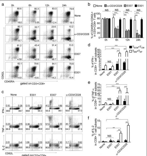
E007, HLA class I mismatched E001, andα-CD3/CD28 beads. Phenotypic analysis revealed that the E007-restimulated TSCM cells showed vigorous
differen-tiation in 6 h, which became more intensive with pro-longed stimulation time. A similar response was observed in the α-CD3/CD28-stimulated TSCM cells.
While the E001-stimulated TSCM cells showed no
significant differentiation after 24 h, the response was similar to that of non-stimulated TSCM cells (Fig.3a, b).
In addition, after 24 h of incubation with E007 and
α-CD3/CD28, we found that TSCM cells in the
incuba-tion showed no significant change in absolute number (Additional file3: Figure S3E),suggesting that TSCMcells
were able to self-renew during proliferation and differentiation.
CD8+ T cell mediated immune effectors include pro-duction of cytokines and killing of target cells. To inves-tigate production of cytokines, the TSCM cells were
stimulated with E007, E001, and α-CD3/CD28 beads, then intracellular IL-2, TNF-α, and IFN-γ production was measured. The E007-restimulated TSCM cells
showed more positive cells of these cytokines. Gating with CD62L, the incubated bulks were divided into CD62L+ (TSCM and TCM) and CD62L- (TEM and TEF)
cells, most of the cytokine positive cells were TEM and
TEFcells (Fig.3c). Meanwhile, gating with CD45RA, we
found that both TEM and TEF cells showed similar
fre-quency of the cytokine positive cells (Additional file 3: Figure S3A-D). Whereas, the TSCM cells in the
incuba-tion produced almost background IL-2, and only a low level of TNF-α and IFN-γ. The TSCM cells responded
againstα-CD3/CD28 in a similar profile of the cytokine production to that against E007. In contrast, the TSCM
cells stimulated with E001 showed frequency of cytokine-positive cells in the same way as that with non-stimulation (Fig.3c–f). Next, we moved to cytotox-icity assay, the TSCM co-cultured with E007 or E001 for
24 h at a ratio 5:1. Killing rate of the TSCMand daughter
cells against E007 was higher than that against E001 after 8 h (Additional file3: Figure S3F, G). These results showed that the prepared TSCM cells were E007-specific
and able to differentiate rapidly into effector T cells after stimulated by the same antigen.
The prepared TSCMcells are able to implant and effectively remove target cells in vivo
To evaluate capacity of persistence and rejecting target cells in vivo of the prepared TSCM cells, human LCL
cells E001 or E007 were inoculated intravenously into NOD/SCID mice to establish LCL-burden mouse model. After 3 days, the LCL-burden mice were treated with ei-ther E007-specific TSCM cells or E007-specific TEMand
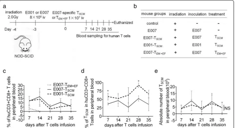
TEFcells (Fig.4a, b). On days 7, 14, 21, 28, and 35 after
T cell infusion, caudal vein peripheral blood samples were taken to detect the frequency and phenotype of hu-man T cells. We found huhu-man CD3+ CD8+ T cells in the peripheral blood of the TSCM-treated mice at all
sampling times. However, in the TEM + EF-treated mice
(E007-TEM + EF), human T cells were detected in
the E007-burden mice (E007-TSCM) were lower than that
in the E001-burden mice (E001-TSCM), but no difference
was found in the absolute number of TSCMcells between
E007-burden and E001-burden mice (Fig.4d, e). On day 35, more frequent human T cells were found in the spleen and bone marrow of the TSCM-treated mice
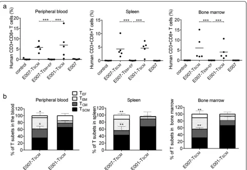
re-vealed by flow cytometry (Fig. 5a), and the immuno-fluorescent staining of the spleen sections showed the similar results (Fig. 6d, e). These findings indicated that
the TSCM cells were able to survive long through
self-renewal in the mice, and differentiate into other T cell subsets as stimulated with the specific antigens of E007.
To investigate the immune reconstitution capacity of the TSCM cells, peripheral blood, spleen, and bone
mar-row of TSCM-treated and TEM + EF-treated mice were
tested for human T cells on day 35. The human T cells in the above specimens consisted of TSCM (CD45RA +
Fig. 2The prepared TSCMcells bear the ability of self-renewing.aTN, TSCM, TCM, and TEMcell subsets in CD8+ T cells were sorted by their corresponding phenotypes from the co-culture on day 7. TREC copy number in the sorted subsets relative to TNcells was shown. Data are represented as mean ± SD of four individual experiments.bThe prepared TSCMcells were relabeled with CFSE and exposed to 200 U/ml of IL-2 for 10 days, the proliferating daughter cells divided into 7 generations (left panel). In each generation, there were a portion of CD8+ T cells expressing CD62L and CD45RA (right panel). Representative FCM plots were shown.cThe frequency of TSCM(CD45RA+ CD62L+) cells in CD8+ T cells was analyzed at each CFSE dilution peak. Data are represented as mean ± SD of four individual experiments. (*p< 0.05; **p< 0.01, and NS,p> 0.05)
CD62L+), TCM (CD45RA-CD62L+), TEM
(CD45RA-CD62L-), and TEF (CD45RA+ CD62L-). The
distribution of T cell subsets revealed more TEMand TEF
cells in the E007-burden mice, whereas more TSCM and
TCM cells in the E001-burden mice (Fig. 5b). As TEM
and TEF cells are at terminal differentiation stage while
TSCM and TCM cells at early differentiation stage, our
findings suggested the E007-specific TSCM cells
differen-tiated into the other T cell subsets when they came cross the same antigen in the E007 burden mice.
To analyze killing efficacy of the TSCM cells in vivo, on
day 35 of the T cell treatment, the residual LCL cells in mice were detected by intracellular LMP1 staining. The LCL cells were mainly found in the spleen and bone mar-row of the LCL-burden mice. In the E007-burden mice, the residual LCL cells in spleen and bone marrow of the TSCM-treated mice were similar to that of the control mice
without LCL infusion (control). Nevertheless, the TEM +
EF-treated mice carried a significant amount of the residual
LCL cells compared to the control mice, but the amount was lower than the E007-burden mice without T cell treat-ment (E007) (Fig.6a–c). More residual LCL cells were also found in the immunofluorescent spleen sections of the TEM + EF-treated mice than those of the TSCM-treated mice
(Fig. 6d, e) These data demonstrated the TSCM cells
equipped with superior capacity of killing targets of specific antigens in vivo compared to the TEMand TEFcells.
In contrast to the E007-burden mice, the E001-burden mice bore not only less TEM and TEFcells after the TSCM
treatment, but also the residual LCL cells as high as that of the E007-burden mice without T cell treatment (Fig.6a–c). Results of immunofluorescence staining of spleen sections for human T cells and LCLs showed residual LCL cells in E007-burden mice treated with TSCMwere similar to those
of the control mice. Although the spleen of the E001-burden mice showed a significant number of human T cells, the residual LCL cells were similar to that of the E007-burden mice without T cell treatment (Fig.6d, e). By the way, the human T cell-treated mice showed no sign of xenogeneic graft-versus-host disease (xeno-GVHD), such as loss of body weight, back arched, and shed. Collectively, these results indicated that the prepared TSCMcells were
able to survive over a long-term in vivo and differentiate into effector T cells to eradicate the target bearing the cog-nate antigens.
Discussion
Mature T cells are comprised of cells that are at various stages of differentiation, which are discernible by the ex-pression of surface molecules. TN cells are
Fig. 4The TSCMare transferred into LCL-burden mice for stem and memory T cell behavior.a bAdoptive transfer protocol of the TSCM. On day−3, irradiated mice were inoculated with E007 or E001 cells to set up the LCL-burden mouse model. On day 0, E007-specific TSCMor TEMand TEF(TEM + EF) were transferred into the mice intravenously (iv). Blood sampling was taken once a week for 5 weeks after the T cell treatment. On day 35, the mice were sacrificed for detection of human T cells and LCL cells in spleen and bone marrow.c–eThe prepared TSCMcells had superior persistence in vivo. The frequency of human CD3 + CD8+ T cells (c), the frequency of TSCMcells (CD45RA+ CD62L+) in human CD3+ CD8+ T cells (d), and the absolute number of TSCMcells in 50μl blood (e) were detected by FCM. Data are represented as mean ± SD of five mice. (*p< 0.05; NS,p> 0.05)
conventionally defined by the co-expression of the CD45RA, CCR7, and the lymph node homing molecules L-selectin (CD62L) [3]. Similar to TN cells, CD62L and
CCR7 are maintained on TCMcells, whereas these
mole-cules are lost on more differentiated TEM cells [3, 35].
TSCMcells, the least differentiated of all distinct memory
populations, are identifiable through the expression of markers, including CD62L, CCR7, CD95, and the che-mokine (C-X-C motif ) receptor 3 [5, 10, 36]. Naïve T cells downregulate CD62L expression after stimulation, as clonal expansion goes on, both CD62LLOW and CD62LHIGHT cell subsets are developed. With real-time tracking of CD8 T-cell divisions in vitro, Kinjyo and co-workers define memory T cells among CD62LHIGH cells. The memory T cells stay CD62LHIGHand prolifer-ate in vitro driven by IL-2, although the division dur-ation is slow, the phenotype and cell cycle durdur-ation are inherited by the progeny of the T cells [37]. We identi-fied CD8+ TSCM subset in the co-culture bulks by the
phenotypes CD3+ CD8+ CD45RA+ CD62L+ CCR7+
CD95+ CD28+, as our results showed the cells with the phenotypes in the co-culture bulks were of stem cell and memory T cell properties.
A frequently used method to enrich TSCM cells is
dif-ferentiation inhibition. With antigen priming, antigen-specific CD8+ TN cells proliferate and develop
into TSCM cells first and then the other subsets. It is
re-ported that TSCM differentiation can be inhibited by
TWS119, which inhibits the GSK-3β and activate Wnt/
β-catenin. The inhibition improves the maintenance of ‘stemness’in mature memory CD8+ T cells which medi-ated a better anti-tumor response after transferred into mice [15, 38]. Rapamycin is also reported to help the formation of T cell subset at an early stage of differenti-ation by moduldifferenti-ation of mTOR signaling [39–41]. How-ever, rapamycin tended to promote the generation of TCMcells in our study (Additional file1: Figure S1C), in
line with other report [41]. In our co-culture of E007 with allogeneic PBLs, the E007-specific TSCM cells were
generated from TN and enriched in the presence of
TWS119 which inhibited the TSCM further
differenti-ation. As inhibition by TWS119 results in a TSCM
phenotype in human CD8+ T cells [5,15,16], our proto-col preferred expansion of CD8+ TSCM to that of CD4+
TSCM.
The administration of cytokines is another method to expand TSCM. Cytokines have important functions
re-lated to T cell expansion, differentiation, survival, and
homeostasis. Common γ-chain (γc) family cytokines are commonly used in clinical trials, including IL-2, IL-7, IL-15, and IL-21. IL-7 is instrumental for the generation of TSCM cells by binding to IL-7 receptor expressing
naive and memory T lymphocytes [42,43]. Expansion of TSCM required either IL-15 or IL-2; IL-15 proves
super-ior to IL-2 in supporting expansion coupled to preserva-tion of the TSCM phenotype [18, 19]. Activated naive T
Fig. 6The TSCMcells are able to eradicate allo-antigen-specific targets in vivo. The spleen and bone marrow were detected for the residual LCL cells by intracellular LMP1 staining using FCM.a–cRepresentative FCM plots (a) and the absolute number of LCLs in bone marrow of two femurs (b) and in 100 mg spleen tissue (c) were analyzed.dSpleen sections were observed for LCLs and human T cells by immunofluorescence. Representative illustrations of immunofluorescent labeling of human CD3 (green), LMP1 (red), and nucleus staining by DAPI (blue) for spleen sections were shown (× 400).eDate showed the mean frequencies of human CD3 and LMP-1-positive cells in 4 fields chosen randomly (100 cells each) for each mouse spleen section. The residual LCLs in the E007-TSCMmice were similar to that in the control mice without LCL inoculation, the rest mouse groups showed relatively more LCL cells. Data are represented as mean ± SD of five mice (*p< 0.05; **p< 0.01; ***p< 0.001, and NS,p> 0.05)
cells show a higher expression of IL-21 receptor, and IL-21 has been reported to be able to enrich less differ-entiated TSCM within the total TSCM subset [20, 22, 23,
44]. However, IL-21 exerted few effects on the TSCM
ex-pansion in our study. Therefore, IL-7 and IL-15 were used to expand the TSCM cells after the proliferation
sorting, resulting in a large number of the TSCM
suffi-cient for the experiments in vitro and in vivo included in our study.
An important feature of stem cells is the self-renewal capacity. Although self-renewal abilities of the prepared TSCM cells were revealed by TREC copy numbers and
proliferative history driven by IL-2 in vitro, long-term in vivo implantation is the gold standard for identification of stem cells. Long-lasting survival of human TSCMcells
in mice is reported. In serial transplantations model, TSCM cells prove able to persist in secondary recipients
suggesting self-renewal abilities [5, 6, 19, 45]. Here, the prepared TSCM migrated to secondary lymphoid organs,
such as bone marrow and spleen, showed long-lasting survival potential at least for 35 days after transferred in our model. Importantly, the number of TSCMcells in the
blood samples was observed consistent during this study, suggesting the self-renewal capacity of the trans-ferred TSCM cells in vivo. The implantation and
long-term existence of the TSCM cells in the host would
be expected to mount durable T cell responses after transferring.
Governing antigen specificity is important for TSCM
preparation; various strategies are used for this purpose. For example, HCMV-specific TSCM cells are isolated
from peripheral blood TSCMcells of HCMV seropositive
donors and enriched in vitro by incubation with HCMV antigen [26]. CD19-specific TSCMcells are prepared from
peripheral blood TN and TSCM by transduction with a
γ-retroviral vector encoding the CD19-chimeric antigen receptor (CAR) [24]. TCR-transgenic mice are also a source of antigen-specific TSCM cells [46]. In our
ap-proach, the E007-specific T cells underwent proliferation in the co-culture bulks, making it feasible to be sorted by CFSE dilution. In our pre-experiment, IL-7 or IL-15 was able to make PBLs proliferation without the E007 stimulation. Hence, no cytokine was added to the co-culture before the sorting, to ensure the E007-specificity of the prepared TSCMcells.
Two HLA class I mismatched LCLs, E007 and E001, were used to examine the antigen specificity of the pre-pared CD8+ TSCM cells. Rapid differentiation into
ef-fector T cells and robust efef-fector functions were observed when the prepared TSCM cells were stimulated
with the E007, but not with the E001. Although the E007-specificity was suggested by these in vitro findings, it would be more important to observe the behavior of the prepared TSCM cells in vivo. In our LCL burden
mouse model, LCL cells could be found in spleen and bone marrow up to 45 days after transfer into the im-munodeficiency mice in our preliminary test. The LCL burden mice showed no measurable side effect during our study, such as loss of body weight, back arched, and shed. Interestingly, the prepared TSCMcells exhibited
in-tensive differentiation into other T cell subsets and ef-fective eradication of LCL targets in the E007-burden mice instead of the E001-burden mice. These observa-tions reflected the prepared TSCM cells were
E007-specific.
Conclusions
Functional allo-specific CD8+ TSCM cells were prepared
from human PBLs in a procedure of allogeneic co-culture, differentiation inhibition, proliferation sort-ing, and cytokine expansion. Although our study pro-vided a practical protocol for allo-specific TSCM cell
preparation, this method would be adapted to prepare TSCM cells specific for antigen of interesting. As TSCM
cells show the implantation and long-term existence in the host after transferring, the preparation of antigen-specific TSCM cells is crucial for T cell adoptive
immunotherapy.
Additional files
Additional file 1:Figure S1. Enrichment of TSCMcells by differentiation inhibitors and the lymphocyte distribution changes in the cultural bulks over the TSCMpreparation course.A,BTWS119 exhibited concentration-dependent enrichment of TSCMin the alloreactive co-culture, 5μM of TWS119 was the best concentration. TSCMcells in frequency (A) and in absolute number (B) were shown.CDifferentiation inhibition by rapamy-cin was more likely to enrich TCMinstead of TSCM.D–FAfter allogeneic activation, differentiation inhibition, proliferation sorting, and cytokine ex-pansion, lymphocyte distribution in the cultural bulks was revealed by FCM over the TSCMpreparation course. CD3 + CD4+ T cells, CD3 + CD8 + T cells and CD3- cells in the cultural bulks were detected, and the propor-tion of CD3 + CD8+ T cells was increased over time (D). The majority of cultural bulks were CD3 + CD8+ TSCM(60.1 ± 11.2%) on day 14 (E). The proportion of CD3 + CD8+ TSCMin CD8+ T cells was increased over time, about 80% of CD8+ T cells were TSCMon day 14 (F). Data are represented as mean ± SD of four individual experiments. (JPG 170 kb)
Representative FCM plots (C). TSCMcells differentiated more when incu-bated with the T2/AFP (D). The daughter cells showed more frequent IFN-γpositive cells when incubated with the T2/AFP (E). Data are repre-sented as mean ± SD of four individual experiments (**p< 0.01). (JPG 404 kb)
Additional file 3:Figure S3. The prepared TSCMdifferentiated into effector T cells stimulated by E007. The prepared TSCMwere stimulated with E007, E001 andα-CD3/CD28, respectively. The T cell subsets and their intracellular IL-2, TNF-αand IFN-γproduction were detected. The TSCMdifferentiated into effector T cells upon the stimulation with E007 andα-CD3/CD28.A–DAfter 4-h stimulation, both TEMand TEFcells exhib-ited the similar frequency of the cytokine positive cells. Representative FCM plots (A). Gating by CD3+ CD8+ CD62L-, the T cells were divided into CD45RA- (TEM) and CD45RA+ (TEF) cells. The TEMand TEFsubsets showed the cytokine positively stained cells of IFN-γ(B), TNF-α(C) and IL-2 (D).EAfter 24-h stimulation, the absolute number of TSCMremained stable during differentiation.F,GThe cytotoxicity of the TSCMand the daughter cells were E007-specific. E007 and E001 labeled with celltrace, co-cultured with the TSCMat ratio 1:5. Dead cells stained by PI dye were detected by FCM. Representative FCM plots (F) and the frequencies of dead cells (G) were shown. Data are represented as mean ± SD of four in-dividual experiments (NS,p> 0.05; **p< 0.01, and ***p< 0.001). (JPG 722 kb)
Abbreviations
ACT:Adoptive cell therapy; GVHD: Graft versus host disease; HLA: Human leukocyte antigen; LCL: B lymphoblastoid cell lines; LMP1: Latent membrane protein 1; PBL: Peripheral blood lymphocytes; PBMC: Peripheral blood mononuclear cells; pMHC: Peptide major histocompatibility complex; TCR: T cell receptor; TREC: T cell receptor rearrangement excision circle
Acknowledgements
The authors apologize to colleagues whose work could not be cited because of space limitation. They thank Dr. Jinghui Zhang (The Union Hospital, Tongji Medical College, Huazhong University of Science and Technology) for the FACS sorting in this study.
Funding
This project was supported by the National Natural Science Foundation of China (no. 31370885).
Availability of data and materials Please contact author for data requests.
Authors’contributions
XWW designed the study, analyzed the data, and wrote the manuscript. LG was a major contributor in the experimental performance, analyzed the data, and wrote the manuscript. XL performed and analyzed the cell culture experiments. JW contributed to the animal experimental performance. XFW contributed to the design of study. ZL contributed to the data analysis. JY contributed to the manuscript preparation. All authors read and approved the final manuscript.
Ethics approval
All donors’samples were obtained after informed consent according to a protocol approved by the Ethics Committee of Tongji Medical College, Wuhan, China. Animal experiments were approved by the Ethics Committee of Tongji Medical College. All animal procedures were performed in strict accordance with the guidelines of the Chinese Council on Animal Care.
Consent for publication Not applicable.
Competing interests
The authors declare that they have no competing interests.
Publisher’s Note
Springer Nature remains neutral with regard to jurisdictional claims in published maps and institutional affiliations.
Received: 1 May 2018 Revised: 12 November 2018 Accepted: 19 November 2018
References
1. June CH. Principles of adoptive T cell cancer therapy. J Clin Invest. 2007;117: 1204–12.
2. Restifo NP, Dudley ME, Rosenberg SA. Adoptive immunotherapy for cancer: harnessing the T cell response. Nat Rev Immunol. 2012;12:269–81. 3. De Rosa SC, Herzenberg LA, Roederer M. 11-color, 13-parameter flow
cytometry: identification of human naive T cells by phenotype, function, and T-cell receptor diversity. Nat Med. 2001;7:245–8.
4. Mahnke YD, Brodie TM, Sallusto F, Roederer M, Lugli E. The who’s who of T-cell differentiation: human memory T-T-cell subsets. Eur J Immunol. 2013;43: 2797–809.
5. Gattinoni L, Lugli E, Ji Y, Pos Z, Paulos CM, Quigley MF, et al. A human memory T cell subset with stem cell-like properties. Nat Med. 2011;17:1290– 7.
6. Cieri N, Camisa B, Cocchiarella F, Forcato M, Provasi E, Bondanza A, et al. IL-7 and IL-15 instruct the generation of human memory stem T cells from naive precursors. Blood. 2013;121:573–84.
7. Biasco L, Scala S, Basso Ricci L, Dionisio F, Baricord C, Calabria A, et al. In vivo tracking of T cells in humans unveils decade-long survival and activity of genetically modifed T memory stem cells. Sci Transl Med. 2015.https:// doi.org/scitranslmed/scitranslmed3010314.
8. Lugli E, Gattinoni L, Roberto A, Mavilio D, Price DA, Restifo NP, Roederer M. Identification, isolation and in vitro expansion of human and nonhuman primate T stem cell memory cells. Nat Protoc. 2013;8:33–42.
9. Flynn JK, Gorry PR. Stem memory T cells (TSCM)-their role in cancer and HIV immunotherapies. Clin Transl Immunology. 2014.https://doi.org/10.1038/cti. 10. Gattinoni L, Klebanoff CA, Restifo NP. Paths to stemness: building the
ultimate antitumour T cell. Nat Rev Cancer. 2012;12:671–84. 11. Xu L, Zhang Y, Luo G, Li Y. The role of stem cell memory T cells in
hematological malignancies. J Hematol Oncol. 2015.https://doi.org/10.1186/ s13045-015-0214-5.
12. Lugli E, Dominguez MH, Gattinoni L, Chattopadhyay PK, Bolton DL, Song K, et al. Superior T memory stem cell persistence supports long-lived T cell memory. J Clin Invest. 2013;123:594–9.
13. Fuertes Marraco SA, Soneson C, Caqnon L, Gannon PO, Allard M, Allard Maillard S, et al. Long-lasting stem cell–like memory CD8+ T cells with a naive-like profile upon yellow fever vaccination. Sci Transl Med. 2015.
https://doi.org/scitranslmed/scitranslmedaaa3700.
14. Morrot A. Lifelong protection mediated by stem cell-like CD8(+) T memory subset cells (TSCM) induced by vaccination. Ann Transl Med 2016; doi:
https://doi.org/10.21037/atm.2016.05.38.
15. Gattinoni L, Zhong XS, Palmer DC, Ji Y, Hinrichs CS, Yu Z, et al. Wnt signaling arrests effector T cell differentiation and generates CD8+ memory stem cells. Nat Med. 2009;15:808–13.
16. Forget MA, Huon Y, Reuben A. Stimulation of Wnt/β-catenin pathway in human CD8+ T lymphocytes from blood and lung tumors leads to a shared young/memory phenotype. PLoS One. 2012.https://doi.org/10.1371/journal. pone. 0041074.
17. Muralidharan S, Hanley PJ, Liu E, Chakraborty R, Bollard C, Shpall E, et al. Activation of Wnt signaling arrests effector differentiation in human peripheral and cord blood-derived T lymphocytes. J Immunol. 2011;187: 5221–32.
18. Cieri N, Camisa B, Cocchiarella F, Forcato M, Oliveria G, Provasi E, et al. IL-7 and IL-15 instruct the generation of human memory stem T cells from naive precursors. Blood. 2013;121:573–84.
19. Xu Y, Zhang M, Ramos CA, Durett A, Liu E, Dakhova O, et al. Closely related T-memory stem cells correlate with in vivo expansion of CAR CD19-T cells and are preserved by IL-7 and IL-15. Blood. 2014;123:3750–9.
20. Lugli E, Goldman CK, Perera LP, Smedley J, Yovandich JL, Creekmore SP, et al. Transient and persistent effects of IL−15 on lymphocyte homeostasis in nonhuman primates. Blood. 2010;116:3238–48.
21. Alvarez-Fernandez C, Escriba-Garcia L, Vidal S, Sierra J, Briones J. A short CD3/CD28 costimulation combined with IL-21 enhance the generation of human memory stem T cells for adoptive immunotherapy. J Transl Med. 2016.https://doi.org/10.1186/s12967-016-0973-y.
22. Zeng R, Spolski R, Finkelstein SE, Oh S, Kovanen PE, Hinrichs CS, et al. Synergy of IL-21 and IL-15 in regulating CD8+ T cell expansion and function. J Exp Med. 2011;201:139–48.
23. Pilipow K, Roberto A, Roederer M, Waldmann TA, Mavilio D, Lugli E. IL-15 and T-cell Stemness in T-cell-based Cancer immunotherapy. Cancer Res. 2015;75:5187–93.
24. Sabatino M, Hu J, Sommariva M, Gautam S, Fellowes V, Hocker JD, et al. Generation of clinical-grade CD19-specific CAR-modified CD8+ memory stem cells for the treatment of human B-cell malignancies. Blood. 2016;128: 519–28.
25. Albrecht J, Frey M, Teschner D, Carbol A, Theobald M, Distler E. IL-21-treated naive CD45RA+ CD8+T cells represent a reliable source for producing leukemia-reactive cytotoxic T lymphocytes with high proliferative potential and early differentiation phenotype. Cancer Immunol Immunother. 2011;60:235–48.
26. Schmueck-Henneresse M, Sharaf R, Vogt K, Weist BJ, Landwehr-Kenzel S, Fuehrer H, et al. Peripheral blood-derived virus-specific memory stem T cells mature to functional effector memory subsets with self- renewal potency. J Immunol. 2015;194:5559–67.
27. Yang I, Weiss L, Abdul-Hai A, Kasir J, Reich S, Slavin S. Induction of early post-transplant graft-versus-leukemia effects using intentionally mismatched on or lymphocytes and elimination of alloantigen-primed donor
lymphocytes for prevention of graft-versus-host disease. Cancer Res. 2005; 65:9735–40.
28. Amir AL, van der Steen DM, Hagedoorn RS, Kester MG, van Bergen CA, Drijfhout JW, et al. Allo-HLA-reactive T cells inducing graft-versus-host disease are single peptide specific. Blood. 2011;118:6733–42.
29. Yu Q, Zhang L, Ouyang LC, Gong YL, Liang ZH, Shen GX, et al. A similarity in peptide cross-reactivity between alloantigen and nominal antigen-induced CD8+ T cell responses in vitro. Immunogenetics. 2013;65:173–84. 30. Tsirigotis P, Shimoni A, Nagler A. The expanding horizon of immunotherapy
in the treatment of malignant disorders: allogeneic hematopoietic stem cell transplantation and beyond. Ann Med. 2014;46:384–96.
31. Negrin RS. Graft-versus-host disease versus graft-versus-leukemia. Hematology Am Soc Hematol Educ Program. 2015;1:225–30.
32. Neitzel H. A routine method for the establishment of permanent growing lymphoblastoid cell lines. Hum Genet. 1986;73:320–6.
33. Bunce M, O’Neill CM, Barnardo MC, Krausa P, Browning MJ, Morris PJ, et al. Phototyping: comprehensive DNA typing for HLA-A, B, C, DRB1, DRB3, DRB4, DRB5 & DQB1 by PCR with 144 primer mixes utilizing sequence-specific primers (PCR-SSP). Tissue Antigens. 1995;46:355–67.
34. Lang PO, Mitchell WA, Govind S, Aspinall R. Real time-PCR assay estimating the naive T-cell pool in whole blood and dried blood spot samples: pilot study in young adults. J Immunol Methods. 2011;369:133–40.
35. Weninger W, Crowley MA, Manjunath N, von Andrian UH. Migratory properties of naive, effector, and memory CD8+ T cells. J Exp Med. 2001; 194:953–66.
36. Sallusto F, Lenig D, Forster R, Lipp M, Lanzavecchia A. Two subsets of memory T lymphocytes with distinct homing potentials and effector functions. Nature. 1999;40:708–12.
37. Kinjyo I, Qin J, Tan SY, Wellard CJ, Mrass P, Ritchie W, et al. Real-time tracking of cell cycle progression during CD8+ effector and memory T-cell differentiation. Nat Commun. 2015.https://doi.org/10.1038/ncomms7301. 38. Gattinoni L, Ji Y, Restifo NP. WNT/β-catenin signaling in T-cell immunity and
cancer immunotherapy. Clin Cancer Res. 2010;16:4695–701.
39. Scholz G, Jandus C, Zhang L, Grandclement C, Lopez-Mejia C, Soneson C, et al. Modulation of mTORC signaling triggers the formation of stem cell-like memory T cells. EBioMedicine. 2016;4:50–61.
40. Chi H. Regulation and function of mTOR signalling in T cell fate decisions. Nat Rev Immunol. 2012;12:325–38.
41. Araki K, Turner AP, Shaffer VO, Gangappa S, Keller SA, Bachmann MF, et al. mTOR regulates memory CD8 T-cell differentiation. Nature. 2009;460:108–12. 42. Tan JT, Dudl E, LeRoy E, LeRoy E, Murray R, Sprent J, et al. IL-7 is critical for
homeostatic proliferation and survival of naive T cells. Proc Natl Acad Sci U S A. 2001;98:8732–7.
43. Boyman O, Letourneau S, Krieg C, Sprent J. Homeostatic proliferation and survival of naive and memory T cells. Eur J Immunol. 2009;39:2088–94. 44. Battaglia A, Buzzonetti A, Baranello C, Fanelli M, Fossati M, Catzola V, et al.
Interleukin-21 (IL-21) synergizes with IL-2 to enhance T-cell receptor-induced human T-cell proliferation and counteracts IL-2/transforming growth factor-beta-induced regulatory T-cell development. Immunology. 2013;139:109–20.
45. Kagoya Y, Nakatsugawa M, Ochi T, Cen Y, Guo T, Anczurowski M, Saso K, et al. Transient stimulation expands superior antitumor T cells for adoptive therapy. JCI insight. 2017.https://doi.org/10.1172/jci. insight. 89580. 46. Stemberger C, Graef P, Odendahl M, Albrecht J, Dossinger G, Anderl F, et al.