PRIMARY RESEARCH
High expression of metabolic enzyme
PFKFB4 is associated with poor prognosis
of operable breast cancer
Ling Yao
1,2,3†, Lei Wang
1,2,3†, Zhi‑Gang Cao
1,2,3, Xin Hu
1,2,3*and Zhi‑Ming Shao
1,2,3,4*Abstract
Background: Enhanced glycolysis in tumors, known as the Warburg effect, provides the metabolic basis of
enhanced cancer cell proliferation and metastasis. The Warburg pathway enzyme 6‑phosphofructo‑2‑kinase/fructose‑ 2,6‑bisphosphatase 4 (PFKFB4) is a newly identified key kinase that regulates transcriptional reprogramming and cell proliferation. Here we show the prognostic value of PFKFB4 expression in patients with operable breast cancer. Methods: PFKFB4 expression was evaluated by immunohistochemistry in surgical specimens retrospectively col‑ lected from 200 patients with histologically proven invasive ductal breast cancer. Kaplan–Meier survival analysis and Cox regression analysis were performed to assess the prognostic significance of PFKFB4 expression.
Results: Kaplan–Meier survival analysis revealed that breast cancer patients with high PFKFB4 expression demon‑ strated unfavorable disease‑free survival (p = 0.008) and overall survival (p = 0.002). PFKFB4 had an hazard ratio (HR) of 7.38 (95% CI 1.69–32.3; p= 0.008) in univariate Cox analysis and retained prognostic power (HR 7.44, 95% CI 1.67–33.2;
p= 0.009) when adjusted by tumor size, lymph node status, grade, estrogen receptor status, human epidermal growth factor receptor 2 status and subtype, which indicated PFKFB4 was an independent prognostic factor in breast cancer.
Conclusions: Together, our findings establish the prognostic value of metabolic enzyme PFKFB4 in patients with operable breast cancer.
Keywords: PFKFB4, Overall survival, Disease‑free survival, Breast cancer
© The Author(s) 2019. This article is distributed under the terms of the Creative Commons Attribution 4.0 International License (http://creat iveco mmons .org/licen ses/by/4.0/), which permits unrestricted use, distribution, and reproduction in any medium, provided you give appropriate credit to the original author(s) and the source, provide a link to the Creative Commons license, and indicate if changes were made. The Creative Commons Public Domain Dedication waiver (http://creat iveco mmons .org/ publi cdoma in/zero/1.0/) applies to the data made available in this article, unless otherwise stated.
Background
Breast cancer is the most common cancer in women can-cer and the second highest cause of cancan-cer-related death in women in the United States [1]. In China, breast can-cer is also the most commonly diagnosed cancan-cer and accounts for 15% of all new cancers in women [2]. Breast cancer is a heterogeneous disease composed of four sub-types: luminal A, luminal B, human epidermal growth factor receptor 2 (HER-2)-enriched and triple-negative [3]. According to current guidelines and consensus, the
selection of an appropriate adjuvant therapy regimen is largely based on the subtype and risk recurrence cate-gory. The value of prognostic biomarkers and gene-based assays has recently been added into the 8th edition of the primary tumor, lymph node, and metastasis (TNM) clas-sification of the American Joint Commission of Cancer for breast cancer [4]. Despite the intensive research into the mechanisms of breast cancer performed over the past decades, very few of the identified critical molecules have been adopted for therapeutic or prognostic approaches in clinical practice. Thus, there is an urgent need to iden-tify novel biomarkers that provide additional risk assess-ment for personalized treatassess-ment.
In recent years, studies of cancer metabolism have provided insight into the process of adaptation of can-cer cells, which alter their metabolic activity to meet
Open Access
*Correspondence: [email protected]; [email protected] †Ling Yao and Lei Wang contributed equally to this work
the increased needs for energy and biosynthetic precur-sors. Enhanced glycolysis, known as the Warburg effect, is observed in many cancers and provides the metabolic basis of cancer cell proliferation [5]. The Warburg path-way enzyme 6-phosphofructo-2-kinase/fructose-2,6-bis-phosphatase 4 (PFKFB4) is a newly identified key factor that regulate transcriptional reprogramming and func-tions as the most dominant kinase that regulates cell pro-liferation [6]. Several studies identified PFKFB4 as a key molecule in multiple cancers, including breast cancer [6], prostate cancer [7] and glioma [8]. However, our current knowledge about PFKFB4 has largely originated from in vitro studies, and the in vivo relevance of this molecule requires further study. Furthermore, the prognostic value of the PFKFB4 protein in breast cancer has not been investigated.
In this study, we evaluated the prognostic power of PFKFB4 expression in 200 tumor samples from patients with stage I to III breast cancer. We examined the asso-ciations between PFKFB4, clinicopathological variables and survival. Our results indicated that elevated PFKFB4 is strongly associated with shorter disease-free survival (DFS) and overall survival (OS) and indicate that PFKFB4 could serve as a novel prognostic factor in breast cancer.
Materials and methods
Patients and specimens
Clinical data and surgical specimens were retrospectively collected from 200 female patients who were diagnosed with stage I to III primary breast cancer at the Depart-ment of Breast Surgery in Fudan University Shanghai Cancer Center (FUSCC, Shanghai, China) between Janu-ary 2004 and JanuJanu-ary 2008. All specimens in this cohort were histologically confirmed with invasive ductal carci-noma and all participants underwent a mastectomy and axillary lymph node dissection or breast conservation surgery. Treatment decisions, including chemotherapy, radiation therapy, target therapy or endocrine therapy, were performed at the discretion of clinicians follow-ing the Chinese Anti-Cancer Association’s guidelines. All patients were regularly followed, with the last update occurring in October 2014. This study was approved by the Ethics Committee of FUSCC, and written informed consent was acquired from all patients.
Data of the clinicopathological variables and expres-sion status of estrogen receptor (ER), progesterone recep-tor (PR) and human epidermal growth facrecep-tor receprecep-tor 2 (HER-2) in the surgical specimens were retrospectively collected from the medical database of FUSCC. Since Ki-67 expression status was not available in all cases, we defined breast cancer subtypes as follows: luminal (ER and/or PR positive, HER-2 negative); HER-2-enriched
(ER and PR negative, HER-2 positive); and triple-negative (ER negative, PR negative, HER-2 negative).
Tissue microarray preparation
Tissue microarrays (TMAs) were constructed as previ-ously described [9]. Briefly, breast cancer specimens from the 200 surgical cases described above were fixed using standard protocols. Archived and de-identified forma-lin-fixed paraffin-embedded (FFPE) samples were then analyzed. After histological examination of the tissue samples by our dedicated pathologist, TMAs were devel-oped by punching two 10-mm-diameter cores out of each tumor at two different sites. The TMAs were prepared using a Quick-Ray tissue arrayer (Unitma Co., Ltd., Seoul, South Korea) at the Department of Pathology in FUSCC. The use of the tissue samples was approved by the Insti-tutional Review Board at FUSCC.
Immunohistochemistry staining and evaluation
As in our previous study [9, 10], the TMAs were sub-jected to immunohistochemical staining for PFKFB4 using a two-step protocol (GTVision™ III). PFKFB4 was detected using the rabbit PFKFB4 polyclonal anti-body [ab137785] (1:50;Abcam). The negative controls were generated using phosphate-buffered saline instead of primary antibody. Positive controls were established according to the instructions provided with the antibod-ies. The PFKFB4 staining intensity was rated according to four scores (0 denoting negative; 1, weak; 2, moder-ate; and 3, strong). The stained TMAs were evaluated independently by two experienced pathologists who were blinded to all clinical data on a case-by-case basis. Because the TMAs represented duplicate samples for each case, the score used in all subsequent analyses was the average across the available scores. The cutoff value for high and low PFKFB4 expression was the median value of the intensity scores.
Statistical analysis
Patient clinicopathological variables were summarized for all participants using standard descriptive statistics. The Pearson χ2 test was performed to compare
qualita-tive variables, and Fisher’s exact test was used when necessary. DFS was defined as the time from the date of primary surgery to the date of recurrence, distant metas-tasis or death. OS was calculated from the date of pri-mary surgery to the date of death. The follow-up period was defined as the time from surgery to relapse or death (for complete observations) or to the last observation (for censored cases).
method was used to calculate the median follow-up time. Univariate Cox regression models were fitted to estimate the effect of clinicopathological variables and PFKFB4 at the time-to-event endpoints (DFS and OS events). Mul-tivariate analyses were performed to estimate the risk of variables in which Wald p were smaller than 0.20 in uni-variate analyses. All statistical analyses were performed using SPSS statistics version 24 (SPSS Inc., Chicago, IL, USA). All reported p values were two-sided, and p < 0.05 was considered statistically significant.
Results
Expression pattern of PFKFB4 in breast cancer
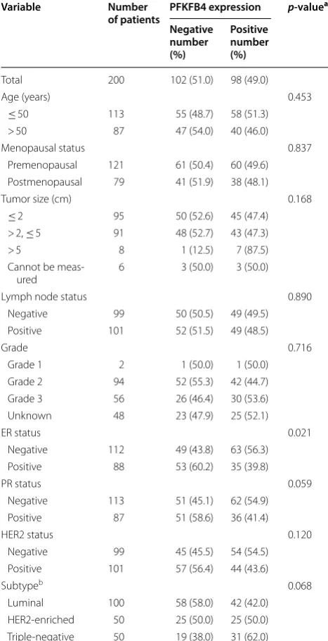
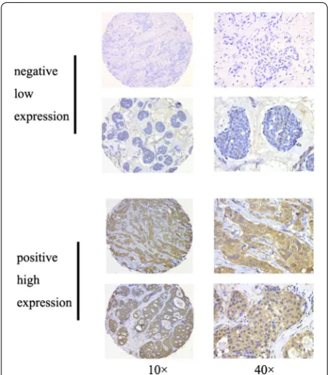
A total of 200 female patients with invasive ductal breast cancers were recruited in this study. The average age of the participants was 50.4 years (standard deviation 10.4, median 50.0, range 26–84). The clinicopathological characteristics of the cohort are summarized in Table 1. After a median follow-up time of 93.9 months, 37 of the 200 patients experienced recurrence or death. To inves-tigate the clinical significance of PFKFB4 in breast can-cer, we conducted immunohistochemical staining for PFKFB4 expression in the tumor samples. As shown in Fig. 1, positive staining for PFKFB4 protein was observed mainly in the cytoplasm of breast cancer cells, and most of the intra- or extra-tumor stromal cells were negative for PFKFB4. Using the semi-quantified scoring criteria, positive PFKFB4 staining was observed in 98 (49.0%) of the 200 cases (Table 1). Higher PFKFB4 expression was associated with ER status (p= 0.021) and had a
border-line relationship with PR status (p= 0.059) and subtype (p= 0.068).
Elevated PFKFB4 expression is associated with poor DFS and OS in breast cancer
To evaluate the clinical implications of PFKFB4 over-expression in breast cancer, we assessed the correlation between PFKFB4 status and DFS and OS using Kaplan– Meier survival analysis. As shown in Fig. 2a, b, patients with high PFKFB4 expression showed unfavorable DFS (p= 0.008) and OS (p = 0.002). After stratification by
molecular subtypes, PFKFB4 predicted poor DFS in luminal breast cancer (p= 0.018; Fig. 2c) and better OS
was observed in TNBC patients with low PFKFB4 expres-sion (p= 0.017; Fig. 2h). However, no survival association
was observed in the HER-2-enriched subgroup (p > 0.05; Fig. 2e, f). There was no positive correlation between PFKFB4 and OS in the luminal subtype (p > 0.05; Fig. 2d) and OS showed no significance in TNBC patients when stratified by PFKFB2 status (p > 0.05; Fig. 2g). These results suggest that the molecular function of PFKFB4 in breast cancer may depend on the molecular subtype, which is largely determined by receptor status.
Moreover, since PFKFB4 can regulate ER [6], we further investigated the survival relationship of PFKFB4 in breast cancer patients stratified by ER status. High PFKFB4 was associated with poor DFS in ER-positive patients (p= 0.020; Additional file 1: Figure S1A), whereas low Table 1 Clinicaopathological variables and the expression of PFKFB4
ER estrogen receptor, HER-2 human epidermal growth factor receptor 2, PFKFB4
6-phosphofructo-2-kinase/fructose-2,6-bisphosphatase 4, PR progesterone receptor
a Based on the Pearson χ2 test (Fisher’s exact test was used when needed) b Definitions of subtypes: luminal (ER- and/or PR-positive), HER-2-enriched (ER-
and PR-negative, HER-2-positive), and triple-negative (ER-negative, PR-negative, and HER-2-negative)
Variable Number
of patients PFKFB4 expression p-value a
Negative number (%)
Positive number (%)
Total 200 102 (51.0) 98 (49.0)
Age (years) 0.453
≤ 50 113 55 (48.7) 58 (51.3) > 50 87 47 (54.0) 40 (46.0) Menopausal status 0.837
Premenopausal 121 61 (50.4) 60 (49.6) Postmenopausal 79 41 (51.9) 38 (48.1) Tumor size (cm) 0.168
≤ 2 95 50 (52.6) 45 (47.4) > 2, ≤ 5 91 48 (52.7) 43 (47.3) > 5 8 1 (12.5) 7 (87.5) Cannot be meas‑
ured 6 3 (50.0) 3 (50.0) Lymph node status 0.890
Negative 99 50 (50.5) 49 (49.5) Positive 101 52 (51.5) 49 (48.5)
Grade 0.716
Grade 1 2 1 (50.0) 1 (50.0) Grade 2 94 52 (55.3) 42 (44.7) Grade 3 56 26 (46.4) 30 (53.6) Unknown 48 23 (47.9) 25 (52.1)
ER status 0.021
Negative 112 49 (43.8) 63 (56.3) Positive 88 53 (60.2) 35 (39.8)
PR status 0.059
Negative 113 51 (45.1) 62 (54.9) Positive 87 51 (58.6) 36 (41.4)
HER2 status 0.120
Negative 99 45 (45.5) 54 (54.5) Positive 101 57 (56.4) 44 (43.6)
Subtypeb 0.068
expression of PFKFB4 predicted favorable long-term out-come in ER-negative patients (p= 0.004; Additional file 1: Figure S1D). No positive correlation between PFKFB4 expression and OS was observed (p > 0.05; Additional file 1: Figure S1B), and comparison of DFS in ER-negative subgroups revealed no significant difference when strati-fied by PFKFB4 status (p > 0.05; Additional file 1: Figure S1C).
Univariate and multivariate Cox regression analyses
Next, we examined the correlation between DFS and the clinicopathological variables in the univariate Cox analyses. As shown in Table 2, PFKFB4 (HR 2.52, 95% CI 1.24–5.11; p= 0.010) and triple-negative subtype (HR 2.19, 95% CI 1.10–4.35; p= 0.025) were associated with a higher risk of relapse and death. After adjustment by variables for which HR were smaller than 0.2, PFKFB4
remained a significant prognostic factor with an HR of 2.42 (95% CI 1.17–5.00; p= 0.017).
Table 3 presents the association between OS and the clinicopathological variables analyzed using univariate and multivariate Cox regression. PFKFB4 had an HR of 7.38 (95% CI 1.69–32.3; p= 0.008) and retained prognos-tic power (HR 7.44, 95% CI 1.67–33.2; p= 0.009) when adjusted by tumor size, lymph node status, grade, ER status, HER2 status and subtypes. Lymph node status, a traditional prognostic factor, was associated with poor OS as well (HR 3.41, 95% CI 1.08–10.8; p= 0.037). Thus, PFKFB4 was established to be an independent prognostic factor in breast cancer.
Discussion
Increasing recognition of the active role of cancer metabolism in tumorigenesis has led to the identifica-tion of novel markers for prognostic predicidentifica-tion [11, 12]. Enzymes participating in core metabolic pathways have proven to be essential for the proliferation and survival of cancer cells [6, 7, 13, 14]. In this study, we evaluated the relationship of the cancer metabolic enzyme PFKFB4 with the risk of recurrence, metastasis and death in operable breast cancer. We demonstrated that elevated PFKFB4 expression from immunohistochemistry analysis is associated with shorter DFS and OS in breast cancer. Our results established that PFKFB4 is an independent prognostic factor in breast cancer.
Dasgupta et al. found that PFKFB4 can phosphoryl-ate steroid receptor coactivator-3 (SRC3) and lead to increased ER co-activation and cell proliferation. The authors examined 80 samples from the Cancer Genome Atlas and demonstrated that breast cancer patients with high SRC3 and PFKFB4 mRNA expression have unfa-vorable prognosis [6]. Using public high-throughput expression data, Ros et al. reported that a high level of PFKFB4 mRNA predicted reduced survival in patients with breast cancer and non-small cell lung cancer [15]. PFKFB4 mRNA expression has been proven to be a prognostic marker in non-muscle-invasive bladder can-cer [16]. However, quantification of mRNA expression is not easy to perform in routine clinical settings. In this study, we confirmed the prognostic value of PFKFB4 pro-tein in breast cancer using immunochemistry, which can be easily performed in FFPE samples. To the best of our
Fig. 1 Representative PFKFB4 immunohistochemical staining in breast cancer specimens in low‑magnification (×100) and high‑magnification (×400) images. The PFKFB4 staining intensities were classified as negative (low expression) and positive (high expression)
Fig. 2 Kaplan–Meier analysis of PFKFB4 in breast cancer. a DFS in all patients (n= 200); b OS (n= 200); c DFS in luminal subgroup (n= 100); d OS in luminal subgroup (n= 100); e DFS in HER‑2‑enriched subgroup (n= 50); f OS in HER‑2 in HER‑2‑enriched subgroup (n= 50); g DFS in triple‑negative subgroup (n= 50); h OS in triple‑negative subgroup (n= 50). DFS, disease‑free survival; OS, overall survival; HER‑2, human epidermal growth factor receptor 2. Definitions of subtypes: luminal (ER‑ and/or PR‑positive), HER‑2‑enriched (ER‑ and PR‑negative, HER‑2‑positive), and triple‑negative (ER‑negative, PR‑negative, and HER‑2‑negative)
Time (months) 120 100 80 60 40 20 Disease-free survival 1.0 0.8 0.6 0.4 0.2 Time (months) 125 100 75 50 25 0 Overal ls urviva l 1.0 0.8 0.6 0.4 0.2 0.0 Time (months) 120 100 80 60 40 20 0 Disease-free surviva l 1.0 0.8 0.6 0.4 0.2 0.0 Time (months) 125 100 75 50 25 0 Overall survival 1.0 0.8 0.6 0.4 0.2 0.0 Time (months) 120 100 80 60 40 20 0 Disease-fre e surviv al 1.0 0.8 0.6 0.4 0.2 0.0 Time (months) 120 100 80 60 40 20 0 O veral lsurviva l 1.0 0.8 0.6 0.4 0.2 0.0
Time (months)40 60 80 100 120
20 0 Disease-f re e survival 1.0 0.8 0.6 0.4 0.2 0.0 Time (months) 120 100 80 60 40 20 0 Overall survival 1.0 0.8 0.6 0.4 0.2 0.0 a b c d e f g h PFKFB4-low PFKFB4-high
p = 0.008 p = 0.002
p = 0.018 p = 0.195
p = 0.866 p = 0.210
p = 0.108 p = 0.017
knowledge, this is the first study supporting the prognos-tic value of PFKFB4 protein in breast cancer.
PFKFB4 plays an important role in regulating glucose metabolism and directing metabolic pathways required for biosynthesis of macromolecules to maintain cancer
cell proliferation [17]. Several groups independently identified PFKFB4 as a key metabolic enzyme in cancer using high-content screening [6–8]. PFKFB4 is required to maintain the balance of glycolytic activity for energy generation and cellular redox in prostate cancer [7]. Using an unbiased RNA interference genome-wide screening assay, Dasgupta et al. discovered PFKFB4 as a dominant modulator of SRC3-dependent cancer cell proliferation [6]. PFKFB4 and SRC-3, an ER co-activator, can hyperactivate ER activity in the presence of estradiol [6], which may explain the correlation between reduced DFS and high PFKFB4 observed in luminal and ER-pos-itive breast cancer. PFKFB4 and SRC-3 are drivers of the growth of basal-subtype breast cancer [6]. This may par-tially explain the prognostic significance of PFKFB4 in triple-negative and ER-negative subgroups. Further study is needed to determine the expression pattern of PFKFB4 and SRC-3 and the activated status of the PFKFB4-SRC-3 axis in breast cancer. Besides, it is also worthy to note the non-metabolic function of PFKFB4 that are relevant in cancer development. Gao et al. reported that PFKFB4 enhances breast cancer migration by induction of hya-luronan production in a p38-dependent manner [18]. Moreover, PFKFB4 can interact with endothelial tyrosine kinase to modulate chemoresistance of small-cell lung cancer by regulating autophagy [19].
Recent studies reported PFKFB4 as a potential tar-get in cancer. Silencing of PFKFB4 induced apoptosis in p53-deficient cancer cells and inhibited tumor growth [15]. A selective PFKFB4 inhibitor, 5-(n -(8-methoxy-4-quinolyl)amino)pentyl nitrate, suppressed the glyco-lysis process and proliferation in human cancer cell lines rather non-transformed epithelial cells in vitro, suggest-ing that targetsuggest-ing PFKFB4 may be a promissuggest-ing therapeu-tic strategy against breast cancer. Our study revealed that almost half (49.0%, 98/200) of the breast cancer cases in our study had a score 3 (the highest) for PFKFB4 stain-ing, which indicate a large population of breast cancer patients deposit the potential therapeutic target.
This study has some limitations. First, this is a ret-rospective study, in which we tested the association between PFKFB4 expression with DFS and OS in breast cancer rather than true prediction. Additional study is needed to validate the prognostic value of the novel marker. Second, the number of stage III patients with breast cancer in our cohort was too small (n= 8). Thus, the effectiveness of this potential prognostic marker should be applied to this subgroup with caution. Third, even though the study included subjects with all sub-types, the composition of the cohort was not representa-tive of the general patient population in the real world. Larger cohorts are therefore required before PFKFB4 can be recommended for clinical practice.
Table 2 Univariate and multivariate analysis
of clinicopathological variables and PFKFB4 associated with disease-free survival
− no data, CI confidence interval, ER estrogen receptor, HER-2 human epidermal growth factor receptor 2, HR hazard ratio, PFKFB4 6-phosphofructo-2-kinase/ fructose-2,6-bisphosphatase 4, PR progesterone receptor
a Definitions of subtypes: luminal (ER- and/or PR-positive), HER-2-enriched (ER-
and PR-negative, HER-2-positive), and triple-negative (ER-negative, PR-negative, and HER-2-negative)
Variable Univariate Multivariate
HR (95% CI) p value HR (95% CI) p-value
Age (years)
≤ 50 1 – – –
> 50 1.12 (0.58–2.14) 0.734 – – Menopausal status
Premenopausal 1 – – – Postmenopau‑
sal 1.13 (0.58–2.17) 0.726 – – Tumor size (cm)
≤ 2 1 – – –
> 2, ≤ 5 1.09 (0.56–2.12) 0.800 – – > 5 1.00 (0.13–7.49) 0.998 – – Cannot be
measured 1.29 (0.17–9.69) 0.807 – – Lymph node status
Negative 1 – – –
Positive 1.46 (0.76–2.81) 0.253 – – Grade
Grade 1–2 1 – – – Grade 3 1.10 (0.50–2.43) 0.810 – – Unknown 1.53 (0.71–3.29) 0.281 – – ER status
Negative 1 – 1 –
Positive 0.58 (0.29–1.14) 0.112 0.51 (0.13–2.09) 0.353 PR status
Negative 1 – – –
Positive 0.66 (0.34–1.30) 0.230 – – HER2 status
Negative 1 – 1 –
Positive 0.62 (0.32–1.22) 0.166 1.26 (0.44–3.61) 0.671 Subtypea
Luminal 1 – 1 –
HER2‑enriched 0.61 (0.20–1.82) 0.373 0.26 (0.059–1.19) 0.083 Triple‑negative 2.19 (1.10–4.35) 0.025 1.22 (0.24–6.06) 0.812 PFKFB4
Negative 1 – 1 –
Table 3 Univariate and multivariate analysis of clinicopathological variables and PFKFB4 associated with overall survival
− no data, CI confidence interval, ER estrogen receptor, HER-2 human epidermal growth factor receptor 2, HR hazard ratio, PFKFB4 6-phosphofructo-2-kinase/ fructose-2,6-bisphosphatase 4, PR progesterone receptor
a Definitions of subtypes: luminal (ER- and/or PR-positive), HER-2-enriched (ER-
and PR-negative, HER-2-positive), and triple-negative (ER-negative, PR-negative, and HER-2-negative)
Variable Univariate Multivariate
HR (95% CI) p-value HR (95% CI) p-value
Age (years)
≤ 50 1 – – –
> 50 1.72 (0.66–4.46) 0.266 – – Menopausal status
Premenopausal 1 – – – Postmeno‑
pausal 1.49 (0.58–3.87) 0.411 – – Tumor size (cm)
≤ 2 1 – 1 –
> 2, ≤ 5 1.68 (0.60–4.71) 0.327 2.07 (0.69–6.18) 0.193 > 5 3.22 (0.39–26.8) 0.279 3.43 (0.33–36.0) 0.304 Cannot be
measured 4.21 (0.50–35.1) 0.184 2.67 (0.24–29.7) 0.425 Lymph node status
Negative 1 – 1 –
Positive 2.64 (0.93–7.49) 0.069 3.41 (1.08–10.8) 0.037 Grade
Grade 1–2 1 – 1 – Grade 3 2.36 (0.75–7.45) 0.142 2.01 (0.59–6.85) 0.264 Unknown 2.22 (0.64–7.65) 0.209 2.56 (0.64–10.2) 0.183 ER status
Negative 1 – – –
Positive 0.64 (0.24–1.72) 0.371 – – PR status
Negative 1 – – –
Positive 0.70 (0.26–1.89) 0.477 – – HER2 status
Negative 1 – 1 –
Positive 0.49 (0.17–1.40) 0.184 0.67 (0.14–3.16) 0.617 Subtypea
Luminal 1 – 1 –
HER2‑enriched 0.71 (0.15–3.44) 0.675 0.47 (0.068–3.21) 0.439 Triple‑negative 2.10 (0.76–5.79) 0.152 1.13 (0.31–4.17) 0.850 PFKFB4
Negative 1 – 1 –
Positive 7.38 (1.69–32.3) 0.008 7.44 (1.67–33.2) 0.009
Conclusion
Our study indicates the prognostic value of PFKFB4 protein in breast cancer. High expression of PFKFB4 was associated with reduced DFS and OS in breast can-cer. Our findings may therefore promote the further
clinical use of PFKFB4 and provides additional prog-nostic information to oncologists with regard to evalu-ating risk via assessing cancer metabolic enzymes. Our results, together with previous mechanistic studies, provide a rationale for further clinical investigation to treat cancer by manipulating PFKFB4 expression.
Additional file
Additional file 1: Figure S1. Kaplan–Meier analysis of PFKFB4 in breast cancer stratified by ER status. (A) DFS in ER‑positive patients (n= 88); (B) OS in ER‑positive patients (n= 88); (C) DFS in ER‑negative patients (n= 112); (D) OS in ER‑negative patient (n= 112). DFS, disease‑free sur‑ vival; OS, overall survival; ER, estrogen receptor.
Acknowledgements
Not applicable.
Authors’ contributions
ZS and XH conceived and designed the study. LY and LW performed the experiments. LY analyzed the data. ZC contributed reagents, materials, and analysis tools. LY and LW wrote the paper. All authors read and approved the final manuscript.
Funding
This study was supported by grants from the National Natural Science Foundation of China (81202082, 81602316), Shanghai Natural Science Foundation(19ZR1411000), the Ministry of Science and Technology of China (MOST2016YFC0900300, National Key R&D Program of China). The funders had no role in study design, data collection and analysis, decision to publish, or preparation of the manuscript.
Availability of data and materials
Not applicable.
Ethics approval and consent to participate
This study was approved by the Ethical Committee of Fudan University Shang‑ haiCancer Center for Clinical Research (Reference Number: 050432‑4‑1212B). The written informed consents were obtained from all the patients.
Consent for publication
Not applicable.
Competing interests
The authors declare that they have no competing interests.
Author details
1 Department of Breast Surgery, Key Laboratory of Breast Cancer in Shanghai, Fudan University Shanghai Cancer Center, Fudan University, 270 Dong‑An Rd., Shanghai 200032, People’s Republic of China. 2 Key Laboratory of Breast Can‑ cer in Shanghai, Fudan University Shanghai Cancer Center, Fudan University, Shanghai, People’s Republic of China. 3 Department of Oncology, Shanghai Medical College, Fudan University, Shanghai, People’s Republic of China. 4 Institutes of Biomedical Sciences, Fudan University, Shanghai, People’s Republic of China.
Received: 15 April 2019 Accepted: 3 June 2019
References
1. Siegel RL, Miller KD, Jemal A. Cancer statistics, 2018. CA Cancer J Clin. 2018;68:7–30.
•fast, convenient online submission •
thorough peer review by experienced researchers in your field • rapid publication on acceptance
• support for research data, including large and complex data types •
gold Open Access which fosters wider collaboration and increased citations maximum visibility for your research: over 100M website views per year •
At BMC, research is always in progress.
Learn more biomedcentral.com/submissions
Ready to submit your research? Choose BMC and benefit from: 3. Curigliano G, Burstein HJ, Winer EP, Gnant M, Dubsky P, Loibl S, et al. De‑
escalating and escalating treatments for early‑stage breast cancer: the St. Gallen international expert consensus conference on the primary therapy of Early Breast Cancer 2017. Ann Oncol. 2017;28:1700–12.
4. Giuliano AE, Connolly JL, Edge SB, Mittendorf EA, Rugo HS, Solin LJ, et al. Breast cancer‑major changes in the American joint committee on cancer eighth edition cancer staging manual. CA Cancer J Clin. 2017;67:290–303. 5. Vander Heiden MG, Cantley LC, Thompson CB. Understanding the
Warburg effect: the metabolic requirements of cell proliferation. Science. 2009;324:1029–33.
6. Dasgupta S, Rajapakshe K, Zhu B, Nikolai BC, Yi P, Putluri N, et al. Metabolic enzyme PFKFB4 activates transcriptional coactivator SRC‑3 to drive breast cancer. Nature. 2018;556:249–54.
7. Ros S, Santos CR, Moco S, Baenke F, Kelly G, Howell M, et al. Functional metabolic screen identifies 6‑phosphofructo‑2‑kinase/fructose‑2,6‑bi‑ phosphatase 4 as an important regulator of prostate cancer cell survival. Cancer Discov. 2012;2:328–43.
8. Goidts V, Bageritz J, Puccio L, Nakata S, Zapatka M, Barbus S, et al. RNAi screening in glioma stem‑like cells identifies PFKFB4 as a key molecule important for cancer cell survival. Oncogene. 2012;31:3235–43. 9. Zheng YZ, Cao ZG, Hu X, Shao ZM. The endoplasmic reticulum stress
markers GRP78 and CHOP predict disease‑free survival and respon‑ siveness to chemotherapy in breast cancer. Breast Cancer Res Treat. 2014;145:349–58.
10. Wang L, Yao L, Zheng YZ, Xu Q, Liu XP, Hu X, et al. Expression of autophagy‑related proteins ATG5 and FIP200 predicts favorable disease‑ free survival in patients with breast cancer. Biochem Biophys Res Com‑ mun. 2015;458:816–22.
11. Qiu Y, Cai G, Zhou B, Li D, Zhao A, Xie G, et al. A distinct metabolic signa‑ ture of human colorectal cancer with prognostic potential. Clin Cancer Res. 2014;20:2136–46.
12. Baek G, Tse YF, Hu Z, Cox D, Buboltz N, McCue P, et al. MCT4 defines a glycolytic subtype of pancreatic cancer with poor prognosis and unique metabolic dependencies. Cell Rep. 2014;9:2233–49.
13. Wang JB, Erickson JW, Fuji R, Ramachandran S, Gao P, Dinavahi R, et al. Targeting mitochondrial glutaminase activity inhibits oncogenic transfor‑ mation. Cancer Cell. 2010;18:207–19.
14. Christofk HR, Vander Heiden MG, Harris MH, Ramanathan A, Gerszten RE, Wei R, et al. The M2 splice isoform of pyruvate kinase is important for cancer metabolism and tumour growth. Nature. 2008;452:230–3. 15. Ros S, Floter J, Kaymak I, Da Costa C, Houddane A, Dubuis S, et al.
6‑Phosphofructo‑2‑kinase/fructose‑2,6‑biphosphatase 4 is essential for p53‑null cancer cells. Oncogene. 2017;36:3287–99.
16. Yun SJ, Jo SW, Ha YS, Lee OJ, Kim WT, Kim YJ, et al. PFKFB4 as a prog‑ nostic marker in non‑muscle‑invasive bladder cancer. Urol Oncol. 2012;30:893–9.
17. Dang CV. Cancer cell metabolism: there is no ROS for the weary. Cancer Discov. 2012;2:304–7.
18. Gao R, Liu Y, Li D, Xun J, Zhou W, Wang P, et al. PFKFB4 promotes breast cancer metastasis via induction of hyaluronan production in a p38‑dependent manner. Cell Physiol Biochem. 2018;50:2108–23. 19. Wang Q, Zeng F, Sun Y, Qiu Q, Zhang J, Huang W, Huang J, Huang X, Guo
L. Etk interaction with PFKFB4 modulates chemoresistance of small‑cell lung cancer by regulating autophagy. Clin Cancer Res. 2018;24(4):950–62.
Publisher’s Note